THE RELATIONSHIP BETWEEN GAIT SPEED AND DYNAMIC STABILITY FOLLOWING A 4-WEEK INTERVENTION IN KNEE OSTEOARTHRITIS PATIENTS
Kayla Vredeveld
A thesis submitted to the faculty at the University of North Carolina at Chapel Hill in partial fulfillment of the requirements for the degree of Master of Arts in the Department of Exercise
and Sport Science (Exercise Physiology).
Chapel Hill 2017
Approved By: Erik Hanson
Brian Pietrosimone
iii ABSTRACT
Kayla Vredeveld: The Relationship Between Gait Speed and Dynamic Stability Following a 4-week Intervention in Knee Osteoarthritis Patients
(Under the direction of Erik Hanson)
PURPOSE: This study explored the relationship of both gait speed and body composition to gait stability following knee osteoarthritis (KOA) treatment. METHODS: 53 diagnosed KOA
patients completed 4 weeks of therapeutic rehabilitation with testing sessions immediately before and after. Testing assessed self-reported disability, body composition, muscle function, and gait mechanics. Paired sample t-tests were completed, followed by correlations comparing gait speed, center of pressure, and ground reaction forces with baseline and change score values. RESULTS: 20 meter gait speed significantly increased following intervention, while pain and self-reported function scores improved. Ground reaction forces showed increased acceleration and
iv
v
ACKNOWLEDGEMENTS
vi
TABLE OF CONTENTS
LIST OF ABBREVIATIONS ... viii
CHAPTER 1: INTRODUCTION ...1
Research Questions ...4
Hypotheses ...4
Limitations ...4
Delimitations ...5
Assumptions ...5
Significance of Study ...5
CHAPTER 2: LITERATURE REVIEW ...6
CHAPTER 3: METHODOLOGY ...12
Research Design Overview ...12
Subjects ...12
Procedures ...13
Intervention Groups ...14
Outcome Measures...15
Statistical Analysis ...18
CHAPTER 4: RESULTS ...19
CHAPTER 5: DISCUSSION ...24
vii
viii
LIST OF ABBREVIATIONS
AP Anteroposterior
BIA Bioelectrical impedance analysis BMI Body mass index
CAR Central activation ratio COP Center of pressure
DEXA Dual energy x-ray absorptiometry FM Fat mass
GRF Ground reaction force KOA Knee osteoarthritis
LM Lean mass
ML Mediolateral
MVIC Maximal voluntary isometric contraction TE Therapeutic exercise
1
CHAPTER 1: INTRODUCTION
Knee osteoarthritis (KOA) is a health condition of the musculoskeletal system that affects 12-16% of the population over 60 years old1 2. The degenerative disease is associated with other health conditions including cardiovascular disease, chronic obstructive pulmonary disease, obesity, diabetes, and depression3-8 at a rate of 50% of the KOA population9. Collectively, these comorbidities along with KOA lead to an increased mortality6 and decreased quality of life9 due to the limitations in independently completing activities of daily living. The ability to complete daily tasks in KOA patients is negatively effected by the resulting loss of balance, which is amplified by the loss of strength, muscle activation, and the compromised body composition.
Falls are a prevalent issue in the healthy elderly, with studies reporting balance deficits as early as 50 years old10 and observations concluding that 1 in 3 adults over 65 years will fall over the course of a year11. Overall, loss of balance and falls are not only more common in KOA patients12-14, but also have significantly greater deficits compared to the healthy elderly averages12 15, with deficits increasing as the disease progresses13. Reliable measurements of dynamic balance are infrequent in KOA studies, but their direct relationship to the ability to complete activities of daily living and consequently quality of life12 15 demands that they are further explored.
2
mediolateral (ML) COP velocity and a decreased anteroposterior (AP) COP velocity in an unhealthy population compared to healthy matches19. This suggests that COP velocity may also increase in the KOA population. COP velocity is influenced by gait speed with studies showing COP velocity decreases when gait speed decreases in elderly subjects19 20. Although COP variables are altered based on foot structure, COP measurements have been shown to have moderate to good reliability18.
Although obesity has been identified as a comorbidity with KOA6, it has not been established if changes in body composition are a potential cause or the result of KOA. Studies have shown that the fat mass (FM) and lean mass (LM) are significantly different in KOA patients compared to healthy matched subjects, but the values could not differentiate between mild and severe level KOA subjects21. This suggests that body composition may be just one of several influencing factors. In comparing men and women with KOA, it has been suggested that increased FM more strongly correlates with KOA in women while decreased LM is more closely associated in men22 23. Increased FM or decreased LM both reduce the relative force production of the muscles22 23 indicating that they are related to functional and balance measures, although KOA studies have not directly assessed these outcomes.
3
the whole muscle is not being used. CAR of the rectus femoris has been able to differentiate between asymptomatic and moderate severity KOA28, with moderate KOA patients having a CAR below 80%27.
Balance measures are correlated to quadriceps strength in the KOA population, making quadriceps function an important factor to consider when looking at function (r=0.37)15. When compared to healthy controls, KOA subjects have lower balance, strength, and activation measures14 15, confirming the existing relationship. Other studies have also shown that both decreased quadriceps strength and CAR are indicative of greater balance deficits in KOA patients15 30-32. Within the KOA population, the strength deficit and number of falls are
significantly greater in surgical candidates compared to non-surgical patients32, suggesting that strength and balance continue to decrease as the disease progresses.
To counteract the physical decline resulting from KOA, therapeutic exercise (TE) and transcutaneous electrical nerve stimulation (TENS) interventions have been used to not only attenuate pain, but also to increase quadriceps CAR with the aim of ultimately improving performance in activities of daily living27 33. A combination of TE+TENS was shown to be significantly more effective at improving CAR than TE alone27. Baseline testing measured a 78±7% CAR in KOA patients, with a 4 week TE+TENS intervention increasing the CAR to 94±4%27. Function and balance measures were not assessed in this study, but the literature suggests balance is related to CAR14. In a study that used only TE as an intervention,
4
To fully determine the results of treatment in KOA patients following an intervention, it must be determined not only if patients can move better, as determined by measures of strength, CAR, and gait speed, but also if patients are improving their dynamic stability mechanisms. COP measurements of sway velocity, sway path, and ML displacement can used to quantify dynamic gait stability so they can be compared to other changes occurring as a result of intervention. An increase in dynamic stability and the ability to move must occur simultaneously to ensure that not only are the patients able to move better, but they can move safer.
Purpose
1. To determine if there is a relationship between the changes in gait speed and dynamic stability following a 4 week therapeutic intervention in KOA patients
2. To establish if there is a relationship between body composition and dynamic stability during KOA treatment
Research Questions
1. Is there an association between the change in gait speed and the change in COP measured immediately before and after a 4 week KOA treatment intervention? 2. Is COP trajectory post intervention associated with lean mass, fat mass, CAR, or pain
level? Hypotheses
1. An improved COP trajectory will be associated with an increase in 20 meter maximal walking speed following a 4 week intervention.
5 Limitations
1. COP data will be based on the single step that contacts the force plate. Delimitations
1. Subjects were limited to those able to walk without an assistive device. 2. Subjects were excluded if they are obese (body mass index (BMI) >35). 3. Participation was limited to subjects with moderate to severe KOA. Assumptions
1. Subjects did not intentionally alter gait pattern during the testing session
2. Subjects did not have medical problems, besides KOA, interfering with gait pattern 3. Subjects answered self-reported data truthfully
4. COP variables were representative of overall stability Significance of Study
6
CHAPTER 2: LITERATURE REVIEW
KOA is one of the top five causes of disability and is associated with an early onset of the inability to independently complete activities of daily living, making it as debilitating as heart disease9 34. This separates those with KOA and the healthy aging. Risk of developing KOA increases with age1, so the rising number of the elderly in the United States population, which is expected to double by 205035, demands that more attention is given to this manageable health condition. Up to 50% of KOA patients have at least one other medical condition9, including depression, cardiovascular disease, obesity, diabetes, and chronic obstructive pulmonary
disease3-8. In most cases, it is unknown if KOA is the cause or effect of other diseases. However, a few research studies have concluded that KOA is shown to be the causative factor of
cardiovascular disease, obesity, and diabetes6.
7
Similar to the general aging population, a significant decrease in gait speed has also been found in subjects with severe KOA28. Additionally, balance deficits are shown to be significantly worse in KOA patients when compared to the healthy elderly12 15, with half of KOA patients sustaining falls within the year prior to receiving surgical intervention32. Loss of balance and falls are more prevalent in KOA patients12 13 because they are exacerbated by changes in muscle strength and activation, especially of the quadriceps15 32 37.
To quantify balance more precisely than clinical tests and to detect minimal changes, balance should be measured through COP. Not only should balance be measured using this method, but it should also be measured during a dynamic task, such as walking, so that the results are more applicable than static posture COP assessments. COP can be quantified by measures using both the position and velocity at any point during foot contact on the force plate and provide information about the dynamic stability of the individual. Although COP is
influenced by foot structure, it has been determined that a healthy ML COP displacement covers 18% of the foot width18. COP measures are highly reliable with ICC between 0.65 and 0.82 ML, and between 0.69 and 0.93 AP, indicating moderate to good reliability for COP position, with similar values for COP velocity18. The lower ICC in ML COP was seen primarily in the middle of stance phase, which is when the foot structure has the highest influence on COP18.
Body composition changes in KOA patients are evident, but it is unclear whether they are the cause or effect of KOA21-23. Although not a true assessment of body composition, BMI calculates the ratio of body weight to height and is commonly used to determine the obesity level in KOA patients42-46. Studies using this methodology indicate that a BMI over 30kg/m2 increases KOA risk 7 to 8 times higher compared to those with a BMI less than 25kg/m242 43 47.
8
advantage of simple testing procedures, encouraging its use in research45 46 48. Multivariate analyses showed that BIA estimates of LM were better predictors of KOA presence and severity compared to BMI46. However, in studies using both BMI and body fat percentage via BIA, an increased BMI indicated a greater risk of KOA compared to an increased body fat percentage45. These inconclusive results in the literature regarding the influence of FM and LM on KOA may be due to the error introduced through BIA estimations, despite using pre-assessment guidelines.
Dual energy x-ray absorptiometry (DEXA) has been shown to provide valid and reliable measures of bone mineral density, FM, and LM49 50, making it the ideal method of assessing body composition. In the studies using DEXA in KOA, FM and LM are both significantly correlated with KOA, but neither could determine the severity of KOA21 22. However, the correlation between KOA and LM is not consistent, as other studies found no correlation between them23. Due to the small number of studies looking at the relationship between body composition and KOA, further study should be done using DEXA to quantify FM and LM to produce valid and reliable data.
Without measuring total body composition, muscle atrophy has been quantified using quadriceps cross sectional area, which was found to be 12% smaller in older women with KOA compared to healthy women of the same age51. Although comparisons of healthy old and young women show that older women without KOA battle against loss of muscle mass, KOA diagnosis significantly increased the loss51. Maintaining muscle mass is important because it determines the maximum capacity of strength production, which is shown to be strongly linked to physical function25 52 53.
9
arthroplasty and it was found that muscle mass and muscle inhibition together explained 85% of the strength deficit, but muscle inhibition was twice as significant as the muscle mass54. This shows that negating atrophy is important, but muscle inhibition must be addressed in order to receive the greatest benefits of therapeutic exercise and to gain the maximum pain free function33.
A true maximal contraction can be produced through electric stimulation of the muscle during MVIC testing. The CAR is determined by comparing the MVIC to the stimulated
maximal contraction26 27 29. It is common for KOA patients to have a CAR below 80%, meaning that 20% of the muscle fibers are not voluntarily recruited27. This inactivation leads to weakness in KOA patients that is evident in both isometric and isokinetic strength testing24. It was
observed that 50% of subjects with mild KOA were unable to fully activate the quadriceps55. When muscle fibers are not voluntarily fired, they are not able to be recruited to complete daily activities. However, when quadriceps activation is increased through an exercise intervention, a significant increase in MVIC results29.
This weakness that is due to both a loss of muscle mass and a loss of muscle activation alters the gait pattern in KOA patients24 28 29 33 51 54. Just as weakness effects function, it also effects balance scores in KOA patients31. When compared to healthy controls, KOA subjects have lower static balance, strength, and activation measures14, confirming the existing
relationship. Several studies have also shown that both decreased quadriceps strength and CAR are indicative of greater balance deficits in KOA patients15 30 31. Within the KOA population itself, the quadriceps strength deficit is significantly greater in surgical KOA candidates
10
KOA directly decreases physical function, specifically gait function, due to
accompanying knee pain56-58. Pain levels are strongly associated with the severity of KOA59. However, there is evidence that these pain levels, based on self-reported pain using a visual analog scale (VAS), are manageable because a hyaluronate injection intervention significantly decreased pain after 5 weeks56. KOA pain inhibits a normal gait pattern, as the expectant double peak typically seen in the ground reaction forces of healthy individuals is not present KOA patients57. Instead, KOA patients exhibit a blunted ground reaction force response and alter their mechanics to reduce loading forces in the lower extremities, shown in both level ground
walking57 and during more demanding tasks such as descending stairs60. Hyaluronate injections, used in KOA to reduce pain levels, allows the ground reactions forces to be restored to those of a healthy individual following a 5-week intervention of hyaluronate injections56 57.
Current treatments for KOA include both TE and TENS interventions with the ultimate goal of increasing normal, pain free function in KOA patients. The focus of TE is typically to increase lower extremity strength, especially in the quadriceps34. Although TE can increase the strength of the muscle by increasing the size and number of muscle fibers61, it may not increase the amount of muscle fibers voluntarily contracting, leaving the patient prone to both losses of muscular strength and muscular atrophy62. Traditionally, TENS was used in treatment to reduce pain63-65. However, more recent applications of TENS have been used to increase the CAR of the quadriceps27 29 33 63 66 67. By applying TENS to increase CAR, the muscle’s true potential may be able to be reached.
11
an immediate acute effect on balance, quantified by a pressure mat68. Park et al. compared TE+TENS to a TE+shamTENS group of stroke patients, finding that the TENS group had greater improvements in balance after a 6 week intervention69. The influence of TENS on
balance needs to be further explored and tested in the KOA population because of its potential to decrease fall risk and increase safe participation in activities of daily living.
Research shows that KOA patients are negatively effected by balance deficits, which are related to the compromised body composition and strength. Treatments targeting strength may result in an improvement in body composition as well as physical function. However, in order to benefit KOA patients to the greatest extent, dynamic stability must also increase to lower fall risk while completing these tasks. By ensuring KOA patients are functioning at a higher level
12
CHAPTER 3: METHODOLOGY
Research Design Overview
The primary purpose of this study was to determine if a relationship exists between the change in gait speed and the change in dynamic stability following an intervention. The secondary purpose was to establish if there is a relationship between body composition and dynamic stability following KOA treatment. Subjects went through a 4 week intervention with baseline and post-intervention testing sessions.
Subjects
All subjects in this study were diagnosed radiographically with moderate to severe KOA on the Kellgren-Lawrence (KL) scale. Subjects were recruited through referral by their
orthopaedic physician at the Orthopaedic Clinic. Subjects were eligible for inclusion if they meet all of the following criteria:
Between the ages of 40 and 75 years old
Exhibit symptomatic KOA, which we will define as a normalized, person based, Western Ontario and McMaster Universities Arthritis Index (WOMAC) function subscale score > 31
Radiographic evidence of tibiofemoral osteoarthritis (2-4 on the KL scale)
Neuromuscular activation deficits, defined as central activation ratio of less than 90% ± 2% in the involved leg
13
Diagnosed with a cardiovascular condition restricting exercise
Have had a corticosteroid or hyaluronic acid injection in the involved knee in the previous 14-days
Have a pacemaker
Have a neurodegenerative condition Have rheumatoid arthritis
Have cancer
Have a neural sensory dysfunction over the knee Have a BMI over 35
Have a history of lower extremity orthopaedic surgery in the past year Have a history of a traumatic knee injury in the past 6 months
Have any history of a total knee arthroplasty in either extremity Have a diagnosed, non-reconstructed knee ligament tear
Need an assistive device to walk
Currently pregnant or planning to become pregnant while enrolled in the study Prior to data collection, subjects signed an informed consent form (Appendix A) which was approved by the University of North Carolina Biomedical Institutional Review Board. Procedures
After a physician confirmed the OA diagnosis, a member of the research team explained the goals of the study. Subjects were also recruited through the Carolina Data Warehouse for Health and UNC-Hospital’s Database. These subjects were contacted via mail with an
14
demographic information and self-reported function based on the Functional subscale of the WOMAC Questionnaire. Subjects were also able to ask any questions related to involvement in the study. If interested subjects were still eligible for the study, an appointment in the
Neuromuscular Research/Sports Medicine Research Laboratory was made. At this appointment, subjects signed the informed consent, and completed the baseline testing before their first appointment at UNC Meadowmont Physical Therapy Clinic.
Intervention Groups
The subjects were randomized into 1 of 3 treatment groups before the first PT visit. Therapeutic Exercise
Subjects in this group completed all 10 visits at UNC Meadowmont, participating in TE with the goal of increasing lower extremity strength and function. Each visit lasted
approximately 45 minutes, and subjects completed 2-3 sessions a week, totaling 10 over a 4 week period. TE included flexibility exercises, lower extremity strengthening exercises, and balance training, with all subjects receiving the same protocol, but progressing on an individual basis.
Therapeutic Exercise + TENS
15 Therapeutic Exercise +shamTENS
Subjects in this group completed the same TE program, and were given a TENS unit programmed to discontinue current output after 20 seconds. Subjects were instructed similarly to the TENS group, but were told that it is normal to not feel the current output after application. No instructions for increasing dosage were given.
Outcome Measures
At all lab visits, subjects underwent the same testing procedures, completed by a blinded member of the research team.
Visual Analog Scales (VAS) were used for current level of knee pain. A solid line from 0 to 100 was shown to the subject, and they were asked to mark how they currently felt. A score of 100 indicated “worst pain imaginable”, and a score of 0 indicated “no pain”. Distance was measured from 0 to the line drawn and reported in centimeters.
Body Composition
A subpopulation in the study completed body composition testing via DEXA (DEXA; GE Lunar DEXA, GE Medical Systems Ultrasound & Primary Care Diagnostics, Madison, WI, USA), using the encore software for analysis (enCORE Software Version 16). Subjects adhered to the following pre-assessment guidelines:
Remove shoes and any jewelry prior to testing Two hours fasted and adequately hydrated
16 Muscle Function
Subjects were positioned on the isokinetic dynamometer, using straps to secure their torso, thigh, and lower leg to the device. The knee was in 90º of flexion and the movement arm was in a fixed position to test strength isometrically. Two electrodes were placed over the quadriceps. The subjects were instructed to ‘kick out’ in order to flex the quadriceps as quickly and as strongly as possible while keeping their arms crossed over their chest to prevent accessory movement. A practice trial was done at approximately 25, 50, and 75% of the subject’s perceived maximum, then three maximal trials were completed and the average score was recorded. For the next 2 trials, an electrical stimulus was given after the torque plateaus at the previously
determined average MVIC. The CAR was calculated by comparing the voluntary force to the electrically stimulated force production.
Gait Biomechanics
Subjects walked along a 6m walkway at a comfortable, self-selected speed. A minimum of 5 practice trials were done to determine the average preferred speed given in real time through 2 sets of timing gates (TF100, Trac Tronix, Lenexa, Kansas, United States). Five valid trials for each limb were completed for data collection. For a trial to be valid, subjects had to fully contact the force plate and be within ± 5% of their preferred gait speed.
Force Plates
17
(Innovative Sports Training, Inc., Chicago, IL, USA) for analysis. The x-axis indicated anteroposterior and the y-axis indicated mediolateral movement of the COP relative to the
participant. Once the data was verified and analyzed in The Motion Monitor, the exported results were run through Matlab (The Mathworks, Inc., R2016a) where the mean for each of the
following variables was determined using the 3 middle trials. Trials were substituted if there were ground reaction forces (GRF) on both plates, indicating their foot was not fully on the plate.
Stance time COP ellipse area COP sway path COP sway velocity ML excursion M displacement L displacement
Vertical ground reaction force (VGRF) maximum
Anteroposterior ground reaction force (APGRF) maximum and minimum Mediolateral ground reaction force (APGRF) maximum and minimum
All GRF variables were calculated for the first and second 50% of stance phase. Initial contact was defined as the point at which the ground reaction force is greater than 10N, and the toe off was the point at which the force is less than 10N.
Maximum Gait Speed
18
safely could without running. Three trials were completed and the average score was used in the data analysis.
Statistical Analysis
SPSS 23.0 (SPSS INC, Chicago, Ill) was used for all statistical calculations with
statistical significance set a-priori at p<0.05. Analysis for outliers was completed by determining the interquartile range for the 20 meter walk test. One outlier was found and removed for all statistical analysis. The descriptive characteristics were reported as the mean ± standard
deviation. A series of T-tests were run to determine if there were significant changes following intervention. In order to answer research question one, a correlation analysis was done
19
CHAPTER 4: RESULTS
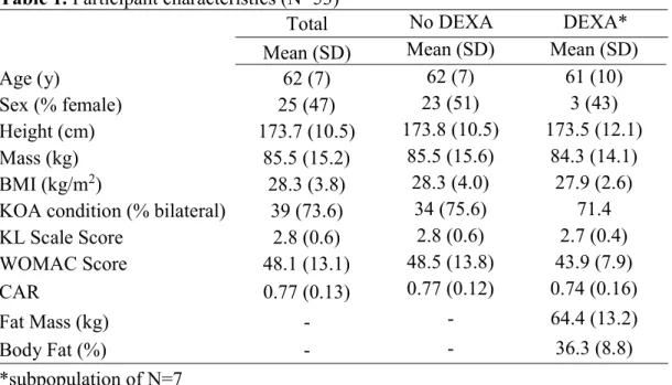
Fifty three older subjects participated in this study who were, on average, overweight, diagnosed radiographically with moderate to severe KOA and experiencing mild to moderate pain and dysfunction (Table 1), one subject was removed as an outlier upon statistical analysis. In a sub-group of individuals with body composition scans (N=7), subjects were found to be obese. No significant differences between the DEXA and no DEXA groups were found.
Table 1. Participant characteristics (N=53)
Total No DEXA DEXA*
Mean (SD) Mean (SD) Mean (SD)
Age (y) 62 (7) 62 (7) 61 (10)
Sex (% female) 25 (47) 23 (51) 3 (43)
Height (cm) 173.7 (10.5) 173.8 (10.5) 173.5 (12.1)
Mass (kg) 85.5 (15.2) 85.5 (15.6) 84.3 (14.1)
BMI (kg/m2) 28.3 (3.8) 28.3 (4.0) 27.9 (2.6)
KOA condition (% bilateral) 39 (73.6) 34 (75.6) 71.4
KL Scale Score 2.8 (0.6) 2.8 (0.6) 2.7 (0.4)
WOMAC Score 48.1 (13.1) 48.5 (13.8) 43.9 (7.9)
CAR 0.77 (0.13) 0.77 (0.12) 0.74 (0.16)
Fat Mass (kg) - - 64.4 (13.2)
Body Fat (%) - - 36.3 (8.8)
*subpopulation of N=7
20
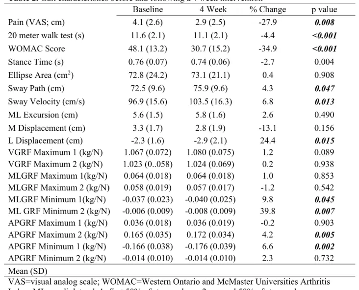
The changes in gait function following a 4 week intervention are displayed in Table 2. Maximum gait speed, represented by the 20 meter walk test, improved significantly following intervention (p<0.001). COP sway velocity and L displacement both significantly increased with intervention, while there was a trend for higher COP sway path (p=0.055). GRFs were measured as the vertical (V), mediolateral (ML), and anteroposterior (AP) forces. Significant increases in minimum MLGRF during the first and second 50% of stance were found. Significant changes
Table 2. Gait characteristics before and following a 4 week intervention
Baseline 4 Week % Change p value
Pain (VAS; cm) 4.1 (2.6) 2.9 (2.5) -27.9 0.008
20 meter walk test (s) 11.6 (2.1) 11.1 (2.1) -4.4 <0.001
WOMAC Score 48.1 (13.2) 30.7 (15.2) -34.9 <0.001
Stance Time (s) 0.76 (0.07) 0.74 (0.06) -2.7 0.004
Ellipse Area (cm2) 72.8 (24.2) 73.1 (21.1) 0.4 0.908
Sway Path (cm) 72.5 (9.6) 75.9 (9.6) 4.3 0.047
Sway Velocity (cm/s) 96.9 (15.6) 103.5 (16.3) 6.8 0.013
ML Excursion (cm) 5.6 (1.5) 5.8 (1.6) 2.6 0.490
M Displacement (cm) 3.3 (1.7) 2.8 (1.9) -13.1 0.156
L Displacement (cm) -2.3 (1.6) -2.9 (2.1) 24.4 0.015
VGRF Maximum 1 (kg/N) 1.067 (0.072) 1.080 (0.075) 1.2 0.089 VGRF Maximum 2 (kg/N) 1.023 (0..058) 1.024 (0.069) 0.2 0.938 MLGRF Maximum 1(kg/N) 0.064 (0.018) 0.064 (0.018) 1.0 0.853 MLGRF Maximum 2 (kg/N) 0.058 (0.019) 0.057 (0.017) -1.2 0.542 MLGRF Minimum 1(kg/N) -0.037 (0.023) -0.040 (0.025) 9.8 0.045 ML GRF Minimum 2 (kg/N) -0.006 (0.009) -0.008 (0.009) 39.8 0.007 APGRF Maximum 1 (kg/N) 0.036 (0.018) 0.036 (0.019) -0.2 0.903 APGRF Maximum 2 (kg/N) 0.165 (0.035) 0.172 (0.034) 4.2 0.005 APGRF Minimum 1 (kg/N) -0.166 (0.038) -0.176 (0.039) 6.6 0.002 APGRF Minimum 2 (kg/N) -0.014 (0.010) -0.014 (0.010) 2.3 0.732 Mean (SD)
21
were also found in APGRF with the minimum during the first 50% of stance time and the maximum during the second 50% increasing following intervention.
To determine the relationship between gait speed and stability in KOA patients (research question 1), correlation coefficients were computed between 20 meter walk test score and COP variables using baseline values. This analysis showed that maximum gait speed was correlated with sway velocity (r(53)=-0.318, p=0.02). No significant correlations were found between maximum gait speed and ML COP position at baseline.
22
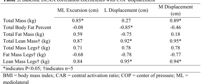
In order to fully answer research question 2 and determine the relationship between the participant characteristics and gait cycle, correlation coefficients between baseline COP variables and subject characteristics were computed (Table 4). No relationships between any of the
variables were observed.
In the subpopulation that completed DEXA scans (n=7), both total body lean mass and lean mass in the legs showed a significant correlation to M and L COP displacement (Table 5). A
Table 3. Change score Pearson’s correlations between COP and GRF variables
Stance Time (s) Ellipse Area (cm2) Sway Path (cm) Sway Velocity (cm/s)
V GRF Maximum 1 -0.25 0.20 0.25 0.27
V GRF Maximum 2 -0.05 0.26 0.04 0.06
ML GRF Maximum 1 -0.27+ 0.20 0.12 0.17
ML GRF Maximum 2 -0.35* 0.21 0.19 0.25
ML GRF Minimum 1 0.05 0.03 -0.00 -0.02
ML GRF Minimum 2 -0.14 0.09 0.27 0.28*
AP GRF Maximum 1 0.12 -0.11 -0.13 -0.14
AP GRF Maximum 2 -0.44* 0.42* 0.38* 0.44*
AP GRF Minimum 1 0.34* -0.15 -0.21 -0.26
AP GRF Minimum 2 0.38* -0.05 -0.06 -0.16
*indicates P<0.05
COP=center of pressure; GRF=ground reaction force; WOMAC= Western Ontario and McMaster Universities Arthritis Index; 1=first 50% of stance phase; 2=second 50% of stance phase; V=vertical; ML=mediolateral; AP=anteroposterior
Table 4. Baseline COP correlations with subject characteristics Maximum Gait Speed (s) Stance Time (s) Ellipse Area (cm2)
Sway Path (cm)
Sway Velocity
(cm/s)
Pain (VAS, cm) -0.09 -0.15 0.16 -0.02 -0.03
WOMAC -0.24 -0.03 0.2 -0.11 -0.14
CAR -0.12 0.10 -0.14 0.07 -0.06
KL Score 0.20 -0.7 0.01 0.00 -0.14
Height (m) -0.17 -0.02 -0.21 0.21 0.08
Weight (kg) 0.04 0.05 -0.08 0.19 -0.04
*indicates P<0.05
23
positive correlation with M displacement shows that the COP has greater movement medially, while L displacement shows that the COP has less movement laterally. An increase in ML excursion would result from an increase in M displacement and a decrease in L displacement.
Table 5. BaselineDEXA correlation coefficients with COP displacement
ML Excursion (cm) L Displacement (cm) M Displacement (cm)
Total Mass (kg) 0.85* 0.27 0.89*
Total Body Fat Percent -0.08 -0.85* -0.46
Total Fat Mass (kg) 0.59 -0.75 0.18
Total Lean Mass† (kg) 0.87 0.92* 0.95*
Total Mass Legs† (kg) 0.71 0.78 0.78
Fat Mass Legs† (kg) -0.68 -0.78 -0.77
Lean Mass Legs† (kg) 0.84 0.95* 0.94*
*indicates P<0.05; †indicates n=5
24
CHAPTER 5: DISCUSSION
The aim of this study was to assess the change in gait speed and COP characteristics following a 4 week intervention and determine their association with participant characteristics. Following intervention, subjects had a significant improvement in pain and WOMAC score, as well as maximum gait speed, assessed using the 20 meter walk test score. Improved GRF scores following intervention included increased maximum and minimum AP GRFs during the second and first half of the gait cycle, respectively. Change score correlations indicated that only
maximum gait speed was inversely related to COP sway velocity. These results contribute to the literature on KOA and the impact of common treatment methods, showing that not only can maximum gait speed improve following intervention, but the health of the effected leg improves since they are able to have higher propulsive and braking forces.
To compare the relationship between gait speed and gait stability, several correlation coefficients were calculated. These were done using the baseline values and change score values with maximum gait speed, stance time, COP sway path, COP sway velocity being the variables of primary interest. Maximum gait speed increased following the intervention, as expected based on another study which included a slightly longer intervention of 6 weeks and showed an
25
Baseline and change score values of gait speed were negatively correlated with baseline and change score values of COP sway velocity. Increased COP sway velocity has been used in previous studies to measure decreased stability10 69. These inverse relationships support other research showing that a slower walking speed is not more stable41 72. The loss of stability indicated with higher COP velocity may be the result of increased ML COP velocity. However, we were unable to differentiate between ML and AP COP velocity in our current study, allowing this to remain speculative and an area for further study.
Although stance time and gait speed changed as expected with stance time decreasing as speed increased, they were not significantly correlated to each other. Both variables were compared to GRF variables to further explore gait changes that occurred. APGRF minimum during the first and second 50% of stance phase were positively correlated with gait speed, supporting previous literature73, and indicating an increased braking force upon initial contact and as weight shifts to the forefoot. As suggested by Peterson et al., an increase in posterior GRF indicates a healthier gait cycle since it is a mechanism by which gait is controlled73. The
maximum MLGRF, which is negatively correlated with stance time, indicates there is a greater medial force. The negative correlation may not be due to an increase in overall GRF, but rather a greater posterior force in the first and second half of stance phase. If the posterior force is a higher relative proportion of the total force, the medial force would consequently decrease.
26
FM is women and LM in men have previously been linked to the presence of KOA22 23. In an exploratory analysis of body composition measure via DEXA (N=7), both total body lean mass and lower extremity lean mass were significantly correlated with an increased L and M COP displacement. A greater M COP displacement value indicates that the COP is further medially, whereas a greater L COP displacement indicates that the COP travels laterally to a lesser extent. Greater medial movement of the COP may not necessarily indicate a decline in COP trajectory. Instead, the increased displacement may suggest that as the lean tissue increases, force production also increases52, and the COP moves to a greater extent because there is now a greater ability to control movement laterally without a loss of balance. Improved balance will consequently result in better physical function and increase quality of life. However, due to a small sample size that completed DEXA scans, we are unable to fully address this question at this time. However, this question should be reevaluated with a larger sample and subjects ranging from healthy controls to severe KOA patients.
The study was limited due to its reliance on the subject walking at a maximum gait speed at each visit. The gait speed selected may be influenced by factors other than the intervention such as fatigue and pain from activities other than TE. Subjects were also asked to self-report for pain and WOMAC scores. Each individual’s interpretation of “mild”, “moderate”, or “severe” may differ on these questionnaires, but the WOMAC has been established as a valid assessment of dysfunction for KOA patients. The implications from the change in COP velocity were also limited because we could not differentiate between ML and AP COP velocity through the Matlab code used.
27
gait speed increases and pain, and WOMAC scores decrease. Other meaningful implications come from the increased maximum AP GRF and the decreased minimum AP GRF, which indicate that the involved limb can withstand higher propulsive and braking forces after
treatment. Further exploration is warranted to determine the implications of FM and LM on the gait cycle in KOA patients. However, this study contributes to the existing literature by
28 APPENDIX A University of North Carolina at Chapel Hill
Consent to Participate in a Research Study Adult Participants
Consent Form Version Date: 7-05-2016 IRB Study # 15-1150
Title of Study: The KNEEhabilitation Study: Improving Disability in Individuals with Knee Osteoarthritis
Principal Investigator: Brian Pietrosimone
Principal Investigator Department: Exercise and Sport Science Principal Investigator Phone number: 919-962-3617
Principal Investigator Email Address: [email protected]
Co-Investigators: Todd Schwartz, Leigh Callahan, Troy Blackburn, Jeffrey Spang, Yvonne Golightly, Daniel Nissman, David Berkoff, Joanne Jordan, Robert Creighton, Daniel Del Gaizo, Ganesh Kamath, Darin Padua, Harry Stafford, Lauren Porras
Funding Source and/or Sponsor: NATIONAL INSTITUTE OF ARTHRITIS AND MUSCULOSKELETAL AND SKIN DISEASES (NIAMS)
Study Contact Telephone Number: 843-2014 Study Contact Email: [email protected]
_________________________________________________________________
What are some general things you should know about research studies?
You are being asked to take part in a research study. To join the study is voluntary.
You may refuse to join, or you may withdraw your consent to be in the study, for any reason, without penalty.
Research studies are designed to obtain new knowledge. This new information may help people in the future. You may not receive any direct benefit from being in the research study. There also may be risks to being in research studies. Deciding not to be in the study or leaving the study before it is done will not affect your relationship with the researcher, your health care provider, or the University of North
Carolina-Chapel Hill (UNC). If you are a patient with an illness, you do not have to be in the research study in order to receive health care.
Details about this study are discussed below. It is important that you understand this information so that you can make an informed choice about being in this research study. You will be given a copy of this consent form. You should ask the researchers named above, or staff members who may assist them, any questions you have about this study at any time.
What is the purpose of this study?
Thigh muscle strength is important for improving function in individuals with knee osteoarthritis. There is evidence that stronger thigh muscles allow people with knee osteoarthritis to be more physically active and to do more activities without pain. One cause of muscle weakness in individuals with knee osteoarthritis stems form the inability of nerves to “turn on” the muscles in the leg. Physical therapy is one method for changing how those leg muscles “turn on”. "There is some evidence that using
29
daily activities changes your ability to “turn on” your thigh muscles as well as how TENS and physical therapy may improve your thigh muscle strength, ability walk and perform activities of daily living, and potentially changes feelings about your knee function. We are evaluating the effect of TENS and physical therapy in people with knee osteoarthritis, who feel some disability related to knee function and have difficulty turning “their muscles on”. You are being asked to be in the study because you have been diagnosed with knee osteoarthritis, you feel some limitations in performing activities because of you have been diagnosed with knee osteoarthritis.
Are there any reasons you should not be in this study?
You should not be in this study if you have: 1) been diagnosed with a cardiovascular condition restricting you from participating in exercise; 2) had a corticosteroid or hyaluronic acid injection in your knee in the past 2-weeks; 3) a pacemaker; 4) a neurodegenerative condition; 5) rheumatoid arthritis; 6) cancer; 7) neural sensory dysfunction over the knee 8) a BMI over 35; 9) history of lower extremity orthopaedic surgery in the past year; 10) a history of a traumatic knee injury in the past 6 months; 11) any history of a total knee arthroplasty (or joint replacement) in either extremity; or 12) a diagnosed, non-reconstructed knee ligament tear. Also you should not be in this study if you need an assistive device, such as a cane or walker, to walk. You should not be in this study if you are currently pregnant or plan to become pregnant during this study.
How many people will take part in this study?
A total of approximately 177 people may be screened to participate in this study while 90 people with knee osteoarthritis from this institution will be enrolled in the intervention portion of the study.
How long will your part in this study last?
The study will consist of a screening/ baseline session, 10 visits of physical therapy over 4-weeks, as well as two follow-up tests at 4 and 8 weeks after beginning your first physical therapy visit. The screening session will last approximately 1 hour. If you qualify for the study and still would like to participate, we will conduct a few extra measurements over an additional 1-hour as part of the baseline session. Each of the 10 physical therapy sessions will last 45-60 minutes. Each of the two follow up testing session will consist of two 1.5 hour sessions at time points occurring at 4 and 8-weeks after your first physical therapy session.
What will happen if you take part in the study?
If you decide to take part in this study, you will be assigned to receive a study group that is provided a TENS unit and physical therapy or a group that only receives physical therapy.
Once included, you will be asked to report to the Sports Medicine Research Laboratory (SMRL)
Laboratory in Fetzer Hall at the University of North Carolina at Chapel Hill for 3 separate testing sessions and to the UNC Meadowmont Physical Therapy Clinic for 10 supervised physical therapy sessions.
30
If you are eligible for the study at this point we will conduct a few additional tests during session one that include knee pain and physical activity surveys, a thigh muscle function assessment, knee range of motion assessment, gait biomechanics assessment and a physical performance assessment.
Testing Sessions 2 and 3 (will last approximately 1.5 hours each) will consist of questionnaires and surveys about your knee function, pain and physical activity as well as the thigh muscle function
assessment, knee range of motion assessment, gait biomechanics assessment and a physical performance assessment. Testing sessions 2 and 3 will take place 4 and 8-weeks after your first physical therapy session.
Body Composition Assessment
During each lab visit, you will have a DEXA full body scan which will measure your fat free mass, fat mass, and bone mineral content. A DEXA uses minimal amounts of radiation, the test lasts 6 minutes and requires you to lie on the table and remain still.
Thigh Muscle Function Assessment
Similar to the screening measurement, you will be asked to perform maximal contractions of the
quadriceps muscle on the front of your thigh. You will be seated and asked to kick out your leg as hard as you can against a device that measures how much force you can produce. During the contraction, a very brief (less than 1/1,000th of a second) pulse of electricity will be sent to your quadriceps muscle. This pulse of electricity will allow us to measure muscle function and is similar to a strong “carpet shock”. You will be allowed to perform at least 2 practice trials to become comfortable and familiar with the electrical stimulus, after which 3 trials will be recorded. You will similarly be asked to push your heel into the pad without an electric pulse as you sit in the chair and side lie on a table.
Range of Motion Testing
We will test how far you are able to bend and straighten your knee as you lie down on a table.
Gait Biomechanics Assessment
You be asked to walk forward along a 20 foot walkway at a comfortable, self-selected speed over a device called a force plate that is embedded in the walkway. You will perform at least 5 practice trials to ensure that you are comfortable walking over the force plate with a natural stride and without “aiming” for the force plate. We will put small reflective markers on different parts of your legs which will allow cameras in the SMRL to track how your knees move while you walk.
Performance Testing
You will be asked to do three different performance tests.
The first is a chair stand test, in which you will start seated in a chair and we will count how many times you can stand up and sit down in a 30 second period. Next, we will determine how fast you can
comfortable walk forty-meters. We will time you 3 times (with at least a 1-minute rest period in between each trial). Finally we will test how fast you can climb 10 stairs at a fast but comfortable pace and you will be timed with a stopwatch. We will time 3 trials with at least a 1-minute rest period in between each trial.
Physical Therapy Intervention at the UNC Meadowmont Clinic
31
will ask that you do not tell the investigator that will be collecting data from you in the SMRL what intervention you received. You can always contact the study coordinator Brittney Luc, or the Principal Investigator Brian Pietrosimone about the TENS unit.
Physical Therapy
All groups will complete a standardized rehabilitation program. You will receive therapeutic exercise 2 to 3 times per week for a total of 10 sessions over a 4 week period. Ten physical therapy sessions over a 4-week period is the current standard of care for patients with knee osteoarthritis at our facility. A lower extremity physical assessment will be conducted bilaterally for each participant to identify deficits and establish clinical baseline measures prior to beginning rehabilitation and following the 4 -week
intervention. The goals of the therapeutic exercise program are to increase thigh muscle strength and your ability to do activities of daily living.
Being Randomized to a TENS group with Physical Therapy
If you are randomized into a TENS group, you will receive a Select System TENS unit, self-adhesive electrodes and batteries. A licensed health care professional will assist you in applying the electrodes on the knee joint and will educate you on how to operate the TENS unit. An instruction manual will be provided to you for reference purposes explaining operation of the device. The sensation of the electrical stimulation may vary among people. The strength of the perceived stimulation will vary among subjects from little or nothing to modest tingling, depending on the dosing you are randomized to receive. You will also complete the standardized physical therapy program under the supervision of your physical therapist at the UNC Meadowmont Clinic. The TENS unit will be worn during all therapeutic exercise sessions and at least 8 hours per day when you are the most active. We will ask you to keep a log that will indicate how long you wore the TENS unit each day. We will collect the log from you at the end of each week as well as ask you how well you are tolerating the intervention each week (you will indicate this on a questionnaire form).
Not Being Randomized to a TENS group with Physical Therapy
If assigned, you will still receive the standard of care of therapeutic strengthening exercises for the 10 sessions over the 4-week period. You will not be provided with any TENS unit.
What are the possible benefits from being in this study?
Research is designed to benefit society by gaining new knowledge. The benefits to you from being in this study may be that you may increase the strength of your leg muscles, which may result in less knee pain and less disability compared to what you may have experienced before you started the study.
What are the possible risks or discomforts involved from being in this study?
32
are comfortable with it before the testing procedures begin. There is also a possibility of acquiring some mild, transient skin irritation from the sticky TENS electrodes if you are randomized to the TENS group. There may be uncommon or previously unknown risks. You should report any problems to the principal investigator, Brian Pietrosimone.
What if we learn about new findings or information during the study?
You will be given any new information gained during the course of the study that might affect your willingness to continue your participation.
How will information about you be protected?
Any information obtained in connection with this research study that can be linked to you will remain confidential. You will be identified only by a subject identification number. A code list that associates your name and information with a specific subject identification number will be kept under lock-and-key on a password-protected computer in the Neuromuscular Research Laboratory. Only the research team will have access to this information. Participants will not be identified in any report or publication about this study. Although every effort will be made to keep research records private, there may be times when federal or state law requires the disclosure of such records, including personal information. This is very unlikely, but if disclosure is ever required, UNC-Chapel Hill will take steps allowable by law to protect the privacy of personal information. In some cases, your information in this research study could be reviewed by representatives of the University, research sponsors, or government agencies (for example, the FDA) for purposes such as quality control or safety.
What will happen if you are injured by this research?
All research involves a chance that something bad might happen to you. This may include the risk of personal injury. In spite of all safety measures, you might develop a reaction or injury from being in this study. If such problems occur, the researchers will help you get medical care, but any costs for the medical care will be billed to you and/or your insurance company. The University of North Carolina at Chapel Hill has not set aside funds to pay you for any such reactions or injuries, or for the related medical care. You do not give up any of your legal rights by signing this form.
What if you want to stop before your part in the study is complete?
You can withdraw from this study at any time, without penalty. The investigators also have the right to stop your participation at any time. This could be because you have had an unexpected reaction, or have failed to follow instructions, or because the entire study has been stopped.
Will you receive anything for being in this study?
You will receive all physical therapy associated with this study free of charge.
You will be compensated a total of $100 for attending all three outcomes assessments. You will not be compensated for the screening portion of the study. After screening into the study, you will be
compensated $20 for baseline measurements and $40 for the 4-week and 8-week post-baseline testing sessions. You will be compensated after your last completed outcome session study. Compensation will be in the form of a $100 Visa gift card.
Will it cost you anything to be in this study?
It will not cost you anything to be in this study. All physical therapy sessions will be fully reimbursed by the grant supported by the National Institutes of Health (NIAMS).
Who is sponsoring this study?
33 What if you have questions about this study?
You have the right to ask, and have answered, any questions you may have about this research. If you have questions, complaints, or concerns about the study; or if a research-related injury occurs, you should contact the researchers listed on the first page of this form.
A description of this clinical trial will be available on www.clinicaltrials.gov, as required by U.S. Law. This website will not include information that can identify you. At most, the website will include a summary of the results. You can search this website at any time.
What if you are a UNC employee?
Taking part in this research is not a part of your University duties, and refusing will not affect your job. You will not be offered or receive any special job-related consideration if you take part in this research
What if you have questions about your rights as a research participant?
All research on human volunteers is reviewed by a committee that works to protect your rights and welfare. If you have questions or concerns about your rights as a research subject, or if you would like to obtain information or offer input, you may contact the Institutional Review Board at 919-966-3113 or by email to [email protected].
Participant’s Agreement:
I have read the information provided above. I have asked all the questions I have at this time. I voluntarily agree to participate in this research study.
______________________________________________________ Signature of Research Participant
____________________ Date
______________________________________________________ Printed Name of Research Participant
______________________________________________________ Signature of Research Team Member Obtaining Consent
____________________ Date
______________________________________________________ Printed Name of Research Team Member Obtaining Consent
34
REFERENCES
1. Jordan JM, Helmick CG, Renner JB, et al. Prevalence of knee symptoms and radiographic and symptomatic knee osteoarthritis in African Americans and Caucasians: the Johnston County Osteoarthritis Project. The Journal of Rheumatology 2007;34(1):172-80. 2. Dillon CF, Rasch EK, Gu Q, et al. Prevalence of knee osteoarthritis in the United States:
arthritis data from the Third National Health and Nutrition Examination Survey 1991-94. The Journal of Rheumatology 2006;33(11):2271-79.
3. Arizono S, Taniguchi H, Nishiyama O, et al. Improvements in Quadriceps Force and Work Efficiency are Related to Improvements in Endurance Capacity Following Pulmonary Rehabilitation in COPD Patients. Internal Medicine 2011;50(21):2533-39.
4. Mador MJ, Bozkanat E, Kufel TJ. Quadriceps Fatigue After Cycle Exercise in Patients With COPD Compared With Healthy Control Subjects. Chest 2003;123(4):1104-11.
5. Shrikrishna D, Patel M, Tanner RJ, et al. Quadriceps wasting and physical inactivity in patients with COPD. European Respiratory Journal 2012;40(5):1115-22.
6. Nüesch E, Dieppe P, Reichenbach S, et al. All cause and disease specific mortality in patients with knee or hip osteoarthritis: population based cohort study. BMJ: British Medical Journal 2011;342(7798):638-38.
7. Swallow EB, Reyes D, Hopkinson NS, et al. Quadriceps strength predicts mortality in patients with moderate to severe chronic obstructive pulmonary disease. Thorax 2007;62 (2):115-20.
8. Kalyani RR, Tra Y, Yeh HC, et al. Quadriceps Strength, Quadriceps Power, and Gait Speed in Older U.S. Adults with Diabetes Mellitus: Results from the National Health and Nutrition Examination Survey, 1999–2002. Journal of the American Geriatrics Society
2013;61(5):769-75.
9. Guccione AA, Felson DT, Anderson JJ, et al. The effects of specific medical conditions on the functional limitations of elders in the Framingham Study. American Journal of Public Health 1994;84(3):351-58.
10. Choy NL, Brauer S, Nitz J. Changes in postural stability in women aged 20 to 80 years. Journals of Gerontology - Series A Biological Sciences and Medical Sciences 2003;58(6):525-30.
11. Sattin RW, Sattin RW, Lambert Huber DA, et al. The incidence of fall injury events among the elderly in a defined population. American Journal of Epidemiology
1990;131(6):1028-37.
35
13. Kim H-S, Yun DH, Yoo SD, et al. Balance Control and Knee Osteoarthritis Severity. Annals of Rehabilitation Medicine 2011;35(5):701-09.
14. Hassan BS, Mockett S, Doherty M. Static postural sway, proprioception, and maximal voluntary quadriceps contraction in patients with knee osteoarthritis and normal control subjects. Annals of the Rheumatic Diseases 2001;60(6):612-18.
15. Mohammadi F, Taghizadeh S, Ghaffarinejad F, et al. Proprioception, dynamic balance and maximal quadriceps strength in females with knee osteoarthritis and normal control subjects. International Journal of Rheumatic Diseases 2008;11(1):39-44.
16. Chiu M-C, Wu H-C, Chang L-Y. Gait speed and gender effects on center of pressure progression during normal walking. Gait and Posture 2013;37(1):43-48.
17. Chiu M-C, Wu H-C, Chang L-Y, et al. Center of pressure progression characteristics under the plantar region for elderly adults. Gait and Posture 2013;37(3):408-12.
18. De Cock A, Vanrenterghem J, Willems T, et al. The trajectory of the centre of pressure during barefoot running as a potential measure for foot function. Gait & Posture 2008;27(4):669-75.
19. Cau N, Cimolin V, Galli M, et al. Center of pressure displacements during gait initiation in individuals with obesity. JOURNAL OF NEUROENGINEERING AND
REHABILITATION 2014;11(1):82-82.
20. Fan Y, Li Z, Han S, et al. The influence of gait speed on the stability of walking among the elderly. Gait & Posture 2016;47:31-36.
21. Ertürk C, Altay MA, Sert C, et al. The body composition of patients with knee osteoarthritis: relationship with clinical parameters and radiographic severity. Aging Clinical and Experimental Research 2015;27(5):673-79.
22. Visser AW, de Mutsert R, Loef M, et al. The role of fat mass and skeletal muscle mass in knee osteoarthritis is different for men and women: the NEO study. Osteoarthritis and Cartilage 2013;22(2):197-202.
23. Ho-Pham LT, Lai TQ, Mai LD, et al. Body Composition in Individuals with Asymptomatic Osteoarthritis of the Knee. Calcified Tissue International 2016;98(2):165-71.
24. Baert IAC, Jonkers I, Staes F, et al. Gait characteristics and lower limb muscle strength in women with early and established knee osteoarthritis. Clinical Biomechanics
2013;28(1):40-47.
36
26. Pietrosimone BG, Saliba SA, Hart JM, et al. Contralateral effects of disinhibitory tens on quadriceps function in people with knee osteoarthritis following unilateral treatment. North American journal of sports physical therapy : NAJSPT 2010;5(3):111-21. 27. Pietrosimone BG, Pietrosimone BG, Saliba SA, et al. Effects of Transcutaneous Electrical
Nerve Stimulation and Therapeutic Exercise on Quadriceps Activation in People With Tibiofemoral Osteoarthritis. The journal of orthopaedic and sports physical therapy 2011;41(1):4-12.
28. Astephen JL, Deluzio KJ, Caldwell GE, et al. Gait and neuromuscular pattern changes are associated with differences in knee osteoarthritis severity levels. Journal of Biomechanics 2008;41(4):868-76.
29. Pietrosimone BG, Saliba SA. Changes in voluntary quadriceps activation predict changes in quadriceps strength after therapeutic exercise in patients with knee osteoarthritis. Knee 2012;19(6):939-43.
30. Takacs J, Carpenter MG, Garland SJ, et al. Factors Associated With Dynamic Balance in People With Knee Osteoarthritis. Archives of Physical Medicine and Rehabilitation 2015;96(10):1873-79.
31. Pua YH, Liang Z, Ong PH, et al. Associations of knee extensor strength and standing balance with physical function in knee osteoarthritis. Arthritis Care & Research
2011;63(12):1706-14.
32. Levinger P, Menz HB, Wee E, et al. Physiological risk factors for falls in people with knee osteoarthritis before and early after knee replacement surgery. Knee Surgery, Sports Traumatology, Arthroscopy 2011;19(7):1082-89.
33. Hopkins JT, Hopkins JT, Ingersoll CD. Arthrogenic muscle inhibition: A limiting factor in joint rehabilitation. Journal of sport rehabilitation 2000;9(2):135-59.
34. French HP, Fitzpatrick M, FitzGerald O. Responsiveness of physical function outcomes following physiotherapy intervention for osteoarthritis of the knee: an outcome comparison study. Physiotherapy 2011;97(4):302-08.
35. Ortman JM, Velkoff VA, Hogan H. An aging nation: the older population in the United States. United States Department of Commerce 2014:1-28.
36. Murray MP, Drought AB, Kory RC. Walking Patterns of Normal Men. The Journal of Bone & Joint Surgery 1964;46(2):335-60.
37. Cromwell RL, Newton RA. Relationship between Balance and Gait Stability in Healthy Older Adults. Journal of Aging and Physical Activity 2004;12(1):90-100.
37
39. Schrager MA, Kelly VE, Price R, et al. The effects of age on medio-lateral stability during normal and narrow base walking. Gait & Posture 2008;28(3):466-71.
40. England SA, Granata KP. The influence of gait speed on local dynamic stability of walking. Gait & Posture 2007;25(2):172-78.
41. Bruijn SM, van Dieën JH, Meijer OG, et al. Is slow walking more stable? Journal of Biomechanics 2009;42(10):1506-12.
42. Toivanen AT, Heliövaara M, Impivaara O, et al. Obesity, physically demanding work and traumatic knee injury are major risk factors for knee osteoarthritis-a population-based study with a follow-up of 22 years. Rheumatology 2010;49(2):308-14.
43. Holliday KL, McWilliams DF, Maciewicz RA, et al. Lifetime body mass index, other anthropometric measures of obesity and risk of knee or hip osteoarthritis in the GOAL case-control study. Osteoarthritis and Cartilage 2011;19(1):37-43.
44. Grotle M, Hagen KB, Natvig B, et al. Obesity and osteoarthritis in knee, hip and/or hand: An epidemiological study in the general population with 10 years follow-up. BMC
Musculoskeletal Disorders 2008;9(1):132-32.
45. Lohmander LS, De Verdier MG, Rollof J, et al. Incidence of severe knee and hip osteoarthritis in relation to different measures of body mass: A population-based prospective cohort study. Annals of the Rheumatic Diseases 2009;68(4):490-96. 46. Sowers MF, Yosef M, Jamadar D, et al. BMI vs. body composition and radiographically
defined osteoarthritis of the knee in women: a 4-year follow-up study. Osteoarthritis and Cartilage 2008;16(3):367-72.
47. Lee R, Kean WF. Obesity and knee osteoarthritis. Inflammopharmacology 2012;20(2):53-58. 48. Purcell S, Thornberry R, Elliott SA, et al. Body Composition, Strength, and Dietary Intake of
Patients with Hip or Knee Osteoarthritis. CANADIAN JOURNAL OF DIETETIC PRACTICE AND RESEARCH 2016;77(2):98-102.
49. Smith-Ryan AE, Mock MG, Ryan ED, et al. Validity and reliability of a 4-compartment body composition model using dual energy x-ray absorptiometry-derived body volume.
Clinical nutrition (Edinburgh, Scotland) 2016.
50. Lohman M, Tallroth K, Kettunen JA, et al. Reproducibility of dual-energy x-ray absorptiometry total and regional body composition measurements using different scanning positions and definitions of regions. Metabolism 2009;58(11):1663-68.
38
52. Pisciottano MVC, Pinto SS, Szejnfeld VL, et al. The relationship between lean mass, muscle strength and physical ability in independent healthy elderly women from the community. The journal of nutrition, health & aging 2014;18(5):554-58.
53. Skelton DA, Skelton DA, Greig CA, et al. Strength, power and related functional ability of healthy people aged 65-89 years. Age and ageing 1994;23(5):371-77.
54. Mizner RL, Petterson SC, Stevens JE, et al. Early quadriceps strength loss after total knee arthroplasty: The contributions of muscle atrophy and failure of voluntary muscle activation. Journal of Bone and Joint Surgery - Series A 2005;87(5):1047-53. 55. Lewek MD, Rudolph KS, Snyder-Mackler L. Quadriceps femoris muscle weakness and
activation failure in patients with symptomatic knee osteoarthritis. Journal of Orthopaedic Research 2004;22(1):110-15.
56. Skwara A, Peterlein CD, Tibesku CO, et al. Changes of gait patterns and muscle activity after intraarticular treatment of patients with osteoarthritis of the knee. A prospective, randomised, doubleblind study. Knee 2009;16(6):466-72.
57. Tang SF, Chen CP, Chen MJ, et al. Changes in sagittal ground reaction forces after intra-articular hyaluronate injections for knee osteoarthritis. Archives of Physical Medicine and Rehabilitation 2004;85(6):951-55.
58. Leopold SS, Redd BB, Warme WJ, et al. Corticosteroid Compared with Hyaluronic Acid Injections for the Treatment of Osteoarthritis of the Knee A Prospective, Randomized Trial. The Journal of Bone & Joint Surgery 2003;85(7):1197-203.
59. Neogi T, Felson D, Niu J, et al. Association between radiographic features of knee osteoarthritis and pain: results from two cohort studies. BMJ: British Medical Journal 2009;339(7719):498-501.
60. Liikavainio T, Bragge T, Hakkarainen M, et al. Gait and muscle activation changes in men with knee osteoarthritis. The Knee 2010;17(1):69-76.
61. Knoop J, Steultjens MPM, Roorda LD, et al. Improvement in upper leg muscle strength underlies beneficial effects of exercise therapy in knee osteoarthritis: secondary analysis from a randomised controlled trial. PHYSIOTHERAPY 2015;101(2):171-77.
62. Stokes M, Young A. The contribution of reflex inhibition to arthrogenous muscle weakness. Clinical science (1979) 1984;67(1):7-14.
63. Pietrosimone BG, Hart JM, Saliba SA, et al. Immediate effects of transcutaneous electrical nerve stimulation and focal knee joint cooling on quadriceps activation. Medicine and Science in Sports and Exercise 2009;41(6):1175-81.
39
65. Palmer S, Domaille M, Cramp F, et al. Transcutaneous Electrical Nerve Stimulation as an Adjunct to Education and Exercise for Knee Osteoarthritis: A Randomized Controlled Trial. Arthritis care & research (2010) 2014;66(3):387-94.
66. Pietrosimone BG, Hertel J, Ingersoll CD, et al. Voluntary Quadriceps Activation Deficits in Patients with Tibiofemoral Osteoarthritis: A Meta-Analysis. PM&R 2011;3(2):153-62. 67. Pietrosimone BG, McLeod MM, Lepley AS. A Theoretical Framework for Understanding
Neuromuscular Response to Lower Extremity Joint Injury. Sports Health 2012;4 (1):31-35.
68. Kang MK, Nam BR, Lee YS, et al. Relationship between the Application of TENS to the Lower Limbs and Balance of Healthy Subjects. JOURNAL OF PHYSICAL THERAPY SCIENCE 2013;25(9):1079-81.
69. Park J, Seo D, Choi W, et al. The Effects of Exercise with TENS on Spasticity, Balance, and Gait in Patients with Chronic Stroke: A Randomized Controlled Trial. Medical Science Monitor : International Medical Journal of Experimental and Clinical Research
2014;20:1890-96.
70. Chang S-Y, Ssu-Yu C, Yi J. Exercise Alters Gait Pattern but Not Knee Load in Patients with Knee Osteoarthritis. BioMed research international 2016;2016:1-12.
71. Börjesson M, Weidenhielm L, Elfving B, et al. Tests of walking ability at different speeds in patients with knee osteoarthritis. Physiotherapy Research International 2007;12 (2):115-21.
72. Espy DD, Yang F, Bhatt T, et al. Independent influence of gait speed and step length on stability and fall risk. Gait & Posture 2010;32(3):378-82.