Endoscopic management of bile leaks after liver
transplantation: An analysis of two high-volume
transplant centers
Oriol Sendino
1,2, Alejandro Ferna´ndez-Simon
1,3, Ryan Law
4,
Barham Abu Dayyeh
5, Michael Leise
5, Karina Chavez-Rivera
1,2,
Henry Cordova
1,2, Jordi Colmenero
2,6, Gonzalo Crespo
2,6,
Cristina Rodriguez de Miguel
1,2, Constantino Fondevila
2,7, Josep Llach
1,2,
Miquel Navasa
2,6, Todd Baron
8and Andre´s Ca´rdenas
1,2,6Abstract
Background:Bile leak after liver transplantation (LT) is commonly treated with endoscopic retrograde cholangiopancreato-graphy (ERCP); however, there are limited data regarding the optimal treatment strategy.
Objective:We aimed to examine the role of ERCP in LT recipients with bile leaks at two large institutions.
Methods: We reviewed all ERCPs performed in LT recipients with bile leak and duct-to-duct biliary anastomosis at two high-volume transplant centers.
Results:Eighty patients were included. Forty-seven (59%) patients underwent ERCP with plastic stent placement (with or without sphincterotomy) and 33 patients (41%) underwent sphincterotomy alone. Complete resolution was obtained in 94% of the stent group vs. 58% of the sphincterotomy group (p<0.01). There was no difference in three-month survival among both groups. Percutaneous transhepatic therapy and surgery were required in 4% and 6% in the stent group vs. 12% and 42% in the sphincterotomy group, respectively (p¼0.22 andp<0.001). The only predictive factor of bile leak resolution was stent placement.
Conclusion:ERCP with plastic stent placement is highly successful and more effective than sphincterotomy alone for post-LT bile leak treatment. These results indicate that ERCP and plastic stent placement should be considered the standard of care for the treatment of bile leaks in LT.
Keywords
Bile leak, liver transplantation, biliary complications, endoscopic retrograde cholangiopancreatography, sphincterotomy, biliary strictures, plastic stents, biliary stenting
Received: 20 February 2017; accepted: 6 May 2017
Introduction
Biliary adverse events constitute an important cause of morbidity and mortality in patients after liver trans-plantation (LT). Among the different types of biliary adverse events after LT, bile leak is the second most
1GI/Endoscopy Unit, Institut de Malalties Digestives i Metabo`liques, Hospital Clı´nic, and University of Barcelona, Spain
2
Institut d’Investigacions Biome`diques August Pi-Sunyer (IDIBAPS), Ciber de Enfermedades Hepa´ticas y Digestivas (CIBEREHD), Barcelona, Spain 3
GI Unit, Hospital Sant Joan Despı´ Moises Broggi, Barcelona, Spain 4
Department of Medicine and Division of Gastroenterology, University of Michigan, MI, USA
5Division of Gastroenterology and Hepatology, Mayo Clinic, Rochester, MN, USA
6Liver Unit, Institut de Malalties Digestives i Metabo`liques, Hospital Clı´nic and University of Barcelona, Spain
7General and Digestive Surgery, Institut de Malalties Digestives i Metabo`liques, Hospital Clı´nic, and University of Barcelona, Spain 8
Division of Gastroenterology and Hepatology, University of North Carolina, Chapel Hill, NC, USA
Corresponding author:
Andre´s Ca´rdenas, Institut de Malalties Digestives i Metaboliques, University of Barcelona, Hospital Clinic-Villarroel 170, Esc 3-2, 08036 Barcelona, Spain.
Email: [email protected]
O.S. and A.F.-S. contributed equally to this manuscript.
United European Gastroenterology Journal 2018, Vol. 6(1) 89–96
!Author(s) 2018 Reprints and permissions:
frequent with an incidence of 2% to 25%.1–7Bile leaks are classified as anastomotic, T-tube-related, cystic duct-related or, in the case of living donor liver transplants (LDLT), cut-surface-related.6 Common risk factors for the development of bile leaks are related to surgical technique, hepatic artery thrombosis, LT from donors after cardiac death, ABO mismatch, pro-longed warm and cold ischemia times, and T-tube use.8 The standard of care for the treatment of bile leaks after LT is not well established. Patients with a peri-T-tube leak can sometimes be managed conservatively by keeping the T-tube drainage open for a prolonged period of time.9Endoscopic retrograde cholangiopan-creatography (ERCP) is also very effective and high-resolution rates are near 90%.1–4,6–8 A variety of ERCP techniques have been described for managing leaks, including: nasobiliary drainage,10–13 sphincterot-omy alone,14–17plastic biliary stent placement with or without out sphincterotomy,3,4,6,7,18–24 and placement of fully covered self-expandable metallic stents (FCSEMS).25–27 Regardless of method used, the sug-gested mechanism of healing relates to the equalization of pressures in the bile duct and duodenum, which allows antegrade bile flow into the duodenum.8,10 Nevertheless, the optimal endoscopic approach has not yet been established and to date there are very few studies that compare the different endoscopic treat-ment options. In addition, there are no data that define factors predictive of endoscopic success in patients with post-LT bile leaks. The aim of this analysis was to evaluate data from two large institutions to determine the role of ERCP in the treatment of bile leaks follow-ing LT.
Materials and methods
This study was approved by the Ethical and Institutional Review Board at the Hospital Clinic of Barcelona (Barcelona, Spain) and Mayo Clinic (Rochester, MN, USA). The protocol, Evaluation of Endoscopic Therapy of Bile Leaks after Liver Transplantation, was approved at both institutions in May and August 2015. The study protocol conforms to the ethical guidelines of the 1975 Declaration of Helsinki as reflected in a prior approval by the institu-tions’ human research committee. Informed consent of the procedures was obtained from all patients included in the study. LT recipients with clinical or radiologic suspicion of a bile leak referred for ERCP were included. We reviewed all ERCPs in patients following LT at Mayo Clinic from July 2000 to June 2013 and at Hospital Clinic from January 2003 through January 2015. Mayo Clinic performs between 100 and 120 LTs and approximately 2000 ERCPs yearly. Hospital Clinic performs between 80 and 90 LTs and
approximately 450 ERCPs yearly. Data were col-lected and entered after each case. The electronic med-ical records and endoscopy database of both institutions were accessed to abstract demographic, clinical, surgical, and endoscopic data. All procedures were performed with sedation, with levels from moder-ate to general anesthesia. All patients received intravenous broad-spectrum prophylactic antibiotics. Administration of pharmacologic agents and pancre-atic stent placement for prevention of post-ERCP pan-creatitis were not routinely performed.
ERCP was performed using standard techniques. After cannulation, cholangiography was performed to confirm the leak. If necessary, an occlusion cholangio-gram using a balloon catheter was performed to deter-mine the leak site. After confirmation of the bile leak, endoscopic intervention (complete sphincterotomy or stent with or without complete sphincterotomy) was performed at the proceduralist’s discretion. Biliary stents were placed across the site of the bile leak, when technically feasible; in some cases patients were treated with biliary sphincterotomy alone.
Definitions
Bile leaks were diagnosed based on the following criteria: (1) clinical symptoms (i.e. abdominal pain, ascites, fever and/or jaundice); (2) the presence of a new fluid collection consistent with a leak on cross-sec-tional imaging; (3) increasing or persistent bilious output from an intra-abdominal drain; (4) extravasa-tion of contrast seen on T-tube cholangiography. Intrahepatic leaks were defined as extravasation of contrast due to bile duct rupture and extravasation of bile into the hepatic parenchyma. Patients with a peri-T-tube leak were referred for an ERCP only if the condition persisted after leaving the T-tube to open drainage. We only included patients with a sus-pected bile leak confirmed by ERCP.
biliary drainage (PTBD) and were categorized as fail-ures. Definitions of individual adverse events and their severity after ERCP (i.e. pancreatitis, cholangitis, hem-orrhage, perforation) were defined by criteria as estab-lished by Cotton et al.28,29Mild events were considered when hospitalization was prolonged by two to three days, moderate by 4–10 days, and severe by more than 10 days.
Statistical analysis
Patient demographics, clinical and procedure data and outcomes were analyzed.2 or Fisher exact test were used for categorical variables and the Studentt-test for continuous variables. Univariate analysis was con-ducted to explore the relationships between the patients’ characteristics, the type of transplantation, the presence of a T-tube, immunosuppression therapy, the type of leak, the location of the leak, the presence of a stricture, resolution of the bile leak, the time of reso-lution and the need for percutaneous transhepatic chol-angiography (PTC) or surgery. Independent factors associated with resolution of bile leak were studied using a stepwise multivariate logistic regression model that used initial inclusion criteria with a significance of
p0.05, using SPSS statistical packages (version 20.0; SPSS Inc, Chicago, IL). For this purpose, patients with a resolved bile leak (resolution group) were compared with those who presented with persistent bile leak after follow-up (persistent leak group), as defined previously. Ap0.05 was considered statistically significant.
Results
A total of 80 patients were included, 45 at Hospital Clinic and 35 at the Mayo Clinic (mean age 55 years, 73% male). The baseline characteristics of patients are shown in Table 1. Overall, 75% were recipients of a cadaveric liver and 24% were living donor recipients. Two-thirds of the patients had a T-tube after LT. The incidence of known risk factors for bile leak was low: hepatic artery thrombosis (9%), donors after cardiac death (5%) and ABO blood group mismatch (1%).
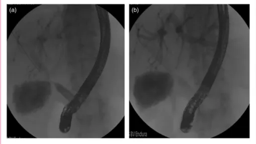
Endoscopic findings are shown in Table 2. The loca-tion of bile leaks was as follows: biliary anastomosis (45%), T-tube (34%), intrahepatic (10%), cystic duct remnant (9%), cut surface (2%). A concomitant anas-tomotic biliary stricture was identified in 28% of patients (Figure 1). The median time from LT to ERCP was 32.25 (16–73.5) days. Biliary stent place-ment was performed in 47 patients, 41 with sphincter-otomy and six without sphinctersphincter-otomy. The median number of biliary stents placed per case was one. All were plastic stents and the majority (31/47) were 10F stents. Thirty-three patients (41%) underwent biliary
sphincterotomy alone (30 at Hospital Clinic and three at Mayo Clinic-Rochester). The mean number of ERCPs performed per patient was 1.8. The majority of patients (60, 75%) required only one ERCP proced-ure for bile leak resolution and 19 (23.8%) required two ERCP procedures. One patient with a non-anastomotic bile leak required three ERCPs. There were 12 cases of a failed attempt to place a biliary stent. Of these 12
Table 1. Patient baseline characteristics.
PATIENT CHARACTERISTICS n(%)
Gender (M/F) 58/22 (72.5%/22.5%)
Age (years) 54.710.3
Indication/etiology of LT
HCV 33 (41.3%)
Alcohol 14 (17.5%)
HCC 23 (28.8)
Others 10 (12.5%)
Type of LT
DDLT 60 (75%)
LDLT 19 (24%)
Split 1 (1%)
T-tube 60 (75%)
Risk factors for bile leakage
Hepatic artery thrombosis. 7 (9%)
Donor after cardiac death 4 (5%)
ABO mismatch 1 (1%)
Immunosuppression therapy
Tacrolimus 61 (76.3%)
Cyclosporine 9 (11.3%)
Prednisone 75 (93.8%)
Mycophenolic acid 48 (60%)
Sirolimus 5 (6.3%)
M: male; F: female; LT: liver transplant; HCV: hepatitis C virus; HCC: hepa-tocellular carcinoma; DDLT: deceased donor liver transplant; LDLT: living donor liver transplant.
Table 2. Endoscopic findings.
TYPE OF LEAK
Anastomotic leak 36 (45%)
Peri-T-tube leak 27 (34%)
Spontaneous 13 (16%)
After removal of T-tube 14 (17%)
Intrahepatic 8 (10%)
Cystic 7 (9%)
Cut surface leak 2 (2%)
cases in two the stent could not be placed because of a large disruption and inability to pass the guidewire into a proximal bile duct. In the remaining 10 cases the stent was not placed because of concomitant stricture that could not be traversed with a guidewire in order to place a stent.
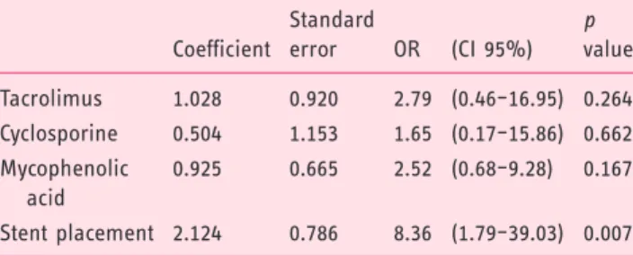
Factors associated with bile leak resolution were analyzed with univariate and multivariate ana-lysis as shown in Table 3 and 4. The only predictive factor of bile leak resolution on the multivariate ana-lysis was ERCP with stent placement (odds ratio, 8.36; 95% confidence interval, 1.79–39.03; p¼0.007)
Figure 1. Characteristic findings of a bile leak with an anastomotic stricture. (a) Bile leak and anastomotic stricture. (b) Stent placement across the bile leak site.
Table 3. Factors associated with bile leak resolution.
Resolution group (n¼63)
Persistent leak group
(n¼17) pvalue
Univariate analysis
Sex (M/F) 45/18 13/4 NS
Arterial thrombosis rate (n(%)) 5 (7.9%) 2 (11.8%) NS
ABO mismatch rate (n(%)) 0 0 1 (5.9%) NS
LDLT (n(%)) 3 (4.8%) 1 (5.9) NS
HCV (n(%)) 27 (42.9%) 6 (35.3%) NS
HCC (n(%)) 19 (30.2%) 4 (23.5) NS
Use of T-tube (n(%)) 48 (76.2%) 12 (70.6%) NS
Tacrolimus (n(%)) 51 (81%) 10 (58.8%) 0.06
Cyclosporine (n(%)) 5 (7.9%) 4 (23.5%) 0.09
Sirolimus (n(%)) 3 (4.8%) 2 (11.8%) NS
Mycophenolic acid (n(%)) 43 (68.3%) 5 (29.4%) .005
Corticosteroids (n(%)) 59 (93.7%) 16 (94.1%) NS
Anastomotic location (n(%)) 26 (41.3%) 10 (58.8%) NS
Anastomotic stricture (n(%)) 15 (23.8%) 7 (41.2%) NS
Time of LT>3 months (n(%)) 12 (19%) 2 (11.8%) NS
Stent placement (n(%)) 44 (69.8%) 3 (17.6%) <0.001
(Table 4). ERCP and patient-related outcomes are out-lined in Table 5. Overall resolution of bile leaks with ERCP therapy was observed in 63 out of 80 (79%) patients. The resolution rate per location was: anasto-motic leak (72.2%), T-tube-related leak (88.9%), intra-hepatic (87.5%), cystic remnant leak (71.4%) and cut surface leak (50%), (Supplementary table). Successful resolution of bile leak occurred in 93.6% of patients in the biliary stent group and 57.6% of the sphincterot-omy group (p<0.01). There was no difference in three-month survival between groups.
Failure of ERCP therapy occurred in 14 (42.4%) patients in the sphincterotomy group and three patients in the stent group (6.4%). In these cases, two patients underwent PTBD, two patients underwent PTBD plus surgery and 11 underwent surgery without prior PTBD. In two patients no further therapy was performed. Eight (10%) patients developed post-ERCP adverse events: mild acute pancreatitis (n¼5), mild bleeding (n¼2), and mild pancreatitis with concomitant cholan-gitis (n¼1). All adverse events were managed conservatively.
Discussion
We sought to analyze ERCP-guided treatment approaches for patients with bile leaks after LT. The results of this analysis from two large academic medical centers indicate that ERCP therapy with placement of a biliary plastic stent (with or without biliary sphincter-otomy) is highly successful and significantly more effective than biliary sphincterotomy alone.
Bile leaks are the second most common biliary adverse event following LT and constitute significant morbidity for LT recipients. In addition, bile leaks are considered an independent risk factor for the development of early or late anastomotic biliary stric-tures and thus require a prompt, safe and highly effect-ive therapy.8,30–32In this analysis 75% of patients had T-tube placement. T-tubes have been routinely placed as a prophylactic measure for anastomotic stricture development. However, the results of several compara-tive studies, systematic reviews and meta-analyses suggest no major differences in the incidence of biliary complications, and the current trend has favored more the abandonment of the use of T-tubes after LT in most centers.33 Most bile leaks can be resolved non-opera-tively with early intervention. The most widely accepted treatment approach in patients with a duct-to-duct bil-iary anastomosis is early ERCP-guided endoscopic therapy.1–4,6–8 ERCP-guided endoscopic therapy of bile leaks can be performed with a combination of bil-iary sphincterotomy and plastic stent placement or with sphincterotomy alone. Some authors propose the use of biliary sphincterotomy alone as it is easy to perform and patients do not require a subsequent ERCP for stent removal; however, most of the available data sup-porting this practice stem from patients with bile leaks following cholecystectomy.14,16,34,35 In the case of bile leaks after LT, available data regarding the use of bil-iary sphincterotomy alone for bile leaks are scant and limited to large series of patients treated for an array of biliary adverse events after LT.14–17The success rate of this approach in LT is poorly understood as no rando-mized controlled trials have directly compared this strategy to sphincterotomy plus biliary plastic stent placement. We believe plastic stent placement for bile leaks after LT has the advantage of preferentially diverting bile flow to the duodenum through the elim-ination of the transpapillary pressure gradient, and per-haps could be the reason why stent placement was responsible for better outcomes when compared to sphincterotomy alone. In this analysis, a center effect was clear as most of the cases with sphincterotomy alone were performed at Hospital Clinic. The reso-lution rate at Hospital Clinic was 69% vs. 91% at Mayo Clinic (p¼0.014). That said, the center effect is driven only by the fact that sphincterotomy alone was
Table 4. Multivariate logistic regression model assessing factors associated with resolution of bile leaks after liver transplantation.
Coefficient
Standard
error OR (CI 95%)
p value
Tacrolimus 1.028 0.920 2.79 (0.46–16.95) 0.264
Cyclosporine 0.504 1.153 1.65 (0.17–15.86) 0.662
Mycophenolic acid
0.925 0.665 2.52 (0.68–9.28) 0.167
Stent placement 2.124 0.786 8.36 (1.79–39.03) 0.007
OR: odds ratio; CI: confidence interval.
Table 5. ERCP and patient-related outcomes.
Stent group (n¼47)
Sphincterotomy alone group
(n¼33) pvalue
RESOLUTION (n(%))
44 (93.6%) 19 (57.6%) <0.001
NEED FOR PTBD (n(%))
2 (4.3%) 4 (12.1%) NS
NEED FOR SURGERY (n(%))
3 (6.4%) 14 (42.4%) <0.001
SURVIVAL (3 months) (n(%))
44 (93.6%) 30 (90.9%) NS
performed mostly at Hospital Clinic. We did not find other differences in relation to the type of surgical technique or risk factors for bile leaks. Therefore, we believe this approach should be preferred over sphincterotomy alone as it appears to increase ante-grade bile flow, subsequently promoting healing at the leakage site.
Several studies have reported outcomes of endoscopic therapy for bile leaks after LT, but to our knowledge this is the only study that has analyzed the effectiveness of biliary sphincterotomy alone vs. plastic stent placement with or without sphincterotomy in two high-volume centers. Among the factors associated with the resolution of the bile leak after LT, multivari-ate analysis identified ERCP with stent placement as the only factor predictive of bile leak resolution (odds ratio, 8.36; 95% confidence interval, 1.79–39.03;
p¼0.007). Simmons et al. investigated whether sphinc-terotomy should routinely be performed along with stent placement when managing bile leaks.36 In this study, the investigators identified a higher rate of post-ERCP pancreatitis after placement of large-bore biliary stents when sphincterotomy was not performed suggesting that sphincterotomy might be protective of post-ERCP pancreatitis when 10Fr stents are placed, and that otherwise sphincterotomy alone did not confer additional benefit for the treatment of bile leak.36Our findings confirm the results of a recent mul-ticenter study of patients with post-cholecystectomy bile leaks.37 In this study, 162 out of 178 patients (91%) had complete resolution of their bile leak after therapy with biliary sphincterotomy and biliary plastic stent placement. Although there are centers that per-form sphincterotomy alone for bile leaks, a recent study in patients with bile leaks after liver resection showed that endoscopic sphincterotomy without stent insertion (p¼0.002) was significantly associated with failure of endoscopic therapy.20
Though controversial, some authors recommend dif-ferent endoscopic techniques based on the size of the bile leak, suggesting that small leaks may be managed with sphincterotomy alone.14,24 In contrast, our find-ings suggest that placement of a biliary stent with or without sphincterotomy for the treatment of bile leaks results in a high rate of resolution compared to sphinc-terotomy alone regardless of the bile leak location. Aside from these two approaches (sphincterotomy alone and sphincterotomy with plastic stent placement), some authors also advocate the use of FCSEMS for patients with bile leaks (mainly post-cholecystectomy leaks) given the high resolution rates (93%–95%) in early studies.26,27,38,39 In our clinical experience FCSEMS are useful in some LT recipients with large or refractory bile leaks, though FCSEMS were not placed during the present study. While FCSEMS may
be effective for bile leaks, some stents carry a risk of new stricture formation in LT recipients, thus more information is needed in these patients.25
A number of studies indicate that the location of the bile leak is a key factor predicting ERCP-directed treat-ment success. Pfau et al.4described a significantly lower success rate of endoscopic treatment of anastomotic leaks (42.9%) compared with T-tube-related leaks (95.2%). In a recent study by Tewani et al.21 that included a variety of surgical bile leaks (i.e. cholecystectomy, hepatobiliary surgery, LT), post-cholecystectomy bile leak was identified as a predictive factor for ERCP therapy success compared with leaks after hepatobiliary surgery. Interestingly, in a subgroup analysis 90% of post-LT bile leaks were successfully treated after a single ERCP with stent placement.21 Finally, one group proposed classifying post-cholecys-tectomy leaks into low-grade and high-grade. Leaks visualized at the intrahepatic biliary branches were con-sidered low grade, while those seen below the intrahe-patics were considered high grade. Low-grade leaks managed with sphincterotomy alone had a high success and high-grade leaks managed with stents and sphinc-terotomy also had very high success.24 Our study focuses only on LT recipients and we found that the majority of bile leaks occurred at the anastomotic site (45%) and peri-T-tube site (34%); however, leak site was not a predictor of endoscopic success.
There are inherent limitations in our study. Although this analysis was carried out at two high-volume centers, it is a retrospective analysis including a relatively low number of patients. The study is a descriptive analysis of the ERCP practices at two cen-ters and not a true comparative study of two endo-scopic approaches. Also, we were unable to record the bile duct mismatch and size of the bile leaks. Finally, the management strategy for patients in which the first ERCP failed to resolve the bile leak was at the discretion of each proceduralist.
In summary, our study indicates that ERCP with biliary plastic stent placement with or without sphinc-terotomy is a highly effective therapy for bile leaks after LT and should be considered the standard of care in this clinical situation.
Acknowledgments
Author contributions are as follows: O Sendino: acquired, analyzed and interpreted the results, performed the statistical analysis and drafted and revised the manuscript.
A Ferna´ndez-Simon: acquired, analyzed and interpreted the results, performed the statistical analysis and drafted and revised the manuscript.
B Abu Dayyeh: acquired, analyzed and interpreted the results; performed the statistical analysis and drafted and revised the manuscript.
M Leise: acquired, analyzed and interpreted the results; per-formed the statistical analysis and drafted and revised the manuscript.
K Chavez-Rivera: acquired, analyzed and interpreted the results and revised the manuscript.
H Cordova: interpreted the results and revised the manu-script.
J Colmenero: interpreted the results and revised the manu-script.
G Crespo: interpreted the results and revised the manuscript. C Rodriguez de Miguel: acquired, analyzed and interpreted the results.
C Fondevila: interpreted the results and revised the manu-script.
J Llach: interpreted the results and revised the manuscript. M Navasa: interpreted the results and revised the manuscript. TH Baron: acquired, analyzed and interpreted the results; conceived the study and participated in its design; drafted and revised the manuscript.
A Ca´rdenas: acquired, analyzed and interpreted the results; conceived the study and participated in its design; performed the statistical analysis and drafted and revised the manuscript. All authors read and approved the final manuscript.
Declaration of conflicting interests None declared.
Funding
Part of the research reported in this article was funded by the Institut d’Investigacions Biome`diques August Pi-Sunyer (IDIBAPS) and Ciber de Enfermedades Hepa´ticas y Digestivas (CIBERHED), Barcelona, Spain.
Ethics approval
This study was approved by the Ethical and Institutional Review Board at the Hospital Clinic of Barcelona (Barcelona, Spain) and Mayo Clinic (Rochester, MN, USA).
Informed consent
Informed consent was obtained from all patients included in the study.
References
1. London˜o MC, Balderramo D and Ca´rdenas A. Management of biliary complications after orthotopic liver transplantation: The role of endoscopy. World J Gastroenterol2008; 14: 493–497.
2. Karimian N, Westerkamp AC and Porte RJ. Biliary com-plications after orthotopic liver transplantation.Curr Opin Organ Transplant2014; 19: 209–216.
3. Rerknimitr R, Sherman S, Fogel EL, et al. Biliary tract complications after orthotopic liver transplantation with choledochocholedochostomy anastomosis: Endoscopic
findings and results of therapy. Gastrointest Endosc
2002; 55: 224–231.
4. Pfau PR, Kochman ML, Lewis JD, et al. Endoscopic management of postoperative biliary complications in orthotopic liver transplantation. Gastrointest Endosc
2000; 52: 55–63.
5. Welling TH, Heidt DG, Englesbe MJ, et al. Biliary com-plications following liver transplantation in the model for end-stage liver disease era: Effect of donor, recipient, and technical factors.Liver Transpl2008; 14: 73–80. 6. Thuluvath PJ, Pfau PR, Kimmey MB, et al. Biliary
com-plications after liver transplantation: The role of endos-copy.Endoscopy2005; 37: 857–863.
7. Thuluvath PJ, Atassi T and Lee J. An endoscopic approach to biliary complications following orthotopic liver transplantation.Liver Int2003; 23: 156–162. 8. Seehofer D, Eurich D, Veltzke-Schlieker W, et al. Biliary
complications after liver transplantation: Old problems and new challenges.Am J Transplant2013; 13: 253–265. 9. Shuhart MC, Kowdley KV, McVicar JP, et al. Predictors of bile leaks after T-tube removal in orthotopic liver transplant recipients.Liver Transpl Surg1998; 4: 62–70. 10. Bjorkman DJ, Carr-Locke DL, Lichtenstein DR, et al.
Postsurgical bile leaks: Endoscopic obliteration of the transpapillary pressure gradient is enough. Am J Gastroenterol1995; 90: 2128–2133.
11. Liao JZ, Zhao Q, Qin H, et al. Endoscopic diagnosis and treatment of biliary leak in patients following liver trans-plantation: A prospective clinical study. Hepatobiliary Pancreat Dis Int2007; 6: 29–33.
12. Ostroff JW, Roberts JP, Gordon RL, et al. The manage-ment of T tube leaks in orthotopic liver transplant recipi-ents with endoscopically placed nasobiliary catheters.
Transplantation1990; 49: 922–924.
13. Sherman S, Jamidar P, Shaked A, et al. Biliary tract com-plications after orthotopic liver transplantation. Endoscopic approach to diagnosis and therapy.
Transplantation1995; 60: 467–470.
14. Llach J, Bordas JM, Elizalde JI, et al. Sphincterotomy in the treatment of biliary leakage.Hepatogastroenterology
2002; 49: 1496–1498.
15. Ward EM, Wiesner RH, Hughes RW, et al. Persistent bile leak after liver transplantation: Biloma drainage and endoscopic retrograde cholangiopancreatographic sphincterotomy.Radiology1991; 179: 719–720.
16. Davids PH, Rauws EA, Tytgat GN, et al. Postoperative bile leakage: Endoscopic management. Gut 1992; 33: 1118–1122.
17. Saraswat VA, Choudhuri G, Sharma BC, et al. Endoscopic management of postoperative bile leak.
J Gastroenterol Hepatol1996; 11: 148–151.
18. Oh DW, Lee SK, Song TJ, et al. Endoscopic management of bile leakage after liver transplantation.Gut Liver2015; 9: 417–423.
19. Solmi L, Cariani G, Leo P, et al. Results of endoscopic retrograde cholangiopancreatography in the treatment of biliary tract complications after orthotopic liver trans-plantation: Our experience. Hepatogastroenterology
20. Decheˆne A, Jochum C, Fingas C, et al. Endoscopic man-agement is the treatment of choice for bile leaks after liver resection.Gastrointest Endosc2014; 80: 626–633.e1. 21. Tewani SK, Turner BG, Chuttani R, et al. Location of
bile leak predicts the success of ERCP performed for postoperative bile leaks. Gastrointest Endosc 2013; 77: 601–608.
22. Morelli J, Mulcahy HE, Willner IR, et al. Endoscopic treatment of post-liver transplantation biliary leaks with stent placement across the leak site.Gastrointest Endosc
2001; 54: 471–475.
23. Wadhawan M, Kumar A, Gupta S, et al. Post-transplant biliary complications: An analysis from a predominantly living donor liver transplant center. J Gastroenterol Hepatol2013; 28: 1056–1060.
24. Sandha GS, Bourke MJ, Haber GB, et al. Endoscopic therapy for bile leak based on a new classification: Results in 207 patients. Gastrointest Endosc 2004; 60: 567–574.
25. Phillips MS, Bonatti H, Sauer BG, et al. Elevated stric-ture rate following the use of fully covered self-expand-able metal biliary stents for biliary leaks following liver transplantation.Endoscopy2011; 43: 512–517.
26. Traina M, Tarantino I, Barresi L, et al. Efficacy and safety of fully covered self-expandable metallic stents in biliary complications after liver transplantation: A pre-liminary study.Liver Transpl2009; 15: 1493–1498. 27. Wang AY, Ellen K, Berg CL, et al. Fully covered
self-expandable metallic stents in the management of complex biliary leaks: Preliminary data—a case series.Endoscopy
2009; 41: 781–786.
28. Cotton PB, Lehman G, Vennes J, et al. Endoscopic sphincterotomy complications and their management: An attempt at consensus. Gastrointest Endosc1991; 37: 383–393.
29. Cotton PB, Eisen GM, Aabakken L, et al. A lexicon for endoscopic adverse events: Report of an ASGE work-shop.Gastrointest Endosc2010; 71: 446–454.
30. Verdonk RC, Buis CI, Porte RJ, et al. Anastomotic bil-iary strictures after liver transplantation: Causes and con-sequences.Liver Transpl2006; 12: 726–735.
31. Balderramo D, Sendino O, Burrel M, et al. Risk factors and outcomes of failed endoscopic retrograde cholangio-pancreatography in liver transplant recipients with anas-tomotic biliary strictures: A case-control study. Liver Transpl2012; 18: 482–489.
32. Sharma S, Gurakar A and Jabbour N. Biliary strictures following liver transplantation: Past, present and prevent-ive strategies.Liver Transpl2008; 14: 759–769.
33. Lo´pez-Andu´jar R, Oro´n EM, Carregnato AF, et al. T-tube or no T-T-tube in cadaveric orthotopic liver trans-plantation: The eternal dilemma: Results of a prospective and randomized clinical trial.Ann Surg2013; 258: 21–29. 34. Binmoeller KF, Katon RM and Shneidman R. Endoscopic management of postoperative biliary leaks: Review of 77 cases and report of two cases with biloma formation.Am J Gastroenterol1991; 86: 227–231. 35. Foutch PG, Harlan JR and Hoefer M. Endoscopic
ther-apy for patients with a post-operative biliary leak.
Gastrointest Endosc1993; 39: 416–421.
36. Simmons DT, Petersen BT, Gostout CJ, et al. Risk of pancreatitis following endoscopically placed large-bore plastic biliary stents with and without biliary sphincter-otomy for management of postoperative bile leaks.Surg Endosc2008; 22: 1459–1463.
37. Canena J, Horta D, Coimbra J, et al. Outcomes of endo-scopic management of primary and refractory postchole-cystectomy biliary leaks in a multicentre review of 178 patients.BMC Gastroenterol2015; 15: 105.