Denefil O. V., Ordynskyi Iu. M. Changes in the content of interleukins in the serum of male and female rates with different resistance to acute hypoxic hypoxia under immobilization stress. Journal of Education, Health and Sport. 2020;10(5):282-289. eISSN 2391-8306. DOI http://dx.doi.org/10.12775/JEHS.2020.10.05.030
https://apcz.umk.pl/czasopisma/index.php/JEHS/article/view/JEHS.2020.10.05.030
https://zenodo.org/record/3930426
The journal has had 5 points in Ministry of Science and Higher Education parametric evaluation. § 8. 2) and § 12. 1. 2) 22.02.2019. © The Authors 2020;
This article is published with open access at Licensee Open Journal Systems of Nicolaus Copernicus University in Torun, Poland
Open Access. This article is distributed under the terms of the Creative Commons Attribution Noncommercial License which permits any noncommercial use, distribution, and reproduction in any medium, provided the original author (s) and source are credited. This is an open access article licensed under the terms of the Creative Commons Attribution Non commercial license Share alike.
(http://creativecommons.org/licenses/by-nc-sa/4.0/) which permits unrestricted, non commercial use, distribution and reproduction in any medium, provided the work is properly cited. The authors declare that there is no conflict of interests regarding the publication of this paper.
Received: 04.05.2020. Revised: 16.05.2020. Accepted: 29.05.2020.
CHANGES IN THE CONTENT OF INTERLEUKINS IN THE SERUM Of MALE
AND FEMALE RATS WITH DIFFERENT RESISTANCE TO ACUTE HYPOXIC
HYPOXIA UNDER IMMOBILIZATION STRESS
O. V. Denefil, Iu. M. Ordynskyi
Ternopil National Medical University named by I.Ya.Horbachevskyi
Ministry of Health of Ukraine
Abstract
Attention of scientists of the world are study the features of increased resistance to hypoxia and stress, different development inflammation in them.
The aim of the study was to trace the features of interleukins level in high- and low-resistance to hypoxic hypoxia rats (HR, LR) in different models of immobilization stress.
Material and methods of investigation. The experiments were performed on 144 outbred HR and LR aged 5.5-6 months, dividing into 3 groups – control and 2 with different model of immobilizing stress. Determine concentration of interleukins 1beta, 2, 4, 6, 10 (IL-1beta, IL-2, IL-4, IL-6, IL-10), tumor necrosis factor alpha (TNF-alpha) in the blood serum.
males, compared with females, higher blood levels of IL-1beta (42.98%; p<0.001), IL-6 (54.32%; p<0.001), TNF-alpha (16,40%; p<0,002), the highest level of pro-inflammatory cytokines under stress with a frequency of every 24 hours. In HR males under these conditions, the level of IL-1beta increases (in 2.35 times; p<0.001), TNF-alpha (in 8.20 times; p<0.001). In exposed to stress every 72 hours in HR males, the level of TNF-alpha increases in 2.88 times (p<0.002), and in LR males in 5.18 times (p<0.001). At the same time, in HR females its level increases in 3.25 times (p<0.01), in LR females - in 5.73 times (p<0.01).
Conclusion. The experiments revealed the changes of pro- and anti-inflammatory interleukins, which depends of sex, resistance to hypoxy and kinds of stress.
Кey wards: interleukins; immobilizing stress; resistence to hypoxia; sex; rats
Introduction. Interleukins play a significant role in stress. They cause chronic low-grade inflammation [1]. There are data in the literature on the growth of interleukin 6 in chronic stress [2], the increase of which is associated with increased secretion of norepinephrine [3]. Interleukin (IL) -10 is a potent activator of the hypothalamic – pituitary – adrenal axis and immobilization stress may induce an increase in rat cytokine IL-10 [4].
Recently, more and more attention of scientists of the world are study the features of increased resistance to hypoxic hypoxia [5, 6, 7, 8]. Hypoxia is lying in the bases of all diseases. It is known that females are more resistant to hypoxia [9, 10, 11]. Also, both healthy individuals and patients have long been in a state of limited physical activity. Under conditions of stress, the inflammation can be developted. In the experiment, it is advisable to study the mechanisms of increased resistance of persons of different sexes under different models of immobilization stress.
The aim of the study was to trace the features of interleukins level in high- and low-resistance to hypoxic hypoxia rats (HR, LR) in different models of immobilization stress.
All experiments were performed in the morning in a specially designated room at a temperature of 18-22 oC, a relative humidity of 40-60% and an illumination of 250 lux. The
experiments were performed in compliance with the norms of the Council of Europe Convention on the Protection of Vertebrate Animals Used for Research and Other Scientific Purposes (Strasbourg, 18.03.1986), the resolution of the First National Congress on Bioethics (Kyiv, 2001) and the Ministry of Health of Ukraine № 690 of 23.09 .2009 р.
Euthanasia of rats was performed by total bloodletting from the heart after previous thiopental-sodium anesthesia (60 mg/kg of body weight intraperitoneally). For further experimental study in the blood serum were determined the concentration of interleukins 1beta, 2, 4, 6, 10 (IL-1beta, IL-2, IL-4, IL-6, IL-10), tumor necrosis factor alpha (TNF-alpha) [14]. Determination of interleukin levels was performed in blood serum using reagent of the "VECTOR-BEST Ukraine" company on the analyzer STAT FAX 303 plus..
Statistical processing of digital data was performed using the program "STATISTICA" 6.0 ("Statsoft", USA).
Results and discussion. In control HR rats-males, compared with LR, the lower level of the concentration of IL-1beta in 2.06 times (p<0.001) and higher level of IL-4 on 24.73% (p<0.001) were determined (Tables 1, 2).
At stress 1, compared with the control, in HR male-rats noted significant increase of IL-1beta in 2.35 times (p<0.001), TNF-alpha in 8.20 times (p<0.001), IL-10 on 40.19% (p<0.01), IL-4 on 22.68% (p<0.001). In LR rats-males it was considered significant decrease of IL-1beta on 54.74% (p<0.001), IL-6 on 51.33% (p<0.001), IL-4 on 20.40% (p<0.01), increase of TNF-alpha in 5.86 times (p<0.001). Compared that two groups with different resistance to hypoxia was determined in HR increase of IL-1beta on 60.38% (p<0.001), IL-6 on 33.97% (p<0.001), IL-4 on 22.49% (p<0.01).
Proinflammatory cytokine IL-1 beta responds to any stressor in the body, it is an activator of T cells, NK cells, NKT cells, stimulates the formation of cytokines by T cells, that indicating tissue damage.
High levels of serum TNF-alpha are a marker of the risk of heart damage. It stimulates the activity of leukocytes, the production of cells IL-1 beta, IL-6 and has a destructive effect on tissues. The increase of TNF-alpha and IL-1 beta was obtained under stress 1 in HR male-rats.
Table 1 - Changes in proinflammatory interleukins in the serum of high- and low-resistance to hypoxia rats of different sexes caused by stress, М ± m (n=12)
Group Index
ІL-1beta, pg/ml ІЛ-2, х10-3, pg/ml ІЛ-6, pg/ml
High-resistance to hypoxia male-rats
Control 40.45 ± 3.83 6.92 ± 1.11 3.33 ± 0.57
Stress 1 95.11 ± 2.77* 6.00 ± 0.61 2.47 ± 0.08
Stress 2 31.25 ± 0.62* 7.75 ± 0.82 3.73 ± 0.46
Low-resistance to hypoxia male-rats
Control 83.26 ± 5.23** 8.25 ± 1.42 3.35 ± 0.39
Stress 1 37.69 ± 5.38*,** 5.00 ± 0.88 1.63 ± 0.11*,**
Stress 2 50.34 ± 3.71*,** 6.67 ± 0.80 1.86 ± 0.10*,**
High-resistance to hypoxia female-rats
Control 55.79 ± 5.04# 8.08 ± 0.84 1.67 ± 0.24#
Stress 1 39.07 ± 3.10*,# 7.17 ± 0.50 0.67 ± 0.05*,#
Stress 2 70.25 ± 3.91*,# 5.00 ± 0.68*,# 0.62 ± 0.07*,# Low-resistance to hypoxia female-rats
Control 47.48 ± 2.28# 9.50 ± 0.73 1.53 ± 0.11#
Stress 1 42.38 ± 4.74 11.08 ± 0.72**,# 1.23 ± 0.19**
Stress 2 60.24 ± 3.85* 9.83 ± 1.20**,# 1.19 ± 0.15**,# Notes: 1. * – indexes are reliable, compared to control; 2. ** – indexes are reliable, compared to
HR rats; 3. # – indexes are reliable, compared to male-rats.
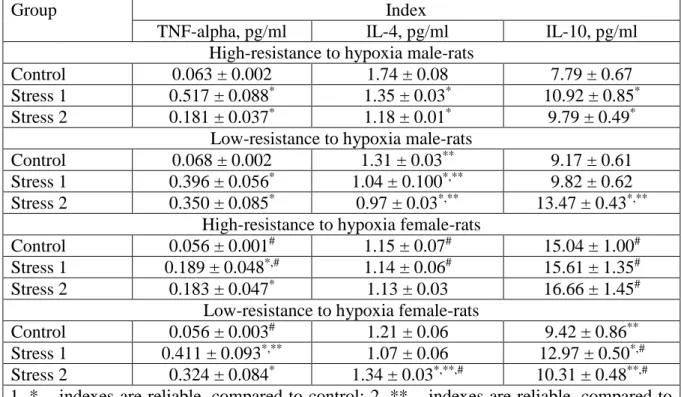
Table 2 - Changes in the concentration of interleukins caused by stress in the serum of high- and low-resistant to hypoxia animals of different sexes, М ± m (n=12)
Group Index
TNF-alpha, pg/ml ІL-4, pg/ml ІL-10, pg/ml
High-resistance to hypoxia male-rats
Control 0.063 ± 0.002 1.74 ± 0.08 7.79 ± 0.67
Stress 1 0.517 ± 0.088* 1.35 ± 0.03* 10.92 ± 0.85*
Stress 2 0.181 ± 0.037* 1.18 ± 0.01* 9.79 ± 0.49*
Low-resistance to hypoxia male-rats
Control 0.068 ± 0.002 1.31 ± 0.03** 9.17 ± 0.61
Stress 1 0.396 ± 0.056* 1.04 ± 0.100*,** 9.82 ± 0.62
Stress 2 0.350 ± 0.085* 0.97 ± 0.03*,** 13.47 ± 0.43*,** High-resistance to hypoxia female-rats
Control 0.056 ± 0.001# 1.15 ± 0.07# 15.04 ± 1.00#
Stress 1 0.189 ± 0.048*,# 1.14 ± 0.06# 15.61 ± 1.35#
Stress 2 0.183 ± 0.047* 1.13 ± 0.03 16.66 ± 1.45#
Low-resistance to hypoxia female-rats
Control 0.056 ± 0.003# 1.21 ± 0.06 9.42 ± 0.86**
Stress 1 0.411 ± 0.093*,** 1.07 ± 0.06 12.97 ± 0.50*,#
Stress 2 0.324 ± 0.084* 1.34 ± 0.03*,**,# 10.31 ± 0.48**,# Notes: 1. * – indexes are reliable, compared to control; 2. ** – indexes are reliable, compared to
In this kind of stress in HR there was an increase of TNF-alpha in 2.88 times (p<0.002), IL-10 – on 25.60% (p<0.02). In LR males-rats there was a significant decrease of IL-1 beta on 39.54% (p<0.001), IL-6 – on 44.54% (p<0.001), IL-4 – on 25.93% (p<0.001), increase of TNF-alpha in 5.18 times (p<0.001), IL-10 – on 46.90% (p<0.001). In HR males-rats, compared with LR, were lower concentrations of IL-1beta in 1.61 times (p<0.001), IL-10 - on 37.61% (p<0.001), higher level of IL-6 on 50.16% (p<0.001), IL-4 – on 18.03% (p<0.001).
In control HR females, compared with LR, found a higher on 37.36% (p<0,001) concentration of IL-10. At stress 1, compared with the control, in HR rats there was an increase in TNF-alpha in 3.37 times (p<0,01), a decrease in IL-1beta - on 29.96% (p<0,001), IL-6 - on 59.93% (p<0.01). In LR rats there was a significant increase of IL-10 on 37.67% (p<0.001), TNF-alpha in 7.28 times (p<0.001). Moreover, in HR females, compared with LR females, were lower IL-2 on 54.65% (p<0.001), IL-6 - on 83.55% (p<0.001), TNF-alpha – in 2.17 times (p<0.05).
Under influences of stress 2, compared with the control, in HR female there was an increase of IL-1beta on 25.92% (p<0.05), TNF-alpha – in 3.25 times (p<0.01), a decrease of IL-2 on 38.14% (p<0.01), IL-6 - on 62.74% (p<0.001). In LR there was a significant increase in the concentration of IL-1 beta on 26.89% (p<0.05), IL-4 - on 10.84% (p<0.001), TNF-alpha – in 5.73 times (p<0,01). In HR female, compared with LR, were lower content of IL-2 on 96.67% (p<0.001), IL-6 – on 91.84% (p<0.001), IL-4 - on 18.50% (p<0.001), higher level of IL-10 on 38.13% (p<0.001).
In control HR males, compared with HR females, were lower content of IL-1beta on 37.91% (p<0.02) and IL-10 on 93.06% (p<0.001), higher level of IL-4 on 33.92% (p<0.001), IL-6 on 49.93% (p<0.01), TNF-alpha on 10.85% (p<0.01). In control LR males, compared with LR females, IL-1beta was higher on 42.98% (p<0.001), IL-6 - on 54.32% (p<0.001), TNF-alpha – on 16.40% (p<0.002).
At stress 1 in HR males, compared with HR females, were lower contents of IL-10 on 42.91% (p<0.02), higher level of IL-1beta on 58.92% (p<0.001), IL-4 - on 15.31% (p<0.02), IL-6 - on 72.94% (p<0.001), TNF-alpha - on 63.41% (p<0.002). At stress 1 in LR males, compared with LR females, were lower contends of IL-10 on 32.08% (p<0.001), IL-2 – in 2.22 times (p<0.001).
level of IL-10 on 23.44% (p<0,001), IL-6 - on 35.87% (p<0,001), lower - IL-2 on 47.50% (p<0.05), IL-4 - on 38.07% (p<0.001).
In comparing the level of IL-1beta in two models of immobilization stress, different changes were found in animals, which depended on the period of time between the immobilizations, sex and resistance of animals to hypoxia. In HR male, the contents increased significantly in stress 1, decreased - in stress 2. Moreover, under stress 1, the indicator was in 3.04 times higher (p<0.001) compared to stress 2. In LR males, the indicator was significantly reduced for both models of stress, but no statistically significant difference in values was found.
In females, the content of IL-1beta decreased significantly compared with the control under immobilization stress 1 only in HR, and increased under stress 2 in both HR and LR. At stress 1, it was significantly lower than in stress 2, in HR on 44.38% (p<0.001), in LR - on 29.65% (p<0.02). The development of inflammation can be said only in HR males at stress 1 and in HR and LR females under at stress 2.
When comparing the values of IL-2 in the two models of immobilization stress, a significant difference between the indexes was not found in males and LR females. In HR female were lower on 43.33% (p<0.05) rates of stress 2.
When comparing the values of IL-4 in the two models of immobilization stress, a significant difference between the indexe in LR males and HR females was not detected. In HR males were lower level on 13.67% (p<0.001) the value at stress 2, and in LR females were lower on 19.97% (p<0.001) the value in stress 1.
When comparing the values of IL-6 in the two models of immobilization stress, no significant difference between the indexes was found in LR males and all females. In HR males were lower on 33.77% (p<0.02) the value of the indexes at stress 1, compared to stress 2. There was a significant decrease of IL-6 in LR males and HR females, regardless of stress.
When comparing the indexes of TNF-alpha in two models of immobilization stress, a significant difference in the LR males and all females between the indexes was not detected. In HR male, the values were in 2.85 times smaller (p<0.002) ate stress 2.
increased in LR males, they did not differ significantly from the values of LR females and HR females.
The difference in interleukin content when comparing values at different models of immobilization stress was insignificant. A greater increase in the content of IL-1beta was in HR males atr stress 1, and females at stress 2. IL-6 was greater only at stress 2 in HR males. TNF-alpha in HR male increased more at stress 1, compared with stress 2. IL-10 values were significantly higher at stress 2 in LR males, and at stress 1 in LR females, in HR females it were always at a high level. The obtained data indicate the dependence of the development of the injury from sex, resistance to hypoxy and the model of immobilization stress.
Thus, intact male rats have a higher pro-inflammatory potential compared to females, and it is greater in LR animals compared to HR, regardless of sex. Most anti-inflammatory interleukins were observed in female HR, which persisted under stress 1. The maximum activity of pro-inflammatory cytokines in the control was observed in LR males. Activation of proinflammatory cytokines under stress 1 occurred in HR male. At stress 2 in males the content of anti-inflammatory cytokines increased and pro-inflammatory – decreased. In females, an increase in proinflammatory interleukins was observed with stable anti-inflammatory parameters.
Conclusion. High congenital resistance to hypoxia is associated with an increased content of anti-inflammatory cytokines (in males – IL-4, in females – IL-10). Сontrol males, compared with females, have a higher production of pro-inflammatory cytokines (IL-6, TNF-alpha). The experiments revealed the changes of pro- and anti-inflammatory interleukins, which depends of sex, resistance to hypoxy and kinds of stress.
References
1. Rohleder N., Aringer M., Boentert M. Role of interleukin-6 in Stress, Sleep, and Fatigue // Ann N Y Acad Sci. 2012;1261:88–96.
2. Niraula A., Witcher K. G., Sheridan J. F., Godbout J. P. Interleukin-6 Induced by Social Stress Promotes a Unique Transcriptional Signature in the Monocytes That Facilitate Anxiety // Biol Psychiatry. 2019;15;85(8):679–689.
4. Effects of Immobilization Stress and Adrenomodullin on Interleukin-10 Levels in Some Rat Tissues / Gadalla A., Al Gendy A., Hashim A. M., Amer M. M., Sabry O. M. O. // Bull. Egypt. Soc. Physiol. Sci. 2013;33 (2):161–171.
5. Altitude Omics: The Integrative Physiology of Human Acclimatization to Hypobaric Hypoxia and Its Retention upon Reascent / A. W. Subudhi, N. Bourdillon, J. Bucher [et al.] // PLoS One. 2014;9(3):e92191.
6. Gibson O. R., Taylor L., Watt P. W., Maxwell N. S. Cross-Adaptation: Heat and Cold Adaptation to Improve Physiological and Cellular Responses to Hypoxia // Sports Med. 2017;47(9):1751–1768.
7. Glutamate receptors in the nucleus tractus solitarius contribute to ventilatory acclimatization to hypoxia in rat / M. E. Pamenter, J. A. Carr, A. Go [et al.] // J. Physiol. 2014;592(Pt 8):1839–1856.
8. Colleen G. Julian, Lorna G. Moore Human Genetic Adaptation to High Altitude: Evidence from the Andes // Genes (Basel). 2019;10(2):150.
9. Shimoda L. A. Let's Talk about Sex: A Novel Mechanism by Which Estrogen Receptor β Limits Hypoxia-Inducible Factor Expression in Pulmonary Endothelial Cells // Am. J. Respir. Cell Mol. Biol. 2018;59(1):11-12.
10. Miller A. J., Cui J., Luck J. C., Sinoway L. I., Muller M. D. Age and sex differences in sympathetic and hemodynamic responses to hypoxia and cold pressor test // Physiol. Rep. 2019;7(2):e13988.
11. Souza G. M. P. R., Amorim M. R., Moraes D. J. A., Machado B.H. Sex differences in the respiratory-sympathetic coupling in rats exposed to chronic intermittent hypoxia // Respir. Physiol. Neurobiol. 2018;256:109-118.
12. Berezovskij V. A. Hypoxia and individual peculiarities of reactivity. – K.: Scientific thought, 1978;2-216. [in Russian]
13. Kulynskyi V. Y., Olkhovskyi Y. A. Two adaptation strategies in adverse conditions: resistant and tolerant. The role of hormones and receptors. Advances of modern biology. 1992;112;697–711 [in Russian].