Case Report
Cervical lymphadenopathies: a diagnostic crisis
Priya Joy*, G. Gayathri, V. Gangadharan
INTRODUCTION
Cervical lymphadenopathy refers to lymphadenopathy of the cervical lymph nodes (the glands in the neck). Cervical lymphadenopathy is a sign or a symptom, not a diagnosis. Lymphadenopathy represents the immune system which reciprocates in the form of enlarged lymph nodes.1 Normally the size of the lymph node is less than
one cm in diameter. Lymphadenopathies can be local or systemic, benign, self-limited. The causes are varied, and may be inflammatory, degenerative, infective or neoplastic.2 If nodes are healing by resolution or scarring
after becoming inflamed, they may remain palpable thereafter.3
The most common cause of cervical lymphadenopathy seen in clinical practice is tuberculous lymphadenopathy especially in developing countries.4 In elderly over the
age of 50, we have to rule out malignancy as a cause for lymphadenopathy.5
Here, we are discussing about two rare causes of cervical lymphadenopathy: Kimura’s disease and Kikuchi- Fujimoto disease.
ABSTRACT
Cervical lymphadenopathy refers to lymphadenopathy of the cervical lymph nodes. The causes are varied, and may be inflammatory, degenerative, infective or neoplastic. The first case presented here is of a 29-year old male presented with h/o recurrent progressive right sided painless swelling in the infraparotid region with cervical lymphadenopathy for past one year. Swelling was 6×6cm, nontender, firm mass. Cervical lymph nodes were palpable. He had raised eosinophil counts and elevated serum IgE levels. Biopsy showed abundant eosinophilic infiltration with microabscess suggestive of Kimura’s disease. Second case reported here is of a 17-year old female patient presented with fever for 3 weeks with cervical lymphadenopathy. The lymph node was 3×2cm, tender and firm. Routine blood investigations showed anemia. Biopsy showed lymph nodes composed of sheets of macrophages with crescent shaped nuclei with areas of necrosis and karyhorrhexis suggestive of Kikuchi’s disease. Kimura’s disease is a relatively uncommon chronic inflammatory benign condition mostly affecting the lymph nodes, subcutaneous tissue and salivary glands of head and neck region. It is seen in males in their second decade of lives. Peripheral eosinophilia, regional lymphadenopathy and elevated serum IgE levels are characteristics of Kimura’s disease. Kikuchi-Fujimoto disease is a benign, idiopathic and self-limited disease. Affects young females between 20-35 years of age. Usually there will be localized painful lymphadenopathy, fever and leukopenia. Kikuchi disease runs a benign course and resolves in several weeks to months. The two cases are presented to make clinicians aware of Kimura’s disease and Kikuchi’s disease as a differential diagnosis of cervical lymphadenopathy and to avoid the diagnostic dilemma both clinically and histopathologically.
Keywords: Cervical lymphadenopathy, Eosinophilic microabscess, Histiocytic necrotizing lymphadenitis, Kimura’s disease, Kikuchi- Fujimoto’s disease
Department of Respiratory Medicine, Saveetha Medical College and Hospital, Thandalam, Chennai, Tamil Nadu, India
Received: 06 August 2017
Accepted: 01 September 2017
*Correspondence:
Dr. Priya Joy,
E-mail: [email protected]
Copyright: © the author(s), publisher and licensee Medip Academy. This is an open-access article distributed under the terms of the Creative Commons Attribution Non-Commercial License, which permits unrestricted non-commercial use, distribution, and reproduction in any medium, provided the original work is properly cited.
CASE REPORT
Case report 1
29-years old male presented with h/o recurrent progressive right sided painless swelling in the infraparotid region with cervical lymphadenopathy for past one year. There was no discharge, skin changes or excessive salivation. No history of weight loss, fever, night sweats/symptoms of facial nerve involvement.
Figure 1: Swelling in the right parotid region before excision.
Figure 2: Right parotid region after excision of the swelling.
Six months ago, he presented with similar swelling. USG neck showed hypoechoic lesion in the right infraparotid region 1.8×1.5 cm with enlargement of level I and II cervical nodes. FNAC was suggestive of reactive lymphadenitis. CT neck showed possibility of right sialadenitis. He was treated conservatively with antibiotics. Two months later he presented with increase in size of swelling 4×3cm. Repeat FNAC showed chronic sialadenitis, treated with another course of antibiotics.
Again, he presented with increase in the size of swelling to 6×6cm, nontender, firm mass. No warmth/redness on the overlying skin. Cervical lymph nodes were palpable. Routine blood investigations were normal except for raised eosinophil counts (1340 cells/cumm) and elevated serum IgE levels. CT scan: suggestive of right
sialadenitis. Biopsy of parotid tissue and lymphnode showed abundant eosinophilic infiltration with micro abscess suggestive of Kimura’s disease. He was started on steroids and antihistamines, swelling decreased in size and he is under regular follow up.
Figure 3: CT scan of the patient with marked arrow showing enlarged right parotid gland with differential
density.
Figure 4: CXR showing antero-medial bulge of the diaphragm. Lung fields are normal.
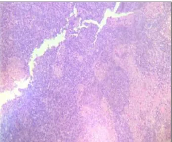
Figure 5: Reactive lymphoid follicles with germinal centre hyperplasia, parafollicular eosinophilic
Case report 2
17-year old female patient presented with fever for 3 weeks with cervical lymphadenopathy. Fever was high grade with evening rise of temperature, history of night sweats present. History of loss of weight and loss of appetite present. No previous history of similar episodes.
On examination, the lymph node was 3×2 cm, tender and firm. There was no warmth or redness. Routine blood investigations showed anemia. Fever workup for infectious causes was negative. USG neck showed few enlarged lymph nodes in the left level II and III. FNAC of left cervical lymph node showed sheets of lymphocytes of varying sizes and transformed lymphocytes, numerous tangible body macrophages, few foamy macrophages and cresentric histiocytes in a background of necrotic material s/o Kikuchi’s disease. Biopsy showed lymph nodes composed of sheets of macrophages with crescent shaped eccentrically placed nuclei with areas of paracortical necrosis and karyhorrhexis suggestive of Kikuchi-Fujimoto disease. She was started on steroids and her fever and lymphnode subsided.
Figure 6: Chest X-ray of the patient; lung fields appear normal.
Figure 7: Reactive lymphoid follicle with circumscribed areas of necrosis.
Figure 8: Infiltration of the histiocytes with karyorrhectic debris; absence of neutrophils is noted.
Figure 9: Lymphocytes of varying sizes and transformed lymphocytes, numerous tangible body macrophages, few foamy macrophages and crescentic
histiocytes in a background of necrotic material.
DISCUSSION
Kimura’s disease is a relatively uncommon chronic inflammatory condition that presents as an unusual allergic or autoimmune response.6 It was first described
by Kimm and Szeto in China as eosinophilic hyperplastic lymphogranuloma.6 Kimura et al from Japan reported a
similar finding and described it as an "unusual granulation and hyperplastic changes of lymphatic tissue", and this condition has since become widely known as Kimura's disease. It is an uncommon disease in India with more preponderance to far eastern countries. There are about 200 reported cases worldwide.6,7
It is a benign inflammatory disease mostly affecting the head and neck region.8 It usually involves the
subcutaneous tissue, lymph nodes and the salivary glands.8 They are rarely reported in other areas like
eyelids, lacrimal glands, orbit, axilla, groin, forearm, and kidneys.9 It presents as solitary or multiple sub-cutaneous
in males in their second decade of lives with a male: female ratio of 3:18. Peripheral eosinophilia, regional lymphadenopathy and markedly elevated serum IgE levels are characteristics of Kimura’s disease.6
Recurrence is very common with upto 25%-40% of surgically treated cases. There are no reports of malignant transformation so far.6 It is a disease that can cause
considerable diagnostic dilemma to clinicians and pathologists.
Aetiology is unknown, but several theories were described such as interference with the immune regulation and an atopic reaction to continuous antigenic stimulation by candida albicans/parasitic infestation/neoplasm is also considered.8 Renal
abnormalities notably nephrotic syndrome and glomerulonephritis are seen in Kimura’s disease.10
Proteinuria is present in 12-16% of cases.9
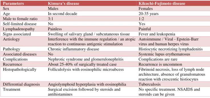
Table 1: Differences between Kimura’s disease and Kikuchi-Fujimoto’s disease.
Parameters Kimura’s disease Kikuchi-Fujimoto disease
Sex Males Females
Age In second decade 20-35 years
Male to female ratio 3:1 1:2
Self-limited disease No Yes
Lymphadenopathy Painless Painful
Signs associated Swelling of salivary gland / subcutaneous tissue Fever and leukopenia Aetiology Interference with the immune regulation / an atopic
reaction to continuous antigenic stimulation
Autoimmune / Viral - Epstein-Barr virus and human herpes virus Pathology Chronic inflammatory disease Histiocytic necrotizing lymphadenitis Associated diseases No Systemic lupus erythematosus Complications Nephrotic syndrome and glomerulonephritis Complications are rare Recurrence About 25-40% of surgically treated case Recurrence is uncommon
Histopathologically Folliculolysis with eosinophilic microabcess Fibrinoid necrosis, loss of lymph node architecture, absence of granulomatous reaction with crescentic histiocytes Differential diagnosis Angiolymphoid hyperplasia with eosinophilia Tuberculosis
Treatment Surgical excision followed by steroids and antihistamines
No specific treatment. NSAIDS and steroids can be given
Diagnosis of Kimura’s disease can be confirmed by open biopsy of the lymph node or swelling.8 CT scan of neck
may aid in finding out the extent of the disease.6
Histopathologically, the most characteristic finding is folliculolysis with eosinophilic microabcess and parafollicular eosinophilic infiltration.9 Peripheral
eosinophilia with elevated serum IgE levels add on to the diagnosis of Kimura’s disease.8 Differential diagnosis are
angiolymphoid hyperplasia with eosinophilia, Reactive lymphadenopathy, Hodgkin’s lymphoma, Castleman’s disease, salivary gland tumors, lymphangioma or haemangioma, nodal metastasis, hamartoma and Mikulicz’s disease.6
Treatment of this disease ranges from conservative management to surgical excision.8 Usually surgical
excision of the lesion followed by steroids and antihistamines are the first line in the management.8 The
second line options are topical tacrolimus twice daily, intralesional administration of steroids, cyclosporine, local radiation therapy and electrodesiccation.8
Kikuchi-Fujimoto disease
Kikuchi-Fujimoto disease (KFD) or histiocytic necrotizing lymphadenitis, is a benign, idiopathic and
self-limited disease.11 It was reported in 1972 in Japan by
Kikuchi and Fujimoto who described the disease independently as “lymphadenitis showing focal reticulum cell hyperplasia with nuclear debris and phagocytosis” and “cervical subacute necrotizing lymphadenitis,” respectively.12 Although KFD was initially thought to be
occurring exclusively in the cervical lymph nodes of young Asian women, it has been seen in patients of any age, sex, and race, and can involve nodal and extranodal locations also.12
Most of the case reports are of Asian origin young females between 20 years and 35 years of age. The male:female ratio is 1:21.The incidence of the disease is unknown.13 It usually presents as localized painful
lymphadenopathy, most commonly in the cervical region with associated fever and leukopenia in up to 50% of patients.11 Kikuchi disease almost always runs a benign
course and resolves in several weeks to months.14 Disease
recurrence is unusual, and fatalities are rare.12
Aetiology of the disease in unknown although autoimmune and viral pathogenesis are suggested.15
Necrosis of the lymph nodes appears to be due to apoptosis.12 Recently studies have suggested that the
protein which is known as perforin.12 Other possible
causes for apoptosis was suggested as Epstein-Barr virus and human herpesvirus.12 Kikuchi-Fujimoto disease has
been associated with systemic lupus erythematosus (SLE) as well as to other autoimmune diseases.11
Clinically, usually there will be unilateral neck involvement. Commonly lymph nodes in the posterior cervical triangle and jugular carotid chain are enlarged. Other areas affected are axillary (14%) and supraclavicular nodal chains (12%). The enlarged lymph nodes will be usually painful and firm in consistency.12
Fever usually occurs in 30% to 50% of the cases. Leukopenia occurs in 30 to 70% of cases. Other nonspecific findings seen are anemia, atypical peripheral blood lymphocytes and increased erythrocyte sedimentation rate with low C-reactive protein.16
Diagnosis usually done by open biopsy.12
Histopathologically there will be extensive fibrinoid necrosis, loss of lymph node architecture, absence of granulomatous reaction with cresentric histiocytes at the margin of the necrotic area with the karyorrhectic foci containing predominantly histiocytes and plasmacytoidmonocytes. Another characteristic feature is that neutrophils will be absent.16 Differential diagnosis
include reactive, infectious and lymphoproliferative diseases, such as Tuberculosis, Toxoplasmosis, infectious Mononucleosis, Sarcoidosis, Kawasaki’s disease, non-Hodgkin's lymphoma.
There is no specific treatment for this disease as it self-limitng.11 Usually NSAIDS are used to alleviate fever and
painful lymphadenopathy. Steroids can be given for speedy resolution.12
CONCLUSION
These two cases are presented to make clinicians aware of Kimura’s disease and Kikuchi’s disease as a differential diagnosis of cervical lymphadenopathy and to avoid the diagnostic dilemma both clinically and histopathologically.
Funding: No funding sources Conflict of interest: None declared Ethical approval: Not required
REFERENCES
1. Batni G, Gaur S, Sinha ON, Agrawal SP, Srivasatva AA. Clinic-pathological study of cervical lymphnodes. Ind J Otolaryngol Head Neck Surg. 2016;68(4):508-510.
2. Mohseni S, Shojaiefard A, Khorgami Z, Alinejad S, Ghorbani A, Ghafouri A. Peripheral lymphadenopathy: approach and diagnostic tools. Iran J Med Sci. 2014;39(2):158-70.
3. Hupp JR, Ellis E, Tucker MR. Contemporary oral and maxillofacial surgery (5th ed). St. Louis, Mo.:
Mosby Elsevier; 2008:428-30.
4. Tanwir F, Amed I, Hasmi S, Ahmed S. Tuberculosis and cervical lymphadenopathy: a study of 175 cases in a tertiary care Hospital. Oral Hyg Health. 2013;1(119):2332-702.
5. Kerawala C, Newlands C, eds. Oral and maxillofacial surgery. Oxford: Oxford University Press; 2010;68:377,392-394.
6. Kumar MU, Yelikar B. Kimura’s disease: a diagnostic challenge. Int J Pharm Bio Sci. 2012;3(4):311-5.
7. Abhay H, Swapna S, Darshan T, Vishal J, Gautam P. Kimura’s disease: a rare cause of local lymphadenopathy. Int J Sci Stud. 2014;2(5):122-5. 8. Bharati VH, Kalaivani V, Bharathi R, Vineetbinu.
Kimura’s disease: a diagnostic and therapeutic dilemma. J Clin Diag Res. 2012;6(2):311-2.
9. EwaOsuch-Wójcikiewicz, Bruzgielewicz A, Lachowska M, Wasilewska A, Niemczyk K. Kimura’s disease in a caucasian female: a very rare cause of lymphadenopathy. Case Reports in Otolaryngol. 2014;2014:1-4.
10. Shetty AK, Beaty MW, McGuirt WF, Woods CR, Givner LB. Kimura’s disease: a diagnostic challenge; Pediatr. 2002;110;e39.
11. Mahajan T, Merriman RC, Stone MJ. Kikuchi-Fujimoto disease (histiocytic necrotizing lymphadenitis): report of a case with other autoimmune manifestations. Proceedings (Baylor University Medical Center). 2007;20(2):149-51. 12. Archibald DJ, Carlson ML, Gustafson RO.
Kikuchi-Fujimoto disease in a 30-year-old Caucasian female. Int J Otolaryngol. 2009;2009:1-4.
13. Deaver D, Horna P, Cualing H, Sokol L. Pathogenesis, diagnosis and management of Kikuchi-Fujimoto disease. Cancer Control. 2014;21(4):313-21.
14. Kampitak T. Fatal Kikuchi-Fujimoto disease associated with SLE and hemophagocytic syndrome: a case report. Clin Rheumatol. 2008;27(8):1073-5.
15. Manchanda A. Kikuchi-Fujimoto Syndrome: an uncommon disease with a familiar presentation. Int J Int Med. 2009;9(1).
16. Bosch X, Guilabert A, Miquel R, Campo E. Enigmatic Kikuchi-Fujimoto disease: a comprehensive review. Am J Clin Pathol. 2004;122: 157-8.