The journal homepage www.jpacr.ub.ac.id p-ISSN : 2302 – 4690 | e-ISSN : 2541 – 0733
Antibacterial Activity and Phytochemical Analysis of Edible
Seaweed
Eucheuma spinosum
Against
Staphyloccocus aureus
Anna Safitri,1* Arie Srihardyastutie,1 Anna Roosdiana1, Sutrisno1 1Chemistry Dept., Brawijaya University, Jl. Veteran, 65145, Malang, Indonesia
*Corresponding email: a.safitri@ub.ac.id
Received 29 October 2017; Revised 9 March 2018; Accepted 28 August 2018
ABSTRACT
The current work aims to determine antibacterial activities of ethanol extracts of Eucheuma spinosum against Staphylococcus aureus. These were showed through inhibition zones formation, minimum inhibitory concentration (MIC), and minimum bactericidal concentration (MBC) values. The MIC was determined using dilution test tube method, and the MBC was calculated using streaking method. Qualitative test was applied to determine phytochemical compounds in ethanol extract of E. spinosum. Results showed that ethanol extracts of E. spinosum at concentrations of 1%; 2%; 5%, and 10% (v/v) resulted in inhibition zones of 1.98; 4.14; 7.42; and 10.27 mm, respectively. In addition, concentration of 10% (v/v) was the MIC, and 15% (v/v) was the MBC values of E. spinosum extracts against S. aureus. The phytochemical analysis showed that alkaloids and saponins were the main components detected. Alkaloids positive tests were observed by the formation of brown sediment with Wagner’s reagent, yellow sediment with Mayer’s reagent, and orange sediment with Dragendorff’s reagent. Saponins positive results were detected by the formation of stable foam when the extracts reacted with water and HCl. Overall, these results suggested that E. spinosum extracts have antibacterial properties against Gram-positive bacteria, i.e. S. aureus.
Key word: antibacterial, alkaloids, Eucheuma spinosoum, Staphylococcus aureus, saponins, MIC, MBC
INTRODUCTION
The development and spread of multidrug-resistant (MDR) bacterial pathogens have substantially threatened the current antibacterial therapy [1]. MDR bacterial infections often lead to increased mortality, higher cost of treatment and care. The most problematic bacteria include, but are not limited to, extended-spectrum 𝛽-lactamase-producing Escherichia coli (ESBL-EC) and Kleb-siella pneumoniae (ESBL-KP), carbapenem-resistant
Enterobacteriaceae, Pseudomonas aeruginosa, and Acinetobacter baumannii, hospital-acquired methicillin-resistant Staphylococcus aureus (MRSA), and vancomycin resistant
Enterococcus (VRE) [2-4]
Therapeutic options for these pathogens are extremely limited and physicians are forced to use expensive or previously discarded drugs, such as colistin, that are associated with significant side effect to the patients’ health [1]. Therefore, it is necessary to search the other alternatives that can potentially be effective in the treatment of these problematic bacterial infections. The usefulness of plant extracts for antimicrobial therapy and/or other diseases have been observed to be promising medications since ancient time [4]. The inclusion of
traditionally used medicines including phytomedicines, if they prove safe and effective, into national health care system is suggested by World Health Organization [4].
In Indonesia, there are numerous studies of use of traditional plants as antibacterial agents [5-8]. Seaweeds, considered as source of bioactive compounds, produce a great variety of secondary metabolites characterized by a broad spectrum of biological activities. Compounds with cytostatic, antiviral, antifungal and antibacterial activities have been detected in green, brown and red algae [9]. Seaweeds have been screened extensively to isolate life-saving drugs or biologically active substances all over the world [10-12].
The aims of this study is to determine the antibacterial activity of red algae,
Rhodophyceae, grown widely in Indonesian seas, Eucheuma spinosum. Eucheuma spinosum
can be used as raw or processed materials, and it is edible. Eucheuma spinosum contains carrageenan, proteins, carbohydrates, fats, and crude fibers [13-14]. Moreover, E. spinosum
may contain bioactive compounds such as flavonoids, alkaloids, saponins, tannins and their derivatives that have antibacterial properties [13]. A previous study has been conducted [15] using methanol for extracting bioactive compounds in E. spinosum from Wongsorejo sea, Banyuwangi, East Java, and tested for their bacterial activity against E. coli and S. aureus.
The highest concentration used, 80 mg/L, had medium to low antibacterial activity in the inhibition zones of 3 mm and 4 mm, for E. coli and S. aureus, respectively [15]. In the current study, the E. spinosum used is cultivated in the Flores Sea, Eastern Nusa Tenggara. The E. spinosum has been extracted using ethanol as solvent, and the bacteria used are S. aureus. These bacteria cause infection. Infection is one of the common causes of disease occurs in tropical climates, such as Indonesia. Growth and reproduction of these bacteria are relatively fast, which is between 20 minutes to 15 hours exponentially [16]. Under normal circumstances, these bacteria are present in the respiratory tract and skin [16]. Therefore, it is important to study the potential of E. spinosum extracts as antibacterial agent against S. aureus, pathogenic bacteria commonly found in tropical country.
EXPERIMENT
Chemicals and instrumentation
Eucheuma spinosum used were cultivated in Flores sea, Eastern Nusa Tenggara, Indonesia. Species taxonomy was identified by Laboratory of Taxonomy and Plant Structure, Department of Biology, Brawijaya University. Staphylococcus aureus cultured were obtained from Faculty of Medicine, Brawijaya University. The sub-culturing of S. aureus and antibacterial test were conducted in the Laboratory of Microbiology, Faculty of Medicine, Brawijaya University.
All reagents of analytical or higher purity grade were purchased from Merck or Sigma-Aldrich and were used as received: HCl (37% aqueous solution), HNO3 (trace pure, 65% w/w
in H2O), NaOH (99.9%), ethanol (96%), n-hexane (95%, HPLC grade), methanol (99%,
HPLC grade), nutrient agar and Mueller Hinton Broth for microbiology, Mayer’s reagent and Dragendorff’s reagent. Water was purified using the Milli-Q technique.
Preparation of E. spinosum extract
One hundred gram (100 g) of dry E. spinosum were wrapped in filter paper, and inserted into a Sohxlet extractor. The Soxhlet flask was filled with 96% ethanol. The Soxhlet extraction was conducted using a rotary evaporator vacuum at 40-45 C for 2 h. The concentrated extracts were collected and diluted with water in order to prepare concentrations of 1%: 2%: 5%: 10: and 15% (v/v).
Determination of Antibacterial Activity – Zones of Inhibition
The antibacterial activity of E. spinosum extracts were evaluated on S. aureus. The bacterial strains were cultured in nutrient agar plate, with Muller Hinton Broth for growth media. The diffusion disc method was applied to define zones of inhibition. Each pathogen was inoculated by spreading onto a solid medium agar plate. The extracts of E. spinosum
were inoculated onto sterile paper discs. Different concentrations of each extract were used: 1%: 2%: 5%: 10: and 15% (v/v), per disc. The petri dishes were incubated for 4 days at room temperature. The diameters of zones of inhibition of microbial growth were measured at day 4[17].
Determination of Antibacterial Activity – MIC and MBC
Different concentrations of E. spinosum extracts: 1%: 2%: 5%: 10: and 15% (v/v), were transferred into sterile test tubes. Each test tube was incubated at 37 C for 24 h. The MIC was determined by the lowest concentration of extract that resulted in clear solution, no bacteria grown. Bacteria suspension from test tubes that contain E. spinosum extracts of: 1%: 2%: 5%: 10: and 15% (v/v) were inoculated onto sterile nutrient agar plate separately. These were incubated at 37 C for another 24 h. The number of S. aureus colony were calculated by colony counter. The MBC value was determined by the lowest concentration that showing no bacteria grown in nutrient agar plate.
Qualitative Phytochemical Analysis Alkaloid Test
E. spinosum extracts (3 mL) were stirred with a few mL of diluted HCl and filtered. Filtrates were transferred into three separated test tubes. A few drops of Wagner’s reagent were added at the side of test tube 1. The formation of reddish brown precipitates showed the presence of alkaloids. A few drops of Mayer’s reagent were added at the side of test tube 2. The formation of yellow precipitates showed the presence of alkaloids. A few drops of Dragendorff’s reagent were added at the side of test tube 3. The formation of orange precipitates showed the presence of alkaloids.
Flavonoid Test
E. spinosum extracts (1 mL) were dissolved in MeOH and a few drops of 10% aqueous NaOH were added to obtain an intense yellow color. The conversion into a colorless solution upon addition of concentrated HCl indicated the presence of flavonoids.
Tannin test
E. spinosum extracts (1 mL) were dissolved in 3 mL of 1% FeCl3. The formation of
green, blue, or black color at the interface indicated the presence of tannins.
Saponin Test
E. spinosum extracts (1 mL) were dissolved in 5 mL of water, and the solution was shake for 10 s. The formation of stable foam for 10 min upon the addition of 2 M HCl, indicated the presence of saponins.
RESULT AND DISCUSSION
There are differences in the positive and negative cell wall bacteria. Gram-positive bacteria have simpler and thinner cell wall than Gram-negative bacteria [18]. It is, therefore, Gram-positive bacteria are more sensitive to antibacterial agent, and as a result,
they are usually chosen for testing the potency of new compound that has antibacterial properties. Staphyloccous aureus are Gram-positive bacteria that commonly cause infection in tropical regions [16], i. e. in Indonesia, thus, are the suitable bacteria for investigating antibacterial properties of promising antibacterial agents.
The production of antimicrobial properties has been considered as an indicator that particular seaweed has the capacity to synthesize bioactive secondary metabolites. Compounds with antiviral, anthelminthic, antifungal, and antibacterial properties have been detected in brow, red, and green algae [11, 19]. Considering that Indonesia has vast area of oceans, and also is the largest island country in the world, investigation related to search of new biologically active compounds from marine products, i.e. algae, can be seen as an almost unlimited field.
The growth of the pathogenic bacteria was inhibited by the release of secondary metabolites from the extracts of E. spinosum. This was indicated by the formation of inhibition zones around the extracts. Inhibition zones of E. spinosum extracts are presented in Figure 1, while the diameters of inhibition zones are listed in Table 1. It has been shown that starting from concentration of 1%, E. spinosum extracts have antibacterial properties, i.e. 6 mm in diameters. Increasing concentrations of E. spinosum extracts caused increasing in the higher inhibition zones. The highest concentration used, 15%, resulted in inhibition zones diameters of 16.3 mm.
Figure 1. Inhibition zones of E spinosum extracts of: 1%; 2%; 5%; 10% and 15% (v/v) from replicate 1, replicate 2, and replicate 3.
Table 1. The diameter of inhibition zones of E. spinosum extracts No E. spinosum extract
concentration Inhibition zones (mm)
1 1% 6
2 3 4
2% 5% 10%
7.9 10.1 13.4
5 15% 16.3
Bacteriostatic and bactericidal properties of the E. spinosum extracts were determined by calculating the MIC and MBC values. The MIC value was determined by observing turbidity levels of bacteria colony in the series of extract concentrations in the separate test tubes. The MIC value was calculated by counting bacteria colony in those series of extract concentrations. Table 2 shows results of turbidity levels and the number of S. aureus with the
addition of E. spinosum extracts in various concentrations. Figure 2 shows the growth of the
S aureus in the petri dishes, under the influences of E. spinosum extracts with concentrations of: 1%, 2%, 5%, 10%, and 15% (v/v).
Table 2. The turbidity levels and the number of S. aureus in the influences of E. spinosum
extracts
Figure 2. The S. aureus growth in petri dishes under the influence of E. spinosum extracts of: 1%; 2%; 5%; 10% and 15% (v/v); and ethanol only (no E. spinosum extracts).
Based on results from Table 2 and Figure 2, at the lowest extracts concentration used, 1%, the growth of bacteria was started to be inhibited. This has been showed as the number of bacteria decreased, from 3x10⁷ to 3x10⁵ CFU/plate, and the turbidity levels also reduced. At 10% concentrations, the minimum growth of the bacteria was observed, and the number of bacteria have been greatly reduced to 1.5x10² CFU/plate. Finally, at extract concentrations of 15%, there were no growth of pathogenic bacteria observed. As a result, the MBC value has been concluded at 15%.
The presence of secondary metabolites in algae is a form of adaptation to the environment. According to Franklin and Snow, 2010 [20], the mechanism of antimicrobial action can be achieved by four different ways. First is by the inhibition of the synthesis or activation of enzymes which damage the bacterial cell wall, thus eliminating the ability to grow and causes lysis of the cell. Second is by direct action on the cell membrane by affecting cell permeability leading to leakage of intracellular compounds. Third is the disruption of the functioning of the bacterial ribosome, thereby inhibiting protein synthesis. Fourth is by interfering with the metabolism of nucleic acids leading to a loss of function of the synthesis of the cell [20].
No E. spinosum extract concentration
Turbidity levels CFU/plate
1 0% +++++ 3x10⁷
2 1% ++++ 3x10⁵
3 4 5
2% 5% 10%
+++ ++
+
2.9x10⁴ 1.7x10³ 1.5x10²
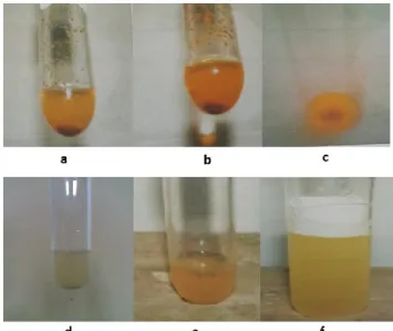
Bioactive compounds detected in the ethanol extracts of E. spinosum were alkaloids and saponins. The qualitative phytochemical test results are presented in Figure 3. The positive results were observed from Figures 3a and 3f. The mechanism of actions of alkaloids and saponins are suggested through inhibition of synthesis of cell wall bacteria. It is known that cell wall bacteria is the outer layer of bacteria, therefore antibacterial agent attacks it first, and subsequently attacks the metabolism system of the bacteria.
Cell wall of Gram-positive bacteria mainly consists of peptidoglycans, teichoic acids, and plasma membranes [20]. Inhibition of synthesis of cell wall bacteria can be through destruction of peptidoglycans or teichoic acids [22]. Peptidoglycans consist of N-acetylglucosamines and N-acetylmuramic acids, and amino acids [23]. The basic structural of peptidoglycans is single strand. Each strand of peptidoglycans is arranged side-by-side and connected to each other by tetra-peptides crosslinking [24]. The proposed mechanism of actions of alkaloids and saponins act as anti-bacterial agent through their interactions with peptidoglycans is illustrated in Figure 4a. Amine groups in alkaloids from E. spinosum
extracts can replace amine groups in tetra-peptides crosslinking. Since there are no carbonyl groups in alkaloids, peptide bonds will not be formed, and as a result, tetra-peptides crosslinking will be disconnected. In addition, saponins from E. spinosum extracts that contain –OH groups can form hydrogen bonds with oxygen in peptidoglycans. Strong interactions between H atoms in saponins and O atoms in peptidoglycans interfered with tetra-peptides crosslinking, as a result, the bonds within peptidoglycans are unstable and, subsequently, the peptidoglycans are easily destroyed.
Another possible mechanism is by the interfere with teichoic acids. Only Gram-positive bacteria have teichoic acids [21]. Teichoic acids contain phosphate groups which connect glycerol with each other [22]. Amine groups in alkaloid and –OH groups in saponins can make hydrogen bonds with O atom in the teichoic acids. The hydrogen bonds between teichoic acids and alkaloids or saponins lead to instability of phosphate groups in teichoic acids, as a result synthesis of cell wall bacteria interrupted, and subsequently causing lysis of cell wall (Figure 4b).
Figure 3. The phytochemical qualitative test results from: (a) alkaloid test using Wagner’s reagent; (b) alkaloid test using Meyer’s reagent; (c) alkaloid test using Dragendorff’s reagent; (d) flavonoid test; (e) tannin test; and (f) saponin test. Positive test results were observed in (a), (b), (c), and (f).
Figure 4. The proposed mechanism of actions of alkaloid and saponin act as anti-bacterial agents through inhibition of synthesis of cell wall bacteria by interactions with: (a) peptidoglycans; (b) teichoic acid.
CONCLUSION
The current work highlighted the antibacterial properties of ethanol extracts of E. spinosum. The ethanol extract was effective antibacterial agent against Gram-positive bacteria, S. aureus. These have been deduced from the formation of inhibition zones around
E. spinosum extract which indicated the inhibition of bacterial growth, and the MIC and MBC values of the extract. The phytochemical study indicated that E. spinosum contained alkaloids and saponins. It is possible that these compounds play important role in antibacterial activities by inhibiting synthesis of cell wall of pathogenic bacteria. Thus, the red algae, E. spinosum is a natural product that could be a new source for applications in the pharmaceutical.
REFERENCES
[1] Boucher, H. W., Talbot, G H, Bradley, J S., Clin. Infect. Dis., 2009,48, 1-12. [2] Giamarellou, H, Int. J. Antimicrob. Agents,2010, 36, S50-S54.
[3] Magiorakos, A. P., Srinivasan, A., Carey, R. B., Microbiol. Infect.,2006, 18, 268-281. [4] WHO, WHO Traditional Medicine Strategy 2002-2005, 2002, Geneva, Switzerland:
WHO Publisher.
[5] Kusmiati, Priadi, D., Rahayu, R. K. B., J Pure App. Chem. Res., 2017, 6,27-33. [6] Lestari, P., Elfrida, N., Suryani, A., Suryadi, Y., Jordan J. Biol. Sci., 2014, 7,75-80. [7] Ernawita, Wahyuono, R. A., Hesse, J., Hipler, U. –C., Elsner, P., Bohm, V.,
Antioxidants, 2017, 6, 11-16.
[8] Putra, M. Y., Hadi, T. A., Murniasih, T., Asian Pacific J. Trop. Dis., 2016, 6,732-735. [9] Shannon, E., Abu-Ghannam, N., Mar. Drugs, 2016, 14, 81 (open access).
[11] Kasanah, N., Seto, D. S., Amelia, W., Isnansetyo, A., Indones. J. Chem., 2015, 15, 201-209.
[12] Williams, P. G., Appavoo, M. R., Lal, N. D., Aravind, A., Williams, G. P., South Indian J Biol. Sci., 2016, 2, 347-353.
[13] Diharmi, A., Fardiaz, D., Andarwulan, N., Heruwati, E. S., Phycol. Res., 2017, 65,
256-261.
[14] Matanjun, P., Mohamed, S., Mustapha, N. M., Muhammad, K., Ming, C. H., J. Appl. Phycol., 2008, 20, 367-340.
[15] Hanapi, A., Fasya, A. G., Mardiyah, U., Miftahurrahmah, Alchemy, 2013, 2, 126-137. [16] Tong, S. Y. C., Davis, J. S., Eichenberger, E., Holland, T. L., Fowler, Jr, V. G., Clin.
Microbiol. Rev., 2015, 28, 603-661.
[17] Kusmiati, K., D. Priadi, R. K. B. Rahayu, J. Pure App Chem. Res.,2017, 6(1), 27-33. [18] Emami-Karyani, Z., Chehrazi, P., African J. Microbiol. Res., 2011, 5,1368-1373. [19] Berri, M., Slugocki, C., Olivier, M., Helloin, E., Jacques, I., Salmon, H., Le Goff, M.,
Collen, P. N., J. Appl. Phycol., 2016, 28, 2999-3008.
[20] Franklin, T. J., Snow, G. A., Biochemistry of microbial action, 2010, 2nd ed. London: Chapman and Hall Ltd.
[21] Brown, S., Santa Maria, J. P., Walker, S., Annu. Rev. Microbiol., 2013,67, 313-316. [22] Silhavy, T. J., Kahne, D., Walker, S., Perspect. Biol.,2010, 1-16.
[23] Jankute, M., Cox, J. A. G., Harrison, J., Besra, G. S., Annu. Rev. Microbiol., 2015, 69, 405-423.