Microbial Pathogen-Induced Necrosis Mediated By NLRP3 and ASC
Stephen Willingham
A dissertation submitted to the faculty of the University of North Carolina at Chapel Hill in partial fulfillment of the requirements for the degree of Doctor of Philosophy in the
Curriculum in Genetics & Molecular Biology
Chapel Hill 2008
Approved by: Dr. Jenny Ting Dr. Al Baldwin Dr. Jeff Dangl
ABSTRACT
STEPHEN B. WILLINGHAM: Microbial Pathogen-Induced Necrosis Mediated By NLRP3 and ASC
(Under the direction of Dr. Jenny P-Y. Ting)
NLRP3 and ASC are important components of the inflammasome, a multi-protein complex required for caspase-1 activation and IL-1β production. NLRP3 mutations underlie autoinflammation characterized by excessive IL-1β secretion. Disease-associated NLRP3 also causes a program of necrosis-like cell death in macrophages, the mechanistic details of which are unknown. We find that patient monocytes carrying disease-associated NLRP3 mutations exhibit excessive necrosis-like cell death by a process dependent on ASC and cathepsin B, resulting in spillage of the proinflammatory mediator HMGB1. Shigella
flexneri and Klebsiella pneumoniae infection also cause NLRP3-dependent macrophage
ACKNOWLEDGEMENTS
Portions of this dissertation have been previously published or are in preparation for publication. I would like to thank the co-authors who have contributed to these works.
Chapter 1: Jenny P.-Y. Ting, Stephen B. Willingham, and Daniel T. Bergstralh.
NLRs at the Intersection of Cell Death and Immunity. Nature Reviews Immunology 2008, Manuscript in press.
Chapter Two: Stephen B. Willingham, Daniel T. Bergstralh, William O’Connor, Amy C. Morrison, Debra J. Taxman, Joseph A. Duncan, Shoshana Barnoy, Malabi M. Venkatesan, Richard A. Flavell, Mohanish Deshmukh, Hal M. Hoffman, and Jenny P.-Y. Ting. Microbial pathogen-induced necrotic cell death mediated by the inflammasome components
CIAS1/cryopyin and ASC. Cell Host & Microbe 2007 Sep 13;2(3):147-59.
TABLE OF CONTENTS
LIST OF TABLES …..……… vi
LIST OF FIGURES ….……….. vii
LIST OF ABBREVIATIONS ………... ix
CHAPTER I. INTRODUCTION 1.1 Summary …………..……… 2
1.2 Introduction ………... 3
1.3 NLR Domain Organization ……….. 6
1.4 Disease Associate Mutations in NLRs ....………... 13
1.5 Cryopyrin Associated Periodic Syndromes (CAPS) ……….. 15
1.6 NLR Inflammasomes ………. 19
1.7 NLRs at the Intersection of Cell Death and Immunity…….……….. 24
1.8 NLRC1 and NLRC2 in Immunity and Inflammation ……… 35
1.9 Conclusions ……… 38
II. MICROBIAL PATHOGEN-INDUCED NECROTIC CELL DEATH MEDIATED BY CIAS1 (NLRP3) AND ASC 2.1 Abstract ……….. 40
2.2 Introduction ……… 41
2.3 Materials and Methods ………... 44
2.5 Discussion ……….. 58
III. KLEBSIELLA PNEUMONIAE INDUCES IL-1β RELEASE AND PYRONECROSIS THROUGH NLRP3 3.1 Abstract ……….. 83
3.2 Introduction ……… 84
3.3 Materials and Methods ………...…… 88
3.4 Results ……… 92
3.5 Discussion ………..…… 98
IV. CONCLUSIONS & FUTURE DIRECTIONS 4.1 Conclusions & Future Directions ……….… 118
4.2 Final Acknowledgements ………. 123
LIST OF TABLES
Table
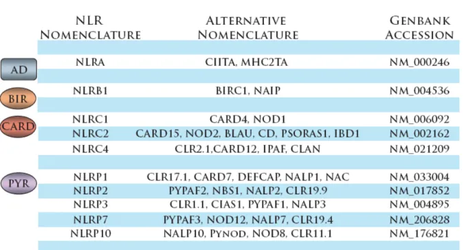
1.1 NLRs discussed in this dissertation, classified by effector
domain ………...………...……… 3 1.2 Summary of symptoms characteristic of CAPS ……….… 17
1.3 A comparison of cell death pathways ……… 28 3.1 Results of all cytokines and chemokines profiled using
RayBiotech G Series 3 cytokine antibody array ………..… 114 3.2 All cytokines and chemokines quantified by RayBiotech
LIST OF FIGURES
Figure
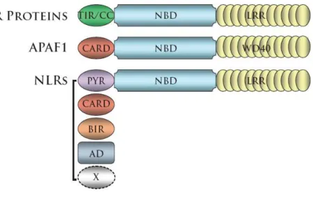
1.1 NLRs are structurally similar to the proapoptotic protein APAF1 and the plant cell death mediating NBS-LRR proteins ……….… 5
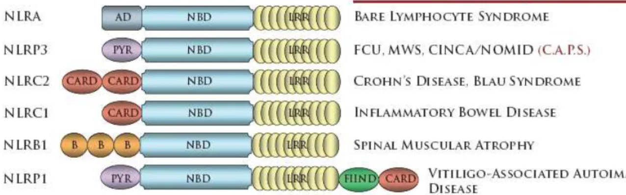
1.2 Mutations in NLRs have been identified in several human
diseases………...… 13
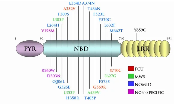
1.3 NLRP3 disease associated mutations cluster within or proximal to the
nucleotide binding domain ………..…………..…… 15
1.4 Depiction of the NLRP3 Inflammasome ………...…… 18 1.5 Comparison of APAF1 and NLRP3 Cell Death Pathways ………... 19 1.6 Specificity of pathogens and pathogen components among
NLRs involved in the induction of cell death and inflammation………….... 23 1.7 Structure of peptidoglycan derivatives recognized by NLRC1
and NLRC2 ……….37 2.1 Disease-associated cryopyrin causes necrotic-like cell death ……… 64 2.2 Disease-associated CIAS1 induces IL-1β release, but cell death
is IL-1β independent ……….. 66 2.3 Mutant CIAS1-induced THP-1 cell death is ASC dependent ……… 68 2.4 HMGB1 is released from THP-1 cells following expression of
disease-associated CIAS1 mutants ……… 70 2.5 Cell death in cells isolated from CAPS patients and in cells
expressing wildtype CIAS1 ……… 71 2.6 Shigella flexneri induced cell death and IL-1β require CIAS1
and ASC but not caspase-1 ……….72 2.7 Shigella flexneri induces cryopyrin-dependent necrosis ……… 74
2.9 Staurosporine induced cell death and mitochondrial membrane
permeability transition controls ………. 78 2.10 Salmonella induced cell death does not require CIAS1 ………. 80
2.11 Glycine fails to abrogate S. flexneri initiated cell death or
IL-1β release ……….. 81 3.1 Klebsiella pneumoniae induced cell death and IL-1β requires
NLRP3 and ASC ………...………...……… 102 3.2 Klebsiella pneumoniae induces NLRP3 and Cathepsin B
dependent pyronecrosis ……….…………..…… 104 3.3 Klebsiella pneumoniae induces inflammatory cytokines and
chemokines in primary macrophages ………...……… 106 3.4 NLRP3 and ASC deficient mice demonstrate increased
mortality following K. pneumoniae airway infection ………..… 108
3.5 NLRP3 deficient mice demonstrate significantly attenuated
airway inflammation f ollowing K. pneumoniae infection …………...…… 109 3.6 NLRP3 regulates Klebsiella pneumoniae induced IL-1β and
necrotic cell death in vivo ………..………..…… 111 3.7 Increased mortality of Klebsiella pneumoniae infected
LIST OF ABBREIVATIONS
ASC Apoptosis-associated speck-like protein containing a CARD BIR Baculovirus inhibitory repeat
CARD Caspase Recruitment Domain
CATERPILLER CARD, transcription enhancer, R(purine)-binding,pyrin, lots of leucine repeats
CIAS1 Cold-Induced Autoinflammatory Syndrome 1
CINCA Chronic Infantile Neurologic Cutaneous and Articular syndrome FCAS Familial Cold Autoinflammatory Syndrome
IAP Inhibitor of apoptosis
ICE Interleukin Converting Enzyme IL-1β Interleukin-1β
IPAF ICE-Protease Activating Factor
LRR Leucine Rich Repeat
MWS Muckle-Wells Syndrome
NACHT NAIP, CIITA, HET-E,and TP-1
NOMID Neonatal-Onset Multisystem Inflammatory Disease NBD Nucleotide Binding Domain
NBS-LRR Nucleotide binding site–leucine rich repeats (aka NB-LRR) NLR Nucleotide-binding, leucine-rich repeat
PYPAF Pyrin-containing APAF1-like proteins
CHAPTER I
INTRODUCTION
1.1
Summary
Inflammation is a crucial element of the host response to cellular insult. Pathogen-induced inflammation includes a molecular pathway which proceeds through activation of the protease caspase-1 to the release of the inflammatory cytokines IL-1β and IL-18.
Importantly, pathogens may also induce forms of cell death with inherently pro-inflammatory features. Here I review recent evidence demonstrating that NLR family proteins serve as a common component of both 1-activated apoptotic pathways and
1.2
Introduction
Table 1.1 - NLRs discussed in this dissertation, classified by effector domain
1.3
NLR Domain Organization
NLR Effector Domains
NLR effector domains, when defined, consists of an N-terminal CARD, pyrin, BIR or Activation Domain, all of which engage in homotypic interactions to mediate signaling downstream of NLR molecule activation. Though the overall domain organization of NLRs has been conserved from the plant NBS-LRR proteins, the R proteins utilize both TIR (Toll-IL-1 receptor) and CC (coiled-coil) domains as effectors. Much insight can be gained through analysis of the individual effector domains, namely the potential involvement of NLRs in the regulation of inflammation and cell death.
NLR Effector Domains: Caspase Recruitment Domain (CARD)
involved in signaling downstream of TNFR1 (Duan and Dixit, 1997)1 . Binding to RIP through its death domain, RAIDD recruits and activates ICH-1 (Duan and Dixit, 1997). More recently, a similar function has been described in which RAIDD recruits ICH-1 to the
PIDDosome, an apoptotic death initiating complex initiated by p53 in response to DNA damage (Tinel and Tschopp, 2004). Interestingly, RAIDD itself was identified in a search for motifs similar to the N-terminus of ICH-1. Within the original report the authors repeatedly note the resemblance of RAIDD and ICH-1 to ced-3, the C. elegans gene required for cell death during development. (Ellis and Horvitz, 1986). Indeed, Tschopp utilized the similarity within the N-terminus of these same 3 proteins to define and identify CARD in other
caspases (Caspase-4, Mch6, ICE), homologues of the viral apoptosis inhibitor lAP (c-lAP1, c-lAP2), and the nematode cell death protein ced-4 (Hofmann et al., 1997). Shortly
thereafter, CARDs were found in many proteins involved in cell death and inflammation. Amongst the more notable examples, the CARDs of APAF1 and caspase-9 interact during formation the apoptosome to initiate apoptosis after cytochrome c release from the
NLR Effector Domains: Pyrin Domain
Like CARDs, the pyrin domain also belongs to the death-domain-fold superfamily. The pyrin domain was initially described by Bertin et al in late 2000 (Bertin and DiStefano, 2000). Based on the resemblance of NLRC1 to plant R-proteins and APAF1, this group reasoned that though NLRC1 contains an identifiable N-terminal CARD, similar proteins may contain novel, non-CARD signaling domains. To identify these, Bertin et al searched for proteins possessing regions similar to the central nucleotide binding domain of NLRC1. Amongst the results were pyrin, NBS1, CARD7, zebrafish caspase-13, and ASC (Bertin and DiStefano, 2000). Ultimately, the term “pyrin domain” was coined to represent the ~95 residue region of shared homology between pyrin, NBS1, CARD7. Within weeks, Pawloski
et al also reported the existence of this domain (Pawlowski et al., 2001) . Unlike Bertin et al,
Pawlowski et al searched for proteins with sequence similarity to the first 100 amino acids of
which utilize the domain to engage in homotypic interactions with ASC to both positively and negatively regulate the activation of caspase-1 (For more information, see chapter 1.6, NLR Inflammasomes)
NLR Effector Domains: Baculovirus Inhibitory Repeats (BIR) Domain
NLRB1 (formerly NAIP5) is unique within the NLR family as the only member containing BIR (Baculovirus IAP Repeat) effector domain (Listen et al., 1996). The ~70 amino acid BIR domain can occur in up to three tandem copies and function as a zinc binding domain (Crook et al., 1993). The BIR domain was identified in 1993 by Lois Miller and her colleagues following a series of experiments involving the Autographa californica virus (Crook et al., 1993). Infection of SF-21 cells with p35 mutant A. californica viruses results in cellular apoptosis before completion of viral replication. Specifically, these mutants are unable to delay cell death sufficiently to allow the formation of polyhedral occlusion bodies. To identify baculovirus genes with the ability to inhibit host cell apoptosis, Crook et
al infected SF-21 cells with p35 mutant A. californica while concurrently transfecting a
involved in the regulation of cell death. Subsequent identification and analysis of similar anti-apoptotic baculovirus genes elucidated a common repeating sequence (GX2YX4DX3
CX2CX6WX9HX6-10C) the authors termed the BIR (Baculovirus IAP Repeat) (Birnbaum et
al., 1994). The BIR was subsequently identified in species ranging from viruses to mammals and now is the defining characteristic of a whole family of anti-apoptotic proteins known as the IAPs (Inhibitors of Apoptosis) (Deveraux and Reed, 1999). Both in vitro and in vivo, NLRB1 has been shown to inhibit apoptosis in response to several stimuli by BIR-dependent inhibition of effector caspases (Diez et al., 2000; Maier et al., 2002). NLRB1 also limits the intracellular replication of Legionella pneumophila, though this function is independent of its ability to inhibit apoptosis and the role of the BIR domains in this process is unclear (Diez et al., 2000; Wright et al., 2003).
NLR Effector Domains: Transactivation Domain (AD)
resembles a CARD, but this region lacks caspase recruitment capabilities (Nickerson et al., 2001). Currently, the AD has no known involvement in initiating or regulating cell death.
NLR Nucleotide Binding Domains
Similar to APAF1, NLRs possess a large central nucleotide binding domain (NBD) which regulates activation and oligomerization of the molecule. Though APAF1 has been shown to bind adenosine-based nucleotides, the nucleotide specificity of few NLRs has been characterized (Kim et al., 2005; Zou et al., 1999). ATP binding by APAF1 is required for caspase-3 activation, thus nucleotide binding and hydrolysis are likely required for NLR activity. Indeed, the binding of ATP is necessary for NLRP3 and NLRC4-mediated cell death and inflammation. (Duncan et al., 2007; Lu et al., 2005). ATP is also preferred by NLRP12 and is required for NLRP12 to inhibit NFκB signalling (Ye et al., 2008). Not all NLRs bind adenosine-based nucleotides, NLRA requires GTP binding for its oligomerization and transactivation functions (Linhoff et al., 2001). Interestingly, some NLRs may bind nucleotides without preference. Such is the case with NLRP1, which binds nucleotide triphosphates indiscriminately while activating caspase-1. (Faustin et al., 2007). NLR
reliance on nucleotide binding suggests nucleotide analogs may have use in modulating NLR cell death and inflammatory pathways and in treatment of NLR associated inflammatory disorders.
NLR Leucine Rich Repeats
1.4
Disease Associated Mutations in NLRs
Figure 1.2 - Mutations in NLRs have been identified in several human diseases.
An understanding of the in vivo role of NLR proteins is aided by the remarkable association of NLR genes with human immune disorders. A summary of diseases associated with NLR mutations is shown in Figure 1.2. NLR molecules were defined based on their resemblance to NLRA. Mutations in NLRA result the absence of MHC class II expression on immune cells, a condition known as Bare Lymphocyte Syndrome (Steimle et al., 1993). Mutations in NLRP3 have been identified in a trio of dominantly inherited autoiflammatory disorders collectively referred as CAPS (CIAS1 Associated Periodic Syndromes)
1.5
Cryopyrin Associated Periodic Syndromes (CAPS)
Figure 1.3- NLRP3 disease associated mutations cluster within or proximal to the nucleotide binding domain. No correlation between location of the mutation and resulting severity of disease. Few mutations span the CAPS spectrum; these are labeled as “non-specific.”
Significant attention has been focused on one NLR family member, NLRP3, which is mutated in a trio of dominantly-inherited periodic fevers: FCAS, Muckle-Wells Syndrome, (Hoffman et al., 2001a), and CINCA/NOMID.(Feldmann et al., 2002) These disorders are characterized by spontaneous, yet recurrent, outbreaks of fever, rash, and the excessive production of IL-1β in the absence of high titers autoantibodies or antigen-specific T cells.
Since several NLRP3 point mutations and clinical symptoms have been identified as overlapping between FCAS, MWS, and CINCA/NOMID, it has been proposed that these fevers comprise a continuum of disease severity collectively referred to as CAPS (Cryopyrin Associated Periodic Syndromes). A summary of symptoms associated with each disorder is presented in Table 1.2. FCAS, the mildest of the three, is characterized by recurring
outbreaks of fever, urticaria rash, and conjunctivitis following generalized cold exposure. Each outbreak typically last less than 24 hours, with symptoms often peaking in the evening and resolving before morning. These attacks begin before 6 months of age and persist through adulthood (Hoffman et al., 2001b). Muckle-Wells syndrome represents an
Table 1.2 – Summary of symptoms characteristic of CAPS
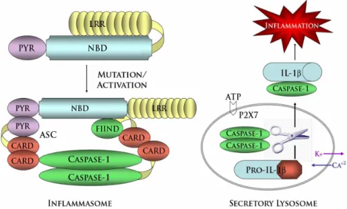
To date, hyperactive formation of the inflammasome (discussed in detail, chapter 1.6, NLR Inflammasomes) has been identified as the predominant defect underlying the excessive IL-1β and inflammation associated with CAPS. On a molecular level, NLRP3 mutations likely weaken the auto-inhibitory interactions between the NBD and LRRs, thereby yielding a hyperactive form of NLRP3 more readily able to form the inflammasome (Fig. 1.4)
of an oral caspase-1 inhibitor has also proven effective in blocking the activation and secretion of IL-1β in CAPS patients (Stack et al., 2005).
1.6
NLR Inflammasomes
Figure 1.5 - Comparison of APAF1 and NLRP3 Cell Death Pathways. Release of cytochrome c from the mitochondria initiates apoptosome-dependent apoptosis. Cytochrome c induces a conformational change in APAF1 to relieve the intramolecular interactions holding the molecule in an auto-inhibited state. Once activated, APAF1 and cytochrome c undergo dATP dependent-oligomerization into a heptameric wheel-like structure termed the apoptosome. Pro-caspase-9 molecules aggregate through homotypic interactions between their CARDs and those of APAF1. Subsequent homodimerization of pro-caspase-9 generates active caspase-9 molecules which cleave and activate the effector caspases -3 and -7 to induce apoptotic cell death. Similarly, NLRP3 activation or disease associated mutations may weaken inhibitory intramolecular interactions between the NLRP3 NBD and its C-terminal LRRs. Utilizing the adaptors ASC and CARDINAL, activated NLRP3 aggregates caspase-1 molecules through the formation of an inflammasome complex. Complex formation potentiates subsequent caspase-1 cleavage and activation. Based on studies of the NLRP1 inflammasome, the complex is likely comprised of 5-7 subunits, each with the ability to recruit pro-caspase-1 molecules. IL-1β is activated by caspase-1 mediated cleavage of the inactive pro-IL-1 β precursor molecule. However, formation of the inflammasome is not the only function of NLRP3. Activated NLRP3 also initiates pyronecrosis, a
molecular pathway of necrotic cell death which is dependent on ASC and proceeds through cathepsin B. This cell death pathway does not rely on caspase-1 or IL-1 β, and thus is independent of inflammasome function.
of cell death, several NLR proteins act to promote a caspase-dependent program of
inflammation. Research on the latter has mainly focused on formation of protein complexes called inflammasomes (Martinon et al., 2002; Srinivasula et al., 2002). In some respects the inflammasomes resemble the apoptosome, which includes APAF1 (Fig. 1.5). APAF1 is composed of an N-terminal CARD, a central NBD, and C-terminal WD-40 repeats. It is thought to be held inactive by intramolecular contact between its WD-40 repeats and N-terminal regions until cytochrome c and dATP relieve this inactive conformation. This enables assembly of the apoptosome, leading to activation of pro-caspase-9 (Schafer and Kornbluth, 2006). The inflammasome NLR proteins appear to act in an analogous manner, in that intramolecular interaction mediated by the C-terminal LRRs is proposed to hold these proteins in an inactive formation until stimulation promotes inflammasome assembly and activating cleavage of pro-caspase-1. (Fig 1.4)
Biochemical studies have identified a number of inflammasomes, which promote inflammation by activating caspase-1, resulting in the release of the pyrogenic cytokine IL-1β and IL-18 from cells treated with different stimuli (Agostini et al., 2004; Duncan et al., 2007) (Fig 1.6). Though differing slightly in their makeup, each inflammasome includes the IL-1β-converting enzyme pro-caspase-1, as well as one of four NLR proteins: NLRP1, NLRP2, NLRP3, or NLRC4. Given that over 20 human NLR genes have been recognized, it is likely that more NLR inflammasomes exist. A potential fourth inflammasome containing the related molecule pyrin has already been identified (Yu et al., 2005).
2007; Mariathasan et al., 2004; Miao et al., 2006). The NLRP1 inflammasome is required for caspase-1 activation in response to anthrax lethal toxin (Boyden and Dietrich, 2006). To date, the NLRP3 inflammasome is associated with the widest range of stimuli, including LPS in the presence of ATP, uric acid crystals, poly I:C, bacterial and viral RNA, and both gram-positive and gram-negative bacteria (Kanneganti et al., 2006a; Kanneganti et al., 2006b; Mariathasan and Monack, 2007; Mariathasan et al., 2006a; Martinon et al., 2006; Sutterwala et al., 2006; Willingham et al., 2007). Though the pathogen recognition steps leading to inflammasome activation continue to be elucidated, recent work has shown that cytosolic bacterial molecules can induce NLRP3-mediated caspase-1 activity independently of TLR signaling (Kanneganti et al., 2007) (For extensive list of activators of NLR inflammasomes, see Fig. 1.6)
Two recent papers describe biochemical stages in inflammasome activation (Agostini et al., 2004; Faustin et al., 2007). Using purified components of the NLRP1 inflammasome, Faustin et al. demonstrated that assembly of this inflammasome complex required the
microbial product muramyl dipeptide (MDP) as well as the presence of nucleotide (Faustin et al., 2007). Surprisingly, and in contrast to the apoptosome, the NLRP1 inflammasome exhibited little nucleotide specificity. However, this does not appear to be true for the NLRP3 inflammasome. Duncan et al. showed that NLRP3 binds specifically to ATP or dATP and acts as an ATPase. NLRP3-catalyzed nucleotide hydrolysis was shown to be vital for NLRP3 self-association, interaction with the inflammasome adaptor protein ASC,
caspase-1 activation and IL-1 release (Duncan et al., 2007).
function by interfering with the recruitment and activation of caspase-1 (Druilhe et al., 2001; Humke et al., 2000; Lee et al., 2001). The pyrin-only proteins POP1 and POP2 also inhibit inflammasome formation through their ability competitively interact with ASC, thereby blocking inflammasome formation (Bedoya et al., 2007; Lee et al., 2001). Finally, several NLR molecules also negatively regulate the inflammasomes. NLRP10 has the potential to inhibit both caspase-1-dependent IL-1β secretion as well as ASC-mediated NF-κB activation. Together, NLRP2 and NLRP7 accomplish the same function. However, NLRP7 is restricted to blocking caspase-1 activation while NLRP2 blocks ASC-mediated NF-κB activation without influencing caspase activity. (Kinoshita et al., 2005)
1.7 NLRs at the Intersection of Cell Death and Immunity
Several routes to cell death
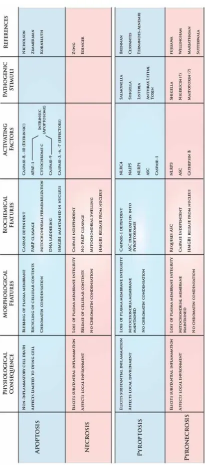
Though cell death is known to play an important role in the immune system, the majority of studies have focused on the role of apoptosis in cell death. Emerging evidence suggests that additional cell-death pathways are crucial for triggering of inflammation and immunity. To begin to understand the contribution of cell death to immunity, it is useful to highlight a number of key differences between these types of death (Table 1.3).
polymerase (PARP), DNA-PK, and other regulatory and structural proteins that maintain cellular and genomic integrity (Nicholson, 1999). Cumulatively, the cleavage of these substrates leads to the death and breakdown of the cell.
In contrast to apoptosis, necrosis has been considered by some to be a passive, and therefore unprogrammed, form of cell death. In general, apoptosis relies on the protease activity of caspases, while necrosis is caspase-independent. Whereas apoptosis is an energy expensive process, necrosis has been described as bioenergetic failure, meaning that the cell lacks sufficient energy resources to maintain its metabolism. This condition may be
triggered by the loss of ion pump activity or overconsumption of ATP (Zong and Thompson, 2006). At the nuclear level, necrosis is distinguished from apoptosis by the persistence of DNA content, which remains uncondensed. Perhaps the most striking difference between these forms of cell death is at the plasma membrane. Apoptosis is a slow process marked by membrane blebbing and the packaging of cellular material for recycling, but necrosis is characterized by rapid loss of plasma membrane integrity with the resultant release of cellular contents into the extracellular medium (Edinger and Thompson, 2004).
This last feature is central to the importance of necrosis in an immune and
released, HMGB1 becomes an agonist for RAGE (receptor for advanced glycosylation endproducts) and the Toll-like receptors (TLRs) TLR2 and TLR4, all of which are expressed by monocytes and some other cell types (Lotze and Tracey, 2005; Park et al., 2004).
Activation of these receptors results in the exacerbation of inflammation in the
microenvironment through the induction of additional pro-inflammatory cytokines (Sunden-Cullberg et al., 2006). Recent work has identified two more forms of cell death, pyroptosis and pyronecrosis, which appear to exploit the pro-inflammatory features of necrosis within the context of immunity. However, the extent to which each resembles apoptosis and necrosis is different.
Pyroptosis is a cell-death pathway activated by microbial pathogens, including
Salmonella and Listeria (Brennan and Cookson, 2000; Cervantes et al., 2008). Pyroptosis is
similar to apoptosis in that DNA damage occurs and the process is caspase-dependent(Fink and Cookson, 2005). However pyroptosis does not rely on the classical pro-apoptotic initiator and effector caspases (caspases 3, 8 and 9), but rather on caspase-1. In addition to its apoptotic qualities, pyroptosis exhibits some features of necrosis. Similar to necrosis, pyroptosis is characterized by plasma membrane breakdown. Moreover, mitochondrial membrane integrity is maintained during pyroptosis (Cervantes et al., 2008; Fink and
Cookson, 2005). Ongoing studies are aimed at identifying additional molecular mediators of pyroptosis. Very recent work has described the pyroptosome, a large complex comprised of ASC dimers that assembles as a feature of this process (Fernandes-Alnemri et al., 2007). ASC is a common binding partner for NLR family proteins. Though it has been
vivo (Fernandes-Alnemri et al., 2007). From an inflammation standpoint, the two
outstanding features of pyroptosis are the activation of caspase-1 and the breakdown of the plasma membrane.
Two very recent studies have identified another NLR-dependent pathway of pro-inflammatory cell death, termed pyronecrosis, which has primarily necrotic features
(Fujisawa et al., 2007; Willingham et al., 2007). This form of cell death is found in genetic autoinflammatory diseases involving mutations in the NLRP3 gene and is also associated with microbial pathogens such as Shigella flexneri (Willingham et al., 2007). Unlike pyroptosis, pyronecrosis is caspase-independent; neither the activating cleavage of effector caspase-3 nor its substrate PARP occur during pyronecrosis, and cell death proceeds in the presence of caspase-1-specific inhibitor and pan-caspase-inhibitor (Fujisawa et al., 2007; Willingham et al., 2007). However, cell death is abrogated in the presence of an inhibitor of the lysosomal protease cathepsin B, implicating lysosome activity in the pathway (Fujisawa et al., 2007). Additional hallmarks of apoptosis are not observed. Pyronecrotic cells demonstrate neither DNA fragmentation nor the loss of mitochondrial membrane potential (Willingham et al., 2007). As determined by electron microscopy, the morphological changes characteristic of pyronecrosis are consistent with necrosis and include membrane degradation and uncondensed chromatin (Fujisawa et al., 2007; Willingham et al., 2007). Similar to classical necrosis, pyronecrosis is accompanied by release of the pro-inflammatory cytokine HMGB1 (Willingham et al., 2007) Recent work suggests an intriguing connection between cell death pathways and the NLR proteins, which are early mediators of
NLR proteins, apoptosis, and pyroptosis
Although a comparison between inflammasome and apoptosome activation may help to illuminate steps in the induction of inflammation, similarities extend to function as well. Much attention has been paid to the role of the NLR protein NLRB1 in determining
susceptibility to Legionella pneumophila (Wright et al., 2003). In addition to inducing the release of IL-1β through the NLRC4 inflammasome, cytosolic L. pneumophila flagellin also activates a caspase-1-dependent form of cell death in macrophages which requires NLRB1 (Suzuki et al., 2007; Zamboni et al., 2006). One characteristic of this cell-death pathway is nuclear condensation, which is typical of apoptosis (Molofsky et al., 2006). Moreover, membrane blebbing, another feature of apoptosis, was observed in NLRB1-expressing HEK293 cells following infection with L. pneumophila (Zamboni et al., 2006). However, unlike classical apoptosis, L. pneumophila flagellin-induced macrophage cell death has been reported to be independent of caspase-3 activity (Molofsky et al., 2006). Thus the observed apoptotic features may instead be related to pyroptosis. Notably, caspase-1 and NLRC4-dependent cell death has been observed at timepoints of less than three hours in Shigella
flexneri infected cells (Suzuki et al., 2007). NLRC4 is also required for cell death in
Salmonella typhimurium-infected macrophages, which is mediated by bacterial flagellin
(Franchi et al., 2007; Mariathasan et al., 2004). Though features of the mechanism remain to be determined, these results demonstrate that an NLR protein is required to mediate an apoptosis-like cell death induced by a bacterial component.
inflammasome is activated by the bacterial product MDP, resulting in the maturation of IL-1β. This process is regulated by two members of the anti-apoptotic BCL2 (B-cell
CLL/lymphoma 2) family of mitochondrial membrane proteins. Both BCL2 and BCLXL
bind to NLRP1 directly to suppress its activity (Bruey et al., 2007). These data illustrate a surprising cross-talk between inflammatory and anti-apoptotic signaling, though the
influence of this interaction on cell survival or death has not yet been determined. Although the regulation of inflammation is an unexpected role for BCL2 family members, the interface between mitochondrial membrane factors and innate immunity is not unprecedented. Recent work has established the mitochondrial outer membrane as a critical staging area for anti-viral signaling through MAVS (mitochondrial antianti-viral signaling, also called IPS-1, VISA, and CARDIF) the RIG-I (retinoic acid inducible gene-I)-like RNA helicases, and the NLR protein NLRX1 (Moore et al., 2008; Seth et al., 2005; Yoneyama and Fujita, 2007).
NLR proteins, necrosis, and pyronecrosis
The induction of necrotic cell death as a crucial component of immunity is well established across phylogenic kingdoms. The plant NBS-LRR disease resistance proteins act in the defense against pathogens by helping to mediate the hypersensitive response, a form of rapid programmed cell death, and it has recently been shown that the NLR family protein NLRP3 mediates a similar pathway in monocytes (Fujisawa et al., 2007; Willingham et al., 2007). NLRP3 was first identified through its association with two dominantly inherited periodic fevers: FCAS (Familial Cold Autoinflammatory Syndrome) and Muckle-Wells Syndrome (Aganna et al., 2002; Hoffman et al., 2001a). It has since been identified as the genetic locus for a third fever syndrome, CINCA/NOMID. (Aksentijevich et al., 2002; Feldmann et al., 2002). These three diseases are now considered to be a spectrum of severity for one single condition, cryopyrin-associated periodic syndrome (CAPS), which is
characterized by spontaneous inflammation (Ting et al., 2006). This suggests that disease-associated variants of NLRP3 may encode a hyperactive version of NLRP3 that promotes excessive production of IL-1β, a possibility that is consistent with the gain-of-function phenotype typically associated with dominant inheritance. Indeed, following stimulation, monocytes isolated from patients with NLRP3 mutations demonstrate hyperactivation of IL-1β {Agostini}(Janssen et al., 2004; Stack et al., 2005).
However, this is not the extent of the phenotypic changes associated with mutant
NLRP3. Peripheral blood mononuclear cells isolated from patients with NLRP3 mutations
lose viability when exposed to lipopolysaccharide (LPS) (Saito et al., 2008; Willingham et al., 2007). To identify more precisely the cellular consequences of mutant NLRP3
NLRP3 (Fujisawa et al., 2007; Willingham et al., 2007). The expression of these variants in the monocytic cell line THP-1 induces excessive IL-1β release, as expected, but also an inflammatory necrosis that we termed pyronecrosis (Willingham et al., 2007). Intriguingly, pyronecrosis is not dependent on IL-1β signaling or caspase-1, though it requires the
presence of the inflammasome component ASC and intact cathepsin B(Fujisawa et al., 2007; Willingham et al., 2007). The binding of ATP to NLRP3 is also necessary for this pathway to proceed (Duncan et al., 2007). Because NLRP3 and ASC appear to act together in a function that is independent of procaspase-1 activation, and hence independent of the inflammasome, we suggest that these two factors comprise an alternate complex to promote pyronecrosis (Fig. 1.5). Cumulatively, these observations offer insight into the consequence of NLRP3 hyperactivity. The inherent function of the protein and its relationship to
pathogen resistance merit further consideration.
flexneri-induced necrotic cell death was shown to be dependent on NLRP3 in both mouse
macrophages and in the human monocytic cell line THP-1. Similar to mutant NLRP3-induced cell death, this process also depends on ASC and requires the protease cathepsin B (Willingham et al., 2007). These findings demonstrate that NLRP3 mediates pyronecrosis as part of its native function within the cell, and show that programmed necrosis can be
activated in response to pathogen invasion of macrophages. We suggest that this program is an adapted response to bacterial invasion. Not only does rapid cell death deny the pathogen an environment in which to replicate, the process of necrosis is inherently pro-inflammatory, leading to release of IL-1β and other factors from surrounding cells. Thus, pyronecrosis is likely to contribute substantially to the disease state in patients with CAPS, who suffer from spontaneous inflammation characterized by IL-1β production.
In its more severe forms, CAPS is also characterized by joint deformities and arthralgias (Ting et al., 2006). NLRP3 expression is not limited to monocytic cells but extends to osteoblasts as well, and the joint-related symptoms of CAPS are likely due to excessive NLRP3 activity in these cells. As with Shigella in macrophages, wild type NLRP3 appears to also regulate pathogen-induced cell death in osteoblasts. Though Salmonella
typhimurium activates the NLRC4 inflammasome in macrophages, mouse primary
osteoblasts do not express NLRC4. In these cells NLRP3 is partly required for maximal S.
typhimurium-induced cell death (McCall et al., 2008). This finding suggests that
pyronecrosis may contribute to the joint-related symptoms of CAPS Moreover, it
The induction of necrosis by Shigella, Salmonella, and other microbial pathogens may be mediated through toxins. Some of these toxins have been examined directly with respect to caspase-1 activation and cell death. Nigericin is a toxin produced by Streptomyces
hygroscopicus. This molecule functions as a potassium ionophore and is a potent inducer of
both IL-1β release and necrosis in monocytes (Hentze et al., 2003; Perregaux et al., 1992). As with pyronecrosis, both functions are dependent on the activity of cathepsin B (Hentze et al., 2003). Another potent toxin, maitotoxin, is produced by the dinoflagellate
Gambierdiscus toxicus. Maitotoxin has been demonstrated to induce necrosis in a manner
dependent on the calcium-activated cysteine protease calpain and also promotes IL-1β release by mouse macrophages (Verhoef et al., 2004; Zhao et al., 1999). Similar to S.
flexneri, both nigericin and maitotoxin activate the NLRP3 inflammasome (Mariathasan et
al., 2006a; Sutterwala et al., 2006). Moreover, these toxins also alter the levels of
1.8 NLRC1 and NLRC2 Function in Immunity and Inflammation
Perhaps the most well studied NLRs are NLRC1 and NLRC2 (formerly NOD1 and NOD2). In 1990, Yuan and Horvitz described a simple pathway in the nematode C. elegans wherein CED-4 interacts with and proteolytically activates CED-3, resulting in cell death (Yuan and Horvitz, 1990). Subsequently, a number of serine proteases, including caspase-1 and caspase-3, had been identified as human orthologs of CED-3, while no ortholog had been found for CED-4 (Fernandes-Alnemri et al., 1994; Xue et al., 1996; Yuan et al., 1993). That changed in 1997 when Zou et al isolated APAF1, a mammalian CED-4 homolog responsible for the cytochrome-C dependent activation of caspase-3 and cell death. (Zou et al., 1997). Upon these reports, NRLC1 was identified near simultaneously in 1999 by two groups searching for proteins resembling the CARD domain of APAF1 (Bertin et al., 1999; Inohara et al., 1999). In turn, NLRC2 identified based on genome search for proteins similar to NLRC1 (Ogura et al., 2001). Interest in NLRC1 and NLRC2 was further intensified when mutations these genes were identified in inflammatory bowel syndrome, Crohn’s disease, and Blau Syndrome (Hugot et al., 2001; Miceli-Richard et al., 2001; Ogura et al., 2001). More than 90% of NLRC2 mutations identified in Crohn’s disease occur within or proximal to the LRR region suggesting a crucial role of for this domain the development of disease (Lesage et al., 2002).
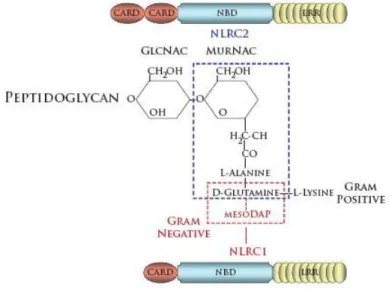
component in the cell wall of Gram positive bacteria, while only existing as a thin layer in the periplasmic space of Gram negative bacteria. Hydrolases constantly degrade PGN into small glycan chains containing alternating acetylglucosamine (GlcNAc) and
N-acetylmuramic acid (MurNAc) sugars. These sugar moieties are linked together by small peptides. Importantly, the composition of gram positive and gram negative PGN differ from each other. In Gram-positive bacteria, the amino acid lysine is found at the third position in the linker peptide, whereas diaminopimelic acid (DAP) is found in most Gram-negative bacteria (Girardin et al., 2003c). Both NLRC1 and NLRC2 recognize naturally occurring PGN degradation products, albeit through distinct mechanisms. The minimal natural structure recognized by NLRC1 is GlcNAc-MurACc-LAla-c-D-Glu-meso-DAP
(GM-triDAP) (Girardin et al., 2003a). The presence of the terminal meso-DAP implicates NLRC1 in the detection of Gram negative bacteria. In contrast, NLRC2 responds to the muropeptide GlcNAc-MurNAc-LAla-D-isoGln (GM-Di), and thus can detect both Gram-positive and Gram-negative bacteria. NLRC2 also responds to muramyl dipeptide (MurNAc-L-Ala-D-isoGln) (Girardin et al., 2003b; Inohara et al., 2003). While this is not a naturally occurring bacterial product, it also can be isolated from both Gram-negative and Gram-positive
Figure 1.7 - Structure of peptidoglycan derivatives recognized by NLRC1 and NLRC2
Initial reports demonstrated that both NRLC1 and NLRC2 utilize homotypic CARD interactions to bind the serine-threonine kinase RICK. RICK interacts with IKKγ, resulting in IκB phosphorylation, and ultimately the activation of NκFB (Bertin et al., 1999; Inohara et al., 1999; Inohara et al., 2000). Indeed, RICK is essential for NLRC1 and NLRC2 activation, as NLRC1 and NLRC2-dependent NFκB activation was abolished in RICK deficient mouse embryonic fibroblasts (Kobayashi et al., 2002). Furthermore, the interaction of NLRC2, RICK, and IKK has also been shown to be essential for the activation of NFκB and JNK pathways following Shigella flexneri infection. Either NLRC1 or NLRC2 also been shown to mediate NFκB signaling in response to several pathogens including Heliobacter pylori,
Pseudomonas aeruginosa, Campylobacter jejuni, Chlamydophila pneumoniae, and Listeria
monocytogenes (Fig. 1.6) (Cervantes et al., 2008; Opitz et al., 2005; Travassos et al., 2005;
1.9 Conclusions
Emerging evidence reveals that NLR proteins contribute to the host cell response to insult by not only facilitating the maturation of IL-1β, but also by mediating cell death (Fig. 1.5). Both processes have a strong impact on immunity. IL-1β release is a well-established signal for the onset of inflammation and initiation of the adaptive immune response. The consequences of pathogen-induced cell death in the context of immunity have not been studied as thoroughly, although a plethora of reports have now shed light on this topic. One obvious result is that invading bacteria are denied an environment in which to replicate. However, cell-death programs which result in a loss of plasma-membrane integrity can also exacerbate inflammation through the discharge of such intracellular inflammatory cytokines and factors such as IL-1β, TNFα, and HMGB1.
CHAPTER II
MICROBIAL PATHOGEN-INDUCED NECROTIC CELL DEATH
MEDIATED BY CIAS1 (NLRP3) AND ASC
2.1
Abstract
Cryopyrin/CIAS1/NLRP3 and ASC are important components of the inflammasome, a multi-protein complex required for caspase-1 activation and cytokine IL-1β production.
CIAS1 mutations underlie autoinflammation characterized by excessive IL-1β secretion. Disease-associated cryopyrin also causes a program of necrosis-like cell death in
macrophages, the mechanistic details of which are unknown. We find that patient monocytes carrying disease-associated CIAS1 mutations exhibit excessive necrosis-like cell death by a process dependent on ASC and cathepsin B, resulting in spillage of the proinflammatory mediator HMGB1. Shigella flexneri infection causes cryopyrin-dependent macrophage necrosis with features similar to the death caused by mutant CIAS1. This necrotic death is independent of caspase-1 and IL-1β, and thus independent of the inflammasome.
2.2 Introduction
The NLR family (Harton et al., 2002a) (formerly CATERPILLER) is comprised of proteins involved in the regulation of innate immunity (Inohara and Nunez, 2003; Martinon and Tschopp, 2005). Functionally similar to the evolutionarily conserved Toll-like receptors (TLRs), increasing evidence suggests that NLRs may serve as intracellular molecules that sense pathogen-derived products (Hoffmann and Reichhart, 2002; Poltorak et al., 1998). Significant attention has been focused one NLR family member, Cryopyrin, which is encoded by the gene CIAS1. CIAS1 is mutated in a trio of dominantly inherited periodic fevers: FCAS (Familial Cold Autoinflammatory Syndrome), MWS (Muckle-Wells
Syndrome), and CINCA/NOMID (Chronic Infantile Neurological Cutaneous and Articular syndrome / Neonatal Onset Multisystemic Autoinflammatory Disease), which are proposed to represent a continuum of severity for a single condition, CIAS1-associated periodic syndrome (CAPS) (Aksentijevich et al., 2002; Feldmann et al., 2002; Hoffman et al., 2001a; Hoffman et al., 2001b).
Recent investigations have highlighted an essential role for IL-1β in the development of mutant-CIAS1-associated periodic fevers. Mutant CIAS1 causes elevated levels of
spontaneous and induced IL-1β both in vitro and in vivo. Indeed, FCAS, MWS, and CINCA/NOMID have all been successfully treated with daily doses of the IL-1β receptor antagonist Anakinra® (Kineret) (Goldbach-Mansky et al., 2006; Hawkins et al., 2004;
mature IL-1β, which is released from the cell. Mutations in cryopyrin result in the
hyperactivation of this pathway, causing excessive IL-1β production and the severe episodes of inflammation.
The functions of cryopyrin in the immune system are not limited to autoinflammatory disorders. Several recent reports have established cryopyrin as an important adaptor capable of organizing the inflammasome to elicit IL-1β release in response to bacterial, viral, and other pro-inflammatory stimuli (Kanneganti et al., 2006a; Kanneganti et al., 2006b;
Mariathasan et al., 2006a; Mariathasan et al., 2006b; Martinon et al., 2006; Sutterwala et al., 2006). However, it is not yet known if the protein has additional biologic functions in the containment of pathogens.
Clues regarding an additional role for cryopyrin in response to pathogen may lie within its makeup. Cryopyrin consists of an amino terminal pyrin domain, a central NACHT (NAIP, CIITA, HET-E, TP1) domain, and seven carboxy terminal LRRs (Leucine Rich Repeats). This architecture is conserved in plants, where similar proteins comprise a
that mediate such a process and the mechanism of this form of cell death have yet been defined.
We report here that cryopyrin and ASC are required for a process of necrotic-like cell death. We furthermore expand the capabilities of cryopyrin by demonstrating that it
mediates both the IL-1β and cell death response to a gram-negative bacterium, S. flexneri, resulting in cellular necrosis and the exacerbation of inflammation. The observation that
2.3 Materials & Methods
Cell lines and reagents – THP-1 cells purchased from American Type Culture Collection (ATCC) and cultured as described previously (Williams et al., 2005). Anti-caspase-3 antibody purchased from Cell Signaling; anti-PARP, anti-ipaB, anti-Actin, and HRP-conjugated secondary antibodies from Santa Cruz Biotechnology; anti-HMGB1 antibody from Abcam; anti-ASC antibody from Immuno Diagnostic Oy; anti-Cryopyrin antibody from Alexis Biochemicals; Super Signal ECL reagent from Biorad; E. Coli LPS from Chemicon. Detailed methods for preparation of retroviral shuttle vectors, transduction, and sorting to generate THP-1 cell lines stably expressing shRNA have been described (Taxman et al., 2006). The shRNA target sequences are as follows: shASC-GCTCTTCAGTTTCACACCA, shCtrl-GCTCTTCctggcCACACCA, shCIAS-GGATGAACCTGTTCCAAAA. Stable expression of shRNA did not induce interferon response as assessed by OAS1 expression (not shown).
Generation of recombinant adenoviruses - Recombinant adenovirus expressing CIAS1 or
LacZ was generated using Adeno-X Expression System (Clontech). Briefly, genes were
Adenovirus transduction of THP-1 cells – THP-1 cells were aliquoted into Falcon 2059 polypropylene tubes at a density of 106/ml in RPMI 1640 containing 10% FBS. After
addition of adenovirus (MOI=1 unless otherwise noted), cells were centrifuged at 2000*g for 2 hours at 37O C. Immediately after centrifugation, cells were resuspended and incubated at 5*105/ml following the addition of fresh RPMI 1640 containing 10% FBS.
.
XTT assay – Cells were plated into 96-well plates at 20,000 cells per well 24 hours after
adenovirus transduction. 50 µl of serum-free media containing 25µM phenazine methosulfate and 1mg/ml XTT was added to each well. Plates were read at 450nM after four hours of incubation.
Mitochondrial membrane potential staining – Cells were stained with
Tetramethylrhodamine ethyl ester, perchlorate (TMRE) for 25 minutes at 37°C at a final concentration of 5nM. After staining, cells were rinsed in PBS, resuspended in 0.5 ml PBS and analyzed using a FACScan (Becton Dickinson) in FL2.
Immunoblotting – Immunoblots were performed as described previously (Williams et al., 2005). Cryopyrin was immunoprecipitated with rabbit anti-CIAS-1 peptide IgG. Expression was confirmed by probing immunoblots with anti-CIAS1. HMGB1 blots were performed directly on culture supernatants and developed as indicated.
Quantitative PCR - Total RNA was isolated, cDNA was reversed transcribed, and
ASC mRNA expression as described (Taxman et al., 2006). Realtime values were
standardized to the expression of 18s rRNA and normalized to 100 in control (untransduced) cells. Primers used for real-time PCR are as follows:
ASC-[AACCCAAGCAAGATGCGGAAG, TTAGGGCCTGGAGGAGCAAG], 18s-[CGGCTACCACATCCAAGG, GCTGCTGGCACCAGACTT].
Viaprobe and 7-AAD cell staining – Cells were collected and rinsed twice in cold PBS.
Pellets were resuspended in 0.5ml PBS with 3 µl Viaprobe (Becton Dickinson) or 1 µl 7-AAD (BD Pharmingen). Cells were incubated in the dark for 15 minutes before analysis on a FACScan (BD).
Propidium Iodide staining – Following treatment, cells were collected and pelleted via
centrifugation. Pelleted cells were fixed in 70% ethanol for a minimum of 2 hours, rinsed once in PBS, then resuspended in PBS containing 1% Triton, 20 µg/ml propidium iodide (Sigma Chemical Co.), and 200 µg/ml RNAse A (Qiagen). The cells were allowed to stain for 15 minutes or longer then analyzed using a FACScan (BD).
Electron microscopy – THP-1 cells were infected with adenoviruses at MOI=1 and fixed in 2% paraformaldehyde, 2.5% glutaraldehyde in 0.15M sodium phosphate, pH 7.4 2 or 6 hours post infection. Electron microscopy was performed at the UNC Microscopy Services Laboratory.
Patient cells – Two female patients (ages 62 and 71) with FCAS were included in the study. Both FCAS patients had classic clinical presentation and met diagnostic criteria,as described previously (Hoffman et al., 2001b). Neither FCAS subjectwas experiencing significant inflammatory symptoms at the timeof study, nor on regular anti-inflammatory medications. Two female controls (ages 39 and 41) were studied simultaneously with the FCAS patients. However, one control was later found to have significantly abnormal inflammatory responses and therefore was not included. An additional 4 male controls aged 27-34 were subsequently studied. PBMCs were isolated and prepared as previously described (Stack et al., 2005).
CIAS1 and caspase-1 deficient mice - CIAS1-/- and Caspase-1-/- mice were described previously and were respectively produced by Millenium Inc. and Dr. Richard Flavell, Yale University (Sutterwala et al., 2006). They were backcrossed for a minimum of six
Bacterial infections – Shigella flexneri strain 12022 was obtained from ATCC. 2457T and BS103 have been described previously (Fernandez-Prada et al., 1997). THP-1 cells were cultured at 106/ml in antibiotic free RPMI. All samples were infected with S. flexneri or S.
2.4 Results
Expression of disease-associated CIAS1 mutants induces a necrotic-like cell death Mutations in CIAS1 are associated with the periodic fever syndromes FCAS, MWS, and CINCA/NOMID. Adenoviral constructs were transduced at a MOI=1 to promote efficient exogenous expression of wild type CIAS1 or CIAS1 containing mutations encoding the disease-associated amino acid changes A439V or R260W (Fig. 2.8A). A fourth construct encoding LacZ was designed as a negative control. Expression of the disease-associated mutants dramatically decreased cell viability in the THP-1 monocytic cell line in three
separate assays: the XTT assay (Fig. 2.1A), trypan blue (Fig. 2.8B) and Viaprobe (Fig. 2.8C). Staurosporine was used to induce apoptosis in all of these assays. To determine the mode of cryopyrin-induced cell death, we examined the activation of caspase-3. During apoptosis, caspase-3 undergoes activating cleavage. In turn, caspase-3 cleaves PARP and other downstream substrates. Neither caspase-3 nor PARP were cleaved in cells expressing a disease-associated mutant cryopyrin, though both were cleaved in staurosporine-treated cells (Fig. 2.1B). Further, pretreatment of cells with the pan-caspase inhibitor (zVAD-fmk) failed to abrogate cell death (Fig. 2.1C). These results indicate that mutant cryopyrin-induced cell death does not require nor proceed via caspase activation. DNA fragmentation, another hallmark of apoptosis, was not observed in mutant cryopyrin expressing cells (Fig. 2.1D), though the positive control, staurosporine, induced DNA fragmentation in a caspase-dependent manner (Fig. 2.1D and Fig. 2.9A). Moreover, in contrast to apoptotic cells, mutant-cryopyrin expressing cells did not demonstrate an increase in mitochondrial
exhibit morphological features consistent with necrosis. Cells expressing mutant cryopyrin demonstrate several of these features: a) degradation of the plasma membrane, b)
dysmorphic/swollen mitochondria, and c) lack of chromatin condensation (Fig 2.1F, middle panel). Staurosporine caused a typical apoptotic morphology (Fig. 2.1F, right panel). Taken together, our results support previous data indicating that disease-associated variants of cryopyrin induce cell death consistent with necrosis (Fujisawa et al., 2006).
Disease-associated cryopyrin mutants induce enhanced IL-1β release, but cell death is independent of caspase-1 and IL-1β signaling
induction of IL-8 by IL-1β was abolished by Kineret®. Together, these results demonstrate that mutant cryopyrin-induced cell death occurs independently of caspase-1 activity and IL-1 β mediated signaling. As previously reported, inhibition of cathepsin B with 50 µM Ca-074-Me substantially blocked cell death caused by disease-associated cryopyrin, but had no effect on staurosporine-induced death (Fujisawa et al., 2006). Of note, Fig. 2.1C shows that the pan-caspase inhibitor caused a slight reversal of disease variant cryopyrin-mediated cell death. This slight improvement in viability may be attributed to cross inhibition of cathepsin B by the zVAD peptide (Schotte et al., 1999).
Disease-associated mutant cryopyrin induced cell death is ASC dependent
Disease-associated cryopyrin mutants induce HMGB1 release
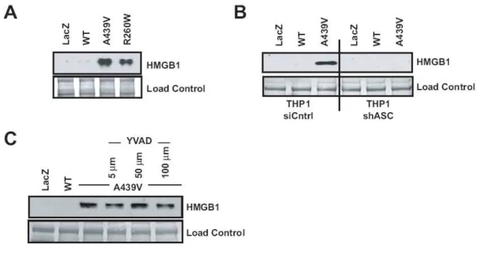
HMGB1 is emerging as an important therapeutic target for sepsis, cancer, and other conditions. Normally maintained as a nuclear factor within the healthy cell, HMGB1 takes on the role of a strong pro-inflammatory factor when released from cells undergoing necrosis (Scaffidi et al., 2002). This prompted us to examine the release of HMGB1 in the presence of disease-associated cryopyrin. As measured by western analysis, HMGB1 release from cells transduced with the wildtype CIAS1-adenovirus is barely detectable, but a high level is released by cells expressing either of two disease-associated forms of CIAS1 (Fig. 2.4A). Though ASC is essential for HMGB1 release (Fig. 2.4B), caspase-1 activity is not (Fig. 2.4C). HMGB1 release following the induction of apoptosis with staurosporine is not observed at the 6 hr timepoint, but is only observed 24 hr post-treatment. This is likely the consequence of secondary necrosis caused by longer treatment time (Fig. 2.9C). Collectively, the results presented in Figs. 2.3 and 2.4 suggest that mutant cryopyrin causes necrotic-like cell death and subsequent HMGB1 release in an ASC-dependent but caspase-1 independent fashion.
LPS induces death of FCAS patient cells.
mononuclear cells (O'Connor et al., 2003). LPS challenge resulted in a dose-dependent decrease in cell viability in patient PBMCs but not healthy controls, supporting the
conclusions made from exogenous expression of disease-associated cryopyrin (Fig. 2.5A). As disease-associated cryopyrin variants are generally accepted as gain-of-function mutants, properties observed with disease-associated cryopyrin are expected to be observed with wildtype cryopyrin, either at a reduced level or under stimulated conditions. While adenovirus with disease-associated CIAS1 caused substantial cell death at MOI=1,
transduction of adenovirus containing wildtype cryopyrin into THP-1 cells also resulted in cell death when a higher MOI was used, (Fig. 2.5B). These findings prompted us to
determine whether wildtype cryopyrin plays a role in cell necrosis associated with bacterial pathogenesis.
Shigella flexneri induced cell death requires cryopyrin and ASC, but not caspase-1.
Necrosis of monocytes and macrophages is a documented response to pathogenic bacteria, although proteins that control this process are not well defined (Golstein and
Kroemer, 2007; Zong and Thompson, 2006) Significant evidence indicates Shigella causes a necrotic-like cell death, although apoptosis has also been reported (Koterski et al., 2005; Nonaka et al., 2003; Suzuki et al., 2005; Zychlinsky and Sansonetti, 1997). This led us to examine the potential of wildtype cryopyrin to mediate necrosis in response to Shigella
flexneri. Stable reduction of cryopyrin protein in THP-1 cells was achieved utilizing
CIAS1 shRNA caused the intended biologic effect (Fig. 2.6B). More importantly, S.
flexneri-induced death was substantially abrogated in shCIAS1 cells (Fig. 2.6C). The presence of shCIAS1 did not affect staurosporine-induced cell death, indicating specificity of cryopyrin for S. flexneri-induced death. To confirm that cryopryin mediated cell death requires ASC, we tested the ability of Shigella to elicit cell death and IL-1β in ASC deficient THP1 cells. As expected, both cell death and IL-1β release were substantially abrogated in the shASC cells (Fig. 2.6D and 2.6E).
To examine the physiologic importance of these results, we utilized macrophages isolated from wildtype and CIAS1 gene-ablated mice. In wildtype bone-marrow derived macrophages, Shigella caused a 2.5-3 fold increase in cell death compared to uninfected macrophages. However, in CIAS1-/- macrophages, Shigella failed to initiate cell death above the level of uninfected cells (Fig. 2.6F). Moreover, the level of IL-1β secretion was reduced approximately 15-fold in peritoneal macrophages from CIAS1-/- mice (Fig. 2.6G). The
requirement of CIAS1 for both cell death and IL-1β release in response to S. flexneri establishes wildtype cryopyrin as a critical host adaptor capable of responding to a gram negative bacterial pathogen. In contrast, bone marrow-derived macrophages isolated from mice lacking caspase-1 demonstrated no differential cell death response to S. flexneri (Fig. 2.6H), though caspase-1 remained essential for IL-1β activation (Fig. 2.6I). These results indicate that cryopyrin and ASC, but not caspase-1, are required for S. flexneri-induced macrophage necrosis.
cryopyrin-induced cell death is caused by virulence factors expressed by S. flexneri, we compared avirulent, plasmid-cured S. flexneri (BS103) and the virulent parental strain (2457T). The former lacks a 230-kb virulence plasmid which encodes the invasion plasmid antigens IpaB, IpaC, and IpaD. These antigens are essential for S. flexneri virulence and entrance into the host cell (Menard et al., 1993). Immunoblot analysis was used to confirm the plasmid-cured Shigella strain lacks the IpaB protein, while this protein is detected in the parental strain (Fig 2.6J, left panel, inset). Cell death was assayed at two (2 and 4 hr)
different time points. At both timepoints, virulent Shigella (2457T) caused cell death, and this process was reduced in macrophages lacking the CIAS1 gene (Fig 2.6J, right and left panels). As expected, when macrophages were infected with the plasmid-cured BS103 strain, cell death is reduced by >80%. This residual level of cell death was not affected by the absence of CIAS1. These results indicate that virulence factors expressed by S. flexneri causes CIAS1-dependent cell death.
Cryopyrin mediates Shigella induced necrotic-like cell death with properties similar to mutant cryopyrin-induced death.
Having established that cryopyrin was essential for Shigella-mediated cell death, we sought to determine the nature of cell death and whether it was consistent with the necrotic-like death induced by mutant cryopyrin. Infection of shCTRL THP-1 cells with S. flexneri for six hours resulted in cell death with features morphologically consistent with necrosis (Fig. 2.7A, compare Panels i and ii). To verify that the necrotic cells contain bacteria, we
bacteria demonstrate the initial loss of cytoplasm, indicating necrosis (Fig. 2.7A, Panel iii). In contrast, cellular morphology consistent with cell death was not observed in shCIAS1 cells, despite the presence of several intracellular Shigella bacteria (Fig. 2.7A, Panel iv). Next, we examined biochemical properties of cryopyrin initiated IL-1β and cell death
response to Shigella. In contrast to the apoptotic control staurosporine, Shigella-induced cell death did not result in the cleavage of PARP after 6 hours (Fig. 2.7B). Earlier reports have suggested that S. flexneri induces apoptosis in macrophages at early timepoints, while necrosis is observed at later points (Zychlinsky et al., 1992; Koterski et al., 2005). We observed that treatment with the pan-caspase inhibitor zVAD slightly diminished cell death after 2 hours of infection, but not after 6 hours of infection. The caspase-1 specific inhibitor YVAD had no effect. Attempts to inhibit cell death and IL-1β release with glycine,
previously shown to reduce Shigella-induced apoptosis, were also ineffective (Fig. 2.11) (Edgeworth et al., 2002). However, the cathepsin B inhibitor Ca-074-Me substantially blocked cell death, further validating that mutant cryopyrin and Shigella-induced necrosis occurred via the same pathway (Fig. 2.7C). Although the caspase-1 inhibitor YVAD failed to block cell death, it substantially abrogated both IL-1β and IL-18 in response to Shigella in shCTRL cells, indicating that cell death is predominantly caspase-1, IL-1β, and IL-18 independent (Fig. 2.7D and 2.7E). As expected, shCIAS1 nearly abolished both IL-1β and IL-18. Finally, Shigella-mediated cell death was associated with the release of HMGB1 (Fig. 2.7F). In contrast, treatment with apoptosis inducing staurosporine for 2 and 6 hrs did not cause HMGB1 release.
2.5 Discussion
We report a necrotic-like cell death caused by disease-associated mutants of CIAS1 which is dependent on ASC but not on caspase-1 or IL-1β. This process results in release of the proinflammatory mediator, HMGB1, which likely propagates the inflammatory response. Additionally, we show that ASC and native, wildtype cryopyrin are required for S. flexneri-induced cell death, which proceeds in a manner identical to that flexneri-induced by
disease-associated cryopyrin. These results implicate cryopyrin as a crucial regulator of pathogen-induced necrotic-like death, a process proposed to play an important role in the pathogenesis of a myriad of infectious diseases, which we propose to call pyronecrosis.
From the perspective of cryopyrin-associated periodic fever, these results and the accumulated data in the literature suggest that cryopyrin mutants directly regulate disease progression through at least two distinct signaling pathways. It is known that cryopyrin participates in the activation of caspase-1, the maturation of IL-1β, and subsequent
underlies disease. This would seem to suggest that the inflammasome function of cryopyrin is solely responsible for periodic fever in these patients. However, given that necrosis allows for the release of pro-inflammatory factors such as HMGB1, which in turn promote further release of IL-1β, it is likely that cryopyrin-mediated cell death also contributes to disease-state in patients.
Both disease-associated cryopyrin expression and S. flexneri infection triggered the release of HMGB1, a chromatin-associated protein released by necrotic cells. Once HMGB1 is released, it acts as a potent danger indicator, inducing several pro-inflammatory cytokines by signaling through the RAGE, TLR2, and TLR4 receptors to elicit a severe inflammatory response (Andersson et al., 2000; Hori et al., 1995; Park et al., 2004). Serum HMGB1 is increased during both endotoxin exposure in mice, and in septic patients who succumbed to infection (Wang et al., 1999). HMGB1 neutralization has been shown to significantly reduce inflammation and improve survival in animal models of established sepsis (Yang et al., 2004). The release of HMGB1 elicited by bacterial-induced cell death further supports the use of HMGB1 antagonists to reduce inflammation during sepsis.
HEK293T cells (Dowds et al., 2004). Overexpressed ASC has also been shown to interact with caspases-8 and-9, and several studies have implicated ASC in the progression of
apoptosis. The concurrent over-expression of a related protein, Ipaf, with ASC in HEK293T cells results in caspase-8 dependent apoptosis, while over-expression of ASC alone induces caspase-9 mediated cell death in HEK293 cells (Masumoto et al., 2003; McConnell and Vertino, 2000). These observations suggest the involvement of apoptotic caspases in ASC-induced cell death in non-monocytic cells (Dowds et al., 2004; Masumoto et al., 2003; Wang et al., 2004). However, co-immunoprecipitation experiments in monocytic THP-1 cells indicate that ASC/caspase interaction is limited to caspase-1, suggesting that ASC is not involved in the initiation of apoptotic caspases in these cells (Stehlik et al., 2003). Our results in a monocytic cell type demonstrate that ASC is important in a necrotic-like cell death pathway that is caspase-independent.
of both pyronecrosis and pyroptosis establish ASC as a key component in the host defense arsenal to invading pathogens.
Several recent reports establish cryopyrin as a pivotal regulator of IL-1 and IL-18 release to both bacterial and viral challenges. Utilizing macrophages isolated from CIAS1 -/-mice, components of the immune response to gram positive bacteria such as Staphylococcus
aureus and Listeria monocytogenes, as well as Sendai and Influenza viruses have been shown
to require cryopyrin (Kanneganti et al., 2006a; Mariathasan et al., 2006b). However, infection with gram negative Salmonella typhimurium or Francisella tularensis elicits caspase-1 activation and IL-1β release in a cryopyrin-independent fashion (Mariathasan et al., 2005; Sutterwala et al., 2006). Macrophage cell death induced by Salmonella and
Francisella are similarly unaffected by CIAS1 deficiency (Mariathasan et al., 2005).
Alternate proteins mediate the response to these pathogens. Ipaf, which participates in its own inflammasome, is responsible for initiating inflammation in response to Salmonella
typhimurium (Mariathasan et al., 2004). It has been suggested that cryopyrin does not
mediate the recognition of gram negative bacteria. However, our results indicate that cryopyrin mediates both IL-1β release and cell death in response to the gram negative bacteria S. flexneri in THP-1 cells and mouse macrophages (Fig. 2.7). Thus, cryopyrin mediates IL-1β processing and secretion in response to specific gram positive and negative bacteria.