GENOME INTEGRITY
Michael Luca Durando
A dissertation submitted to the faculty of the University of North Carolina at Chapel Hill in partial fulfillment of the requirements for the degree of Doctor of Philosophy in the
Department of Pathology and Laboratory Medicine
Chapel Hill 2013
Approved by:
Cyrus Vaziri
William Kaufmann
Kristy Richards
Monte Willis
Dale Ramsden
ii ABSTRACT
MICHAEL DURANDO: Novel Mechanisms through which Translesion Synthesis Protects Genome Integrity
(Under the direction of Cyrus Vaziri, Ph.D.)
Exposure to ubiquitous environmental carcinogens, such as polycyclic aromatic
hydrocarbons and UV light, is a major cause of human disease. It is well accepted that
genetic mutations are an important step in the development of cancer. It has become clear
that such mutations are introduced in part by error-prone DNA polymerases. In response to
many environmental genotoxins, eukaryotic cells have evolved alternative methods of
replicating damaged DNA via the Translesion DNA synthesis (TLS) Polymerases, consisting
of DNA Polymerase eta (Polη), DNA Polymerase kappa (Polκ), DNA Polymerase iota (Polι),
and Rev1. TLS is a DNA damage tolerance mechanism that uses low-fidelity DNA
polymerases to replicate damaged DNA. The inherited cancer-propensity syndrome
Xeroderma Pigmentosum Variant (XPV) results from error-prone TLS of UV-damaged
DNA. TLS is initiated when the Rad6/Rad18 complex monoubiquitinates PCNA, but the
basis for recruitment of Rad18 to PCNA is poorly understood. This dissertation studies
several aspects of regulatory mechanisms that contribute to the damage-induced activation of
Rad18 E3 ligase activity at PCNA. First, we report a novel role for Pol η, the XPV gene
product that is mutated in XPV, in targeting Rad18 to PCNA to initiate TLS. Using
structure-function analyses and immunofluorescence microscopy, we identified a C-terminal domain
iii
stalled replication forks and promote PCNA monoubiquitination. This scaffold function is
unique to Polη among Y-family TLS polymerases and dissociable from its catalytic activity.
Importantly, XPV cells expressing full-length, catalytically inactive Polη exhibit increased
recruitment of error-prone TLS Polymerases after UV irradiation, indicating that maintaining
the bridging function of Polη in the absence of its catalytic activity greatly predisposes to
mutagenesis. These findings define a molecular basis for TLS pathway activation and
provide a new mechanism for mutagenesis and genomic instability in XPV individuals. Next,
this dissertation reports novel mechanisms of regulating TLS via stress-activated protein
kinase (SAPK) phosphorylation of Rad18 and via Chk1-dependent phosphorylation events.
Finally, this dissertation presents data indicating that TLS is involved in the tolerance of
iv
ACKNOWLEDGEMENTS
Express gratitude to the following persons for their contribution to this work:
Dr. Cyrus Vaziri: Thesis Advisor Extraordinaire
Dr. Monte Willis: Committee Member
Dr. Nader Rahimi: Mentor
Dr. Bob Bagnel: Microscopist Maximus
Dr. Eugene Orringer: Director
Dr. Anthony Muscat: Mentor
Dr. Roberto Z. Guzman: El Jefe
Sally E. Peach: Vita socium
v
TABLE OF CONTENTS
LIST OF TABLES ... viii
LIST OF FIGURES ... ix
LIST OF ABBREVIATIONS ... xiii
Chapter I. INTRODUCTION ... 1
1. UV light, benzo[a]pyrene, and environmental carcinogenesis ... 1
2. DNA Damage Tolerance and Trans-Lesion Synthesis (TLS) ... 4
3. TLS and mutagenesis ... 9
4. Checkpoints and the DDR ... 14
5. TLS activation and Rad18 and DNA damage tolerance pathways ... 18
6. TLS activation and Rad18 ... 20
7. XPV and TLS ... 24
8. Oncogenic signaling and TLS ... 29
9. Remaining major unkowns ... 33
II. MATERIALS AND METHODS ... 36
1. Cell Culture and Transfection ... 36
2. Materials, siRNA, plasmad and adenovirus construction ... 36
3. Adenovirual expression and titration ... 38
4. Fluorescence microscopy ... 38
vi
6. Genotoxin treatments ... 40
7. In vitro Binding and Ubiquitination assays ... 40
8. UV cytotoxicity assays ... 41
9. In vitro kinase assays ... 42
10. Clonogenic survival assays ... 43
11. Statistics ... 43
III. A NOVEL NON-CATALYTIC FUNCTION OF DNA POLYMERASE η IN PROMOTING PCNA MONOUBIQUITINATION AT STALLED REPLICATION FORKS ... 44
1. Introduction ... 44
2. Results ... 47
2.1. Polη promotes Rad18-mediated PCNA monoubiquitination ... 47
2.2. Rad18-Polη interactions drive PCNA monoubiquitination ... 51
2.3. Polη-PCNA binding drives PCNA monoubiquitination ... 55
2.4. Polη scaffolding mediates Rad18-PCNA association ... 58
2.5. Polη-induced PCNA-Ub is dissociable from catalytic activity ... 59
2.6. Specificity of Y-family Pol–dependent PCNA-Ub ... 62
2.7. p53 promotes PCNA monoubiquitination through Polη ... 66
3. Discussion ... 70
IV. PHOSPHORYLATION-MEDIATED REGULATION OF POLη -DEPENDENT PCNA MONOUBIQUITINATION AND RAD18 ... 74
1. Checkpoint signaling and Polη-dependent PCNA monoubiquitination ... 74
1.1. Introduction ... 74
1.2. Results ... 76
vii
2.1. Introduction ... 81
2.2. Rad18 is a JNK phosphorylation target ... 82
2.3. JNK-mediated Rad18 phosphorylation promotes Rad18-Polη binding ... 84
2.4. JNK-mediated Rad18 phosphorylation drives Polη to nuclear foci ... 86
2.5. Discussion ... 88
V. ONCOGENIC SIGNALING DRIVES MISREGULATION OF TLS ... 94
1. Introduction ... 94
2. Results ... 96
2.1. TLS is activated differently by different oncogenes ... 96
2.2. Cyclin E and Ras activate TLS ... 98
2.3. TLS and Checkpoints drive tolerance of oncogenic stress ... 101
2.4. RNR-induced tumors and Rad18 expression in vivo ... 102
2.5. Rad18 modulates Ras-driven tumorigenesis ... 103
3. Discussion ... 104
VI. FUTURE DIRECTIONS AND CONCLUSIONS ... 109
1. Future Directions ... 109
1.1. Mutagenesis in the context of catalytically inactive Polη ... 109
1.2. Contribution of TLS and Oncogene-induced Mutagenesis ... 111
1.3. Damage-specificity of TLS Pol recruitment to DNA damage sites ... 114
1.4. Endogenous Impediments to Normal Replication ... 117
2. Conclusions ... 121
REFERENCES ... 124
viii LIST OF TABLES
Table 1.1. Properties of solar ultraviolet (UV) radiation ... 1
Table 1-2. Major DNA repair pathways and associated cancer propensity
syndromes. ... 5
Table 1-3. Properties of Y-family Pols in comparison to replicative Pols. ... 12
Table 1-4. Xerderma Pigmentosum complementation groups and their
defective gene products. ... 26
Table 5-1. Properties of select eukaryotic polymerases. ... 96
Table 5-2. Properties of oncogenes and replication proteins considered in this
study. ... 97
ix
LIST OF FIGURES
Figure 1-1. Schematic representation of major UV-induced DNA lesions. ... 2
Figure 1-2. Metabolic processing of Benzo[a]pyrene to the genotoxin BPDE,
which covalently binds to deoxyguanine. ... 3
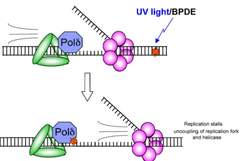
Figure 1-3. Diagram of a processive polymerase that has stalled upon
encountering a bulky DNA adduct (yellow) in its active site. ... 6
Figure 1-4. Uncoupling of the replication fork and helicase after encountering
a bulky adduct DNA lesion. ... 7
Figure 1-5. High fidelity replicative polymerase is replaced by a low-fidelity Y-family Polymerase (A) to perform TLS across the bulky adduct
DNA lesion (B). ... 8
Figure 1-6. Crystal structure of S. cerevisiae Rad30 in complex with DNA showing the right-hand conformation consisting of palm, thumb and finger domains common to all DNA polymerases, as well as
the little finger domain, or PAD, unique to Y-family polymerases ... 11
Figure 1-7. Replication-stalling lesions, such as those induced by UV light and BPDE, uncouple the replication fork forming long stretches of
single-stranded DNA, which is coated by RPA. ... 15
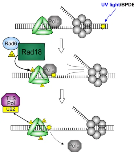
Figure 1-8. Rad18-mediated PCNA monoubiquitination drives binding of TLS
polymerases to PCNA. ... 22
Figure 1-9. Rad18 redistributes to nuclear foci after UV exposure and
x
Figure 1-10. Polη redistribution to nuclear foci after UV exposure is
compromised after depletion of Rad18. ... 24
Figure 1-11. Polη deficiency and processing of UV-induced DNA damage ... 29
Figure 1-12. Oncogene-induced DNA replication leads to a DNA damage
response that may involve TLS. ... 31
Figure 2-1. Killing curves for XPV cells complemented with Polη. ... 42
Figure 3-1. Polη promotes damage-induced Rad18 redistribution and PCNA
monoubiquitination.. ... 51
Figure 3-2. Physical interaction between Rad18 and Polη drives efficient
damage-induced PCNA ubiquitination. ... 54
Figure 3-3. Polη overexpression drives Rad18 redistribution to nuclear foci. ... 55
Figure 3-4. Polη physically bridges Rad18 and PCNA to promote efficient
PCNA monoubiquitination after DNA damage. ... 57
Figure 3-5. Physical bridging of Rad18 and PCNA by Polη is dissociable from
its DNA Polymerase activity. ... 62
Figure 3-6. High-affinity interaction with PCNA drives Polη-specific
induction of PCNA monoubiquitination. ... 66
Figure 3-7. Rad18-Polη interaction is checkpoint sensitive and p53-regulated
in response to DNA damage. ... 69
Figure 3-8. Contributions of p53 and Chk1 signaling to Polη-facilitated PCNA
monoubiquitination. ... 70
Figure 4-1. Hypothesized regulation of Polη recruitment to stalled RFs due to
xi
Figure 4-2. Immunoblot of lysates from H1299 cells that were treated with
non-targeting control siRNA or siRNA against Chk1 ... 77
Figure 4-3. Immunoblot analysis of anti-HA precipitates from HA-Rad18-expressing H1299 cells transfected with non-targeting control siRNA or siRNA against Chk1 followed by treatment with UV (10 J/m2) or sham irradiation ... 78
Figure 4-4. Representative images of H1299 cells expressing CFP-Rad18 and UV- or sham-treated. ... 80
Figure 4-5. Quantification of CFP-Rad18 expressing H1299 cells as a function of RPA and Chk1. ... 80
Figure 4-6. Rad18 is a phosphorylation target of JNK. ... 83
Figure 4-7. Serine 409 phosphorylation drives UV-inducible interaction of Rad18 with Polη. ... 85
Figure 4-8. JNK-mediated Rad18 phosphorylation contributes to UV-inducible redistribution of Polη. ... 87
Figure 4-9. Schematic of JNK-mediated regulation of Rad18-Polη binding in response to UV light. ... 89
Figure 5-1. Expression of different oncogenes affects TLS differently. ... 98
Figure 5-2. Impact of TLS and checkpoints in the response to different oncogenes. ... 99
Figure 5-3. Oncogene-induced redistribution of Ra18 and Polη to nuclear foci. ... 101
Figure 5-4. Percent survival of oncogene-expressing cells. ... 102
Figure 5-5. Rad18 expression is increased in RNR-induced lung tumors. ... 103
xii
Figure 5-7. TLS promotes tolerance of oncogene-induced replication stress. ... 108
Figure 6-1. Hypothesized increased mutagenesis in the context of catalytically
inactive Polη. ... 110
Figure 6-2. Hypothetical models of oncogene-induced activation of TLS that
results in mutagenesis. ... 113
Figure 6-3. Hypothetical models of damage-responsive Y Polymerase selection. ... 115
Figure 6-4. Schematic of G4 DNA. ... 118
Figure 6-5. Potential function of Rad18 in facilitating Polη and Polκ-mediated
xiii
LIST OF ABBREVIATIONS
Polη DNA Polymerase η
Polκ DNA Polymerase κ
Polι DNA Polymerase ι
XP Xeroderma Pigmentosum
XPV Xeroderma Pigmentosum Variant
RPA Replication Protein A
ssDNA single stranded DNA
DSB Double Strand Breaks
DDR DNA Damage Response
RF Replication Fork
OIS Oncogene Induced Senescence
PCNA Proliferating Cell Nuclear Antigen
PIP box PCNA Interacting Peptide domain
IF Immunofluorescence
PAH Polycyclic Aromatic Hydrocarbon
B[a]P Benzo[a]pyrene
BPDE Benzo[a]pyrene-Dihydrodiol Epoxide
ii
TLS Translesion DNA Synthesis
NER Nucleotide Excision Repair
BER Base Excision Repair
HR Homologous Recombination
NHEJ Non-homologous End-joining
CPD Cyclobutane Pyrimidine Dimer
6-4-PP 6-4 Pyrimidine-pyrimidone Photoproduct
RNR Ribonucleotide Reductase
1
CHAPTER 1
INTRODUCTION
1. UV light, benzo[a]pyrene, and environmental carcinogenesis
UV light and polycyclic aromatic hydrocarbons (PAHs) are ubiquitous
environmental mutagens and carcinogens. Solar UV light is omnipresent and is a
major cause of morbidity worldwide.1,2 Solar UV radiation is divided into three types:
UVC, UVB, and UVA (Table 1-1).3 UV radiation causes DNA damage in a manner
inversely proportional to wavelength.4 Solar radiation below 290 nm carries the
highest energy and is most detrimental to DNA and other biomolecules but is
primarily absorbed by stratospheric ozone. Approximately 90% of the UV radiation
that traverses atmospheric ozone is relatively low-energy UVA.3
Table 1.1. Properties of solar ultraviolet (UV) radiation
UV radiation Wavelength (nm) Energy (eV) earth surface % reaching
UVC (<280) >4.4 0 UVB (280-315) 3.9-4.4 10 UVA (315-400) 3.1-3.9 90
UV radiation causes DNA damage through different mechanisms depending
on wavelength.5 The genotoxic effects of UVC and UVB are predominantly due to
absorption by adjacent pyrimidine moieties in DNA, causing fusion of their double
bonds to form cyclobutane pyrimidine dimers (CPDs) and also to a lesser extent 6-4
pyrimidine-pyrimidone photoproducts (6-4-PPs) (Figure 1-1).6 UV-induced cell
2
at greater wavelengths (UVA), cell killing remains high while the relative number of
DNA lesions decreases. In fact, irradiation at 385 nm and higher still induces high
relative cytotoxicity, even when CPDs are virtually undetectable,7 indicating that
DNA damage mechanisms other than CPDs or 6-4PPs contribute to UVA-induced
killing at these wavelengths. Consistently, depletion of glutathione to cause oxidative
stress leads to a many-fold increase in UVA-induced DNA mutations but no change
in UVB-induced mutations, indicating that the genotoxic effects of UVA are
predominantly due to production of Reactive Oxygen Species (ROS), such as
hydrogen peroxides and hydroxyl radicals.8 Thus, UVC and UVB induce mutations
primarily via direct modification of DNA whereas UVA is mutagenic primarily via
ROS.
3
PAHs are a major source of environmental pollutant that are produced
primarily through the processing and consumption of coal and crude oil but also
through combustion of wood and tobacco products.9 Human exposure to PAHs has
been associated with increased cancer risk, and many PAHs are tumorigenic in
animal models.9 PAHs such as Benzo[a]pyrene (B[a]P) require activation to
electrophilic metabolites to exert mutagenic or carcinogenic effects. In vivo, such
PAHs are metabolized in mammalian cells by cytochrome P450s to generate the
highly reactive and mutagenic metabolites. B[a]P, for example is metabolized to its
highly reactive form BPDE, which binds covalently to genomic DNA to form bulky
adducts at the N2 position of guanine (Figure 1-2).10,11 Such bulky adducts resemble
UV-induced DNA lesions and induce mutations and cell killing via similar
mechanisms, although PAHs illicit distinct cellular responses as well.12
Figure 1-2. Metabolic processing of Benzo[a]pyrene to the genotoxin BPDE, which covalently binds to deoxyguanine.
Mutations are a fundamental step in the progression of many human diseases,
including cancer, and propagation of cells that have acquired mutations in oncogenes
or tumor suppressor genes contributes to multi-step carcinogenesis. Mutations can be
4
depurination or deamination, and endogenous or environmental mutagens.13 DNA
harboring bulky DNA lesions, such as CPDs or dG-BPDE, adopts a distorted
conformation that impedes normal replication. The presence of such damaged DNA is
strongly correlated with the acquisition of mutations. For example, the location of
UV- or BPDE-induced mutations in the p53 gene correlates strongly with hotspots of
CPDs and BPDE target sites, respectively.14, 15 Thus, replication or repair of DNA
containing CPD and dG lesions carries a high risk of introducing mutations.14
2. DNA Damage Tolerance and Trans-Lesion Synthesis (TLS)
Living cells are continually exposed to endogenous and environmental DNA
damaging agents, such as UV light and BPDE. It has been estimated that cells
encounter approximately 10,000 DNA lesions/day16. Such an onslaught of DNA
damage poses a major threat to genetic instability, and cells have evolved numerous
mechanisms of tolerating it. Major DNA repair pathways in human cells include base
excision repair (BER), nucleotide excision repair (NER), homologous recombination,
non-homologous end-joining, Fanconi Anemia, and translesion synthesis (TLS)17.
Deficiencies in any of these pathways compromises genetic stability, as evidenced by
5
Table 1-2. Major DNA repair pathways and associated cancer propensity syndromes.
Pathway Syndrome Deficient protein
NER XP many
BER Various cancers OGG1, XRCC118 HR Breast, ovary cancer Brca1, Brca219 NHEJ LIG4 syndrome Ligase IV20
FA Fanconi Anamia 15 FANCs21
TLS XPV Poleta
Each of these DNA repair pathways protect against the deleterious effects of
DNA damage. With regard to UV light and PAHs, CPDs and BPDE adducts greatly
predispose to mutagenesis. Replicative polymerases replicate undamaged DNA with a
high processivity and fidelity, but they are unable to replicate past UV- and
BPDE-induced DNA lesions. Instead, replicative polymerases stall after encountering such
DNA lesions, as the tight catalytic active site of a replicative polymerase is incapable
of accommodating such bulky DNA-distorting adducts (Figure 1-3). Stalling of the
replicative polymerase causes uncoupling of the replication fork, in which the
MCM-containing helicase complex continues unwinding DNA after the polymerase ceases
replication (Figure 1-4). This leads to long stretches of single-stranded DNA that is
rapidly coated by Replication Protein A (RPA), thus initiating a series of signaling
cascades that halts replication and turns on DNA repair pathways (see Checkpoints
6
Figure 1-3. Diagram of a processive polymerase that has stalled upon encountering a bulky DNA adduct (yellow) in its active site.
Unlike other DNA repair pathways that do not necessarily distinguish between
the nature of DNA damage, the translesion synthesis (TLS) pathway of DNA damage
tolerance is specifically directed toward replication-stalling bulky DNA adducts, such
as those induced by UV light and BPDE. TLS utilizes a group of low-processivity,
low-fidelity polymerases (compared to replicative polymerases) that are capable of
replicating past bulky DNA adducts. Bypass of bulky DNA adducts is performed
primarily by a specialized group of four DNA polymerases called the Y-family
Polymerases (Y Pols),22 consisting of DNA Polymerase eta (Polη)23, DNA
7
Figure 1-4. Uncoupling of the replication fork and helicase after encountering a bulky adduct DNA lesion.
In response to bulky DNA adducts like CPDs, these polymerases are recruited
to replication forks where they interact with proliferating cell nuclear antigen, PCNA,
a homotrimeric replication processivity factor to which DNA polymerases bind as
they replicate DNA. Polη, Polκ, and Polι contain highly conserved PCNA Interacting
Peptide domains (PIP boxes) that facilitate physical interaction with PCNA,28
whereas Rev1 interacts with PCNA via a BRCT domain.29 For TLS to occur, a
Y-family polymerase must switch places with the replicative polymerase that has stalled
upon encountering a bulky adduct (Figure 1-5a).30,31 The mechanisms dictating this
polymerase switch and the selection of the appropriate polymerase are poorly
understood, but engagement of the appropriate polymerase with PCNA facilitates
replicative bypass of the bulky adduct (Figure 1-5b). Such replicative bypass of
8
Figure 1-5. High fidelity replicative polymerase is replaced by a low-fidelity Y-family Polymerase (A) to perform TLS across the bulky adduct DNA lesion (B).
TLS across replication-stalling lesions protects against DNA breakage in the
wake of uncoupled replication forks. After replicative polymerases stall upon
encountering bulky DNA adducts, re-priming of the leading strand downstream of the
stalled polymerase allows replication to continue but leaves ssDNA gaps between the
site of re-priming and the stalled polymerase.32 TLS facilitates replication restart at
the site of the stalled polymerase and subsequent completion of replication
throughout these ssDNA gaps; loss of TLS and checkpoints (see below) increases the
number and persistence of such ssDNA gaps,32, 33 which predisposes to chromosome
instability.34
TLS is thus a crucial mechanism of preserving genomic integrity in the wake
of environmentally induced DNA damage. However, the ability of TLS Pols to
replicate past bulky adducts is counterbalanced by their reduced fidelity,
incorporating errors at a rate of approximately 1 per 103 bp, substantially higher than
replicative Pols, such as Polδ.35,36,37 This relatively low fidelity of TLS Pols leads
9 3. TLS and mutagenesis
The existence of error-prone DNA repair pathways was first uncovered in
early studies on bacterial mutants that were found to be nearly resistant to
UV-induced mutations.38 The first genes associated with UV-induced mutagenesis were
recA and laxA in E. coli, which, when deleted, eliminated a global DNA damage
response (SOS response) and also suppressed UV mutability38. Over 30 genes have
since been linked to the DNA damage-inducible SOS response in bacteria, but the
mutagenic portion of SOS is linked almost exclusively to those genes involved in
TLS39. First discovered in screens of E. coli mutants that are not mutable by UV
light,40, 41 members of the UmuC/DinB family were found to exhibit polymerase
activity across DNA lesions.39 Specifically, Pol V in E. coli, consisting of UmuC and
UmuD’2, is capable of replicating across UV-induced lesions such as CPDs42
whereas DinB1 (Pol IV) can replicate across bulky adducts at the N2 position of
guanine.43 Eukaryotic homologues of the SOS genes were later discovered. In S.
cerivisiae, the TLS activities of E. coli Pol V are performed by Rad30, which
processes UV-induced lesions,44 and Rev1, which processes abasic sites and drives
mutagenesis.45 In humans, these TLS activities are performed by the Y-family
polymerases: Polη,46 (homologue of E. coli Pol V and yeast Rad30), Rev147
(homologue of E. coli Pol V and yeast Rev1), Polκ48 (homologue of E. coli DinB1;
no yeast homologue), and Polι49 (originally called Rad30B, no yeast or bacterial
10
Much research has been dedicated to the nature and the significance of
low-fidelity replication by TLS polymerases. Important concepts of debate involve (i)
whether the infidelity is “beneficial” or “deleterious” for the cell and (ii) whether the
infidelity is accurately defined as “error-prone.” The following discussion will
address both of these points.
The infidelity of TLS Pols is attributed to several structural features of
Y-family polymerases that differ substantially from replicative polymerases. Although
the Y-family polymerases share almost no sequence homology with replicative DNA
polymerases, they maintain structural similarities common to replicative polymerases,
such as the classic right-hand polymerase fold that wraps around a DNA template
(Figure 1-6) and highly conserved aspartate and glutamate residues in the active sites
that coordinate magnesium ions and stabilize incoming dNTPs.50, 51 These
similarities, common to nearly all polymerases, are perhaps not surprising considering
the replicative capabilities of the TLS polymerases, but several important structural
and functional differences from replicative polymerases are responsible for their
inaccurate replicative potential and in turn their important roles in error-prone
11
Figure 1-6. Crystal structure of S. cerevisiae Rad30 in complex with DNA showing the right-hand conformation consisting of palm, thumb and finger domains common to all DNA polymerases, as well as the little finger domain, or PAD, unique to Y-family polymerases.50
Key among these differences is their lack of 3’ to 5’ exonuclease proofreading
activity that is common to all replicative polymerases and that removes mismatched
bases.50 Lack of such proofreading capabilities allows TLS Pols to continue
replication even after insertion of the incorrect base, whereas replicative polymerases
can remain stalled in futile insertion-excision cycles.52
Next, although Y-family polymerases contain palm, thumb, and finger
domains analogous to replicative polymerases, structural nuances that are common to
the Y-family Pols but different from replicative pols impart unique functional
characteristics that directly impact their processivity and accuracy (Figure 1-6).52 The
finger domain in Y-family pols uniquely contains a so-called polymerase-associated
domain (PAD), also called a little finger domain, which is normally loose and flexible
but adopts a stable conformation after engagement with the cognate lesion on DNA.53
The PAD thus determines the lesion specificity of the Y-family Pols, allowing certain
12
example, perform TLS across CPDs and dGs, respectively, with relatively higher
accuracy compared to replication across undamaged templates or other lesions. The
PAD also contributes to catalytic efficiency and the mutational spectra.53 Compared
to replicative Pols, the palm, thumb and finger domains of Y-family Pols hold DNA
with a more “open” grip, resulting in a dramatically reduced processivity.50
Regarding replicative fidelity, the active sites of Y-family polymerases are
larger and more open than in replicative DNA polymerases, they form fewer contacts
with template and nascent DNA strands, and they fail to exhibit the “induced-fit”
conformational changes that drive dNTP specificity in replicative polymerases.50 All
of these characteristics combined yield a family of polymerases that, compared to
replicative polymerases, have (i) reduced fidelity, (ii) reduced processivity, (iii)
greater flexibility in accommodating aberrant DNA structures, (iv) unique mutational
spectra, and (v) favored cognate lesions (Table 1-3). These properties have important
implications for both the tolerance of replication-stalling DNA lesions and the genesis
of mutations.
Table 1-3. Properties of Y-family Pols in comparison to replicative Pols.
Property Y-family Pols relative to Replicative Pols Phenotypic outcome
Fidelity Low Mutagenesis Processivity Low Short-lived activation
Active site Loose, non-discriminatory Mutagenesis Preferred
template Lesion specific
Mutagenic for non-preferred lesion
Regarding the discussion of whether the Y-family polymerases are beneficial
or deleterious for a cell, one must consider the context in which they are utilized.
13
uncoupling of replication forks can lead to fork collapse, single-strand DNA breaks,
and double-strand breaks. Lesion bypass after replacement of the replicative Pol with
a TLS Pol can be considered beneficial as it prevents the deleterious effects of DNA
breaks by allowing replication to continue. Of note, a single unrepaired double strand
break is sufficient to mediate cell death.54 However, TLS can also be considered
deleterious if the wrong polymerase is utilized for a specific lesion, thus promoting
mutagenesis. For example, use of Polκ, whose preferred lesion is a bulky adduct at
dG, to bypass a CPD instead of Polη (whose preferred lesion is a CPD), will be
highly mutagenic, whereas use of Polη to bypass a CPD will be relatively accurate.
Experiments in human cells have in fact demonstrated Polη-mediated error-free TLS
across CPDs and highly mutagenic TLS across CPDs by Polκ.55 It is critical,
therefore, for cells to select the proper polymerase for the proper lesion.
Activation of the TLS Pols in the absence of damage, use of the wrong TLS
polymerase, or absence of the proper polymerase after acquisition of DNA damage
conferring its preferred lesion will all predispose to mutagenesis. Such scenarios
highlight the unique and often confusing aspect of the TLS pathway: the TLS
Polymerases are absolutely necessary to prevent genetic instability, but their aberrant
activation will unequivocally lead to genetic instability. In other words, the TLS Pols
must be activated only in response to DNA damage and the cell must ensure that only
the correct polymerase is used for the right type of damage. The consequences of
failed TLS regulation is best exemplified by the cancer propensity syndrome
Xeroderma Pigmentosum Variant, in which Polη is functionally absent, leading to
14
XPV section). The properties of TLS Pols summarized in Table 1-3 and the
phenotypic outcome of aberrant TLS activity demonstrate the importance of proper
regulation of TLS. The molecular mechanisms that regulate TLS are still poorly
understood and are also largely the focus of this dissertation.
One final important distinction of TLS involves the replication past damaged
bases without removing the damage. Whereas DNA repair processes such as BER
and NER remove damaged bases from the genome, TLS does not actually repair
damaged DNA. Rather, it promotes tolerance of damaged DNA by allowing the
continuation of replication without actually removing the damage. TLS thus promotes
genomic stability primarily by preventing the catastrophic consequences of
replication fork collapse, not by actually repairing or removing damaged DNA.
4. Checkpoints and the DDR
Central to the proper regulation of TLS is a cell’s ability to detect and respond
to DNA damage. DNA damage responses are activated by a cascade of intracellular
signals that are elicited by the recognition of DNA damage. These signals, called
checkpoints, utilize DNA damage sensors, signal transducers, and effector proteins to
coordinate processes of DNA repair. Generally, sensor proteins (e.g. the MRN and
9-1-1 complexes) detect DNA damage on chromatin and activate signal transducers
(e.g. ATM/ATR and Chk1/Ch2), which amplify the checkpoint signal to activate
proximal and distal effector proteins (e.g. BRCA1). Such effector proteins are
recruited to chromatin, where they facilitate damage repair or tolerance at the site of
15
replication cell cycle proteins such as cyclin-dependent kinases. Checkpoints thus
coordinate cell cycle progression with genome maintenance, helping ensure that the
cell cycle does not proceed if genome integrity is compromised.
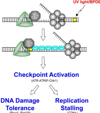
Figure 1-7. Replication-stalling lesions, such as those induced by UV light and BPDE, uncouple the replication fork forming long stretches of single-stranded DNA, which is coated by RPA. RPA recruits ATR, which activates ATRIP to phosphorylate Chk1, which then
simultaneously turns on DNA damage tolerance processes and turns off normal replication.
The S-phase checkpoint response to replication stress is typically divided into
two main pathways: an ATM-mediated checkpoint that responds to DNA
double-strand breaks and an ATR-mediated response to stalling and uncoupling of replication
forks.56, 57 However, much cross-talk exists between the two pathways; over 700
proteins have been identified as targets of damage-induced phosphorylation by ATM
and ATR, almost all of which are involved in DNA damage repair or checkpoints and
16
Double-strand breaks are processed within cells after recognition by the
Mre11-Rad50-Nbs1 (MRN) complex. ATM is rapidly recruited to these DSBs via
physical interactions between the HEAT motifs in ATM and the C-terminus of
Nbs1.59 Activated ATM phosphorylates a variety of targets, including KAP1, which
induces chromatin relaxation,60 and Chk2, which promotes cell cycle arrest. Once
activated by ATM, Chk2 phosphorylates Cdc25A, a dual-specificity
threonine/tyrosine phosphatase that promotes cell cycle progression by activating
cyclin dependent kinase complexes, such as CDK2-Cyclin A, via
dephosphorylation.61 Chk2-mediated phosphorylation of Cdc25A targets Cdc25A for
ubiquitin-dependent degradation, thus decreasing activation of CDKs and inhibiting
cell cycle progression.62
Regarding DNA damage from UV light and PAHs, current models depict that
damage-induced stalling of replication forks leads to uncoupling of the helicase and
polymerase, yielding long stretches of single-stranded DNA (ssDNA)63, 64 (Figure
1-7). ssDNA is rapidly coated by RPA, a heterotrimeric complex consisting of 70, 30,
and 14 kDa monomers, RPA1, RPA2, and RPA3, respectively. In addition to its high
affinity to ssDNA, RPA also binds to many other proteins involved in DNA repair,
including the checkpoint signal transducer ATR-interacting protein (ATRIP). ATRIP
binds to the N-terminus of RPA1 and recruits its binding partner, ATR, to
chromatin65-67 Once docked at RPA-coated ssDNA, ATR-ATRIP is activated by the
presence of Rad9-Rad1-Hus1 (9-1-1), a clamp that encircles DNA and recruits
17
ATR activation domain and strongly stimulates ATR kinase activity to initiate a
series of signaling cascades that amplify the DNA damage response.68, 69
One important signal transducer downstream of ATR is the S-phase kinase
Chk1. ATR-mediated phosphorylation of Chk1 at serines 317 and 345 activates Chk1
via two mechanisms: uncovering of its N-terminal kinase domain70 and release of
inactive chromatin-bound Chk1 to redistribute to centrosomes and block mitotic
entry.71 Like Chk2, activated Chk1 targets Cdc25A for degradation to block cell cycle
progression and suppress origin firing via inhibition of the cyclin-dependent kinase
Cdk2.72 Chk1 also blocks cell cycle progression through Cdc25A-independent
mechanisms involving via direct phosphorylation of Dbf4.73 Phosphorylation of Dbf4
is thought to promote removal of Dbf4 from chromatin and inhibit its binding to
Cdc7, thus inhibiting the kinase activity of the Dbf4/Cdc7 complex.73 Inhibition of
Cdc7 decreases loading of the MCMs to the pre-replicative complex74 and also blocks
Cdc45 loading75 and interaction between Cdc45 and MCM7,76 thereby inhibiting
initiation of DNA synthesis at origins of replication.
Although ATM and ATR are frequently described as independent responses to
double-strand breaks and stalled replication forks, respectively, extensive cross-talk
exists between the two pathways. For example, DSBs can lead to ATR activation in a
manner that is dependent on ATM,77 whereas UV-induced replication stalling can
activate ATM via ATR-mediated phosphorylation.78 Similarly, ionizing radiation can
ATR-18
independent mechanisms.79 Therefore, ATM and ATR function in parallel but also in
concert to mediate checkpoint responses to DNA breaks and fork-stalling lesions.
Numerous links between checkpoints and TLS have been demonstrated. First,
damage-induced Chk1 activation leads to the phosphorylation-mediated activation of
Cdc7 (though Chk1 does not directly phosphorylate Cdc7), which itself
phosphorylates Rad18 to drive Rad18-Polη binding and help promote tolerance of
UV damage (see section on Rad18 and TLS).80 Next, Chk1 is reported to facilitate
maximal damage-induced PCNA monoubiquitination, albeit via kinase-independent
mechanisms.81 Altogether, DNA damage-induced checkpoints contribute to
TLS-mediated DNA damage tolerance by both stalling replication to allow for TLS to
occur and through direct stimulation of TLS via Chk1.
5. TLS activation and Rad18 and DNA damage tolerance pathways
Although the precise mechanisms that regulate activation of TLS remain
poorly understood, it is generally accepted that TLS activation depends in large part
upon physical docking of replicative or TLS polymerases with the homotrimeric
replication factor PCNA. Which polymerase interacts with PCNA is determined in
large part by the post-translational modification of PCNA by Rad18.
RAD18 is a highly conserved gene that was first identified in yeast in the same
epistatis group as RAD6 on the basis of increased sensitivity of UV and ionizing
radiation.82 Human Rad18 codes for a 495 amino acid protein with several important
19
catalytic activity and Rad6 binding (amino acids 26-64),83 a C-terminal Rad6 binding
domain (amino acids 340-395),84 a Zinc finger domain that mediates accumulation at
sites of DNA damage (amino acids 201-225),84 a SAP domain that promotes
assembly at stalled replication forks (amino acids 248-284),85 a C-terminal Polη
binding domain (amino acids 402-445),86 and a C-terminal nuclear localization signal
(amino acids 488-494).85
Rad18 is an E3 ubiquitin ligase that plays a central role in DNA damage
tolerance by catalyzing the damage-induced monoubiquitination of PCNA.87 In
conjunction with its E2 ubiquitin-conjugating enzyme and binding partner, Rad6,
Rad18 redistributes to stalled replication forks after DNA damage, where it catalyzes
the monoubiquitination of PCNA at lysine 164 (Figure 1-8).88,89 The Rad6-Rad18
complex is highly selective for PCNA monoubiquitination, a signaling event that has
important implications for many aspects of the DNA damage response.
Rad18-mediated monoubiquitination of PCNA at lysine 164 serves as a signal
to turn on both TLS and template switching. TLS is activated in part due to increased
affinity of Y-family polymerases to monoubiquitinated PCNA compared to
unmodified PCNA (see below). Template switching is an error-free post-replication
repair process that is thus dependent on Rad18 for activation. Template switching is
activated by HTLF/SHPRH-mediated (Rad5 in yeast) polyubiquitination of K164 that
takes place only after Rad18-mediated K164 monoubiquitination.90 Rad18 thus
stimulates both error-free (template switching) and error-prone (TLS) post-replication
20
Rad18 also contributes to DNA damage tolerance through E3
ligase-independent functions. In contrast to post-replication repair of fork-stalling lesions,
Rad18 confers tolerance of double-strand breaks by promoting homologous
recombination through physical interactions with Rad51C, a key protein in the initial
steps of HR.91 Damage-induced Rad18-Rad51C interactions promote recruitment of
Rad51 to DNA DSB, where Rad51 may begin strand invasion to initiate HR.92 This
HR-promoting activity of Rad18 requires the RING and zinc finger domains but not
the SAP or Rad6-binding domains. The RING domain of Rad18 facilitates physical
interaction with Rad51C, and the zinc finger domain mediates Rad18-binding to
ubiquitinated proteins at sites of DNA damage. These Rad18-binding proteins are
ubiquitinated at DSBs by RNF8, another damage-inducible E3 ligase. Rad18
recruitment to DSBs thus depends on RNF8-mediated ubiquitination events; Rad18
appears to chaperone Rad51C to such DSB sites. Interestingly RNF8-mediated
ubiquitination events seems necessary for recruitment of Rad18 to DSB sites but not
stalled replication forks. Rad18 thus uses the same domains for entirely different
functions (RING domain for PCNA monoubiquitination in TLS and for Rad51C
recruitment in HR), thereby contributing to activation of distinct DNA damage
tolerance pathways.
6. TLS activation and Rad18
As illustrated above, Rad18-mediated PCNA monoubiquitination serves as a
signal to activate TLS at stalled replication forks. TLS requires replacement of stalled
21
This switching mechanism is attributed to a higher affinity of TLS polymerases to
monoubiquitinated PCNA compared to unmodified PCNA. In addition to their highly
conserved PIP-boxes (and BRCT domain for Rev1), the Y-family polymerases
contain ubiquitin-binding motifs (UBZ domains) that confer high-affinity binding to
monoubiquitinated PCNA.93, 94 Replicative polymerases, conversely, display reduced
affinity to monoubiquitinated PCNA. By creating a docking site on PCNA suitable
for TLS Pol binding, it has been proposed that PCNA monoubiquitination by Rad18
constitutes the basis for the engagement of TLS Pols with the replication machinery
and in turn activation of TLS (Figure 1-8).95,96,97 Such a hypothesis is supported by
the findings that TLS Pol mutants in which the UBZ domains have been mutated
demonstrate reduced redistribution to stalled replication foci,94 that XPV cells
complemented with UBZ-mutant Polη exhibit UV survival defects,93, 94 and that UV
survival is compromised in knock-in PCNA mutants in which lysine 164 has been
mutated to eliminate Rad18-mediated PCNA monoubiquitination.98 The reliance of
TLS on PCNA monoubiquitination is not unanimously accepted, however, as other
investigators have reported no UBZ-dependent effect of Polη on UV survival,
22
Figure 1-8. Rad18-mediated PCNA monoubiquitination drives binding of TLS polymerases to PCNA.
The mechanisms regulating the monoubiquitination of PCNA by Rad18 are
poorly understood, and, specifically, it is not known what regulates the redistribution
of Rad18 to stalled replication forks or how it interacts with its monoubiquitination
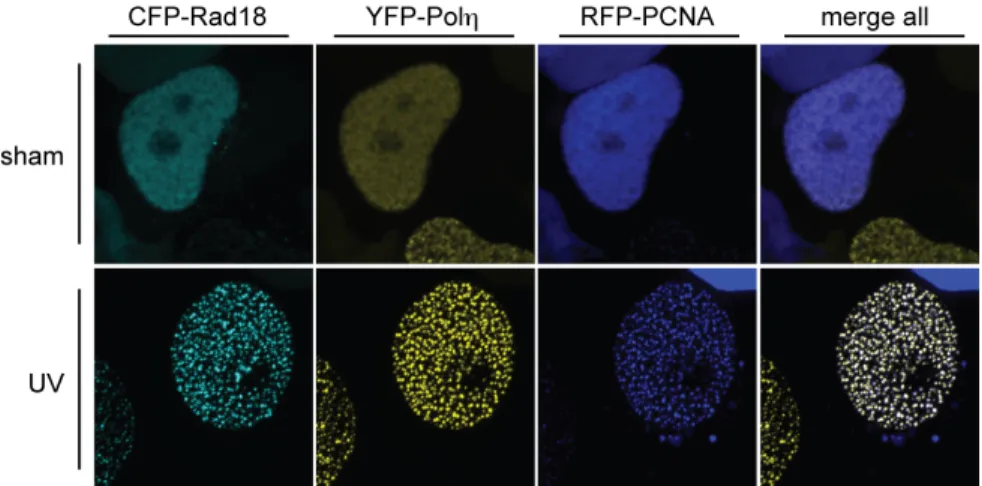
target, PCNA. Rad18 is normally distributed diffusely throughout the nucleus, but it
redistributes to discrete nuclear foci and colocalizes with PCNA after DNA damage
(Figure 1-9).101 Rad18 redistribution is intimately coupled with Polη redistribution
and activity; in addition to its E3 ligase activity, Rad18 physically associates with
23
1-9).86 Rad18 thus drives TLS by physically chaperoning Polη to stalled RFs in
addition to its accepted role of monoubiquitinating PCNA. Indeed, loss of Rad18
eliminates PCNA monoubiquitination and severely compromises redistribution of
Polη to replication nuclear foci after UV damage (Figure 1-10), though
distinguishing between these two functions – E3 ligase activity at PCNA and physical
chaperoning – has proven technically challenging (see Chapter 2).
Figure 1-9. Rad18 redistributes to nuclear foci after UV exposure and colocalizes with Polη and PCNA.
(images by MD; RFP-PCNA pseudo-colored in blue for clarity).
Although Rad18 plays an important role in driving TLS polymerase
recruitment to sites of DNA damage, the regulatory mechanisms that coordinate this
process are only beginning to be delineated. As mentioned above, Rad18-Polη
binding is stimulated by Chk1-dependent Cdc7 phosphorylation of Rad18.80 In
addition to the kinase-independent role of Chk1 that potentiates PCNA
monoubiquitination, direct interactions between RPA and Rad18 contribute to Rad18
24
Rad18 activity and cellular distribution at several levels, ensuring tight coordination
between the acquisition of DNA damage and TLS activation.
Figure 1-10. Polη redistribution to nuclear foci after UV exposure is compromised after depletion of Rad18.
(images by MD)
7. XPV and TLS
Xeroderma pigmentosum (XP) is a rare cancer propensity syndrome defined
by exquisite sensitivity to sunlight and extremely high predisposition to skin cancer.
XP affects both males and females equally and follows a pattern of autosomal
recessive transmission. XP has been reported in nearly all racial groups with
incidences that vary from 1 in 20, 000 in Japan to 1 in 250, 000 in the USA.103 About
60% of XP individuals first show symptoms of an extreme sunburn reaction to
25
sunburn reactions, but rather develop an unusually high number of freckle-like lesions
in sun-exposed areas, typically by age 2.
XP tends to follow a pattern of three stages. While the skin appears healthy at
birth, the first stage usually begins after about 6 months and is characterized by
diffuse erythema, scaling, and areas of increased pigmentation in sunlight-exposed
areas.103 The second stage is characterized by skin atrophy, telangiectasias, and mixed
hyperpigmentation and hypopigmentation, a combination of symptoms known as
poikiloderma. The third stage involves the progression to malignancy. XP patients are
estimated to have a 10,000-fold increased risk of non-melanoma skin cancer and a
2,000-fold increased risk of melanoma before the age of 20.104 Additionally, ocular
abnormalities, including photophobia, conjunctivitis, and ocular neoplasms, occur in
approximately 80% of XP patients, and approximately 20% develop neurologic
problems, including intellectual deficiency, spasticity or ataxia, and microcephaly.
The cutaneous symptoms of XP, including pigmented skin lesions
sunlight-exposed skin, were first documented in 1874 in Vienna by a dermatologist named
Moriz Kaposi,105 and neurologic abnormalities were first described in the 1880’s.106
However, it wasn’t until the 1960’s that the connection between XP and DNA
damage tolerance was appreciated when cultured XP cells were shown to exhibit
deficiency of excision repair.52 In 1971, an excision repair-proficient XP strain was
described107 and subsequently named “variant.”108 Complementation experiments in
the 1970’s led to identification of 7 XP subtypes, complementation groups A-G, all
deficient in nucleotide excision repair, and XP-Variant, which exhibited normal
26
responsible for each complementation group and their function were subsequently
identified (Table 1-4).
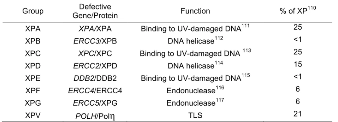
Table 1-4. Xerderma Pigmentosum complementation groups and their defective gene products.
Group Gene/Protein Defective Function % of XP110
XPA XPA/XPA Binding to UV-damaged DNA111 25 XPB ERCC3/XPB DNA helicase112 <1 XPC XPC/XPC Binding to UV-damaged DNA 113 25 XPD ERCC2/XPD DNA helicase114 15 XPE DDB2/DDB2 Binding to UV-damaged DNA115 <1 XPF ERCC4/ERCC4 Endonuclease116 6 XPG ERCC5/XPG Endonuclease117 6
XPV POLH/Polη TLS 21
XP-Variant (XPV) is unique among the XP complementation groups for
several reasons. Unlike groups A-G, which are deficient in proteins that mediate
repair of UV-damaged DNA via NER, XPV is characterized by deficient
TLS-mediated tolerance of UV-induced DNA damage while maintaining normal NER.
DNA lesions are not actually repaired or removed after TLS, but rather remain in the
genome until other mechanisms, like NER, remove them. TLS, rather, allows
replication to continue after a replicative polymerase encounters a DNA lesion, thus
promoting tolerance of such lesions by preventing replication fork collapse and
subsequent DNA breaks. The deleterious effects of XPV are thus due to an entirely
different mechanism of coping with UV-damaged DNA (tolerance vs. repair), in
addition to a different pathway (TLS vs. NER).
Next, mutations in XPV individuals arise via different mechanisms than the
other XP subtypes. Let’s first consider NER. NER can be considered as the first-line
27
removed via relatively error-free processing. As mentioned earlier, the daily burden
of DNA lesions is enormous, estimated in the range of greater than 10,000 DNA
lesions per day.16 In a healthy cell, only a very small percentage of these lesions
escape NER to be processed by other DNA damage tolerance mechanisms. Due to the
inherent infidelity of TLS enzymes, lesion bypass by TLS will introduce some level
of mutation, even when each lesion is processed by the appropriate polymerase (e.g.
CPDs by Polη). In other words, TLS of CPDs by Polη carries a greater risk of
mutation than normal replication of undamaged DNA by replicative polymerases.
Such TLS accounts for at least a large part of the basal mutation rate experienced by
all cells over time, even those with intact DNA damage tolerance pathways.
However, in the absence of NER, the burden of DNA lesions that must be
processed by secondary damage tolerance mechanisms is enormous. Again in light of
the relatively error-prone nature of TLS, lesion bypass via TLS, even when the
appropriate TLS polymerase is present, will greatly increase the probability of
introducing mutations. In the absence of NER, one can assume that Polη is still
performing the vast majority of TLS across UV-induced lesions. Even though Polη
-mediated TLS of CPDs is relatively accurate compared to TLS of CPDs by other TLS
enzymes, it is still far less accurate than replication of undamaged templates by
replicative polymerases. Therefore, tolerance of UV-induced DNA damage carries a
dramatically higher risk of mutagenesis in XP A-G, even though Polη is present to
perform TLS of CPDs. Indeed, one can assume that the majority of these mutations
28
The situation in XPV is entirely different and conceptually much simpler. The
overwhelming majority of UV-induced lesions are removed by NER. Rarely, a CPD
that escapes NER will be processed by TLS. However, the preferred polymerase for
CPDs is absent (Polη), so polymerases that are far more error-prone than Polη on
CPDs (Polι and Polκ) are used instead. Indeed, Polι and Polκ have been implicated in
mutagenic TLS in the absence of Polη in XPV cells.118 Mutagenesis in XPV is thus
due to TLS by polymerases other than Polη, whereas Polη plays an important role in
mutagenesis in XP A-G.
On a molecular level in XPV cells, the effect of Polη deficiency in the
response to CPDs that escape NER can be viewed as having two most likely
outcomes. In the first, CPDs are simply not bypassed and persistent stalling of
replicative polymerases leads to replication fork collapse and single and
double-strand DNA breaks, with obvious deleterious consequences for genomic stability
(Figure 1-11, left). Alternatively, TLS may occur via the inappropriate TLS
polymerase, which will bypass CPDs with a much lower fidelity than Polη and a high
probability of introducing mutations (Figure 1-11, right). Both potential outcomes
severely compromise the genetic integrity of the cell and predispose to malignant
29
Figure 1-11. Polη deficiency and processing of UV-induced DNA damage
Lack of Polη leads to two main outcomes in XPV cells after UV irradiation. Persistent stalling of replicative polymerases (left) leads to long stretches of ssDNA that are vulnerable to double strand breaks, which can be repaired by non homologous end-joining, predisposing to DNA translocations. Alternatively recruitment of inappropriate TLS polymerases (predominantly Polι and Polκ) predisposes to mutagenic replication (right).
8. Oncogenic signaling and TLS
Mutations are well-accepted to play an important role in the initiation and
progression of many types of cancer.119 Mutations contribute to malignant
30
however, the rate of de novo mutations is significantly higher in transformed cells
compared to normal. Whereas mutation rates in non-transformed cells appear to be in
the range of 1X10⁻¹⁰ mutations/base pair/cell generation,120,119 mutation rates in
neoplastic cells have been shown to be over 20-fold higher,121 and gene amplification
has been shown to be >106-fold higher in tumor compared to normal cell lines.122
These data suggest that normal processes of genome maintenance are derailed in
malignant cells, leading to inhibited or mis-regulated damage tolerance mechanisms
in the context of oncogenic signaling and, in turn, increased mutagenesis.
Central to this idea is the hypothesis that pre-malignant cells acquire somatic
mutations that lead to aberrant expression of genes involved in processes important
for malignancy, namely cell growth, arrest, invasion, replication, and
angiogenesis.123,124 In particular, aberrant expression and regulation of proteins
involved in replication and genome maintenance predisposes to additional subsequent
mutations in a feed-forward cycle carrying a dramatic risk of cancer.125 Because TLS
is an error-prone replication process, mis-regulation of TLS in oncogene-expressing
cells would run a high risk of introducing early genetic mutations.
One can thus hypothesize that oncogene-expressing cells exhibit a reduced
ability to respond properly to environmental DNA damaging agents (e.g. UV light
and PAHs) or endogenous replication impediments (G4 structures). Similarly, one
may hypothesize that TLS alone could be improperly activated or regulated in
oncogene-expressing cells, leading to excessive error-prone replication. Malignant
cells would thus acquire mutations at an increased rate due to overactive or
31
and the well-accepted role of environmental DNA damaging agents in carcinogenesis
can be further explained by such mechanisms.127,128 Additionally, oncogene
expression is known to induce stalling and collapse of DNA replication forks,129 as
well as replication stress that activates DNA damage responses.130 For example,
oncogenes have been shown to induce re-replication,131, 132 activate DNA damage
response pathways,132 and induce the formation of reactive oxygen species (Figure
1-12).133
Figure 1-12. Oncogene-induced DNA replication leads to a DNA damage response that may involve TLS.
All of these processes contribute to genetic instability in the context of
oncogenic signaling; however, the mechanisms through which oncogenes lead to
32
seemingly contradictory cellular responses. Oncogenes such as Ras are known to
induce cellular proliferation, but only when expressed in combination with the
inactivation of tumor suppressors, such as p53 or Rb.134 Rather, expression of
oncogenes leads to an initial wave of proliferation that is followed by cellular
senescence. Such oncogene-induced senescence is characterized by irreversible
growth arrest that is associated with accumulation of tumor suppressor genes such as
p53, p16, or p21.135 Therefore, cells respond to oncogenic signaling by expressing
proteins that halt cellular proliferation. Importantly, expression of such tumor
suppressor genes is a consequence of an oncogene-induced DNA damage response.131
Specifically, oncogene-expressing cells demonstrate DNA replication stress in the
form of stalled replication forks and DSBs, together with a concomitant activation of
checkpoint proteins, such as ATM and ATR, and their downstream effectors, Chk2
and Chk1.136 Importantly, abrogation of these checkpoint responses suppresses
oncogene-induced senescence.131 Therefore, DNA damage response and checkpoint
pathways play an integral role in the cellular response to oncogenic signaling;
however, the mechanisms of oncogene-induced checkpoint activation, the
consequences of such activation, and the DNA damage response pathways affected
by such checkpoint activation remains unknown.
As shown blow (see chapter 4) and in numerous reports,67, 80 there is a clear
relationship between checkpoint signaling and TLS, and several lines of evidence
suggest that TLS may be involved in the cellular response to oncogene-induced DNA
damage. First, because activation of Chk1 stimulates TLS, oncogene-induced
33
DNA damage. Next, TLS is known to contribute to cellular tolerance of abnormal
DNA structures, such as G4 DNA, and oncogene-induced replication stress leads to
aberrant topological DNA structures, such as reversed replication forks;130 TLS may
also play a role in the processing of such oncogene-induced DNA structures.
Considering the predilection for mutagenesis in the context of oncogenic signaling
and the connections between TLS and checkpoint signaling, we have hypothesized
that TLS is involved in the tolerance of oncogene-induced replication stress and that
TLS plays an important role in oncogene-dependent mutagenesis (Figure 1-12).
Important unexplored questions include whether TLS is activated in the
context of oncogenic signaling, whether checkpoint activation impacts
oncogene-dependent TLS activity, how TLS contributes to mutagenesis in the context of
oncogene expression, and whether oncogene-expressing cells are sensitized in the
absence of TLS-mediated DNA damage tolerance.
9. Remaining major unkowns
The preceding text is intended to provide a background of specific areas
relevant to the studies presented in this dissertation. For each area, however, several
important processes have remained unknown:
DNA Damage Response Checkpoints and TLS
Although it is clear that checkpoints facilitate activation of TLS via signaling
34
interactions (kinase-independent Chk1-mediated potentiation of PCNA
monoubiuqitination), a broader understanding of the relationship between checkpoint
signaling and TLS is needed. Specifically, it remains entirely unknown whether
checkpoint signaling contributes to binding of other TLS polymerases with Rad18,
the mechanism through which Chk1 facilitates recovery from genotoxin-induced
S-phase checkpoints, and what additional effector proteins function upstream and
downstream of Chk1 to initiate a TLS-stimulatory checkpoint response.
Understanding these concepts will help explain the mechanisms through which
TLS-mediated damage tolerance and mutagenesis take place. These concepts are addressed
in Chapter 4 of this dissertation.
Rad18 and TLS Activation
Although Rad18/Rad6 is known to catalyze damage-responsive
monoubiquitination of PCNA, the mechanisms that regulate this process remain
entirely unknown. It is known that Rad18 does not physically interact with PCNA, so
the processes that drive redistribution of Rad18 to PCNA and subsequent physical
binding of the two are unknown. Additionally, whereas UV-induced binding of
Rad18 and Polη has been clearly demonstrated, the specific mechanistic aspects of
this interaction remain unknown: where does Polη bind to Rad18 and vice versa? Do
known effector domains of Polη, such as the UBZ domains, contribute to Rad18
binding? Where does this interaction take place? Is the interaction direct or are other
binding partners required? Do the components of the Rad18-Polη complex change as
a function of genotoxin stimulation or cell cycle? Further, details of analogous Rad18
35
necessary for Rad18-Polκ interactions? Are these interactions promoted only by
BPDE? Does Rad18 interact at all with Polι or Rev1? Finally, the mechanisms that
dictate selection of the proper polymerase for the appropriate DNA lesion are
completely unknown. This point is of crucial importance because use of the wrong
polymerase greatly predisposes to mutagenesis. Additionally, because of the role that
TLS plays in cellular tolerance anticancer drugs such as cisplatin,137 understanding
the mechanisms that regulate TLS initiation and activation may help guide the
development of therapeutic targets.
Oncogenes and TLS
It is widely accepted that oncogenic signaling activates a DNA damage
response and checkpoints and simultaneously promotes mutagenesis in a manner that
feeds into the mutator phenotype. However, it remains entirely unknown what are the
consequences of such oncogene-induced activation of the DDR and what mechanisms
are responsible for the oncogene-induced increase in mutagenesis. A putative role for
TLS in this process is conceptually feasible but has never been explored. A role for
TLS in the tolerance of oncogenic replication stress concepts is addressed in Chapter
5. Understanding the mechanisms that dictate mutagenesis and survival in the context
of oncogenic signaling is necessary for the discovery of novel targets for
36
CHAPTER 2
MATERIALS AND METHODS
1. Cell Culture and Transfection
H1299, HDF, XP115LO (GM0235946, 138), and HCT-116 WT and Rad18-/- cells139
cells were cultured in Dulbecco’s modified Eagle medium (DMEM) supplemented
with 10% fetal bovine serum (FBS) and penicillin–streptomycin. SiRNA and pcDNA,
pACCMV, and pCAGGS plasmid transfections were done using Lipofectamine 2000
(Invitrogen) as previously described80.
2. Materials, siRNA, plasmad and adenovirus construction
siRNA oligonucleotide sequences were as follows: non-targeting Control, 5′–
UAGCGACUAAACACAUCAAUU–3′; Polη, 5′–GCAGAAAGGCAGAAAGUUA–
3′; Polη-3′ UTR, 5′–CCAUUUAGGUGCUGAGUUA–3′; Polη-5′ UTR, 5′–
GAAUAAAUCUCGCUCGAAA–3′; Chk1, 5′–GCGUGCCGUAGACUGUCCA–3′;
USP1, 5′–TCGGCAATACTTGCTATCTTA–3′; Polκ, 5′–
GUAAAGAGGUUAAGGAAA–3′; Rad18 3′ UTR, 5′–
UUAUAAAUGCCCAAGGAAAUU–3′; Spartan 5′–
ACCGGACUUGCAGGCACUGUUUGUU–3′. CFP was cloned onto the C-terminus
of Rad18 in pACCMV using BamH1 and Xba1 restriction sites. Rad18 and CFP were
37
ACCTCTTCCGGTTCCAGTCCCTGTTCCGGGTCCTGCTCCTATGCGTATGGC
TCC–5′. Rad18-Δ(402-225) and Rad18-C28F were generated as described
previously86 and cloned into pACCMV using EcoRI and BamHI restriction sites.
Polη-ΔPIP was cloned into pACCMV EcoRI and BamHI restriction sites and a
C-terminal primer containing phenylalanine to alanine mutations at AA 705 and 707.
Catalytically-inactive Polη was generated by mutating codons D13, E22, D115, and
E116 to alanine in the N-terminal catalytic active site to disrupt coordination of Mg2+
ions between dNTP, primer, and active site moieties and block nucleotide
incorporation140; this construct was then cloned into pACCMV using EcoRI and
BamHI restriction sites. N-terminal Polη truncations were generated with 5′ and 3′
primers containing EcoRI and BamHI restriction sites, respectively, and cloned into
pACCMV. The Rad18-Polη fusion was constructed by PCR amplification of Polη
with primers containing 3′ BamHI and 5′ XbaI restriction sites, followed by ligation
into pACCMV-Rad18. Polη-ΔPLTH and Polκ+PLTH were generated with
C-terminal primers omitting or adding, respectively, codons for the PLTH domain,
followed by a BamHI restriction site for ligation into pACCMV.
pDEST-SFB-Spartan was obtained from Lee Zou (MGH Cancer Center). Adenovirus constructions
were performed by recombination of pACCMV constructs with pJM17 as described
previously141. UV-C irradiation using a UV cross-linker (Stratagene, Santa Clara,
CA) and BPDE (NCI Carcinogen Repository) treatments were performed as
![Figure 1-2. Metabolic processing of Benzo[a]pyrene to the genotoxin BPDE, which covalently binds to deoxyguanine](https://thumb-us.123doks.com/thumbv2/123dok_us/8290535.2195434/17.918.158.797.667.826/figure-metabolic-processing-benzo-pyrene-genotoxin-covalently-deoxyguanine.webp)