MOLECULAR REGULATION OF ZEBRAFISH CARDIAC MATURATION
Leigh Ann Samsa
A dissertation submitted to the faculty at the University of North Carolina at Chapel Hill in partial fulfillment of the requirements for the degree of Doctor of Philosophy in the Department of Cell Biology and
Physiology in the School of Medicine (Cell and Molecular Physiology).
Chapel Hill 2016
ABSTRACT
Leigh Ann Samsa: Molecular Regulation of Zebrafish Cardiac Maturation (Under the direction of Jiandong Liu)
Congenital heart diseases (CHDs) are the most common type of human birth defect and often feature structural abnormalities that arise during development and maturation. Many CHDs have a genetic component which provides a molecular basis for the cellular defects underlying structural malformations. During embryonic development, the vertebrate heart expands and remodels to meet the cardiovascular needs of the developing embryo in a process called cardiac maturation. In particular, the ventricular chamber matures to optimize the internal architecture for efficient conduction and contraction. Chamber maturation features formation of luminal muscular protrusions, called trabeculae, which
increase myocardial mass and are often malformed in CHD. Here, zebrafish (Danio rerio) are used as an optically accessible, genetically tractable, vertebrate model to explore the conserved, molecular basis of chamber maturation
Accumulating evidence indicates a critical role for cardiac contraction and the resulting fluid forces in shaping the developing heart, yet the molecular basis of this function is largely unknown. Data reported in Chapter 2 describe an essential role for cardiac contraction-responsive transcriptional changes in endocardial cells for regulating trabeculation. Cardiac contraction causes blood flow, which is likely mechanotransduced into intracellular signaling cues by endocardial primary cilia. Contraction stimulates notch1b transcription, and Notch1 activation induces expression of downstream genes ephrinb2a (efnb2a) and neuregulin-1 (nrg1) in the endocardium. Forced Notch activation rescues efnb2a and nrg1 expression in non-contractile hearts, and efnb2a is essential for trabeculation.
essential for establishing the cardiac nerve plexus. Likely as a consequence of impaired cardiac innervation, nrg1 mutants have cardiac malformations and experience early mortality.
ACKNOWLEDGEMENTS
This dissertation would not be possible without the institutional support provided by UNC-Chapel Hill and the countless efforts of my mentors, committee, collaborators, colleagues, family, and friends.
There is simply not enough space to convey the depth and breadth of my gratitude to the people and organizations who have contributed to my success. I considered making a chart to categorize these contributions, but that seemed a little dispassionate, even for me. Instead, I sketched a schematic illustrating the interrelationships between the groups and individuals who have lent me support. Unfortunately, formatting constraints restricted its inclusion of such a schematic. That said, and with apologies for the inadequacy of this list, I would like acknowledge my funding sources through the NIH to the BBSP and IVB training programs. I am grateful to the University of North Carolina at Chapel Hill for providing the infrastructure and core facilities essential for the work described in this dissertation including the Michael Hooker and Olympus Imaging Centers and the Zebrafish Aquaculture Core. I am incredibly grateful to the Department of Cell Biology and Physiology (CBP) for being my home for the past 5 years. It has been a wonderfully supportive place to train and grow, and I am particularly grateful to Michael Goy and Carol Otey for their guidance and perspectives. I would like to thank my first graduate mentor, Arjun Deb, for providing a firm foundation in conducting logical, rigorous science. I am grateful to my current mentor, Jiandong Liu for his constant support and opportunities for growth, and I have truly enjoyed working with incredible labmates on the Jiandong Liu and Li Qian team. Furthermore, would like to thank my current and former committee members, Kathleen Caron, Vicki Bautch, Drew Dudley, Eleni Tzima, James Faber, and Frank Conlon for their guidance throughout the progression of this dissertation.
PREFACE
Chapter 1
The contents of Chapter 1 are derived from two review articles that I wrote while completing graduate studies in Dr. Jiandong Liu’s laboratory. Please note that figures and text have been reproduced and reformatted with permission from the publishers.
“Embryonic Chamber Maturation,” was published online May 29, 2013 in the American Journal of Medical Genetics Part C: Seminars in Medical Genetics (Samsa et al., 2013). This article provides a broad overview of the gross morphological changes that occur during chamber maturation and the molecular mechanisms that regulate these events in vertebrates. As lead author, I wrote and revised the document and generated figures. Betsy Yang contributed to writing the manuscript. Jiandong Liu, Ph.D revised the manuscript.
“Advances in the study of heart development and disease using zebrafish,” was published online April 9, 2016 in the Journal of Cardiovascular Development and Disease (JCDD) (Brown et al., 2016). This article is a detailed report on how zebrafish are used, historically and currently, as a model system to study heart development and disease. Daniel Brown, Ph.D conceptualized and wrote the manuscript. As second author, I contributed sections of written content, generated figures and revised the text. Li Qian, Ph.D and Jiandong Liu, Ph.D conceptualized and revised the manuscript. Though the majority of this review is not germane to this dissertation, the full text is accessible in an open access format, and the sections included in Chapter 1 are primarily my work. These sections provide important background information for Chapters 2 and 3 and are reproduced as Chapter 1.2, below.
Chapter 2
on endocardial cells, stimulating notch1b and downstream signaling components essential for initiating cardiac trabeculation. Prior to this work, little was known about these relationships in the zebrafish heart, and this work represents the most comprehensive assessment of molecular epistasis regulating zebrafish trabeculation.
The study was a combined effort in which I designed and performed the majority of experiments, analyzed data and wrote the manuscript. Chris Givens designed and assisted with the in vitro flow experiment. Eleni Tzima, Ph.D, Didier Stainier, Ph.D, and Li Qian, Ph.D provided intellectual input and supervised the work. Jiandong Liu, Ph.D additionally designed and performed experiments, analyzed data and revised the manuscript. All authors commented on the manuscript.
Chapter 2.2 has been modified from its original version to conform to formatting standards. All figures that were previously reported in online-only supplemental materials have been reproduced in this work. Additionally, the in-text and online-only methods have been combined into a single section. Chapter 2.3 highlights the significance of Chapter 2.2 and discusses future directions.
Chapter 3
However, it plays an essential role in development of the cardiac nerve plexus, which has later life consequences in zebrafish.
Chapter 3.2 is a manuscript in preparation and a combined effort of myself and co-authors. I generated and validated all the novel mutant lines included in the study, designed experiments, analyzed data, and have drafted the manuscript. While I produced the majority of data reported in this document, this study is being completed by Daniel Brown, Ph.D as a co-first author, whose contributions include designing experiments (included in this work and in progress), analyzing data, and manuscript revisions. Other author contributions include data collection and technical assistance from Cade Ito, expected data collection and analysis from Hong Ma, as well as intellectual input, supervision, and manuscript
commentaries from Li Qian, Ph.D and Jiandong Liu, Ph.D. We expect to submit for publication in late 2016 or early 2017.
TABLE OF CONTENTS
LIST OF TABLES ... xi
LIST OF FIGURES ... xii
LIST OF ABBREVIATIONS... xiv
CHAPTER 1 INTRODUCTION ... 1
1.1 Embryonic Cardiac Chamber Maturation: Trabeculation, Conduction and Cardiomyocyte Proliferation ... 1
Introduction ... 1
Embryonic chamber maturation ... 3
Biomechanical forces in cardiac wall maturation ... 12
New areas for investigation ... 13
Chapter 1.1 Figures ... 15
1.2 Advances in the Study of Heart Development and Disease Using Zebrafish ... 19
Introduction ... 19
Modeling cardiovascular development and disease in zebrafish ... 19
Cardiovascular development in zebrafish ... 20
Zebrafish cardiac morphogenesis ... 20
Chapter 1.2 Figures ... 24
CHAPTER 2 CARDIAC CONTRACTION ACTIVATES ENDOCARDIAL NOTCH SIGNALING TO MODULATE CHAMBER MATURATION ... 27
2.1 Historical Context ... 27
2.2 Cardiac Contraction Activates Endocardial Notch Signaling to Modulate Chamber Maturation ... 28
Introduction ... 28
Results ... 30
Materials and methods ... 40
Chapter 2.2 Figures ... 45
2.3 Significance and Future Directions ... 73
Significance ... 73
Future directions ... 73
CHAPTER 3 ISOFORM-SPECIFIC MUTAGENESIS INDICATES MUTIPLE ROLES FOR NEUREGULIN 1 IN ZEBRAFISH CARDIAC MATURATION ... 80
3.1 Historical Context ... 80
3.2 Isoform-Specific Mutagenesis Indicates Multiple Roles for Neuregulin-1 in Zebrafish Cardiac Maturation ... 82
Introduction ... 82
Results ... 83
Discussion and future directions ... 89
Conclusions ... 94
Materials and methods ... 95
Chapter 3.2 Figures ... 99
3.3 Significance and Additional Interpretations ... 121
Significance ... 121
Additional interpretations... 121
CHAPTER 4 CONCLUSIONS... 125
Insights into zebrafish chamber maturation ... 125
Insights into congenital heart disease modeling ... 126
LIST OF TABLES
LIST OF FIGURES
Figure 1 Cardiac chamber maturation ... 15
Figure 2 Signaling pathways in cardiac chamber maturation ... 17
Figure 3 Biomechanical forces in cardiac wall maturation ... 18
Figure 4 Zebrafish model system ... 24
Figure 5 Zebrafish heart development ... 26
Figure 6 Cardiac contraction is required for myocardial trabeculation ... 45
Figure 7 Verapamil treated embryos do not form trabeculae ... 46
Figure 8 Cardiac contraction is required for endocardial Notch activation and notch1b transcription ... 47
Figure 9 Notch signaling is not active in the hearts of blebbistatin treated embryos ... 48
Figure 10 Notch activation in the ventricular endocardium ... 49
Figure 11 Notch activation at 4 weeks post-fertilization ... 50
Figure 12 Notch activation in the developing endocardium is not inhibited by PTK787 ... 51
Figure 13 Notch1b and efnb2a splice blocking MO gene expression quantification ... 53
Figure 14 Cardiac contraction promotes trabeculation through notch1b/efnb2a/nrg1 epistasis ... 54
Figure 15 Trabeculation in mib1 mutant embryos... 55
Figure 16 Gene expression at 48hpf ... 56
Figure 17 Notch activation pattern in efnb2a morphants ... 57
Figure 18 Notch1 activation rescues efnb2a and nrg1 expression in non-contractile hearts ... 58
Figure 19 Primary cilia are detectable in myocardium and endocardium ... 60
Figure 20 Shear stress promotes notch1 expression in a primary-cilia dependent manner ... 61
Figure 21 Control and tnnt2a morphants have endocardial primary cilia ... 63
Figure 22 Ciliobrevin D treatment within a short time window prevents Notch activation ... 64
Figure 23 Cyclopamine treatment does not inhibit Notch activation in the endocardium ... 65
Figure 24 Reducing shear stress or retrograde flow fraction via gata1a, gata2a, and gata 1a/2a knockdown does not prevent Notch activation or trabeculation ... 66
Figure 26 Notch activation requires klf2a. ... 69
Figure 27 Cardiac contraction activates endocardial Notch signaling in a primary cilia-dependent manner to regulate trabeculation. ... 70
Figure 28 Zebrafish neuregulin 1 ... 99
Figure 29 Cross-species comparison Nrg1 ... 102
Figure 30 Nrg1 alleles ... 104
Figure 31 Predicted translations Nrg1-I mutant alleles ... 105
Figure 32 Supernumerary neuromasts in Nrg1-III deficient mutants ... 106
Figure 33 Nrg1 mutants require ErbB2 tyrosine kinase activity to form trabeculae... 107
Figure 34 Adult nrg1nc28mutant phenotype ... 108
Figure 35 Survival and gross phenotype of nrg1 mutants ... 109
Figure 36 Gross phenotype of nrg1-III-deficient mutants ... 110
Figure 37 Ventricle surface innervation defect emerges in juvenile stage ... 111
Figure 38 Reduced surface innervation in Nrg1-III-deficient hearts ... 113
Figure 39 Innervation in the myocardial wall... 115
Figure 40 Peri-mortem cardiac morphology of nrg1nc26 fish ... 116
Figure 41 Altered cardiac morphology in nrg1nc26mutants ... 117
Figure 42 Compact myocardial wall derangements ... 118
LIST OF ABBREVIATIONS
A Atrium
AVC Atrio-Ventricular Canal
BA Bulbous Arteriosus
btc betacellulin
CBD Ciliobrevin D
CHD Congenital Heart Disease
CRD Cysteine Rich Domain
CRISPR/Cas9 Clustered regularly interspaced short palindromic repeats associated / CRISPR associated protein 9
dpf Days post-fertilization
efnb2 ephrin-B2
EGF Epidermal Growth Factor
erbb2 erb-b2 receptor tyrosine kinase 2 ERBB2/4 ERBB2 and ERBB4 heterodimer erbb4 erb-b2 receptor tyrosine kinase 4 FACS Fluorescence Activated cell Sorting H&E Hematoxylin and Eosin
hpf Hours post-fertilization
ift88 intraflagellar transport protein 88
IgG Immunoglobulin-like domain
kdrl vascular endothelial growth factor receptor kdr-like; vegfr2 klf2a krüppel-like factor 2
MO Morpholino
mpf Months post-fertilization myl7 myosin light chain 7, regulatory NICD Notch1 Intracellular Domain
nrg1 neuregulin-1
TM Transmembrane
tnnt2a troponin T type 2a (cardiac)
V Ventricle
CHAPTER 1 INTRODUCTION
1.1 Embryonic Cardiac Chamber Maturation: Trabeculation, Conduction and Cardiomyocyte Proliferation1
Introduction
Cardiovascular malformation is one of the leading causes of human birth defects (Parker et al., 2010), and cardiovascular diseases are the number one cause of adult morbidity and mortality in the developed world (Go et al., 2013). During development, in order to increase cardiac output, the vertebrate embryonic heart undergoes a series of complex morphogenic processes known collectively as cardiac chamber maturation. Alterations in these processes are linked to many cardiac diseases such as non-compaction cardiomyopathy (also known as hypertrabeculation), diastolic dysfunction, and arrhythmias (Teekakirikul et al., 2013). Monogenic alterations that lead to human congenital heart defects have been valuable in identifying key regulators of heart development (Teekakirikul et al., 2013). Yet, these rare mutations do not explain the heterogeneity in cardiovascular defects observed clinically in both children and adults. Clearly, genetic mutations that completely impair heart development do not appear clinically. Advancements in our understanding of the mechanisms that govern cardiac chamber maturation and patient-specific genetic information are necessary for developing improved and personalized therapeutics for these congenital defects. In this review, we present evidence that collectively suggest a wide range of signaling pathways are involved in orchestrating cardiac chamber maturation.
Left ventricular non-compaction
One of the most widely recognized disorders of cardiac maturation is left ventricular non-compaction (LVNC) (Jenni et al., 2007). LVNC is characterized by prominent trabeculae and large recesses/sinuses between trabeculae (Jenni et al., 2001; Stollberger and Finsterer, 2004). Patients with LVNC may be symptomatic or asymptomatic, and LVNC leads to heart failure, thromboembolic events,
1
arrhythmias and/or sudden cardiac death (Bhatia et al., 2011; Ichida et al., 1999; Paterick and Tajik, 2012). LVNC can occur as an isolated disease (called isolated left ventricular non-compaction or ILVNC) or in conjunction with other congenital defects (Peters et al., 2012; Stanton et al., 2009), suggesting multiple etiologies for LVNC. Due in large part to variable diagnostic criteria (Paterick and Tajik, 2012; Thavendiranathan et al., 2013), the true burden of LVNC is unknown, but is estimated to be 0.014–1.3% of children referred to echocardiography laboratories (Oechslin et al., 2000).
The morphology of LVNC hearts closely resembles early embryonic hearts. Because of this resemblance, frequent comorbidity with other congenital cardiac malformations, and prevalence in infants, LVNC is widely considered to be caused by the embryonic arrest of cardiac wall maturation (Angelini et al., 1999; Chin et al., 1990; Sedmera et al., 2000). However, this hypothesis has been challenged recently (Ichida et al., 1999) given the identification of LVNC in adults (Murphy et al., 2005; Oechslin et al., 2000; Stollberger and Finsterer, 2004) and observation that some of the morphological features of LVNC are distinct from the embryonic heart (Stanton et al., 2009; Wessels and Sedmera, 2003). Nevertheless, whether LVNC is strictly congenital, acquired, or some combination of the two, there is a clear genetic component to LVNC (Oechslin and Jenni, 2011; Teekakirikul et al., 2013). Indeed, the American Heart Association classifies LVNC as a genetic cardiomyopathy (Maron et al., 2006). LVNC is associated with mutations in sarcomere-encoding genes, calcium handling genes, genes that encode proteins of the dystrophin-associated glycoprotein complex (DTNA), nuclear lamina, Nkx2.5 and with mutations that cause compromised mitochondrial function [reviewed in (Teekakirikul et al., 2013)]. Although the genetic studies of LVNC and other trabecular disorders have identified some genes associated with LVNC, we know little about how mutations in these genes lead to altered cardiac morphogenesis.
Basic research in cardiac chamber maturation biology
powerful vertebrate model organism for studying early heart development (Beis and Stainier, 2006; Liu and Stainier, 2012). The zebrafish heart is simpler than that of higher vertebrates, but recapitulates early cardiac development. Moreover, accumulating evidence suggests that genes responsible for essential steps of cardiovascular development and morphogenesis are conserved throughout vertebrates
(Moorman and Christoffels, 2003). Unlike the mouse or chicken, the zebrafish embryo develops entirely externally, and the transparency of the embryos enables direct, non-invasive observation of heart development at a cellular resolution. External development also makes it highly accessible for forward genetic approaches and for screening drug targets. Zebrafish embryos are particularly useful for studying developmental cardiac defects because they do not initially require a functioning cardiovascular system. Since their early oxygen needs can be met by passive diffusion, phenotypes that are lethal in other model systems can be studied in greater detail in zebrafish. In addition, technologies for creating zebrafish knockout, transgenic, and reporter lines are readily available.
Embryonic chamber maturation
During development, in order to increase cardiac output, the vertebrate embryonic heart
undergoes a series of complex morphogenic changes known collectively as cardiac chamber maturation. The early embryonic heart is a smooth, two-layered linear heart tube composed of a luminal endocardial endothelial layer and an immature myocardial layer (Fig. 1A). This tube later undergoes extensive growth and topological remodeling to generate the mature vertebrate heart. Though the ultimate cardiac wall topology is somewhat different between cardiac chambers and between species, the patterning and processes of wall maturation is well conserved (Sedmera et al., 2000). Alterations in these processes are linked to many cardiac diseases such as non-compaction cardiomyopathy, diastolic dysfunction, and arrhythmias. A basic understanding of these processes is necessary to appreciate the morphological defects observed in non-compaction and trabecular disease and to identify how mutations in genes regulating these processes can lead to non-compaction phenotypes.
describe the anatomical changes in the cardiac chamber associated these processes. In the sections following these descriptions, we will then discuss known regulators of chamber maturation and propose future directions for research in chamber maturation.
Trabeculation
We direct interested readers to the reviews by and references in Sedmera et al. (2000) and (Moorman and Christoffels, 2003) for in depth presentation of the morphological changes associated with trabeculation. Cardiac trabeculation begins after the cardiac looping stage to form a network of luminal projections called trabeculae which consist of myocardial cells covered by the endocardial layer.
Trabeculae increase cardiac output and permit nutrition and oxygen uptake in the embryonic myocardium prior to coronary vascularization without increasing heart size (Liu and Stainier, 2010; Minot, 1901; Rychter and Ostadal, 1971). As the cardiac wall matures, trabeculae undergo extensive remodeling concomitant with compact myocardial proliferation, formation of the coronary vasculature and maturation of the conduction system. In humans, infants born with either hypotrabeculated or hypertrabeculated ventricles have impaired function (Breckenridge et al., 2007; Weiford et al., 2004). The anatomical
changes associated with trabeculation can be divided into three distinct steps—emergence, trabeculation, and remodeling.
Emergence.In the human at Carnegie stage 12, chicken at stage 16/16, mouse at E9.5 and zebrafish around 60 hpf (hour post-fertilization), myocardial protrusions begin to appear extending into the lumen (Moorman and Christoffels, 2003; Peshkovsky et al., 2011; Sedmera et al., 2000) (Fig.1B). Recent work using zebrafish embryos have described this process in greater detail (Peshkovsky et al., 2011). Trabeculae begin to develop in the outer curvature of the ventricle in a stereotypical manner starting on the outer curvature ventrally across from the AV nodes. It is not clear whether these protrusions form by buckling of the myocardial wall, active invagination of cardiomyocytes into the lumen, active evagination of the endocardium into the myocardial layer, or some combination of these actions (Icardo and
Trabeculation. After the initial trabecular ridges form, trabecular projections propagate radially to form a network of trabeculae and also increase in length (Peshkovsky et al., 2011; Sedmera et al., 2000). During this stage, the majority of the myocyte mass is contained within trabeculae rather than within the compact wall (Fig. 1C). Cells along the longitudinal axis of each trabecula are more differentiated at the luminal side and less differentiated at the mural side (Sedmera and Thompson, 2011). Defects at this stage manifest as either over or under trabeculated myocardial walls populated by thin trabeculae, and this stage is considered complete when the first signs of trabecular remodeling begin.
Remodeling. The final stage of trabecular growth is a period of remodeling also known as consolidation or compaction (independent of expansion of the compact myocardial layer, discussed below). Species and cardiac chamber-specific differences in adult trabecular morphology are generally attributed to differences in remodeling. This stage is characterized by trabeculae ceasing growth in the luminal direction and thickening radially (Fig. 1D). The bases of the trabeculae thicken and/or collapse to the point that they are indistinguishable from the myocardial wall proper. As the trabeculae compact, the spaces between trabeculae are transformed into capillaries. This compaction stage is considered complete when a “mature” trabeculated network is evident at Carnegie stage 22, chicken stage 34, and mouse at E14.5 (Sedmera et al., 2000). Defects at this stage manifest as overly long, thin trabecular projections that are separated by deep invaginations in the wall.
Compact myocardium proliferation
Temporally, around the remodeling/compaction step of trabeculation (above), the epicardium invades the myocardial wall, forming the coronary vasculature and contributing cardiac fibroblasts to the myocardial wall. The appearance of coronary vasculature is accompanied by rapid proliferation of the compact layer. As the compact layer grows in size and complexity, it supplants trabecular myocardium as the major contractile force (Wessels and Sedmera, 2003). This proliferation is concomitant with trabecular remodeling, so it is often difficult to distinguish whether altered myocardial wall structure is from
maladaptive trabecular compaction or compact myocardium proliferation. Factors modulating the spatial and temporal growth of the compact layer are reviewed in Sedmera and Thompson (2011) and include FGFs, Wnts, Retinoic acid (RA), and erythropoietin (Chen et al., 2002; Merki et al., 2005; Pennisi et al., 2003; Stuckmann et al., 2003). Further growth and rearrangement of the compact myocardium occur in post-natal development.
Conduction system
The mature PVCS consist of PFs of myogenic origin, insulating fibers, and nervous input and is located within trabeculae in the subendocardial space between the endocardial cells and underlying differentiated cardiomyocytes. PFs do not require the insulating fibers and or external innervation as they can propagate the cardiac AP when trabeculae have just been formed (Christoffels and Moorman, 2009). The morphological appearance of the PFs varies somewhat across vertebrates, but are generally
characterized by underdeveloped sarcomeres, sarcoplasmic reticulum, and mitochondrial network; insulation by connective tissue (mature fibers only); and connected as an electrically excitable network detectable by retrograde tracing (Munshi, 2012). Based on lineage tracing data using different markers in different species, conduction cells are derived from cardiac progenitor cells and not from endocardial or epicardial cells (Miquerol et al., 2011). There is some debate as to whether conduction cells arise from direct progenitor differentiation into conduction cells, from cardiomyocyte differentiation into conduction cells, or if both derivations are possible (Munshi, 2012; Yelon et al., 1999). Christoffels and Moorman (2009) have suggested a model of PF development in which differentiation of cardiomyocytes on the epicardial side of trabeculae into working cardiomyocytes is opposed by differentiation of cardiomyocytes on the endocardial side into working PF cells.
Vertebrate small animal models including mouse, chicken, and zebrafish have been used to study the basic cell signaling involved in cardiac wall maturation. Much of what we do know is from genetic loss of function approaches in which removal of a gene results in a cardiac chamber maturation phenotype. While gene networks governing general cell survival and proliferation are essential for cardiac
development, at the molecular level, cardiac maturation requires specific signaling networks including Notch, Neuregulin, Ephrin, BMP, FGF, Semaphorin, RA, Endothelin, and extracellular matrix signaling (ECM). As of yet, no comprehensive model has emerged describing the specific molecular regulators of chamber maturation. In the following section, we will review the evidence implicating each of these pathways in wall maturation.
Notch
cleaved by ADAM17 (ADAM metallopetidase domain 17) and the intracellular portion is cleaved by a γ
-secretase. This releases the NOTCH intracellular domain (NICD) into the cytoplasm there it translocates into the nucleus and associates with transcription factors to activate downstream target genes
(MacGrogan et al., 2010) (Fig. 2A). Notch activation is upstream of Ephrin and Neuregulin-based modulation of trabeculation and BMP10 modulation of cardiomyocyte proliferation (Fig. 2A).
Consistent with the involvement of Notch signaling in multiple aspects of cardiac development, components of the Notch pathway show dynamic spatial and temporal expression patterns in the developing vertebrate heart and both endocardial and myocardial expression have been described (MacGrogan et al., 2010). The endocardium expresses DELTA4, NOTCH1, and NOTCH4 (Del Amo et al., 1992; Krebs et al., 2000; Uyttendaele et al., 1996) while the myocardium expresses JAGGED1 and NOTCH2 (Loomes et al., 1999; McCright et al., 2002). NOTCH1 is expressed in the endocardium and its activated form shows strongest expression at the base of the ventricular trabeculae. In
addition, Notch1 or RBPjk (effector transcription factor) deficient mice display deficient cardiac wall maturation including failure of cardiac trabeculation, reduced marker genes expression, and decreased cardiomyocyte proliferation (Grego-Bessa et al., 2007). The myocardially expressed NOTCH2 has also been shown to play a role in chamber maturation. NOTCH2 is down-regulated in the compact
myocardium layer during mouse cardiac development. Overexpression of Notch2 in the myocardium leads to hypertrabeculation, reduced compaction, and septal defects (Yang et al., 2012). Double knockout of Numb and Numblike (suppressors of NOTCH2) leads to a comparable phenotype as NOTCH2 overexpression and increases BMP10 expression which modulates trabeculation (discussed below) (Yang et al., 2012).
Neuregulin/ERBB
Neuregulin-1 (NRG1) is a Type 1 transmembrane protein and a member of the epidermal growth factor (EGF) family of ligands. It is highly expressed in the cardiovascular system and has been
C-terminal domains, leading to downstream signaling modulation of gene expression through (Fig. 2A) (Yarden and Sliwkowski, 2001).
In the heart, endocardial derived Neuregulin signaling through ERBB2/4 heterodimers in the myocardium is essential for proper chamber maturation (Fuller et al., 2008). Mice lacking NRG1 (Meyer and Birchmeier, 1995) or functional NRG1 Ig-like domain 1 (Kramer et al., 1996), die before birth due to defective cardiac trabeculation. Likewise, mice lacking either ERBB2 (Lee et al., 1995) or ERBB4 (Gassmann et al., 1995) also die in early gestation due to defective trabeculation. Similar to mice, zebrafish devoid of functional ERBB2 protein (Liu et al., 2010) or with ERBB activity pharmacologically inhibited (Peshkovsky et al., 2011) do not form trabeculae (Liu et al., 2010). Detailed lineage tracing and transplantation studies in zebrafish embryo has suggested that initiation of trabeculation is driven by directional migration of cardiomyocytes regulated by ERBB2 signaling (Liu et al., 2010).
Since NRG1, ERBB2, and ERBB4 deficiency results in embryonic lethality in multiple model organisms, it is unlikely that complete loss-of-function mutations are present in the human populace with congenital heart malformations. However, it is possible that partial loss of function in these genes or in the up or downstream mediators of Neuregulin signaling could manifest within the cardiac wall malformation disease etiology.
Ephrin B2/B4
Ephrin signaling is essential for normal endothelial cell function and thus heart development. In the heart, Ephrin-B2 (EFNB2) and one of its receptors, EPHB4, are expressed in the endothelial cells lining trabeculae (Wang et al., 1998). Eph4 tyrosine kinase activity leads to downstream signaling that modulates cell shape, migration, and adhesion (Salvucci and Tosato, 2012) (Fig. 2A). In mice, trabeculae fail to form in the absence of EFNB2 (Wang et al., 1998) or EPHB4 (Gerety et al., 1999).
BMP
deletion of BMP10 is embryonic lethal with severely reduced cardiomyocyte proliferative capacity (Chen et al., 2004). BMP10 appears to modulate cardiomyocyte differentiation through activation of transcription factors NKX2.5, MEF2C (Chen et al., 2004), and TBX20 (Zhang et al., 2011).
FGF
Fibroblast growth factor (FGF) family of secreted ligands binds to fibroblast growth factor receptors (FGFR) in either a cell autonomous or non-cell autonomous manner, leading to complex, cell- and context-specific intracellular signaling (Fig. 2A). Proliferation of the compact myocardium requires FGF signaling (Mikawa, 1995; Mima et al., 1995). In mouse embryos, the FGF9 family members FGF9, FGF16, and FGF20 are expressed in both the endocardium and epicardium and are important regulators of regulate cardiomyocyte proliferation (Lavine et al., 2005; Lu et al., 2008).
Semaphorins
Semaphorin signaling through Plexin receptors modulate cell behavior and gene transcription through complex intracellular signaling cascades (Zhou et al., 2008b) (Fig. 2B). Though Semaphorins are more typically known for their role in axonal migration, members of the semaphorin family, such as SEMA6D (Semaphorin-6D) have been shown to play a role cardiac patterning, and SEMA6D loss-of-function leads to trabeculation phenotypes (Toyofuku et al., 2004a; Toyofuku et al., 2004b). In mouse and chicken embryos, knockdown of SEMA6D or its receptor PLXNA1 (PlexinA1) in mouse and chicken embryos leads to the typical non-compaction phenotype with a thin compact myocardium and expansive spongy trabeculated myocardium (Toyofuku et al., 2004a; Toyofuku et al., 2004b).
Retinoic acid
RA is derived from Vitamin A. Both Vitamin A deficiency (Wilson et al., 1953; Wilson and Warkany, 1949) and exposure of embryos to excess Vitamin A leads to cardiac defects (Morriss-Kay, 1992). Canonically, lipophilic RA diffuses into cells and binds to the RA receptor RXRs on the nuclear membrane (Fig. 2B). RXRs directly bind DNA to regulate gene transcription (Duester, 2008). Genetic
ablation of the RA receptor RXRα is embryonic lethal in mice due to failed proliferation of the compact
RA signaling on cardiomyocytes. Rather, RA appears to induce the epicardium to secrete trophic factor(s) that mediate cardiomyocyte proliferation (Chen et al., 2002; Stuckmann et al., 2003).
Endothelin
In the developing heart, cardiomyocyte differentiation into PFs is regulated in part by endothelin signaling (Takebayashi-Suzuki et al., 2000). Pro-endothelin (Pro-ET), the precursor of active endothelin-1 (ET1), is produced by endothelial cells in response to shear stress (the force of fluid flow parallel to the endocardial surface). Presumably due to higher levels of shear stress, in the heart endocardial and arterial endothelial cells, but not venous endothelial cells or cardiomyocytes, produce the endothelin-converting enzyme-1 (ECE1) necessary to convert Pro-ET into ET1. ET1 interacts with the G-protein coupled receptor EDNRA (endothelin receptor type A) to induce cardiomyocytes to differentiate into PFs (Gourdie et al., 1998; Takebayashi-Suzuki et al., 2000) (Fig. 2B). ECE1 is required for normal PF formation (Hall et al., 2004).
Extracellular matrix molecule signaling
Each of the cardiac layers—endocardial, myocardial, and epicardial—are separated by layers of extracellular matrix (ECM). ECM–cell interactions are coupled cell signaling though transmembrane proteins called integrins which, upon binding undergo conformational changes that lead to complex cell and context-depending cell signaling (Fig. 2B). Integrins modulate intracellular signaling cascades to modulate many cellular processes including growth, migration, survival, and differentiation. Integrins exist as heterodimers, and ligand specificity is conferred by different combinations α and β subunit isoforms.
The α4 integrin is essential for cell adhesion during cardiac development (Yang et al., 1995). The exact
Biomechanical forces in cardiac wall maturation
Though genes regulating cell signaling are clearly essential for cardiac chamber maturation, epigenetic factors such as the biomechanical forces influence heart development. We direct interested readers to a recent review for an analysis of mechanical forces in heart development (Granados-Riveron and Brook, 2012). During development, the heart is exposed to many biomechanical forces including those exerted on the wall by blood flow (shear stress), by fluid pressure (cyclic strain), within the wall by cell–cell attachments, and on the wall by extracardiac pressures (Fig. 3). The signaling mechanisms through which cells translate biomechanical forces into changes in the signaling events that modulate cardiac wall patterning are poorly understood.
Flow
Fluid flow plays an important role in trabeculation, cardiomyocyte proliferation, and establishment of the cardiac conduction system. While flow exerts a force parallel to the vessel wall called shear stress, fluid pressure exerts force on the developing heart to the vessel wall. This pressure, also known as mechanical load, can be manipulated ex vivo in developing hearts. Reduced mechanical load is mimicked by maintaining hearts in normal atmospheric pressure, while increased mechanical load is mimicked by filling the ventricles to end diastolic volume by injection of silicon oils. In chick, changing the mechanical load leads to altered development of the conduction system, impaired growth, and disorganized
trabeculae (Sankova et al., 2010). Likewise, zebrafish carrying an atrial sarcomere mutation,
the wea mutant, have weak blood flow in the ventricles (Berdougo et al., 2003). Wea mutants exhibit reduced trabeculation (Peshkovsky et al., 2011). One way that cells sense flow is through the bending of primary cilia. Primary cilia are sensory organelles that protrude from the normal plane of the cell
membrane and are found on nearly every cell type, including endothelial cells. Interestingly, mice that do not have primary cilia have decreased cardiac trabeculation and abnormal outflow tract development (Clement et al., 2009), suggesting a role for shear stress sensing in chamber maturation.
Stretch
channels leads to decreased expression of ECE1 in the endocardium and decreased expression of the PF specific marker connexin40 in the developing chick ventricle (Hall et al., 2004). Conversely, pressure overload by truncal banding increases PF formation (Hall et al., 2004). Thus, mechanical forces play a role in PF development at least in part through modulation of ET1 signaling.
Inward forces
The developing heart in its entirety is contained within the splanchnopleural cavity. Advances in four-dimensional optical coherence tomography (OCT) have permitted study of the complex
interrelationship between cardiac layers during the cardiac cycle. (Garita et al., 2011) used OCT imaging in chick and mouse embryos to demonstrate that the splanchnopleural membrane interacts with the myocardial wall. This study is the first to demonstrate a direction interaction of the developing heart interacts with its boundaries, suggesting that inward transduction of this mechanical interaction could play a role in final positioning of the heart.
New areas for investigation
Though a few genes have been implicated as necessary for trabeculation, much work remains to fully characterize cardiac trabeculation. Greater understanding of the normal morphogenesis of the heart will inform treatment efforts and could play a role in developing personalized therapeutics.
Chapter 1.1 Figures
Figure 1 Cardiac chamber maturation
A–D features a schematized cross-section of a theoretical ventricle wall with the developing atrio-ventricular canal represented as the open break in the ventricle wall, such that the outer curvature is on the left and the inner curvature is on the right. A: Early in development, the cardiac chamber wall is smooth and consists of endocardial cells and myocardial cells. B: Emergence: Myocardial protrusions called trabeculae begin to appear in the outer curvature of the ventricle, projecting into to the lumen. The trabeculae are lined by a continuous layer of
endocardium. C:Trabeculation: Trabeculae increase in length and the chamber wall becomes topologically more complex as additional trabeculae form throughout the outer curvature, creating a meshwork network of
Figure 2Signaling pathways in cardiac chamber maturation
Several signaling pathways have been identified as key regulators of cardiac chamber morphogenesis. Please see below for abbreviations. A: Canonical NOTCH ligands including Delta and Jagged family members bind to NOTCH
family receptors. Upon binding, ADAM17 cleaves the extracellular domain of NOTCH and γ-secretase cleaves the
intracellular domain of NOTCH, releasing the NICD into the cytoplasm. NICD translocates into the nucleus and modulates gene transcription. NOTCH activation leads to stimulation of EphrinB2 signaling through EPH4 and NRG1 signaling through ERBB2/4, both of which are essential for trabeculation. NOTCH activation also leads to activation of BMP signaling through BMP10/BMPR interactions and FGF signaling through FGFR. BMP and FGF signaling are essential for cardiomyocyte proliferation and expansion of the compact myocardium. B: Other signaling pathways essential for cardiac chamber maturation. SEMA6D signaling though PLXNA1 activates the enabled homolog MENA, modulating both trabeculation and compact myocardium proliferation/expansion. Vitamin A is oxidized into retinoic acid. Retinoic acid family members, RXRs, bind retinoic acid and translocate into the nucleus where they influence gene transcription involved in compact cardiomyocyte proliferation. Pro-endothelin secreted into extracellular space is converted into ET-1 by ECE1. ET-1 binding activates the G-protein coupled receptor EDNRA, leading to downstream signaling and gene transcription essential for Purkinje fiber formation. Diverse extracellular matrix molecules
collectively referred to as ECM, either whole or after proteolysis by MMPs, interact with α/β integrin heterodimers. This induces conformation changes in the integrin heterodimer that activate downstream signal transduction that ultimately modulates all elements cardiac chamber maturation. Abbreviations: NRG1, Neuregulin-1; ERBB2/4, heterodimer of ERBB2 (v-erb-b2 erythroblastic leukemia viral oncogene homolog 2) and ERBB4 (v-erb-a
erythroblastic leukemia viral oncogene homolog 4); EFNB2, Ephrin-B2; EPHB4, EPH receptor B4; ADAM17, ADAM
metallopeptidase domain 17; NOTCH, NOTCH family receptors; γ-secretase, gamma-secretase; NICD, NOTCH
Figure 3 Biomechanical forces in cardiac wall maturation
1.2 Advances in the Study of Heart Development and Disease Using Zebrafish2
Introduction
Congenital heart diseases (CHDs) are the most common type of human birth defect and frequently exhibit structural abnormalities that arise from defective cardiac development and maturation (Moran et al., 2014; Mozaffarian et al., 2015; Vos et al., 2015). These defects compromise cardiac output and lead to poor clinical outcomes. Though Mendelian genetics can explain some CHDs, differential penetrance of CHD phenotypes in affected families underscores the need for a better understanding of the cellular and molecular events of cardiac development (Teekakirikul et al., 2013). The genetic networks that regulate vertebrate heart development are highly conserved across species enabling modeling of human heart developmental disorders in zebrafish (Fahed et al., 2013; Moorman and Christoffels, 2003). Additionally, the zebrafish model system is relatively inexpensive and can be used in high-throughput compound screens to identify novel therapeutics for personalized medicine (Asnani and Peterson, 2014; Barros et al., 2008; Delvecchio et al., 2011; Kitambi et al., 2012; Stewart et al., 2015; Vos et al., 2015; Yozzo et al., 2013).
Modeling cardiovascular development and disease in zebrafish
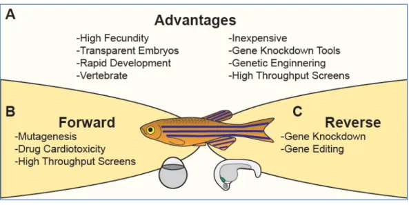
The zebrafish, Danio rerio, has emerged as a premier vertebrate model system for investigating the molecular basis of heart development and assessing therapeutic potential of small molecules (Kessler et al., 2015; Liu and Stainier, 2012; Ruzicka et al., 2015). Zebrafish have several key advantages over other vertebrate model systems that are inherent to their biology (Fig. 4A) (Westerfield, 2000). A single breeding pair produces hundreds of eggs weekly, facilitating genetic and statistical analysis. These externally fertilized eggs develop rapidly, and by 24 hours post fertilization (hpf), the embryonic heart has initiated cardiac contraction. Though the zebrafish heart has a simpler structure than the human
counterpart (two chambers instead of four chambers), it possesses analogs of the major components of the human heart and utilizes similar cellular and molecular strategies to assemble the heart (Moorman and Christoffels, 2003; Stainier et al., 1993). Due to the transparency of the embryos, the morphology and function of the developing hearts can be directly observed by light microscopy. This optical
2
This chapter previously appeared in part in the Journal of Cardiovascular Development and Disease. The original citation is
transparency can also be leveraged by the use of transgenic reporters in which cardiac cells are labeled with fluorescent markers (Huang et al., 2003; Jinn et al., 2005; Long et al., 1997; Perner et al., 2007). Importantly, zebrafish embryogenesis does not require a functional cardiovascular system during the first week of life because the zebrafish embryo is small enough to meet oxygenation needs by diffusion (Bang et al., 2004; Chen et al., 1996; Sehnert et al., 2002; Stainier et al., 1996; Strecker et al., 2011). This allows for examination of severe cardiovascular defects that usually cause embryonic lethality in other model organisms such as mice. These advantages allow for robust forward and reverse genetic approaches to study the genetic and molecular basis of heart development and disease (Fig. 4B-C).
In this review, we will provide an overview of zebrafish heart development, and discuss how zebrafish are leveraged to study cardiovascular development and disease.
Cardiovascular development in zebrafish
The heart is the first organ to form and function during vertebrate embryo development. The key steps of heart development are conserved across vertebrates, and the gross morphological changes associated with cardiac morphogenesis have been well described in detail in previous reviews (Kirby, 2007; Liu and Stainier, 2012; Moorman and Christoffels, 2003; Samsa et al., 2013; Sedmera et al., 2000). Additionally, we refer the interested reader to the online Zebrafish Atlas (http://zfatlas.psu.edu) for
histological details of zebrafish development from embryo to adult. Histology methods are also readily available for zebrafish (Sabaliauskas et al., 2006; Tsao-Wu et al., 1998). Below, we will overview zebrafish cardiac morphogenesis and disease phenotypes placing an emphasis on parallels to human heart disease.
Zebrafish cardiac morphogenesis
(Bussmann et al., 2007; de la Pompa et al., 1998; Keegan et al., 2004). These progenitors migrate during gastrulation to reside in the posterior half of anterior lateral plate mesoderm (ALPM) by 15 hpf (Fig. 5B) (Bussmann et al., 2007; Holtzman et al., 2007; Palencia-Desai et al., 2015; Trinh and Stainier, 2004; Yelon et al., 1999). Subsequently, these bilateral CPCs initiate differentiation program and fuse into a disk with endocardial cells in the center lined by ventricular and atrial myocytes (Fig. 5C). The disk elongates into a linear tube with distinct expression profiles for atrial CMs, ventricular CMs and endocardial cells (Fig. 5D) (Bussmann et al., 2007; Garavito-Aguilar et al., 2010; Holtzman et al., 2007; Palencia-Desai et al., 2015; Rohr et al., 2008; Yelon et al., 1999).
The linear heart tube is originally composed of cells from the first heart field (FHF). Additional cardiac cells are recruited to the heart tube in a second wave of differentiation as late-differentiating CPC populations called the second heart field (SHF) extend the linear heart at its arterial and venous poles starting at around 28 hpf (de Pater et al., 2009; Hami et al., 2011; Lazic and Scott, 2011; Zhou et al., 2011). Concurrent with addition of the SHF-derived cardiac cells, the linear heart tube migrates leftward and begins looping (Fig. 5E) (Baker et al., 2008; Chen et al., 1997; Rohr et al., 2008; Stainier et al., 1993). By 48 hpf, the looped heart is located in the pericardial cavity and is clearly divided into a two-chambered heart by constriction of the atrio-ventricular (AV) canal (Fig. 5F) (Beis et al., 2005; Stainier et al., 1993). Although at 48 hpf, the major components of the heart have formed, the heart is still immature and lacks auxiliary cell types and additional structures that are important for function as the organism grows (Martin and Bartman, 2009). These structures include the bulbous arteriosus, valve cushions and leaflets, myocardial protrusions called trabeculae, and epicardium (Figure 5G-H). These are discussed in detail, below.
Cardiac outflow tract
Epicardium
The epicardium develops from an extra-cardiac population of cells called the pro-epicardium. The pro-epicardium can be distinguished morphologically at 48 hpf as a group of spherical cells located in close proximity to the ventral wall of the looped heart at the level of AV junction (Hofsteen et al., 2013; Liu et al., 2010; Serluca, 2008; Zhou et al., 2008a). At approximately 72 hpf, the pro-epicardium expands and starts to spreads over the myocardial surface to form the epicardium (Fig. 5H) (Peralta et al., 2014; Plavicki et al., 2014). The epicardium is an important source of signals to the underlying myocardium (Kikuchi et al., 2011). It also is a source of epicardial-derived, cardiac resident cells such as cardiac fibroblasts (Gonzalez-Rosa et al., 2012; Peralta et al., 2014).
Trabeculation
Cardiac trabeculae are highly organized, luminal, muscular ridges lined by endocardial cells in the ventricular lumen. Trabeculae increase myocardial surface area for blood oxygenation and are critical for cardiac function (Icardo and Fernandez-Teran, 1987; Liu et al., 2010; Samsa et al., 2013; Sedmera et al., 2000). Following cardiac looping and chamber ballooning, CMs delaminate from the ventricle wall to initiate cardiac trabecular formation, and the ventricular has obvious, stereotyped trabecular ridges by 72 hpf (Liu and Stainier, 2010; Samsa et al., 2015; Staudt et al., 2014). The trabecular myocardium rapidly expands in the developing heart and as the cardiac wall matures, the trabeculae undergo extensive remodeling in association with compact myocardial proliferation, formation of the coronary vasculature and maturation of the conduction system (Samsa et al., 2013). Remodeling, also known as consolidation or compaction, marks the final stage of trabecular growth such that species-specific differences in adult trabecular morphology are generally attributed to differences in remodeling (Sedmera et al., 2000).
Valvulogenesis
Cardiac valves are a critical component of the vertebrate heart. Valves function to ensure
readily detectable during looping morphogenesis. Around 40 hpf, AV CMs expand their luminal surface while constricting their abluminal surface (Beis et al., 2005). The underlying AV endocardial cells undergo an epithelial-to-mesenchymal transition to form the endocardial cushion, which subsequently remodels to create primitive valve leaflets allowing for complete block of retrograde blood flow at 76 hpf (Beis et al., 2005; Scherz et al., 2008; Timmerman et al., 2004). These leaflets continue to thicken and lengthen to form the mature valve (Martin and Bartman, 2009).
Late maturation
Chapter 1.2 Figures
Figure 4 Zebrafish model system
Figure 5 Zebrafish heart development
CHAPTER 2 CARDIAC CONTRACTION ACTIVATES ENDOCARDIAL NOTCH SIGNALING TO MODULATE CHAMBER MATURATION
2.1 Historical Context
An increasing body of evidence highlights the importance of an active interplay between the biomechanical forces generated by the functioning embryonic heart and cardiac structure in regulating cardiac maturation. Mutations in genes that influence force production or detection in the heart are associated with a wide range of CHDs, underscoring the importance of this interplay [reviewed in Granados-Riveron and Brook (2012)]. However, the underlying mechanisms connecting cardiac function and form are largely unknown.
Early studies in chicken embryos were foundational in establishing a role for biomechanical forces in regulating chamber maturation. Partial ligation of the right lateral vitelline vein disrupts
intracardiac fluid dynamics and leads to later chamber maturation defects (Hogers et al., 1997). Similarly, altering ventricular afterload by conotruncal banding or changing preload and afterload dynamics through atrial ligation or clipping, respectively, leads to dramatic alterations in the ventricular myocardial
architecture (Sedmera et al., 1999).
2.2 Cardiac Contraction Activates Endocardial Notch Signaling to Modulate Chamber Maturation3
Introduction
Congenital heart diseases often feature structural abnormalities that arise from defects in the development of the heart during embryogenesis (Chin et al., 2012; Samsa et al., 2013). The heart is the first organ to function, yet the details of its formation are only partially understood. In vertebrates, cardiac morphogenesis commences as the two bilateral cardiac primordia fuse at the ventral midline to form the linear heart tube, which is composed of a luminal endocardial layer and an immature myocardial layer (Fishman and Chien, 1997; Harvey, 2002; Olson and Srivastava, 1996; Staudt and Stainier, 2012; Yelon, 2001). Concomitant with cardiac contraction, the primitive heart tube develops into a multi-chambered functional organ that grows and matures through a series of complex morphogenic processes collectively known as cardiac chamber maturation (Moorman and Christoffels, 2003; Samsa et al., 2013).
As a part of chamber maturation, cardiac trabeculation is a tightly regulated process by which ventricular cardiomyocytes protrude and expand into the lumen of the ventricular chambers to form ridge-like muscular structures called cardiac trabeculae (Liu et al., 2010; Peshkovsky et al., 2011). Trabeculae increase cardiac output and allow for nutrition and oxygenation of the myocardium prior to coronary vascularization, and are required for establishment of the mature conduction system of the developing ventricle (Lai et al., 2010; Liu and Stainier, 2010). Thus, failure to form cardiac trabeculae causes embryonic lethality, and subtle perturbations of this process could lead to congenital cardiomyopathy (Jenni et al., 1999).
Crosstalk between endocardial and myocardial cells is important for cardiac maturation.
Zebrafish cloche mutants that do not form endocardial cells fail to develop trabeculae and ultimately die, presumably from heart failure (Peshkovsky et al., 2011; Stainier et al., 1995). Mice deficient in the epidermal growth factor (EGF) receptor ligand Neuregulin 1 (Nrg1), which is expressed in the
endocardium and signals through the myocardial ErbB4/ErbB2 receptors complex, fail to form trabeculae (Gassmann et al., 1995; Lee et al., 1995; Meyer and Birchmeier, 1995). Likewise, inhibition of Nrg1/ErbB signaling in zebrafish embryos completely blocks trabeculation (Liu et al., 2010; Peshkovsky et al., 2011;
3
This part of chapter 2 previously appeared in the journal Development. The original citation is Samsa, L. A., Givens, C., Tzima, E., Stainier, D. Y., Qian, L., and Liu, J. (2015) Cardiac contraction activates endocardial notch signaling to modulate chamber
Samsa et al., 2013; Staudt et al., 2014). Notch ligands and receptors are expressed in endocardial cells during development, and Notch signaling regulates many aspects of endothelial biology including artery-vein specification, angiogenesis, and proliferation (Benedito and Hellström, 2013; Corada et al., 2014; de la Pompa and Epstein, 2012; Gridley, 2010). Upon ligand binding, the cleaved Notch receptor intracellular domain (NICD) translocates to the nucleus, where it acts as a cofactor to promote transcription of Notch effectors including EphrinB2, an essential upstream regulator of Nrg1 signaling (Grego-Bessa et al., 2007). Despite this knowledge, questions remain on whether this epistasis is a requirement for all vertebrate cardiac trabeculation, the precise spatiotemporal roles of these genes, and the roles of mediators upstream of Notch.
An increasing body of evidence suggests that the biomechanical forces generated by the
functioning embryonic heart could influence cardiac chamber maturation, underscoring the importance of a dynamic relationship between cardiac form and cardiac function (Auman et al., 2007; Bartman et al., 2004; Dietrich et al., 2014; Hove et al., 2003; Kalogirou et al., 2014; Lee et al., 1995; Lin et al., 2012; Peralta et al., 2013; Stainier et al., 2002; Vermot et al., 2009; Yang et al., 2014). Interestingly, in zebrafish and chick embryos, reducing blood flow through the ventricle significantly impairs cardiac trabeculation (Auman et al., 2007; Bartman et al., 2004; Chen et al., 1996; Dietrich et al., 2014; Hove et al., 2003; Kalogirou et al., 2014; Lin et al., 2012; Peralta et al., 2013; Stainier et al., 2002; Vermot et al., 2009; Yang et al., 2014). But, how mechanical stimulus is sensed and translated into spatial and temporal signals to regulate cardiac trabeculation through regulatory interaction with other myocardial signals remains largely unexplored.
downstream effectors, suggesting a role for primary cilia flow detection in endocardial Notch activation. Together, our findings suggest that in early cardiac morphogenesis, endocardial cells respond to cardiac contraction by detecting flow with primary cilia to regulate trabeculation by epistasis of
notch1b/efnb2a/nrg1.
Results
Cardiac contraction is required for myocardial trabeculation
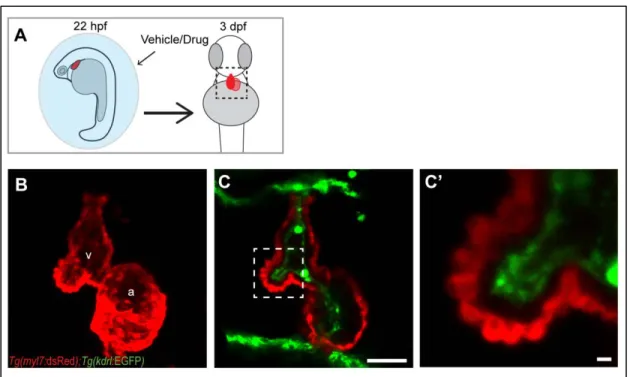
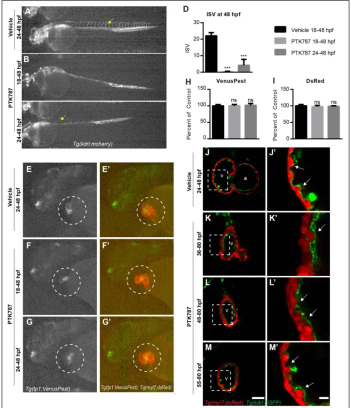
One of the earliest signs of cardiac chamber maturation is the formation of muscular, luminal protrusions called cardiac trabeculae. In zebrafish, cardiac trabeculae begin to form around 55 hpf (hours post fertilization) and are easily detectable at 3 dpf (days post fertilization) by examining cross sections of ventricle outer curvature (Liu et al., 2010; Peshkovsky et al., 2011; Staudt et al., 2014). Though the basic structure of the heart can form in the absence of myocardial function, some morphogenic events require cardiac contraction. To determine if cardiac contraction is required for trabeculation, we examined embryos deficient in cardiac troponin T type 2a (tnnt2a), which encodes an essential component of the contractile apparatus (Sehnert et al., 2002). We injected Tg(myl7:dsRed); Tg(kdrl:GFP) double transgenic embryos (labeling cardiomyocytes and endothelial cells, respectively) with standard control or tnnt2a morpholinos and assessed the presence of trabeculae at 3 dpf (Fig 6A). Like tnnt2a-/- hearts, the non-contractile tnnt2a morphant hearts underwent relatively unaltered looping morphogenesis and chamber formation at 3 dpf, but notably, failed to form trabeculae (Chi et al., 2008; Staudt et al., 2014) (Fig. 6B-C’’).
To determine if failure to form trabeculae is due to a direct role of Tnnt2a or is more generally associated with contraction deficiency, we tested if the trabeculation defects observed in tnnt2a morphants could be recapitulated by chemical inhibition of cardiac contraction. We thus treated
Tg(myl7:dsRed); Tg(kdrl:GFP) embryos with vehicle or a pharmacological inhibitor of contraction from 22 hpf to 3 dpf (Fig. 6D). The hearts of embryos treated with blebbistatin, a myosin II ATPase inhibitor, were non-contractile on day 3 and strongly resembled tnnt2a morphants in that both myocardial and
Cardiac contraction is required for endocardial Notch activation and notch1b transcription Cardiac contraction exerts mechanical forces on both myocardium and endocardium, and trabeculation requires crosstalk between these two layers (Tian and Morrisey, 2012; Wagner and
Siddiqui, 2007). Thus, we next sought to determine if any signaling pathways are activated in the ventricle in response to cardiac contraction. Using Tg(Tp1:EGFP) Notch reporter embryos in which the Notch1 responsive TP1 module drives EGFP as a fluorescent readout for active Notch1 signaling (Parsons et al., 2009), we assessed Notch activation in control and tnnt2a morphants (Fig. 8A). At 48-50 hpf, Notch activation in control morphants hearts was robust in the ventricular endocardium and atrioventricular canal (AVC) with occasional weak signal detectable in the atrium (Fig. 8B-C’’). Interestingly, Notch activation was below detection in tnnt2a morphant hearts of embryos examined in whole mount or in confocal images (Fig. 8D-E’’). In contrast, Notch signaling was robust in the brains of control and tnnt2a morphants, indicating a specific role for cardiac contraction in regulating endocardial Notch signaling (Fig. 8B,D). Similar results were observed with DMSO and blebbistatin-treated embryos compared to control and tnnt2a morphants, respectively (Fig. 9A-C’’). These data indicate that cardiac contraction is required for Notch activation in the endocardium at 2 dpf.
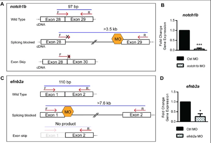
We next sought to identify the primary Notch receptor mediating Notch activation in the
endocardium. Using a previously described morpholino to knockdown notch1b (Milan et al., 2006; Wang et al., 2010) in Tg(Tp1:EGFP); Tg(myl7:dsRed) double transgenic embryos, cardiac Notch activation was below detection in notch1b morphants hearts at 48-50 hpf (Fig. 8F-G’’). To determine whether cardiac contraction regulates Notch1 activation by controlling notch1b gene transcription, we performed in situ hybridization at 48-50 hpf. notch1b expression mirrors Tg(Tp1:EGFP) Notch reporter expression in the heart at 48 hpf , but was below detection in tnnt2a-/- hearts (Fig. 8H,I). Thus, cardiac contraction controls Notch1 signaling by regulating notch1b expression. Together, these findings indicate that cardiac
contraction regulates notch1b at the transcript level in the heart. Our studies do not exclude the possibility that the mechanical forces associated with contraction may also regulate Notch receptor activation.
Spatiotemporal pattern of Notch activation
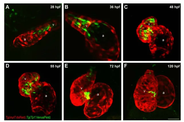
Notch reporter transgenic line which express, under control of the Tp1 Notch response element, a partially-destabilized fluorescent protein with a half-life around 2 hours as compared to 24 hours half-life of GFP protein (Aulehla et al., 2008; Ninov et al., 2012). The use of this reagent afforded us greater spatial and temporal resolution to determine the time window of Notch activation in the ventricular endocardium. VenusPest expression was first detectable in the endocardium at 28 hpf, just 4 hours after initiation of contraction, with substantial spatial bias towards the end of the heart tube containing
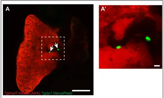
ventricular cardiomyocytes (Fig. 10A). VenusPest expression in the ventricular endocardium persisted until 55 hpf, after which it declined in parts of ventricular endocardium lining the outer curvature (Fig. 10B-D). By 72 hpf, VenusPest expression was primarily restricted to AVC endocardium, and was retained in the AVC through at least 4 weeks post fertilization (Fig. 10E,F, Fig. 11A-A’). Thus, within the limits of reporter expression, Notch signaling is activated throughout the ventricular endocardium shortly after initiation of contraction, but becomes inactive shortly after the initiation of cardiac trabeculation and is completely restricted to the AVC by 3 dpf.
Cardiac contraction promotes trabeculation through notch1b/efnb2a/nrg1 epistasis
Our data demonstrate that, while Notch is required for trabeculation, it is not active in the
function point mutation in the gene mindbomb E3 ubiquitin protein ligase 1. mib1 encodes an E3 ubiquitin ligase required for canonical trafficking of Notch ligands (Itoh et al., 2003). At 3 dpf, trabeculae were completely lacking in the ventricle of these mutant embryos (Fig 15A-B).
To test if ephrinb2a and nrg1 could act downstream of Notch signaling to control cardiac
trabeculation, and to establish the epistasis of these genes in zebrafish cardiac maturation, we assessed their expression in the zebrafish heart and found that notch1b, efnb2a, nrg1 and their principle ligand or receptor partners are expressed at 48-50 hpf (Fig. 16). We isolated hearts at 48-50 hpf and found that tnnt2a morphants had significant reduction in notch1b, efnb2a, and nrg1 expression, while notch1b morphants had significant reduction in efnb2a and nrg1 transcripts levels (Fig. 14D,E). In contrast, notch1b expression and Notch activation pattern was not affected in efnb2a morphants, but nrg1
expression was significantly reduced (Fig. 14F, Fig.17A-D). Notably, these defects in gene expression do not reflect failure of the endocardium to form, as both endocardium and myocardium are present at 3 dpf (Fig. 14A-C’). Combined, these data suggest that cardiac contraction is required for trabeculation by activating a regulatory notch1b/efnb2a/nrg1 pathway.
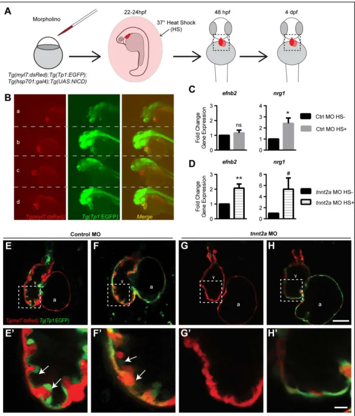
Next, we asked if forced activation of Notch signaling could bypass the requirement for cardiac contraction in trabeculation. To this end, we overexpressed the Notch intracellular domain to activate Notch signaling using the Tg(hsp701:gal4); Tg(UAS:NICD) system in control and tnnt2a morphants carrying the Tg(tp1:EGFP) and Tg(myl7:dsRed) transgenes (Fig. 18A). All embryos were exposed to 37°C heat shock to activate Gal4 expression and, as an indication of forced Notch activation, we
observed a dramatic increase in tp1:EGFP across all somatic tissues in approximately 25% of control and tnnt2a morphants at 48 hpf (Fig. 18B). We isolated hearts from control and tnnt2a morphants and found significant upregulation of efnb2a and nrg1 expression in tnnt2a morphants hearts with NICD
overexpression at 2-3 dpf (Fig. 18C-D). However, this upregulation was not sufficient to induce cardiac trabeculation in tnnt2a morphants (Fig. 18E-H).
Primary cilia are required for Notch activation in endocardial cells
Endocardial cells are particularly well positioned to detect hemodynamic forces including shear stress (the frictional force of parallel flow). Endothelial primary cilia—microtubule based sensory organelles that, in certain hemodynamic environments, protrude into the lumen of blood vessels—have a well-established role in detecting low levels of flow (Culver and Dickinson, 2010; Hahn and Schwartz, 2009; Slough et al., 2008; Van der Heiden et al., 2006). Recently, Goetz et al., demonstrated that endothelial primary cilia in the zebrafish vasculature are highly sensitive to low magnitude shear forces from 24-28 hpf to regulate vascular development (Goetz et al., 2014). Thus, we hypothesized that primary cilia play a key role in flow detection, leading to notch1b upregulation and Notch1 activation in endocardial cells.
We validated the presence of primary cilia on endocardial cells around the time of Notch1 activation using Tg(actb2:Arl13b-GFP) transgenic embryos where Arl13b-GFP localizes to primary cilia (Borovina et al., 2010). Though promoter activity for this transgene is higher in myocardial than
endocardial cells, primary cilia can be detected in both cell layers (Fig. 19A-B). Owing to their potential role in flow detection, we focused on characterizing endocardial primary cilia. At 30 hpf, individual endocardial cells of un-injected embryos, control morphants, and tnnt2 morphants possess a single, primary cilium projecting into the lumen of the heart (Fig. 20A-B’, Fig. 21A-B’’). This indicates that endocardial cells possess primary cilia, and that ciliation is independent of cardiac contraction.
Mutations in intraflagellar transport 88 (ift88) cause ciliopathies in zebrafish (Kramer-Zucker et al., 2005; Lunt et al., 2009; Neugebauer and Yost, 2014; Tsujikawa and Malicki, 2004) and ift88 morphants have deficiencies in endothelial primary cilia formation (Goetz et al., 2014). Similarly, we observed mislocalization of Arl13b-GFP in ift88 morphants (arrowheads, Fig. 21A-C). To test if endocardial primary cilia are involved in zebrafish ventricular endocardial Notch1 activation, we knocked down ift88 in
isolated hearts from ift88 morphants and found a significant reduction in notch1b and nrg1 expression (Fig. 20H,I).
Primary cilia are required at the onset of flow for Notch1 activity
Though our data supports a model in which primary cilia respond to luminal flow to activate Notch transcription, the loss of endocardial Notch activation in ift88 morphants could be secondary to a role for primary cilia in early embryogenesis (Gerdes et al., 2009; Sasai and Briscoe, 2012). To address this possibility, we used a pharmacological approach to define when primary cilia are necessary for endocardial Notch activation. Ciliobrevin D (CBD) inhibits the AAA+ ATPase motor cytoplasmic dynein and significantly reduces the microtubule cycling necessary to construct and maintain primary cilia (Firestone et al., 2012). We treated Tg(Tp1:VenusPest) positive embryos with DMSO or CBD starting at 18-24 hpf and assessed VenusPest expression at 42-48 hpf (Fig. 22A-D). DMSO injection at any of these times had no effect on VenusPest expression level (Fig. 22A,B,E). Embryos injected with CBD at 18 or 20 hpf had reduced VenusPest expression (Fig. 22C,E), while embryos injected with CBD from 22 or 24 hpf were indistinguishable from DMSO injected controls (Fig. 22D,E). Given a time delay between initiation of treatment and sufficient accumulation for biological effects, these data suggest that Notch1 expression in the endocardium requires primary cilia in a short time window coinciding with the onset of flow.
Primary cilia likely detect low magnitude shear stress to upregulate Notch in ventricular endocardial cells
Primary cilia have two well-defined, independent functions—facilitating Hedgehog (Hh) signaling and detecting low magnitude shear stress (Anderson, 2006; Egorova et al., 2012; Goetz et al., 2014; Hierck et al., 2008; Roy, 2012; Van der Heiden et al., 2011; Wilson and Stainier, 2010). To determine whether Hh signaling is necessary for endocardial Notch activation, we treated Tg(Tp1:VenusPest); Tg(myl7:dsRed) Notch reporter embryos with cyclopamine to antagonize Hh signaling downstream of primary cilia (Chen et al., 2002; Stanton and Peng, 2010). Embryos treated with cyclopamine from 4-48 hpf exhibited severe body axis deformities indicative of successful inhibition of Hh signaling (Fig. 23A,B). Embryos treated with cyclopamine from 18-48 hpf and 24-48 hpf did not exhibit defects in Notch