DEVELOPING HIGH THROUGHPUT ORGANOID-BASED PLATFORMS TO STUDY ENTERIC PHYSIOLOGY IN VITRO
Ian Andrew Williamson
A dissertation submitted to the faculty of the University of North Carolina at Chapel Hill in partial fulfillment of the requirements for the degree of Doctor
of Philosophy in the Joint Department of Biomedical Engineering in the School of Engineering.
Chapel Hill 2018
ii © 2018
iii ABSTRACT
Ian Williamson: Developing High Throughput Organoid-based Platforms to Study Enteric Physiology In Vitro (Under the direction of Scott T. Magness)
iv
ACKNOWLEDGMENTS
The work presented in this dissertation is the result of many collaborative projects made possible by the support of the UNC research community. I would like to thank my mentor Dr. Scott Magness for introducing me to laboratory science and believing in my pursuit of an advanced education in bioengineering. I would like to thank the UNC Center for Center for Gastrointestinal Biology and Disease for providing an interesting and diverse environment, facilitating my growth as a basic science researcher. Drs. Susan Henning and Adam Gracz for their guidance throughout my graduate studies. The technologic innovations making up the majority of this work would not have been possible without the help and guidance of Drs. Nancy Allbritton, Christopher Sims, and Yuli Wang in the UNC Chemistry Department. I would like to thank my fellow researchers in the Magness lab and Allbritton lab: Dr. Kyle Roche, Dr. Bailey Zwarycz, Dr. Asad Ahmad, Dr. Pete Attayek, Dr. Matthew DiSalvo, and Jennifer Speer for their contributions to the work detailed here. Finally, I would like to acknowledge the exceptional undergraduates in the Magness lab, including Xiao Fu Liu, Liam Gaynor, and Malvika Pillai.
v
TABLE OF CONTENTS
ABSTRACT ... iii
ACKNOWLEDGMENTS ...iv
TABLE OF CONTENTS ... v
LIST OF FIGURES ... xii
Chapter 1: Introduction to Intestinal Physiology ... 1
The Intestinal Lumen is our Primary Interface with the Environment ... 1
Organization and physiology of the intestinal mucosa ... 1
Microbiota colonizing the intestinal lumen affect host physiology ... 5
Enteropathies involve aberrant renewal and/or barrier function dysregulating the intestinal luminal interface. ... 6
Inflammatory bowel diseases and the enteric microbiota ... 8
Colitis associated colorectal cancer. ... 11
Gastrointestinal organoids are a reductionist in vitro platform to model intestinal physiology and enteropathies ... 12
Organoid Structure and physiology. ... 12
Modeling human diseases in organoids. ... 14
Significant technical barriers have limited applications of organoid-based assays .... 16
vi
Chapter 2: Engineering Platforms to Expand Organoid Applications ... 19 Introduction ... 19
Reconstructing basal-luminal signaling gradients in vitro and measuring
gradient influences on organoid compartmentalization ... 20 Developing self-renewing monolayers of primary colonic epithelial cells ... 21 Developing a high throughput microfabricated IESC culture
platform to describe intrinsic and extrinsic regulation of stem cell growth ... 23 Methods/Materials ... 24
Reconstructing basal-luminal signaling gradients in vitro and measuring
gradient influences on organoid compartmentalization ... 24 Developing self-renewing monolayers of primary colonic epithelial cells ... 25 Developing a high throughput microfabricated IESC culture platform
to describe intrinsic and extrinsic regulation of stem cell growth ... 27 Results ... 34
Reconstructing basal-luminal signaling gradients in vitro and
measuring gradient influences on organoid compartmentalization... 34 Developing self-renewing monolayers of primary colonic epithelial cells ... 35 Developing a high throughput microfabricated IESC culture platform
to describe intrinsic and extrinsic regulation of stem cell growth ... 36 Conclusions ... 41
Reconstructing basal-luminal signaling gradients in vitro and measuring
gradient influences on organoid compartmentalization ... 41 Developing self-renewing monolayers of primary colonic epithelial cells ... 41 Developing a high throughput microfabricated IESC culture platform to
vii
Chapter 3: A High throughput Organoid Microinjection Platform to
Study Gastrointestinal Microbiota and Luminal Physiology ... 44
Synopsis ... 44
Overview ... 44
Background & Aims ... 46
Results ... 49
Development of an organoid injection system ... 49
Validating the organoid injection system ... 52
Automated computer vision facilitates high-throughput organoid identification, injection and quantification of injected cargos ... 54
Growth dynamics of transplanted microbial communities and colonoid barrier integrity can be monitored by computer vision ... 56
The colonoid lumen supports the growth of aerobic and obligately anaerobic human microbiota taxa ... 59
Conclusions ... 63
Methods ... 67
Automated Imaging System ... 67
Microinjection Hardware Custom Fittings ... 67
Optimized Microinjection Needle Processing ... 68
Colonoid culture and expansion ... 68
Plating Colonoids on CRA devices ... 69
viii
Computer Vision Quantification of Injected Cargos ... 71
Assessing Efficiency of Microinjection Device ... 71
Retrieving Bacteria from the Organoid Lumen ... 72
Bacterial Cultivation ... 72
High Resolution Imaging of Microinjected Colonoids ... 73
Mock Community Response to Media Antimicrobials ... 73
Relating Fluorescent Signal to Microbial Load ... 74
Stool Sample Preparation ... 75
Colonoid Fecal Microbiota Transplantation ... 75
DNA Isolation ... 76
16S rRNA Gene amplicon sequencing ... 76
Sequencing data analysis ... 77
Colonoid Anaerobe Monoculture Assay ... 78
Statistics ... 78
Figures ... 80
Appendix A: In Vitro Polarization of Colonoids to Create an Intestinal Stem Cell Compartment ... 103
Introduction ... 103
Materials and Methods ... 106
ix
Colonoid Culture ... 107
Diffusion-Based Gradient Generation and Characterization ... 108
Culture of Colonic Cells in the Microchannel of the Gradient Device ... 109
Microscopy ... 109
Immunofluorescence and EdU Assays ... 110
Colonoid Segmentation using DsRed or Hoechst 33342 ... 110
Measurement of the Percentage of Colonoids Positive for a Fluorophore ... 111
Measurement of Sox9EGFP Polarization in a Colonoid ... 111
Measurement of EdU Polarization in a Colonoid ... 112
Statistics ... 113
Results and Discussion ... 114
Design and Characterization of Gradient-Microdevice ... 114
Microchannel devices support colonoid development similar to conventional cultures ... 115
Colonoids do not demonstrate overall polarization in the absence of an external gradient ... 117
A Wnt-3a gradient is sufficient to polarize the stem/progenitor cell compartments ... 119
A Wnt-3a/R-spondin1 gradient enhances polarization of the stem/ progenitor cell compartment ... 121
Effect of a Wnt-3a and R-spondin1 Gradient on Growth and Polarization of Colonoids Derived from a Single Stem Cell ... 122
x
Appendix B: Self-renewing Monolayer of Primary Colonic or Rectal Epithelial Cells .. 136
Introduction ... 136
Materials and Methods ... 138
Isolation of crypts from mouse colon and human rectal biopsies. ... 138
Preparation of the collagen hydrogel in a 6-well plate. ... 139
2D monolayer culture on a collagen hydrogel. ... 140
Characterization of the 2D monolayer. ... 141
High-throughput screening of dietary metabolites and natural products. ... 142
Assaying for hit dietary compounds on mouse 3D organoids and human 2D monolayers. ... 142
Image Acquisition. ... 143
Image Analysis. ... 144
Hit Selection. ... 144
Other Statistics/Methods. ... 145
Results ... 146
A murine colonic epithelial monolayer proliferates on the surface of a collagen hydrogel ... 146
Equivalency of murine colonic 2D monolayers to 3D organoids... 149
Lineage tracing confirms the presence of stem cells in the murine 2D monolayer ... 150
xi
Screening dietary components and food metabolites on murine colonic monolayers reveals specific impacts on
cell proliferation and differentiation ... 152 Screening dietary metabolites and natural products on human
xii
LIST OF FIGURES
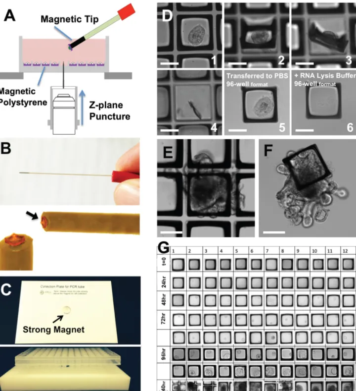
Table 1: Factors limiting the use of organoid technologies. ... 80 Figure 1. Modified CRAs are compatible with long-term culture of primary IESCs. ... 81 Figure 2. Retrieval of magnetic rafts for downstream gene expression analysis. ... 83 Figure 3. Software-assisted post hoc analysis identifies initial well contents of CRA culture. ... 85 Figure 4. Cell-to-cell contact is required for PC-influenced survival
of IESCs in vitro. ... 87 Figure 5. Single cells do not form cell–cell contacts in microwells
after initial plating. ... 89 Figure 6. Robotically Articulated Colonoid Microinjection Maintains
Atmospheric Control Facilitating Long-term Sampling of Large
Batches of Colonoids: ... 92 Figure 7. Increasing Microinjection Throughput Using Computer
Vision to Quantify Cargo Retention, Organoid Morphology and
Injection Success: ... 94 Figure 8. The colonoid lumen forms a discrete compartment
compatible with specific microbial growth: ... 97 Figure 9: Monolayer Respiration Makes the Colonoid Lumen a
Hypoxic Environment Capable of Supporting the Growth
of Anaerobic Enteric Microbes: ... 99 Figure 5. The Colonoid Lumen Is Compatible with Patient Derived
Microbial Communities and Non-sporulating Anaerobes: ... 101 Appendix A: Fig 1. Characterization of the gradient-generating microdevice. ... 126 Appendix A: Fig 2. Colonoid properties in the absence of a gradient. ... 128 Appendix A: Fig 3. Incorporation of EdU into colonoids
after a 2 h pulse in the absence of a gradient. ... 129
Appendix A: Fig 4. Colonoid growth in the presence of a
xiii
Appendix A: Fig 5. Colonoid growth in the presence of a
dual Wnt-3a/R-spondin1 gradient. ... 132 Appendix A: Fig 6. Growth of single stem cells in the
presence of a Wnt-3a/R-spondin1 gradient. ... 134 Appendix B: Figure 1. Proliferative 2D monolayer culture of
murine colonic crypts on the surface of collagen hydrogel. ... 158 Appendix B: Figure 2. Proliferative capacity, lineage composition, and
compartmentalization are highly similar between 2D murine
colonic monolayers and 3D organoids. ... 160 Appendix B: Figure 3. Lineage tracing of mouse colonic epithelial
cells in the 2D monolayer. ... 161 Appendix B: Figure 4. Human rectal epithelial cells can be
cultured as a proliferative 2D monolayer. ... 162 Appendix B: Figure 5. The impact of dietary compounds and natural
products on primary murine colonic monolayers. ... 163 Appendix B: Figure 6. Assaying a subset of the dietary compounds
and metabolites on murine 3D organoids. ... 164 Appendix B: Figure 7. The impact of seven dietary compounds
xiv
LIST OF ABBRIVATIONS
3D Three-dimensional
BMP Bone Morphogenetic Protein
CBCs Crypt-base Columnar Cells
CD Crohn’s Disease
CF Cystic Fibrosis
CHGA Chromogranin A
CRC Colorectal Cancer
CTFR
Cystic Fibrosis Transmembrane Conductance Regulator
DSS Dextran Sulfate Sodium
ENS Enteric Nervous System
FACs Fluorescence Activated Cell Sorting
GF Germ-free
GI Gastro Intestinal
IBD Inflammatory Bowel Disease
IESC Intestinal Epithelial Stem Cell
Lct Lactase
LPS Liposaccharide
MAMPs Microbe Associated Patterns
MVID . Microvillus Inclusion Disease
PBS Phosphate Buffered Saline
xv
PEG Polyethylene Glycol
PRRs Pattern Recognition Receptors
RFP Red Fluorescent Protein
SCFAs Short-chain Fatty Acids
SIS Sucrase-isomaltase
TA Transit-amplifying
TLR Toll-like Receptor
UC Ulcerative Colitis
1
Chapter 1: Introduction to Intestinal Physiology
The Intestinal Lumen is our Primary Interface with the Environment
Absorption of ingested nutrients occurs in the intestine where components of digested food are both actively transported and passively diffuse into the body. The intestine is a complex tissue consisting of mucosa, muscularis, and serosa layers with distinct cellular composition and function. The innermost mucosa layer of the intestine is responsible for the bulk of absorptive processes. The intestinal mucosa consists of a supportive lamina propria of fibroblasts, myofibroblasts, capillaries, and lymphocytes underlying a monolayer of columnar epithelium (intestinal epithelium) that directly interfaces with the luminal space. The outer submucosa, muscularis, and serosa layers of the intestine drive the movement of ingested material down the gastrointestinal (GI) tract and regulate the transport of absorbed nutrients to the rest of the body. This dissertation focuses on the mucosa layer and developing in vitro approaches to study physiology and renewal of the epithelial surface of the lumen.1,2
Organization and physiology of the intestinal mucosa
2
titrated thymidine and their migration was observed at various times following administration1. These approaches identified a pluripotent intestinal epithelial stem cell
3
exclusively in the base of small intestinal crypts where they secrete protective antimicrobials like lysozyme that form a niche for resident stem cells.1–3
Components of the mucosa produce secreted and membrane bound ligands forming localized signaling gradients that regulate renewal. The Wingless-related integration site (Wnt), Bone morphogenic protein (BMP), and Notch signaling pathways are the most substantially linked to epithelial proliferation and differentiation. Niche cells near the crypt base secrete Wnt ligands that bind to receptors on crypt-based cells, stabilizing cytoplasmic β-catenin. Stabilized β-catenin translocates to the cell nucleus, where it activates the TCF and LEF transcription factors that drive targets generally associated with cellular growth and proliferation. Wnt signaling decreases as cells migrate away from the crypt base reducing access to niche ligands. BMP signaling acts opposite of Wnt signaling, driving differentiation as cells migrate up the crypt villus axis. Bmp4 and related ligands are strongly expressed in the villus mesenchyme, where it binds to serine/threonine kinase receptors on epithelial cells to activate genes through the SMAD transcription factor4–7. Notch signaling is mainly restricted to the crypt
4
The dynamics of intestinal epithelial renewal have been well understood for decades but direct studies of IESCs were hampered by a lack of specific genetic biomarkers. In 2007, the G-protein coupled receptor Lgr5 was validated by in vivo lineage-tracing as an IESC biomarker expressed in crypt-base columnar cells (CBCs) intercalated between PCs in the crypts of the small intestine11. Tracing the progenitors of Lgr5 cells through
genetic lineage tracing showed that progeny formed long-lived clonal units containing all post-mitotic lineages persisting for 60 days or longer. These studies confirmed the self-renewal and multipotency of Lgr5 CBCs and established the gold standard of in vivo lineage tracing used to identify additional CBC IESC biomarkers including Olfm4, Ascl2, and Sox912–15. Following the identification of the CBC IESC population, a second IESC
population residing above the PC compartment was described by lineage tracing from Bmi1 expressing cells16. Subsequently, other genes associated with the super-Paneth
position were validated as in vivo IESC biomarkers, including mTert and Hopx17. While
all the super-Paneth IESC populations have some stem cell capacity under normal physiological conditions, reports show increased lineage tracing from super-Paneth IESC biomarkers following damage. The field consents that super-Paneth IESCs represent a reserve population, normally quiescent, that activates to initiate repair following damage to CBC IESCs16–18. This model has been confirmed by genetically
ablating Lgr5 IESCs in animal models, which were able to compensate by activating super-Paneth IESC populations.
5
to act as a dynamic, selectively permeable barrier to allow absorption of essential nutrients while sealing the body from harmful toxins and microbial invasion. The intestinal mucosa responds dynamically to nutrient availability in specific and non-specific manors to increase or decrease absorption. Villi projections extend further into the lumen in response to feeding, grossly increasing the absorptive surface area and potential19. The apical surface of absorptive epithelial cells is coated with membrane
projections called microvilli that are highly concentrated with transport proteins and digestive enzymes. Digestive enzymes on the apical surface break down macromolecules like carbohydrates into monomers for active transport by absorptive enterocytes into the body. Other polar nutrients like ions are also actively transported by proteins on the apical membrane of enterocytes. Lipids are digested into monoglycerides and free fatty acids that passively diffuse into the cytosol. Absorptive enterocytes change their expression of transport proteins and digestive enzymes to increase or decrease specific nutrient uptake in response to nutritional requirements and availability.20
Microbiota colonizing the intestinal lumen affect host physiology
6
absorbed by the epithelium, where they act as energy sources, mitogens, and morphogens effectively preserving normal physiology. The classical example of host-microbiota molecular interactions is butyrate, produced by bacterial fermentation of dietary fiber in the colonic lumen. Enterocytes prefer butyrate to glucose as an energy source promoting stem cell driven renewal. Butyrate also acts as a morphogen by inhibiting epigenetic changes in histone methylation. The microbiota also produce neuroactive peptides like serotonin and gaba that grossly effect host digestion and peristalsis.21–23
The bacterial component of the enteric microbiota is dominated by gram-positive species of the Firmicutes, Bacteroidetes, Actinobacteria, or Proteobacteria phylum that compete for suitable luminal space. The composition of the microbiota is stabilized by competition for key nutritional and environmental factors. Changes in the luminal environment caused by pathogens, antimicrobial treatment, or dietary changes can lead to the overgrowth of specific members inside or outside of their normal habitat, termed dysbiosis. Dysbiosis is particularly important in modern health care due to an epidemic of Clostridium difficile colitis caused by the overgrowth of an anaerobic motile bacteria normally inhabiting the small intestine. C. difficile dysbiosis occurs in about 500,000 Americans every year, reoccurring in nearly 25% and causing mortality in about 5% of cases due to severe colon inflammation and diarrhea.
Enteropathies involve aberrant renewal and/or barrier function dysregulating the intestinal luminal interface.
7
characterized non-pathogenic enteropathies result from aberrant renewal or differentiation caused by mutations in Wnt, BMP, or Notch signaling components. Mutations causing IESC populations to become hyperproliferative lead to crypt hyperplasia and adenomas, arising in 20-50% of Americans by age 70. Adenomas lead to the formation of polyps, which dramatically increase cancer risk. Nearly 1 million new cases of colorectal cancer (CRC) are diagnosed each year with familial risk associated with about 30% of cases. Some cases are caused by autosomal dominant mutations that drive polyposis or nonpolyposis forms of hereditary colorectal cancer. These two conditions cause 5-10% of CRC and are directly inherited by an effected individual. The other 20% of new cases with concurrent familial risk have recessive or partially functional mutations in key tumor-suppressor and oncogenes. In these cases, cancer is initiated through subsequent mutations in the Wnt or BMP signaling pathways. Accumulation of cell signaling changes, rather than the order of occurrence, initiates tumor formation.24–27
8
carbonate ions, leading to a syndrome termed cystic fibrosis (CF). CF causes thickening of the mucous secreted into the lumen, causing blockages and preventing ingested material from reaching the apical surface. More than 5,000 specific mutations in CTFR are known to cause CF though the effect of CTFR dysfunction that leads to the CF phenotype is still under debate.28–30
Inflammatory bowel diseases and the enteric microbiota
There is considerably less consensus on the kinetics of enteropathies driven by the dysregulation of host immune responses. In these syndromes, inappropriate responses by enteric immune cells cause prolonged inflammation of the mucosa that can disturb nutrient absorption and barrier function. In Celiac disease, dietary glutens that are absorbed by the mucosa interact with type II major histocompatibility complexes to activate lymphocytes. Prolonged lymphocyte activation leads to lesions of flat villi and hyperplastic crypts to form. Similar lesions occur in cases of inflammatory bowel disease (IBD) where microbial cues activate and prolong inflammation. The specific microbial taxa, epithelial receptors, and cell signaling pathways initiating and prolonging IBD are still under debate despite extensive investigations. Our understanding of the microbiota’s role in IBD has been reviewed in detail elsewhere and will be summarized here.31,32
9
like peptidoglycan and flagellin are known TLR ligands that increase the expression of nuclear factor-KB regulated targets when bound to TLRs33,34. In healthy states the host
intestine becomes habituated to commensal microbiota taxa and reduces its inflammatory response. Habituation is accomplished through the expression of redundant inhibitory peptides (reviewed by 28,32,35–38) regulating paracrine interactions
between the intestinal epithelium and regulatory lymphocytes in the lamina propria. Various metabolic biproducts produced by commensal microbiota enhance the expression of anti-inflammatory peptides by the host epithelium. In this way the microbiota can directly modulate host immunity to relieve inflammatory responses and maintain homeostasis.
10
mucous production, increasing mucosa barrier integrity and reducing interactions between luminal contents and the mucosa.28,32,38–40
Considerable evidence links dysbiosis to IBD pathogenesis but direct evidence has not been convincingly demonstrated. IBD cases are sub-classified by the tissues that they affect. Ulcerative Colitis (UC) primarily effects the distal colon and rectum, while Crohn’s disease (CD) is more systemic with affects observed in the small intestine, colon, mouth, esophagus, and stomach. The microbial load of the intestinal lumen is increased in both forms of IBD, with significantly more anaerobic and aerobic bacteria recovered from IBD patient stool samples compare to controls. IBD symptoms are most severe in the distal colon where the highest load of microbiota is found. Rodents with mucosal defects in PRRs or downstream ligands have chronic intestinal inflammation. These rodent models fail to achieve pathogenesis when raised in germ-free (GF) conditions further implicating the enteric microbiota in IBD. Microbiota composition is considerably altered in both IBD types with increases in Bacteroidetes, Lactobaccillus, and Proteobacteria taxa observed. Outgrowth of Bacteroidetes taxa coincides with decreases in Firmicutes taxa, significantly increasing the ratio of Bacteroidetes compared to Firmicutes comprising the microbiota. This ratio has become key in diagnosing IBD and measuring theopoetic relief. More direct evidence links dysbiosis to CD than UC. CD is associated with polymorphisms in PRRs by genome-wide association studies. Also abundant taxa like Escherichia coli isolated from CD patients shows enhanced virulence compared to strains from healthy controls.29,38,41,42
11
found that CD symptoms were relieved when the luminal fecal stream was diverted and reoccurred once fecal flow was restored. Microbial treatments for IBD have expanded since that finding to include antibiotic and probiotic treatments aimed at correcting dysbiosis. IBD patients experience symptom relief when treated with antibiotics, which is lost with long term use. This fits the model of acquired antibiotic resistance observed in many bacterial pathogens. Probiotic treatments delivering live Bifidobacterium and Lactobacillus microbes can prevent IBD lesion reoccurrence in some patients and rodent models. But the relief is not ubiquitous across patients and symptomatic profiles. Expanding microbiota-targeted IBD treatments has been difficult, in part due to the interpersonal variability in microbiota profile and symptom presentation in IBD patients.43–48
Colitis associated colorectal cancer.
12
Microbiota composition also varies within individual CRC patients when sampled at tumor sites or healthy regions of mucosa. Despite the high variability in CRC-associated dysbiosies, several microbiota species meet the Bradford Hill criteria for epidemiologic causality of CRC. Enterotoxigenic Bacteroides fragilis, Enterococcus faecalis, Escherichia coli, and Fusobacterium necrophorum can produce Wnt and pro-inflammatory ligands that can dysregulate intestinal renewal and repair potentially driving carcinogenesis.23,44,49–53
Gastrointestinal organoids are a reductionist in vitro platform to model intestinal physiology and enteropathies
The lack of techniques to culture primary intestinal tissues has limited the mechanistic description of GI biology during health and disease states. Many of the early studies on molecular regulation of intestinal renewal relied on in vivo models that limit the scope of longitudinal IESC studies. Transplanting human fecal microbiota samples into GF animal models have been fruitful in describing human diseases, but GF facilities are rare and GF animal housing is costly, limiting scale and the use of transgenic animals54–56. Recently, concerns have been raised around the metabolic
profiles of mice raised GF, and their differential expression of epithelial transport proteins confounds interpretations made following colonization52,55,57.
Organoid Structure and physiology
.
13
limiting their power as physiologic models56. Recent reports utilize advanced,
three-dimensional (3D) culture techniques that produce intestinal epithelial organoids from isolated IESCs58. Organoids recapitulate the native tissue patterning, renewal,
membrane transport, and other physiologic properties. Embedding IESCs in protein hydrogel mimics the extra cellular matrix (ECM) of the lamina propria. Ligand peptides and small molecule inhibitors targeting the Wnt, Bmp and Notch are delivered in the culture media driving niche signaling. All these components are necessary to drive IESC renewal in vitro forming complex multicellular monolayers in 3D space. 59
Organoids self-pattern into hollow structures as they grow, with proliferative IESCs restricted to specific regions. Organoid IESC populations are often concentrated in crypt-like ‘buds’ projecting from the enclosed organoid ‘lumen’ where they produce adjacent progenitors that differentiate as they migrate toward the organoid lumen. In this way, organoids recapitulate normal renewal with cycling stem cells producing all the differentiated lineages of the native epithelium60. Importantly, organoid cells maintain
native polarity, exhibited through differential expression of transporter proteins on their inner apical and outer basolateral membranes61.
14
refined. Synthetic Polyethylene glycol (PEG) hydrogels are replacing the animal derived ECM hydrogels employed in early reports. Groups are also expanding the use of small molecules to replace peptide ligands that have variable activity, confounding studies. Small molecule treatment can enrich organoids with specific lineages by changing cell signaling kinetics.58,62,63
Modeling human diseases in organoids.
Organoids represent an important bridge between traditional 2D cultures and in vivo mouse/human models. Organoids are genetically stable and recapitulate traits of the source tissue more completely than conventional immortalized cell cultures. IESC niche conditions can be more easily manipulated in organoids than animal models. Organoids can be grown from transgenic animals to study the genetic involvement in renewal processes specifically in the epithelium. Genetic manipulation of organoids using viral and CRISPR/Cas9 vectors has also been demonstrated allowing for novel transgenic lines to be produced in vitro. The self-renewal capacity of organoids can be used to expand specific, rare mucosa populations that are not easily collected from animal models.64–68
15
restitution by treating defective organoids with small molecules to drive CTFR activity, which causes healthy organoids to swell in size. Induced swelling of CTFR mutant organs following gene editing demonstrated a gain of CTFR function effectively curing CF.57,63,66,69,70
Introducing mutations to oncogene and tumor suppressor alleles causes human and murine organoids to adopt a cancer-like phenotype. Serial mutations to Wnt and BMP signaling components drives organoid IESC proliferation without the normally essential stimulation by niche signaling. The cancer-like phenotype of organoids is assessed by starving established cultures of Wnt and BMP ligands usually delivered in the media. Organoids with mutations in oncogenes driving WNT and BMP signaling survive long-term without normally essential ligands. Cancer-like human and murine organoids form tumors when transplanted into animal models demonstrating the extent that organoids can be used to model disease.15,27,71,72
Co-cultures of intestinal organoids and microbes are being pursued to study host-microbiota interactions in health and disease. These studies look to leverage similarities between the lumen of organoids and the native tissue to cultivate microbiota taxa in a physiologic context. Protective mucins and antimicrobial peptides accumulate in the organoid lumen along with exfoliated cells that migrated from the bud tips. Starving organoids of Wnt signaling factors or accelerating differentiation with small molecules increases the rate exfoliated cells accumulate in the organoid lumen. Inhibitors can be used to force organoid secretory cells to release vesicle content into the organoid lumen delivering a variety of antimicrobial and anti-inflammatory peptides.
16
Organoids also display many PRRs on their enclosed apical surface providing the major molecular components mechanistically involved in host-microbe interactions. Organoids at least express TLR2, TLR3, TLR4, TLR7, TLR8, and TLR9 that demonstrate various functions when activated in vitro. Activating TLR3 by peptidoglycans increases the expression of inflammatory cytokines. Certain peptidoglycans also interact with CD14 causing IESCs to divide more quickly and expand in number, increasing organoid outgrowth. These effects were lost in organoids from mouse models with defects in TLR-mediated signaling pathways. These properties make self-renewing intestinal organoids an attractive, new option for co-culture studies with enteric microbes.32,64,69,73–77
Significant technical barriers have limited applications of organoid-based assays A variety of factors have limited the use of organoid technologies in studying in variety of GI health and disease processes, detailed in Table 1: Factors Limiting the use of organoid technologies
17
presence of synaptic connections between ENS neurons and enteroendocrine cells77–79.
Less progress has been made incorporating immune cells into organoid culture platforms, limiting their use to study IBD mechanistically. Organoid technology has instead been employed to assess epithelial barrier function and PC dysfunction in IBD contexts using genetic models and inflammatory cytokine treatment.62,63,79–83
Organoids also lack mesentery components limiting their use in modeling fibrosis, a process that is also difficult to illicit in rodent models. Mesentery cell types are sufficiently compatible with organoid culture conditions for robust co-culture assays. Incorporating specific myofibroblast populations into organoid cultures allowed researchers to identify specific subpopulations of myofibroblasts driving fibrosis and evaluate the potency of anti-fibrotic drugs.68,84,85
Adding non-epithelial cell types and tissue layers to organoid culture platforms is a prolific area of biotechnology innovation that will be integral in expanding the application of organoids to study disease. This dissertation instead focuses on mechanically refining organoid platforms to expand their applications.
Culture heterogeneity and complicates organoid sampling in 3D space
18
Parts of this chapter previously appeared as an article in the journals NCB, Plos1, and CMGH the original citations are as follows: Gracz, A. D. et al. A high-throughput platform for stem cell niche co-cultures and downstream gene expression analysis. Nat. Cell Biol. 17, 340–349 (2015).
Attayek, P. J. et al. Automated microraft platform to identify and collect non-adherent cells successfully gene-edited with CRISPR-Cas9. Biosens. Bioelectron. 91, 175–182 (2017).
Wang, Yuli, DiSalvo, Matthew, Gunasekara, Dulan B., Dutton, Johanna, Proctor, Angela, Lebhar, Michael S., Williamson, Ian A., Speer, Jennifer, Howard, Riley L., Smiddy, Nicole M., Bultman, Scott J., Sims, Christopher E., Magness, Scott T. & Allbritton, Nancy L. Self-renewing Monolayer of Primary Colonic or Rectal Epithelial Cells. Cmgh 4, 165–182.e7 (2017).
Williamson, Ian A., Arnold, Jason W., Samsa, Leigh Ann, Gaynor, Liam, DiSalvo, Matthew, Cocchiaro, Jordan L., Carroll, Ian, Azcarate-Peril, M. Andrea, Rawls, John F., Allbritton, Nancy L. & Magness, Scott T. A High-Throughput Organoid Microinjection Platform to Study Gastrointestinal Microbiota and Luminal Physiology. Cell. Mol. Gastroenterol. Hepatol. 1– 38 (2018). doi:10.1016/j.jcmgh.2018.05.004
Chapter 2: Engineering Platforms to Expand Organoid Applications Introduction
Organoid cultures require defined growth conditions that mimic the IESC niche, making organoid growth a physiological readout for the activity of niche factors delivered in vitro. Organoid formation and self-renewal has been used as a measure of Wnt activity and its effect on IESC activity. Mice genetically lacking the PC produced niche factor Wnt3 form normal crypts but fail to grow organoids in standard conditions. Adding exogenous Wnt driving ligands to the culture media rescues organoid formation allowing researchers to assess the effects of redundant signaling sources on IESC renewal. Inhibiting notch signaling in vitro phenocopies the manipulation in vivo, resulting in massive goblet cell production. Deletion of Lrig1, an inhibitor of BMP signaling, increases IESC numbers in vivo and in vitro. These results show that organoids accurately recapitulate Wnt, BMP, and Notch’s influence on IESC driven renewal and the effects of perturbation on epithelial physiology. Closing the gap between organoid culture technologies and high throughput conventional monolayer culture technologies will better leverage organoids to mechanistically describe intestinal
20
Reconstructing basal-luminal signaling gradients in vitro and measuring gradient influences on organoid compartmentalization
In standard platforms, organoids are exposed to systemically high concentrations of signaling ligands more closely resembling the crypt environment than the more differentiated epithelial compartments. Culture media ligand levels can be manipulated to effect extrinsic signaling but cannot recapitulate the opposing Wnt-BMP signaling gradients that normally regulate epithelial renewal. A tight correlation between Wnt activity and IESC proliferation is observed in vivo, with the highest levels of both observed at the basal-lateral pole, but it has been difficult to specifically describe Wnts role in regulating epithelial patterning. Transgenic animal models inactivating or stimulating Wnt signaling have noteworthy phenotypes. Loss of signaling arrests IESC proliferation and constitutive activation causes hyperproliferation and crypt hyperplasia. Loss of Wnt target genes like EphB2 and EphB3 causes defects in PC migration with lysozyme expressing cells observed throughout the mucosa. 87,92–96
21
gradient. The absence of IESC segregation within a single colonoid was likely due to the shallow gradient imposed across each colonoid, effectively placing the entire colonoid within the same chemical environment. The microfluidic device to increase the slope of the produced Wnt gradient and test if chemical gradients of signaling factors can pattern individual colonoids under controlled conditions (Appendix A: Fig1).87,92–96
Initial attempts employing a gradient of only Wnt3a were mildly successful. Colonoids grown under a steep linear gradient of Wnt3a demonstrated observable by Sox9EGFP polarity but failed to significantly affect the pattern of proliferation (Appendix A:
Fig4). I included the Wnt co-factor R-spondin1, normally systemically delivered in culture, to the linear gradient in hopes of exaggerating the polarity observed under Wnt3a gradients. Furthermore, IESCs were being cultured in the microchannel device from whole crypt units containing PCs and other niche components. I hypothesized that the native niche components being used in the microchannel cultures might be masking the effects of the ligand gradients. I sought to remove these artifacts by sorting individual IESCs from dissociated crypts by fluorescence activated cell sorting (FACs). Developing self-renewing monolayers of primary colonic epithelial cells
22
question their predictive ability in assays designed to understand normal epithelial physiology. Although organoid culture technology has had a major positive impact on the in vitro study of primary gut epithelium, the 3D geometry of organoids prevents access to the apical aspect of the epithelium, making luminally relevant studies challenging. The spheroidal architecture of the organoids prevents access of exogenously delivered compounds to the luminal surface, limiting studies focused on apical transporters, receptors, and metabolic enzymes.
23
Developing a high throughput microfabricated IESC culture platform to describe intrinsic and extrinsic regulation of stem cell growth
Due to the difficulties concerning sampling in 3D space, conventional organoid-growth based assays preclude statistically meaningful studies of clonal IESCs or IESC– niche cell co-cultures, limiting genetic analysis of single IESCs and their organoid progeny. Platforms facilitating clonal stem cell studies have driven our understanding of renewal in the hematopoietic system and mammary glands. The lack of similar tools in the IESC field has hindered our understanding of key niche interactions like IESC-PC signaling. PCs have been shown to increase IESC organoid formation in vitro but studies relied on the co-culture of hundreds of IESCs with hundreds of PCs. These interactions may not reflect physiologically normal signaling, where much smaller numbers of IESCs (~15) and PCs (~8) interact in the crypt base. 7,106–110
24
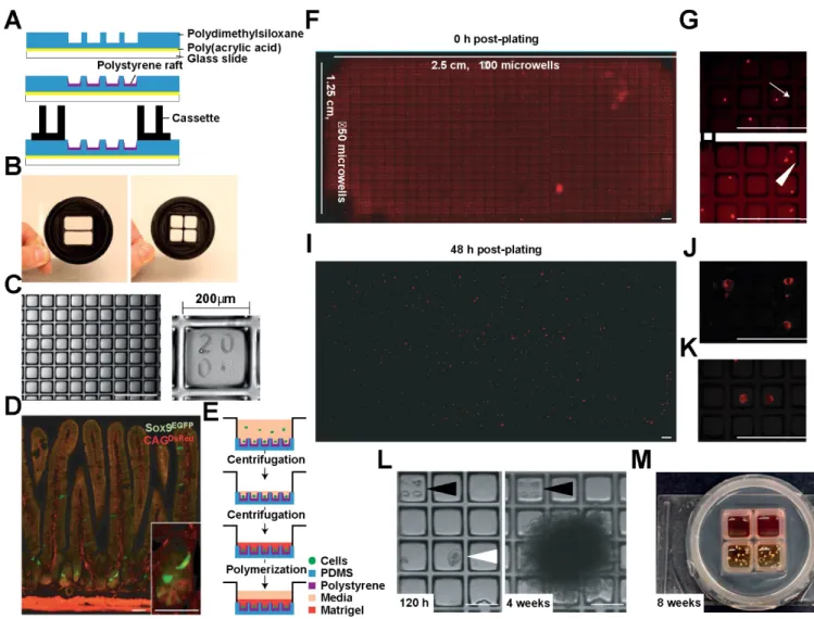
Microfabricated culture arrays were modified for long-term 3D culture to capture and functionally assay clonal IESCs and IESC–niche cell co-cultures, effectively providing a platform for high throughput niche reconstruction using primary IESCs and niche cells. Rafts fabricated into the array wells allows for efficient retrieval of single IESCs and developed organoids for direct integration into conventional high throughput biochemical assays (Fig. 2).7,106–110
Methods/Materials
Reconstructing basal-luminal signaling gradients in vitro and measuring gradient influences on organoid compartmentalization
The Sox9EGFP: CagDsRED mouse model
The CagDsRED mouse line ubiquitously expresses the red fluorescent protein
DsRed driven by the chicken beta-actin promoter. The Sox9EGFP mouse line possesses
the Sox9 promoter controlling EGFP (enhanced green fluorescent protein) expression integrated using a modified bacterial artificial chromosome. Crossing these two transgenic lines provides an epithelium source where DsRED expression marks all cell types and low expression levels of EGFP marks CBC IESCs (Fig. 1D).92
Isolation of Crypts from Mouse Colon
Epithelial cells were dissociated from whole murine colons using previously described techniques with some modifications. Colons were resected, opened longitudinally and rinsed in PBS. Colons were cut into 10 mm segments and transferred
to 3 mM EDTA (Sigma), 10 μM Y-27632 (Selleck) and incubated for 45 min at 4oC with
25 Isolation of Single Colonic Stem Cells
Epithelial cells were dissociated from whole murine colons using previously described techniques with some modifications111. Colons were resected, opened
longitudinally and rinsed in PBS. Colons were cut into 1 mm segments and transferred
to 3 mM EDTA (Sigma), 10 μM Y-27632 (Selleck) and incubated for 45 min at 4oC with
gentle agitation. Fragments were transferred into fresh PBS with 10 μM Y-27632 and shaken by hand for 2 min to release the epithelial layer from the submucosa. Epithelium was separated from the remnant mucosa, washed twice in phosphate buffered saline (PBS) and dissociated to single cells in 0.3 U/mL dispase (Life Technologies), 10 µM Y-27632 in Hank’s Buffered Saline Solution (HBSS, Life Technologies) incubated at 37 °C for 10-14 min while shaking every 2 min. Cell
solutions were passed sequentially through 100 μm, 70 μm, and 40 μm pore-size nylon filters before being transferred to intestinal stem cell (IESC) culture media (Advanced DMEM/F12 (Life Technologies), N2 (1×, Life Technologies), B27 (1×, Life Technologies), Glutamax (1×, Life Technologies), penicillin (100 unit/mL, Life Technologies), and streptomycin (100 µg/mL, Life Technologies), 10 mM HEPES (Life
Technologies), 10 μM Y27632 (Selleck Chemicals) and 500 mM N -acetyl- cysteine (Sigma)).
Developing self-renewing monolayers of primary colonic epithelial cells
Lgr5EGFPCreERT2xR26confetti mouse
Lgr5EGFP-IRES-creERT2 mice were generated by targeted insertion of the
26
crossed onto the Rosa26Confetti transgenic line where a strong CAGG promoter was
integrated into the Rosa26 allele followed by a LoxP-flanked NeoR-cassette blocking transcription. This was upstream of the Brainbow-2.1 cassette112. Cre-mediated
recombination removes the transcriptional roadblock allowing expression of one of the four fluorescent marker proteins, stochastically driven by the CAGG promoter, allowing discrimination between the clonal progeny of neighboring IESCs within the same crypt. Monolayer culture of colonic crypts
The crypts were placed on top of the collagen hydrogel at a density of 1000 crypts/cm2 (unless otherwise stated) and cultured in 4 mL medium per well in the 6-well plate. The medium was changed every 48 hours, and Y-27632 was added for the initial 48 hours of culture. When the cell coverage was greater than 80% (typically after 3–4 days for mouse cells, 5–7 days for human cells), the monolayers were sub-cultured by a gentle 2-step dissociation method. The first step was to lift the monolayer from the collagen hydrogel by scraping the collagen (with cells) from the well and transferring the hydrogel to a 15-mL conical tube containing 1 mL culture medium with 500 U/mL collagenase (type IV; Worthington Biochemical, USA). The gel was broken into small pieces by pipetting using a 5-mL serologic pipette, followed by a 1-mL pipet tip. The tube was then incubated at 37oC for 10 minutes to completely digest the collagen gel.
27
pipet tip. The cell fragments were resus- pended in medium and subcultured on a new collagen hydrogel at a passage ratio of 1:3. To convert 3D organoids to a 2D monolayer, the organoids were extracted from Matrigel by detaching the Matrigel patty, breaking it into coarse pieces, and then pipetting the suspension by using a 200-mL pipet tip. The cells were then cultured on a collagen gel at a density of 10 organoids/cm2.
Genetic lineage tracing from IESCs in 2D culture
Lineage tracing was induced in adult Lgr5EGFP-IRES-creERT2 mice by interparitanial
injection of 2mg of 4-hydroxy tamoxifen 24 hours prior to sacrifice and resection.
Developing a high throughput microfabricated IESC culture platform to describe intrinsic and extrinsic regulation of stem cell growth
Clonal stem cell culture
Isolated IESCs were plated in CRAs (Cell Microsystems, USA) at ratios of ∼1.5 cells per microraft. Sorted cells were added to array reservoirs in culture media and cells were seeded into microrafts by centrifugation at 51g for 5 min at 4oC. Following
centrifugation, medium was gently aspirated and arrays were overlaid with 600 µl (2 reservoir array) or 200 µl (4 reservoir array) of Matrigel and growth factors. Arrays were centrifuged a second time at 51g for 5 min at 4oC, to recapture any cells displaced by
the addition of Matrigel. Matrigel was allowed to polymerize for 30min at 37oC before
28
(Selleck Chemicals, USA). Initial media in validation experiments contained 2.5 µM CHIR99021 (Selleck Chemicals, USA) and 2.5 µM Thiazovivin (Selleck Chemicals, USA). Growth factors were added at two-day intervals: 1 µM Jagged-1 peptide, 50 ng/mL EGF, 100 ng/mL Noggin and 1 µg/ml RSPO1 (R&D, USA). Medium was changed every four days, and no CHIR99021 or Thiazovivin was used past initial plating.57,113
Stem cell niche co-culture
Isolated IESCs and PCs were plated in CRAs (Cell Microsystems, USA) at ratios of ∼1.5 cells per microraft for IESCs and ∼1 cell per 2 microrafts for PCs. Sorted cells were added to array reservoirs in culture media and cells were seeded into microrafts by centrifugation at 51g for 5 min at 4oC (Fig. 1E). Following centrifugation, medium was
gently aspirated and arrays were overlaid with 600 µl (2 reservoir array) or 200 µl (4 reservoir array) Matrigel and growth factors. Arrays were centrifuged a second time at 51g for 5 min at 4oC, to recapture any cells displaced by the addition of Matrigel.
Matrigel was allowed to polymerize for 30 min at 37oC before being overlaid with 1 ml (2
reservoir array) or 600 µl (4 reservoir array) IESC Sort/Culture Media. In IESC/PC co-culture experiments, growth factors, minus Jagged-1 peptide and Y27632, were supplemented every two days, and medium was changed every four days, as previously described.57,113
CRA image acquisition
29
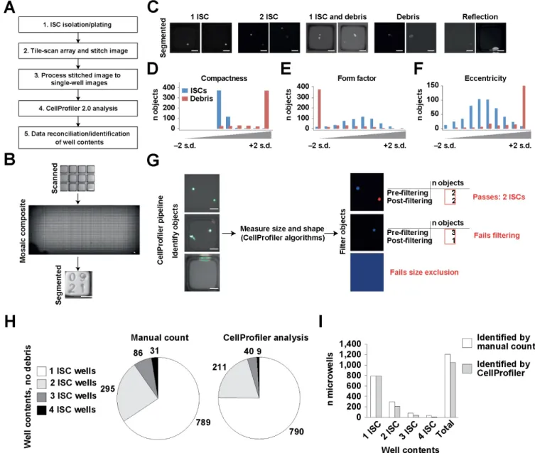
imaging, to prevent cell death due to the imaging procedure. Scanned images were stitched into a single composite image using the open source image analysis suite FIJI and then segmented into address-associated individual well images using an algorithm, ‘Segmenter.m’ , designed in MATLAB (MathWorks, USA). The ‘Segmenter.m’ is available for download at http://www. magnesslab.org/#!vstc1=page-1/vstc0=software-downloads.
CellProfiler 2.0 analysis.
30
parameters for cell identification and debris discrimination, we randomly selected 500 individual microraft images containing single IESCs and 500 individual microraft images containing debris or fluorescent noise from a two-chamber microraft array containing Sox9EGFP: CagDsRed IESCs (Fig. 3C). All analysis was conducted on the DsRed
wavelength, because the CAG promoter produced a significantly stronger fluorescent signal than the Sox9EGFP BAC transgene. These images were subjected to general
31
allowed us to identify the microraft images containing only intact stem cells and no debris (Fig. 3G). To validate our IESC identification pipeline using unbiased microraft images, we manually scored the contents of 2,254 randomly selected microrafts. This panel of images was then subjected to analysis using our newly developed pipeline, which was able to identify initial microraft contents with a high degree of accuracy, especially for microrafts containing a single IESC (99.87% accurate; Fig. 3H). Owing to stringency settings, this accuracy was reduced for wells containing multiple cells. Source files for ‘Segmenter.m’ , CellProfiler 2.0 pipeline ‘SCPipeline.cp’ and ‘WellContents.xls’ are available here as Supplementary Software and for download at http://www. magnesslab.org/#!vstc1=page-1/vstc0=software-downloads.
Data analysis of CellProfiler results
32
and ‘WellContents.xls’ are available here as Supplementary Software and for download at http://www. magnesslab.org/#!vstc1=page-1/vstc0=software-downloads.
Manual analysis of initial microraft contents.
For experiments examining organoid survival relative to initial cell-to-cell contact, tile-scanned images of CRAs in EGFP and DsRed wavelengths were overlaid and manually scored by microraft address. DsRed+ cells (isolated as Sox9EGFPneg
:CAGDsRed+ :CD24High:SSCHigh) were considered ‘PCs’ and EGFP+ cells (isolated as
Lgr5EGFPhigh or Sox9EGFPlow ) were considered ‘IESCs’. Two or more cells were scored
as ‘in contact’ only if their fluorescent signatures were contiguous in the overlaid images. For manual quantification of well contents and organoid survival, all investigators were blinded to prior well contents and survival outcomes.
Statistical analysis of organoid formation.
33
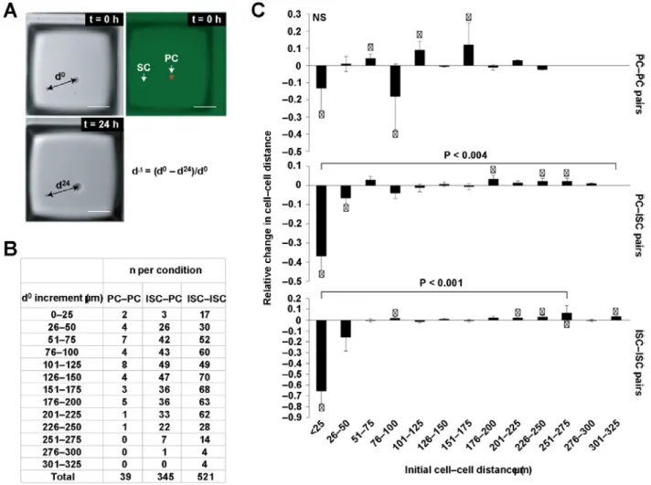
overall significance was found then pairwise tests were also performed. Wells were considered independent and therefore it was not necessary to model the correlation between them. No statistical method was used to predetermine sample size and experiments were non-randomized. All analyses were performed using SAS Version 9.3 (SAS Institute). P values less than 0.05 were considered statistically significant. Quantification of cell movement in microrafts. For cell movement experiments, pairs of PCs, IESCs, and IESC–PC pairs were identified in microraft images at t = 0 hr. Distance between each cell in a pair was measured at t = 0 and t = 24 hr. Relative change in distance (d∆) was calculated by subtracting distance at t = 24 hr (d24) from distance at t
= 0 hr (d0) and normalizing to d0 (Fig. 4). Pairs of cells were included in the experiment
34 Results
Reconstructing basal-luminal signaling gradients in vitro and measuring gradient influences on organoid compartmentalization
A Wnt3a/R-Spondin1 gradient enhances polarization of the stem/progenitor cell compartment
The highest levels of Wnt activity are thought to exist in the crypt base with the Wnt concentration tapering off in a gradient toward the luminal surface. R-Spondin1 (Rspo1) is co-expressed with Wnts in the stem cell zone and functions to potentiate Wnt activity through its receptor LGR5, which is G-protein coupled receptor expressed almost exclusively in colonic stem cells114. To determine whether a dual gradient of
Wnt-signaling along the microchannel might promote enhanced polarization of proliferative and differentiated cellular compartments, R-Spondin1 and Wnt3a were placed at high concentration in the source reservoir (75 ng/mL Wnt3a, 110 ng/mL R-Spondin1) to generate an environment with a steep factor gradient. To assess whether a Wnt3a/R-Spondin1 gradient enhanced polarization of the stem/progenitor and differentiated compartments, Sox9EGFP and EdU location was measured in each
colonoid under the dual factor gradient. Nearly all the 24 colonoids assessed displayed IESC patterning with a Sox9EGFP vector that orientated toward the source reservoir
35
Effect of a Wnt3a/R-Spondin1 Gradient on Growth and Polarization of Colonoids Derived from a Single Stem Cell
The experiments above utilized multicellular colonoid fragments or whole crypts as the source material for development of colonoids. While the fragments were small
(~30 μm diameter with ~25 cells), the fragments did contain many cell types
(differentiated, stem, and TA cells), and thus may have had pre-established cellular interactions that might impact spatial lineage allocation of a colonoid developing under an externally imposed growth-factor gradient. All evidence to- date indicates that the cells within these colonoids are representative of those in vivo and maintain a normal karyotype, it is conceivable that the cultured colonoids differ in an as yet unknown manner from their in vivo counterparts64,90,115. For this reason, single stem cells were
isolated from freshly obtained Sox9EGFP-CAGDsRed mouse crypts by
fluorescence-activated cell sorting of the stem cells (Sox9EGFPHi:CAGDsRED)120. The stem cells
suspended in Matrigel were loaded into a microchannel and cultured for 5 days in the presence of a Wnt3a/R-Spondin1 gradient. The Sox9EGFP polarization was significantly
exaggerated in the single-cell-derived colonoids with expression vectors of 20 of the 23 colonoids assessed pointing towards the gradient source. (Appendix A: Fig. 6)
Developing self-renewing monolayers of primary colonic epithelial cells
Lineage Tracing confirms the presence of stem cells
To determine whether self-renewing colonic stem cells persisted in the 2D monolayers, we performed genetic lineage tracing by using the Lgr5EGFPCreERT2xR26Confetti transgenic mouse. Colonic crypts were isolated from a
36
collagen hydrogel at a low density of 30 crypts/cm2 (to track the growth of individual
crypts). At 24 hours after plating, tracing events (4 out of 3000 crypts) expressed red fluorescent protein (RFP), which marked them as derived from Lgr5 stem cells (Appendix B. Fig. 4). The RFP+ regions expanded into large red fluorescent patches inter-mixed with the progeny of non-fluorescent stem cells. RFP+ cells were isolated from 2D patches and subcultured to determine whether the RFP+ patches contained cells with colonic stem cell properties. The RFP+ cells continued to expand into patches composed only of RFP+ cells (Appendix B. Fig. 4). The RFP+ cell monolayers possessed proliferative (EdU+) cells and all the differentiated cell types: goblet cells (Muc2+), enteroendocrine cells (ChgA+), and absorptive colonocytes (ALP+) (Appendix B. Fig. 4). These data demonstrate that the 2D monolayers possessed Lgr5+ IESCs that expanded as a proliferative monolayer and produced differentiated descendants in vitro. (Appendix B: Fig. 5)
Developing a high throughput microfabricated IESC culture platform to describe intrinsic and extrinsic regulation of stem cell growth
CRAs are adaptable to intestinal stem cell culture and imaging
Previously described polydimethylsiloxane (PDMS)/polystyrene CRAs could be used to isolate and culture single IESCs in 3D embedded in ECM hydrogel. As IESCs require several days to develop into organoids, CRAs had to be amenable to media changes13,57. To meet these requirements, polycarbonate cassettes, with dividers to
37
intervals, were included in the array design to allow for tracking. Conventional IESC cultures are capable of supporting organoid growth for many weeks. IESCs were maintained up to 8 weeks in CRAs, with retention of organoids in their original microrafts (Fig. 1L). At 8 weeks, organoids had grown into large structures containing many crypts (Fig. 1L,M). These observations demonstrate feasibility for long-term CRA-based culture of primary IESCs.
Tile-scanning microscopy produced high-resolution images of whole CRAs for downstream analysis.
Tile-scanning of the CRA in the DsRed wavelength immediately after plating and at 48 hr revealed that isolated IESCs had begun to produce primitive organoids, indicative of biocompatibility (Fig. 1F,I). Large, high-resolution mosaic images of the entire array surface could be segmented into indexed individual microraft images using the segmentor.m script, referencing the microraft location on the array surface in the microraft image file name.
Post hoc image analysis can quantify microraft contents and identify microrafts containing specific numbers of cells
38
single IESCs (99.87%; n=2,258 visually validated; Fig. 3H). Owing to stringency settings adapted specifically for clonal analysis, the percentage of identified microrafts was reduced for wells containing multiple cells, but the incidence of falsely identified microrafts remained 0% for all cell numbers examined (Fig. 3H,I).
Cell-cell contact is required for increased organoid formation in vitro
As previous studies have speculated that PC-secreted Wnts are responsible for enhancing IESC growth in vitro, IESC–PC co-culture experiments were carried out in the absence of exogenous Wnt, to avoid ‘masking’ the potential impact of PCs on organoid formation 57,113,116. The GSK3β inhibitor CHIR99021, a Wnt agonist, was also
excluded from co-culture experiments. To address the possibility that a PC in one microraft might affect the growth of an IESC in an adjacent, but separate microraft, we modelled diffusion dynamics of cell-secreted molecules in CRAs. Diffusion between microrafts was deemed negligible under models relying on liberal rates of diffusion and decay115. We reasoned that increased numbers of PCs would result in increased
secretion of Wnts and IESC growth, and examined microrafts with initial contents consisting of any combination of 1–5 IESCs and 0–2 PCs. IESCs were isolated from Sox9EGFP or Lgr5EGFP mice and PCs from Sox9EGFP:CAGDsRed mice (Fig. 4A). DsRed
39
revealed no statistically significant trends, regardless of whether IESCs were isolated using Sox9EGFP or Lgr5EGFP (Fig. 4B,C). To investigate the overall effect of PCs on
organoid survival, we next analyzed the percentage of organoids formed in microrafts containing any number of IESCs or any number of IESCs with any number of PCs (Fig. 4D). These analyses also failed to produce statistically significant differences between IESC-alone and IESC–PC microrafts, suggesting that PC presence alone is insufficient to increase organoid formation in vitro at physiologically relevant numbers. Previous studies have suggested that cell-to-cell contact between IESCs and PCs may influence organoid formation, but this has not been formally tested by comparison between touching and non- touching IESCs and PCs113. Using the same data generated in our
40
Single cells do not form de novo contacts after plating
The contact-dependent effects on organoid formation prompted us to examine whether de novo cell–cell contacts occurred after initial classification as ‘non-touching’ at t = 0 hrs. To assay this, we measured the distance between IESC–IESC, IESC–PC and PC–PC pairs in microraft images acquired at t = 0 hrs and calculated positive or negative changes in cell–cell distance at t = 24 hrs (Fig. 5A). Microwells in which both cells were alive at 24 hrs were included for analysis, resulting in n=905 pairs (521 IESC–IESC; 345 IESC–PC; 39 PC–PC; Fig. 5B). To control for background movement due to known changes in extracellular matrix integrity that occur over time, measurements were taken on pairs of fixed intestinal epithelial cells for comparison (n=50). Analysis of change in distance in alive pairs demonstrated that: cell movement was statistically significant compared with measurements taken on pairs of fixed cells; and no observed cells formed de novo contacts after initial plating of CRAs. Interestingly, non-touching pairs of cells with an initial cell–cell distance ≤ 25 µm seem to grow further apart within the first 24 hr of culture (Fig. 5C). Together, these data demonstrate that single cells migrate within Matrigel cultures and that cell–cell signaling may repel single cells from one another over short distances in vitro. Previous studies demonstrate significant organoid movement and merging in vitro124. To further examine
41 Conclusions
Reconstructing basal-luminal signaling gradients in vitro and measuring gradient influences on organoid compartmentalization
Microfabricated platforms can produce steep linear gradients of morphogenic factors across individual colonoids or single stem cells recapitulating the crypt axis. A simple linear gradient of Wnt3a/Rspondin1 was sufficient to induce polarization of the proliferative and differentiated cellular architecture in colonoid tissue constructs derived from both single colonic stem cells and small multicellular colonoids composed of heterogeneous cell types. Combining a R-Spondin1 (110 pg/mL/μm) gradient with a Wnt3a (75 pg/mL/μm) gradient acted synergistically to produce enhanced polarization of the colonoid body. Maintenance of colonoids derived from single stem cells under the combined gradient condition produced the most highly polarized colonoid structure suggesting that the multicellular fragments possessed some internal patterning which limited the ability of derived colonoids to maximally respond to morphogenic gradients.(Appendix A: In Vitro Polarization of Colonoids to Create an Intestinal Stem Cell Compartment)
Developing self-renewing monolayers of primary colonic epithelial cells
42
in the GI field to validate the presence of potent IESC populations11. Confirming that
Lgr5+ IESCs are present in the monolayer culture systems validates their use as an alternative to embedded organoid cultures in a platform to study the apical mucosa surface more readily manipulated by addition of compounds to the media. This structure enables facile assay of drugs, toxins, and metabolites using conventional cell-based assay systems, not possible in the embedded organoid systems.(Appendix B: Appendix B: Self-renewing Monolayer of Primary Colonic or Rectal Epithelial Cells) 38
Developing a high throughput microfabricated IESC culture platform to describe intrinsic and extrinsic regulation of stem cell growth
CRAs have broad applicability to assaying stem cell–niche interactions and organoid development and serve as a high throughput culture platform to interrogate gene expression at initial stages of stem cell fate choices. Stem cell niches provide critical extrinsic signals that govern stem cell self-renewal and differentiation, but their anatomical locations and complexity often present significant challenges to the study of stem cell renewal in vivo. PCs express soluble and insoluble IESC niche signaling components, including Wnt and Notch ligands113,116. We examined the impact of PC
43
contact dependency of PCs in a microscale format. in maintaining stemness. Importantly, the CRA platform was critical in testing dose and contact dependency of PCs in a microscale format.
Array-based stem cell culture platforms are growing in use and present an efficient and cost-effective alternative to conventional cell culture126,127. However, most
platforms are not amenable to long-term cultures, such as required for the development of IESC-derived organoids and other self-assembled, stem-cell-derived organoids. CRAs facilitate the culture of thousands of primary stem cells over many days and weeks as well as high throughput reconstitution of the stem cell niche at physiologically relevant cell numbers. The power of the CRA platform is further highlighted by the ability to retrospectively ‘mine’ existing high throughput CRA data sets to test new hypotheses, such as niche cell dose dependency, cell-cell contact and cellular movement within microrafts.106,107,114
Parts of this chapter previously appeared as an article in the journals NCB, Plos1, and CMGH the original citations are as follows: Gracz, A. D. et al. A high-throughput platform for stem cell niche co-cultures and downstream gene expression analysis. Nat. Cell Biol. 17, 340–349 (2015).
Attayek, P. J. et al. Automated microraft platform to identify and collect non-adherent cells successfully gene-edited with CRISPR-Cas9. Biosens. Bioelectron. 91, 175–182 (2017).
Wang, Yuli, DiSalvo, Matthew, Gunasekara, Dulan B., Dutton, Johanna, Proctor, Angela, Lebhar, Michael S., Williamson, Ian A., Speer, Jennifer, Howard, Riley L., Smiddy, Nicole M., Bultman, Scott J., Sims, Christopher E., Magness, Scott T. & Allbritton, Nancy L. Self-renewing Monolayer of Primary Colonic or Rectal Epithelial Cells. Cmgh 4, 165–182.e7 (2017).
Williamson, Ian A., Arnold, Jason W., Samsa, Leigh Ann, Gaynor, Liam, DiSalvo, Matthew, Cocchiaro, Jordan L., Carroll, Ian, Azcarate-Peril, M. Andrea, Rawls, John F., Allbritton, Nancy L. & Magness, Scott T. A High-Throughput Organoid Microinjection Platform to Study Gastrointestinal Microbiota and Luminal Physiology. Cell. Mol. Gastroenterol. Hepatol. 1– 38 (2018). doi:10.1016/j.jcmgh.2018.05.004
Chapter 3: A High throughput Organoid Microinjection Platform to Study Gastrointestinal Microbiota and Luminal Physiology
Synopsis
A high-throughput organoid microinjection platform was developed to study gastrointestinal physiology and the microbiome. Monitoring and quantification of injected microbes and other cargos was achieved by automated imaging. Human fecal microbiota including highly oxygen-sensitive anaerobic taxa were transplanted into the organoid lumen and maintained over time in stable monocultures or microbial communities.
Overview
45
microinjection system for cargo delivery to the organoid lumen and high-content sampling. Methods: A microinjection platform was engineered using off-the-shelf and 3D-printed components. Microinjection needles were modified for vertical trajectories and reproducible injection volumes. Computer vision (CVis) and microfabricated CellRaftTM Arrays (CRAs) were used to increase throughput and enable high-content
46 Background & Aims
The human gastrointestinal tract contains a remarkably dense and diverse microbial community38,118. The interactions between gut microbiota and host are
becoming increasingly recognized as key factors in homeostasis and disease43. Many
studies indicate that community imbalances, known as dysbioses, are associated with onset and progression of diseases including diabetes119, obesity120–122, colorectal
cancer27,50,51, and inflammatory bowel disease (IBD)44. Despite tight statistical
associations between dysbiosis and disease, the ability to formally test cause-and-effect relationships is severely limited by lack of in vitro experimental models that enable controlled interrogation of host-microbe interactions.
Sequencing of the 16S rRNA gene is routinely used to characterize microbial communities and is a powerful tool to identify bacteria that may contribute to disease32.
While 16S rRNA gene sequencing provides a ‘signature’ of microbial composition within a community, alone it is insufficient to define specific microbial mechanisms that impact host biology. Germ-free (gnotobiotic) animal models are commonly used to investigate host-microbe interactions in a physiologically relevant system, but germ-free animal models are often impractical for researchers to employ because of the scarcity of gnotobiotic facilities and high cost of gnotobiotic experimentation 13. Additionally, the
inherent low throughput nature of germ-free rodent studies limits the ability to decipher the individual role that each microbial species plays in health and disease.
A recent assessment of microbiota research in the United States identified the development of high throughput tools as a key, common unmet need for this field 14.