ORIGINAL ARTICLE
A randomized controlled trial to assess the central
hemodynamic response to exercise in patients with transient
ischaemic attack and minor stroke
J Faulkner1, Y-C Tzeng2,3, D Lambrick4, B Woolley5, PD Allan2,3, T O’Donnell2, J Lanford6, L Wong6and L Stoner7
Early exercise engagement elicits meaningful changes in peripheral blood pressure in patients diagnosed with transient ischaemic attack (TIA) or minor stroke. However, central hemodynamic markers may provide clinicians with important diagnostic and prognostic information beyond that provided by peripheral blood pressure readings. The purpose of this single-centre, randomized, parallel-group clinical trial was to determine the effect of a 12-week aerobic exercise intervention on central and peripheral hemodynamic variables in patients with TIA or minor stroke. In this study, 47 participants (66 ± 10 years) completed a baseline assessment, which involved the measurement of central and peripheral hemodynamic parameters, undertaken in the morning, in a fasted state. Participants were randomized to either a 12-week exercise or control group on completion of the baseline assessment. An identical follow-up assessment was completed post intervention. Central hemodynamic variables were assessed using an oscillometric device at both assessments. Analysis of covariance demonstrated a significant interaction for central and peripheral blood pressure and augmentation index (allPo0.05;ηp2.09–.11), with the exercise group presenting lower values than the control
group post intervention (118 ± 17 vs 132 ± 28 mm Hg for central blood pressure; 125 ± 19 vs 138 ± 28 mm Hg for peripheral blood pressure; 104 ± 49 vs 115 ± 67% for augmentation index). The present study demonstrates that participation in an exercise program soon after stroke/TIA diagnosis may elicit significant beneficial changes to a patient’s central systolic blood pressure and augmentation index. This may positively impact upon the treatment strategies implemented by clinicians in the care of patients with TIA and minor stroke.
Journal of Human Hypertension(2017)31,172–177; doi:10.1038/jhh.2016.72; published online 29 September 2016
INTRODUCTION
Blood pressure is an important risk factor for stroke,1 and is widely cited as a marker that needs to be controlled post stroke by pharmacological and lifestyle management.2 A recent
meta-analysis with stroke and transient ischaemic attack (TIA) patients has shown that significant reductions in peripheral blood pressure responses may be evident following participation in behavioural interventions (that is, exercise, lifestyle management programs).3 However, the measurement of central hemodynamic parameters, including central systolic blood pressure (cSBP) and arterial wave reflection (that is, augmentation index, AIx), hold the potential to provide stroke clinicians with important diagnostic and prognostic information beyond that of peripheral blood pressure readings.
Peripheral blood pressures may not accurately reflect the effects of peak arterial blood pressure on centrally located organs such as the heart and brain.4This is important because individuals who experience a stroke or TIA are at heightened risk of experiencing subsequent vascular events, whether from a cerebrovascular (that is, stroke/TIA) or cardiovascular (that is, myocardial infarction) origin.5,6Thus, the assessment of cSBP is particularly important as it reflects the stress and loading on the left ventricle and coronary arteries.7,8 Yet to date, the effects of secondary prevention
behavioural interventions on central blood pressure responses in
stroke or TIA patients have not been assessed. As behavioural interventions which have incorporated exercise training, counseling and lifestyle education reduce the risk of subsequent cardiac events in patients with stroke,3 further research is
necessary to elucidate the underlying central hemodynamic response to these types of intervention for patients with stroke or TIA.
The purpose of the current randomized control study was to determine the effect of implementing an exercise program soon after minor stroke/TIA diagnosis on cSBP and AIx. It was hypothesised that those individuals who engaged in the exercise program would demonstrate greater changes in central hemodynamic responses compared with those individuals randomized to a usual care control group.
METHODS
Participants
Forty-seven participants (Male,n= 35; 66 ± 10 years; 90 ± 19 kg; 173 ± 7 cm; Female,n= 12; 64 ± 12 years; 80 ± 20 kg; 159 ± 4 cm) diagnosed with either TIA or minor stroke completed the baseline assessment, of which 43 took part part in a post-intervention assessment (Figure 1). Recruitment commenced in September 2013 and lasted 12 months with participants being randomized to either an exercise or usual care control group
1
Department of Sport and Exercise, University of Winchester, Winchester, UK;2
Centre for Translational Physiology, University of Otago, Wellington, New Zealand;3
Department of Surgery and Anaesthesia, University of Otago, Wellington, New Zealand;4
Faculty of Health Sciences, University of Southampton, Southampton, UK;5
School of Sport and Exercise, Massey University, Massey, New Zealand;6
Neurology, Wellington Hospital, Wellington, New Zealand and7
Department of Exercise and Sport Science, University of North Carolina at Chapel Hill, Chapel Hill, NC, USA. Correspondence: Dr J Faulkner, Department of Sport and Exercise, University of Winchester, Sparkford Road, Winchester SO22 4NR, UK. E-mail: [email protected]
(exercise group, n= 25; 66 ± 12 years; 88 ± 19 kg; 168 ± 10 cm; control group, n= 22; 68 ± 10 years; 90 ± 21 kg; 171 ± 8 cm). Follow-up (post-intervention) assessments were completed between December 2013 and December 2014.
TIA and minor stroke diagnosis was based upon criteria from the New Zealand assessment and management guidelines.9At the time of hospital presentation within the inpatient setting, stroke severity was assessed using the National Institutes of Health Stroke Scale, functional severity was assessed using the Barthel Index, and stroke risk of TIA patients was assessed using the ABCD2score. Participants were eligible if they had been diagnosed with theirfirst high-risk TIA (ABCD2score of⩾2) or minor stroke (defined as an National Institutes of Health Stroke Scale of⩽5), lived within the local district health board catchment, and if they did not meet exclusion criteria (Figure 1). Exclusion criteria were: unstable cardiac conditions, uncontrolled diabetes mellitus, severe claudication, oxygen dependence, significant dementia, inability to communicate in English or unable to take part in exercise. All participants complied with drug treatment and standardised therapy in accordance with stroke physician recommendations. The trial was approved by New Zealand’s Central Regional Health and Disabilities Ethics Committee and registered with the Australian and New Zealand Clinical Trials Registry (Trial Registration Number: ACTRN12613000869774), and conformed to the guidelines established in theDeclaration of Helsinki. Written informed consent was obtained before participation.
Sample size
On the basis of the findings of Woolley et al.,10 and when examining changes in peripheral systolic blood pressure (pSBP) in TIA patients following participation in an exercise intervention, and when using a two-sided 5% significance level and a power of 80%, a minimum sample size of 19 participants per group was calculated.
Study design
The study was a single-centre, randomized, parallel-group design. TIA or minor stroke was confirmed by a specialist stroke physician at Wellington Hospital within 7 days of symptom onset. Eligible participants were invited to attend a baseline assessment conducted within a thermoneutral laboratory (21–22 °C) typically between 0700 and 1000 hours All participants were fasted (overnight fast), euhydrated, had abstained from caffeine and supplement intake during the morning, and from strenuous physical activity and alcohol consumption for 24 h before assessment. Following completion of a health history questionnaire, and 15 min of quiet supine rest, central blood pressure responses were obtained with the participant in the supine position. Identical assessments were completed post-intervention.
On completion of the baseline assessment, participants were randomized to either an experimental group (usual care plus participation in a 12-week aerobic exercise intervention) or to a control group (usual care, only) using computerized random numbers. Details pertaining to each allocated group were provided on a piece of paper contained within sequentially numbered, opaque sealed envelopes. The randomization procedures were prepared by an investigator with no clinical involvement in the trial. Although participants and the health and exercise practitioner were aware of the allocated treatment condition, data analysts were kept blinded to the allocation.
Subjects randomized to the exercise program completed twice weekly, group-based (three to five participants) exercise sessions for 12 weeks. Exercise was prescribed on a one-to-one basis by health and exercise practitioners. In accordance with recommendations for moderate physical activity participation, participants completed 30–60 min of aerobic exercise at each exercise session.11 This included up to 30 min of continuous walking and up to 30 mins of continuous cycling, with an interim period of ~ 5 min of passive recovery between bouts. Blood pressure, heart rate and ratings of perceived exertion were measured prior to, during and following each bout of aerobic exercise. Participants commenced their exercise
Figure 1. Participant recruitment.
program at 50% of age-predicted maximal heart rate during all aerobic exercise tasks. The exercise intensity and/or volume typically increased by ~ 5% each week, up to a maximal heart rate training intensity of 90% of age-predicted maximal heart rate. The rate of progression was dependent upon how the subject felt during each session. Subjects were instructed not to exercise beyond an ratings of perceived exertion of 15 (‘hard’ feeling of exertion) during both walking and cycling exercise.12 Exercise practitioners ensured that subjects did not exercise above 90% of their age-predicted maximal heart rate.
The control group (usual care) received standard secondary prevention and educational information from the hospital on discharge and were requested to adhere to their prescribed medication for the duration of the study.
Pulse wave analysis
An oscillometric device (BP+, Uscom Lts, Sydney, NSW, Australia) was used to conduct pulse wave analysis, from which central and peripheral blood pressures and AIx were derived. The AIx% is an independent predictor of cardiovascular risk and mortality.13Pressure waveforms were recorded on the upper arm, following standard manufacturer guidelines.14Briefly, the BP+incorporates an oscillometric blood pressure module which complies with the Association for the Advancement of Medical Instrumentation (AAMI SP10) requirements and receives an A/A rating from the British Hypertension Society evaluation protocol.15 Each measurement cycle (~40 s) records brachial blood pressures and then one set of suprasystolic (~30 mm Hg4systolic) recordings for 10 s. The suprasystolic pressure signals were recorded by a high-fidelity pressure transducer, and the central pressure waveform was derived in the time-domain from the relationship between the total oscillatory pressure in the aorta and the total oscillatory pressure under the occlusion cuff.15,16This estimation was scaling-independent (that is, the estimated aortic wave shape did not depend on brachial blood pressure). AIx% was calculated using the formula: AIx% = (P3–P0)/(P1–P0), where P0 is pressure at the onset of the pulse (diastolic), P1 is peak pressure of the incident wave (systolic), P3 is peak pressure of the reflected wave. This index describes the relative height of the reflected pressure wave when compared with the incident waveform. Only recordings with a high signal quality were accepted. Signal quality was assessed using the signal to noise (S/N) ratio (in decibels), where S/N values 43 dB were considered acceptable. Two measurements were taken, with 5-min interval. If AIx varied by44% or blood pressures by45 mm Hg, a third recording was made and the two closest recordings were averaged.
Statistical analysis
Independent samplest-tests (age, certain lifestyle factors) and Pearson Chi-squared tests (descent, family history of cardiovascular disease (CVD), personal history of CVD, signs and symptoms of CVD, medication) were used to compare baseline data between conditions (exercise, control). A similar analysis was used to assess central and peripheral hemodynamic properties (that is, cSBP, pSBP, peripheral diastolic blood pressure, central pulse pressure, peripheral pulse pressure, AIx) at baseline, and to assess medication use post-intervention. To assess the effect of the exercise intervention on the aforementioned central and peripheral hemodynamic properties, and following checks for normality to determine that these outcome variables were parametric, a series of a-priori repeated-measures analysis of covariance (Test [baseline, post intervention] by Condition [exercise, control], with baseline inserted as a covariate) were conducted. An intention-to-treat analysis was used on all repeated-measures statistical procedures, whereby the last recorded data from a participant’s subsequent assessment was carried forward and used in place of any missing assessments thereafter. Partial eta squared (ηp2) was used to demonstrate the strength of the effect of exercise on the various outcome measures with .0099, .0588 and .1379 representing a small, medium and large effect, respectively.17Partial eta squared was calculated using the following formula:
η2 p¼
SSEffect
SSEffectþ SSError
Whereby, SSEffectis the estimated variance for a given outcome measure, and SSErroris the error variance that is attributable to the effect. Alpha was set at 0.05. Statistical analyses were performed using Statistical Package for Social Sciences version 22 (SPSS, Inc., Chicago, IL, USA). All data are reported as means (s.d.), unless otherwise specified.
RESULTS
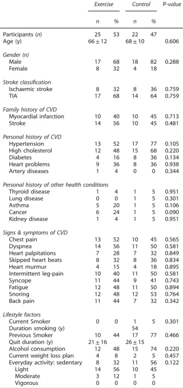
Participant recruitment, adherence and recurrent TIA risk There were no differences in subject characteristics, medication use or CVD risk factors at baseline between groups (P40.05; Tables 1 and 2 and Supplementary Table). Baseline assessments were undertaken 8 ± 3 days post diagnosis. Mean medication use post-intervention was approaching significance (P= 0.09) with
Table 1. Baseline demographics for both exercise and control groups
Exercise Control P-value
n % n %
Participants (n) 25 53 22 47
Age (y) 66±12 68±10 0.606
Gender (n)
Male 17 68 18 82 0.288
Female 8 32 4 18
Stroke classification
Ischaemic stroke 8 32 8 36 0.759
TIA 17 68 14 64 0.759
Family history of CVD
Myocardial infarction 10 40 10 45 0.713
Stroke 14 56 10 45 0.481
Personal history of CVD
Hypertension 13 52 17 77 0.105
High cholesterol 12 48 15 68 0.220
Diabetes 4 16 8 36 0.134
Heart problems 9 36 8 36 0.938
Artery diseases 1 4 0 0 0.344
Personal history of other health conditions
Thyroid disease 1 4 1 5 0.951
Lung disease 0 0 1 5 0.301
Asthma 5 20 1 5 0.106
Cancer 6 24 1 5 0.090
Kidney disease 1 4 1 5 0.951
Signs & symptoms of CVD
Chest pain 13 52 10 45 0.565
Dyspnea 14 56 11 50 0.581
Heart palpitations 7 28 7 32 0.849
Skipped heart beats 8 32 8 36 0.834
Heart murmur 4 15 4 18 0.895
Intermittent leg-pain 10 40 11 50 0.581
Syncope 11 44 9 41 0.743
Fatigue 12 48 11 50 0.894
Snoring 12 48 12 53 0.764
Back pain 11 44 7 32 0.342
Lifestyle factors
Current Smoker 0 0 1 5 0.301
Duration smoking (y) 54
Previous Smoker 10 44 17 77 0.466
Quit duration (y) 21±16 26±15
Alcohol consumption 12 48 15 74 0.220 Current weight loss plan 4 8 2 5 0.457 Everyday activity: sedentary 8 32 11 56 0.122
Light 14 56 10 45
Moderate 3 12 1 5
Vigorous 0 0 0 0
participants in the exercise group typically taking less medication compared to those in the control group (Mean difference (95% CI); 0.67 (−1.48 to 0.13); Table 2). Participants randomized to the exercise program attended, on average, 96% of the available sessions. There were no adverse events from participation in the exercise program. Central and peripheral hemodynamic variables
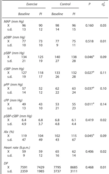
Table 3 summarises the mean values for the central and peripheral hemodynamic properties measured at baseline and post inter-vention for both exercise and control conditions. There were no statistical differences in any of the peripheral or central hemodynamic variables reported at baseline between the exercise and control group (all P40.05). Analysis of covariance demon-strated a significant interaction for cSBP, pSBP and AIx (all
Po0.05), with the exercise group presenting lower values than the control group post intervention. A significant interaction was also observed for peripheral pulse pressure and central pulse
pressure (both Po0.05). An increase in pulse pressure was observed for the control group between baseline and post intervention, but a decrease was observed with the exercise group.
DISCUSSION
This is the first study to demonstrate that cSBP and AIx are significantly reduced in participants with minor stroke and high-risk TIA following participation in an exercise intervention, soon after diagnosis. In the present study, despite similarities in baseline demographics, the 7% improvement in cSBP following participation in a 12-week exercise program equated to a large effect size (ηp2= 0.11). The significance of this finding is further
enhanced when considering that there was a large reduction in medication use for those participants randomized to the exercise
Table 2. Number and percentage of people taking medication at baseline and PI for both conditions (exercise and control)
Exercise Control Baseline PI
Baseline PI Baseline PI Difference Ex vs Con (P)
Difference Ex vs Con (P)
Statins
n 21 18 17 17 0.329 0.830
% 84 72 77 77
Anti-thrombotica
n 21 17 21 20 0.174 0.050b
% 84 68 95 91
Angiotensin converting enzyme inhibitor
n 13 13 12 13 0.968 0.724
% 52 52 55 59
Diuretics
n 2 1 2 2 0.926 0.508
% 8 4 9 9
Calcium blockers
n 2 2 5 5 0.179 0.179
% 8 8 23 23
Beta blockers
n 4 4 5 5 0.608 0.608
% 16 16 23 23
Anticoagulants
n 1 1 2 2 0.345 0.345
% 4 4 9 9
Angiotensin receptor blocker
n 2 1 0 0 0.174 0.160
% 8 4 0 0
Other anti-hypertensives
n 2 2 2 2 0.926 0.926
% 8 8 9 9
Mean medication use
X 3.78 3.08 3.76 3.76 0.947 0.094
s.d. 1.04 1.53 1.00 1.04
Abbreviations: Con, control; Ex, exercise group; PI, post-intervention. Mean (±s.d.) medication use is also reported.aNumber of patients taking two anti-thrombotics weren=18 (exercise, baseline),n=19 (control, baseline), n=8 (exercise, PI), n=14 (control, PI). bSignificant difference between groups (Po0.05).
Table 3. Mean (±s.d.) values for peripheral and central hemodynamic variables at baseline and PI for both conditions (exercise and control)
Exercise Control P ηp2
Baseline PI Baseline PI
MAP (mm Hg)
X 96 90 98 96 0.160 0.05
s.d. 13 12 14 15
pDBP (mm Hg)
X 77 73 77 75 0.518 0.01
s.d. 10 10 9 11
pSBP (mm Hg)
X 134 125 140 138 0.046a 0.09
s.d. 21 19 27 28
cSBP (mm Hg)
X 127 118 133 132 0.027a 0.11
s.d. 19 17 26 28
pPP (mm Hg)
X 57 52 62 63 0.037a 0.10
s.d. 14 12 22 24
cPP (mm Hg)
X 49 43 53 55 0.011a 0.14
s.d. 13 10 21 23
pSBP-cSBP (mm Hg)
X 6.4 6.8 6.8 6.1 0.419 0.02
s.d. 4.9 3.7 4.8 4.4
AIx (%)
X 119 104 102 115 0.045a 0.09
s.d. 47 49 43 67
Heart rate (b.p.m.)
X 59 59 65 62 0.406 0.02
s.d. 9 12 16 14
DP
X 7591 7429 7795 8685 0.468 0.01
s.d. 2359 1985 3737 3111
Abbreviations: AIx, augmentation index; b.p.m., beats per minute; cPP, central pulse pressure; cSBP, central systolic blood pressure; DP, double product; MAP, mean arterial blood pressure; pDP, peripheral double product; pDBP, peripheral diastolic blood pressure; PI, post-intervention; pPP, peripheral pulse pressure; pSBP, peripheral systolic blood pressure. aSignificant difference between groups (Po0.05).
group, and when considering that there were no changes in cSBP for those individuals who received usual care.
Of the various CVD risk factors that are often assessed in patients with minor stroke and TIA following engagement in an exercise program, peripheral blood pressure responses have received the greatest attention.3,18–21 However, it has been suggested that peripheral blood pressures may not accurately reflect the effects of peak arterial blood pressure on centrally located organs,4nor the stress and loading on the left ventricle and coronary arteries.7,8 Accordingly, in the present study, the significant reduction in cSBP is of pragmatic importance for this population group, as individuals diagnosed with stroke and TIA are at a heightened risk of experiencing further cardiovascular complications.5,6It is interesting to note that the 9 mm Hg mean reduction in cSBP is greater than that reported in a recent meta-analysis, whereby, on average, a 4 mm Hg reduction in peripheral blood pressure is suggested to occur following lifestyle and behavioural interventions.3The prescription of exercise at a
moderate- to high-intensity in the current study may be an underpinning reason for this difference. Nevertheless, as the reduction in pSBP was identical to that reported for cSBP for those in the exercise group, the ecological benefit of monitoring cSBP may be somewhat reduced. In the current study, the exercise group also demonstrated a significant reduction in central and peripheral pulse pressure in comparison to the control group (ηp2= 0.10 to 0.14). Because the statistically similar diastolic blood
pressures reported between conditions (exercise and control) and assessment sessions (baseline, post-intervention), the ~ 5 mm Hg reduction in pulse pressure for the exercise group is largely derived by the aforementioned changes in systolic blood pressure (Table 3).
A further novelty of the present study relates to the change in AIx; an indicator of arterial wave reflection.22 In this study, participants who took part in the exercise program demonstrated a 15% reduction in AIx (Table 3). This finding is of significant importance for those with stroke/TIA as AIx is an indicator of systemic arterial stiffness23 and demonstrates the contribution
made by the reflected pressure wave to the ascending aortic pressure waveform.22Conversely, and unexpectedly, AIx increased for the control group. Increased arterial wave reflections have been reported to have adverse effects on ventricular afterload and coronary perfusion, have been significantly correlated to the degree of coronary artery disease,24 and independently predict cardiovascular risk and mortality.25 When considering that stroke and TIA patients have an elevated risk of experiencing cardiovascular and cerebrovascular events post stroke/TIA,1 the measurement of central hemodynamic parameters, including cSBP and AIx, could provide clinicians with important diagnostic and prognostic information beyond that provided by traditional peripheral blood pressure readings. From a pragmatic standpoint, oscillometric devices, like that used in the current study, can provide clinicians with a quick, non-invasive and valid assessment of such hemodynamic parameters.25–28 Despite this study being
sufficiently powered, it is plausible that the observed differences in AIx between the experimental and control groups may be associated with the relatively small sample size and thus, future research should assess such hemodynamic parameters in a larger TIA/minor stroke population.
A recent study reported that the monitoring of central blood pressure, compared with peripheral blood pressure, aided in the management of hypertension and led to decreased medication use without adverse effects of left ventricular mass.29Therefore, future research should assess whether the monitoring of central hemodynamic variables in those with minor stroke or TIA may lead to similar, positive clinical outcome changes, which may lead to more targeted treatment strategies for these population groups. However, it is important to recognize that in the current study there are some important findings with regards to
medication use (Table 2). At the time of follow-up, participants who engaged in the exercise program were shown to be taking significantly fewer anti-thrombotic medications, and there was a trend for taking fewer medications overall, compared to those randomized to the usual care control group. When considering that medicinal management is primarily carried out to have an impact on the cardiovascular system, thisfinding is pertinent, and reflects the value of engaging patients in an exercise program soon after diagnosis.
In conclusion, the present study has demonstrated that participation in an exercise program soon after stroke/TIA diagnosis may elicit significant changes in cSBP and AIx. It may be important for stroke clinicians to consider central hemodynamic parameters at diagnosis and during follow-up assessments as these parameters may provide important information beyond that provided by traditional peripheral blood pressure readings. As automated oscillometric devices can provide a valid, quick and non-invasive assessment of central hemodynamic parameters, clinicians could consider the use of such measurements in providing more targeted treatment strategies for those with TIA or minor stroke.
What is known about this topic ?
● Behavioural interventions (that is, exercise and education) have been shown to improve physiological and psycho-social health in patients with TIA and minor stroke.
● Previous research with this population group has often focused on the effect of exercise on peripheral hemodynamic parameters (that is, peripheral blood pressure).
● The measurement of central hemodynamic parameters, including central systolic blood pressure (cSBP) and arterial wave reflection (that is, augmentation index, AIx), hold the potential to provide stroke clinicians with important diagnostic and prognostic informa-tion beyond that of peripheral blood pressure readings.
What this study adds?
● Participation in an exercise program soon after stroke/TIA diagnosis may elicit significant improvements in cSBP and AIx.
● Stroke clinicians may want to consider central hemodynamic parameters at diagnosis and during follow-up assessments as these parameters may provide important information beyond that provided by traditional peripheral blood pressure readings.
CONFLICT OF INTEREST The authors declare no conflict of interest.
ACKNOWLEDGEMENTS
Funding was provided by the Massey University Research Fund. Funding was used to support consumable purchase and participant travel expenses.
REFERENCES
1 Go AS, Mozaffarian D, Roger VL, Benjamin EJ, Berry JD, Borden WBet al.Heart disease and stroke statistics-2013 update: A report from the American Heart Association.Circulation2013;127: e6–e245.
2 Adams HJ, Adams R, Brott T, del Zoppo G, Furlan A, Goldstein Let al.Guidelines for the early management of patients with ischemic stroke: a scientific statement from the stroke council of the American Stroke Association.Stroke2003;324: 1056–1083.
3 Lawrence M, Pringle J, Kerr S, Booth S, Govan L, Roberts NJ. Multimodal secondary prevention behavioural interentions for TIA and stroke: a systematic review and meta analysis.PLos ONE2015;10: e0120902.
5 Touze E, Varenne O, Chatellier G, Peyrard S, Rothwell P, Mas J. Risk of myocardial infarction and vascular death after transient ischemic attack and ischemic stroke: a systematic review and meta-analysis. Stroke 2005; 36: 2748–2755.
6 Albers GW, Caplan LR, Easton JD, Fayad PB, Mohr JP, Saver JLet al.Transient ischemic attack-proposal for a new definition.N Engl J Med2002;347: 1713–1716. 7 Roman MJ, Devereux RB, Kizer JR, Lee ET, Galloway JM, Ali Tet al.Central pressure more strongly relates to vascular disease and outcome than does brachial pres-sure: the strong heart study.Hypertens2007;50: 197–203.
8 Young Y, Abdolhosseini P, Brown F, Faulkner J, Lambrick D, Williams MAet al. Reliability of oscillometric central blood pressure and wave reflection readings: effects of posture and fasting.J Hypertens2015;33: 1588–1593.
9 Stroke-Foundation. New Zealand Guidelines for the Assessment and Management of People with Recent Transient Ischaemic Attack (TIA). 2008. 10 Woolley B, Stoner L, Lark S, Wong L, Lanford J, Faulkner J. Effect of early
exercise engagement on arterial stiffness in patients diagnosed with a transient ischaemic attack.J Human Hypertens2015;29: 87–91.
11ACSM's Guidelines for Exercise Testing and Prescription.8th edn. Lippincott, Williams and Wilkins: Philadelphia, 2010.
12 Borg G.Borg's Perceived Exertion and Pain Scales. Human Kinetics: Leeds, UK, 1998. 13 Vlachopoulos C, Aznaouridis K, Stefanadis C. Prediction of cardiovascular events and all-cause mortality with arterial stiffness: a systematic review and meta-analysis.J Am Coll Cardiol2010;55: 1318–1327.
14 Stoner L, Lambrick DM, Faulkner J, Young J. Guidelines for the use of pulse wave analysis in adults and children.J Atheroscler Thromb2013;20: 404–406. 15 Lowe A, Harrison W, El-Aklouk E, Ruygrok P, Al-Jumaily AM. Non-invasive
model-based estimation of aortic pulse pressure using suprasystolic brachial pressure waveforms.J Biomech2009;42: 2111–2115.
16 Park CM, March K, Ghosh AK, Tillin T, Mayet J, Lowe Aet al.A method comparsion of central blood pressure measurements by pulsecor and sphygmocor devices. Artery Res2010;4: 171.
17 Cohen J. A power primer.Psychol Bull1992;112: 155–159.
18 Faulkner J, Lambrick D, Woolley B, Stoner L, Wong L, McGonigal G. The long-term effect of exercise on vascular risk factors and aerobicfitness in those with TIA; a randomized controlled trial.J Hypertens2014;32: 2064–2070.
19 Faulkner J, Lambrick D, Woolley B, Stoner L, Wong L-K, McGonigal G. Effects of early exercise engagement on vascular risk in patients with transient ischemic attack and nondisabling stroke. J Stroke Cerebrovasc Dis 2013; 22: e388–e398.
20 Lennon O, Carey A, Gaffney N, Stephenson J, Blake C. A pilot randomized controlled trial to evaluate the benefit of the cardiac rehabilitation paradigm for the non-acute ischaemic stroke population.Clin Rehabil2008;22: 125–133.
21 Prior PL, Hachinski V, Unsworth K, Chan R, Mytka S, O'Callaghan C et al. Comprehensive cardiac rehabilitation for secondary prevention after transient ischemic attack or mild stroke: I: feasibility and risk factors.Stroke 2011;42: 3207–3213.
22 Oliver JJ, Webb DJ. Noninvasive assessment of arterial stiffness and risk of atherosclerotic events.Arterioscl Throm Vas2003;23: 554–566.
23 Stoner L, Faulkner J, Lowe A, Lambrick D, Young M, Love R et al.Should augmentation index be normalized to heart rate?J Atheroscl Throm2014;21: 11–16.
24 Weber T, Auer J, O'Rourke MF, Kvasm E, Lassnig E, Lamm Get al.Increased arterial wave reflections predict severe cardiovascular events in patients undergoing percutaneous coronary interventions.Eur Heart J2005;26: 2657–2663. 25 Weber T, O’Rourke MF, Lassnig E, Porodko M, Ammer M, Rammer Met al.Pulse
waveform characteristics predict cardiovascular events and mortality in patients undergoing coronary angiography.J Hypertens2010;28: 797–805.
26 Lowe A, Harrison W, Wl-Aklouk E, Ruygrok P, Al-Jumaily AM. Noninvasive model-based estimation of aortic pulse pressure using suprasystolic brachial pressure waveforms.J Biomech2009;42: 2111–2115.
27 Hwang MH, Yoo JK, Kim HK, Hwang CL, Mackay K, Hemstreet Oet al.Validity and reliability of aortic pulse wave velocity and augmentation index determined by the new cuff-based SphygmoCor Xcel.J Hum Hypertens2014;28: 475–481. 28 Papaioannou T, Karatzis EN, Karatzi KN, Gialafos EJ, Protogerou AD,
Stamatelopoulos KS et al. Hour-to-hour and week-to-week variability and reproducibility of wave reflection indices derived by aortic pulse wave analysis: implications for studies with repeated measurements. J Hypertens 2007; 25: 1678–1686.
29 Sharman JE, Marwick TH, Gilroy D, Otahal P, Abhayaratna WP, Stowasser M. Randomized trial of guiding hypertension management using central aortic blood pressure compared with best-practice care: principal findings of the BP GUIDE study.Hypertens2013;62: 1138–1145.
This work is licensed under a Creative Commons Attribution 4.0 International License. The images or other third party material in this article are included in the article’s Creative Commons license, unless indicated otherwise in the credit line; if the material is not included under the Creative Commons license, users will need to obtain permission from the license holder to reproduce the material. To view a copy of this license, visit http://creativecommons.org/licenses/ by/4.0/
© The Author(s) 2017
Supplementary Information accompanies this paper on the Journal of Human Hypertension website (http://www.nature.com/jhh)