MOLECULAR REGULATION OF CARDIOVASCULAR DEVELOPMENT AND DISEASE
Nicole D. Fleming
A dissertation submitted to the faculty at the University of North Carolina at Chapel Hill in partial fulfillment of the requirements for the degree of Doctor of Philosophy in the Department of
Pathology and Laboratory Medicine in the School of Medicine.
Chapel Hill 2019
ABSTRACT
Nicole D. Fleming: Molecular Regulation of Cardiovascular Development and Disease (Under the direction of Jiandong Liu)
Congenital heart disease (CHD) represents the most common type of human birth defect, accounting for approximately one-third of all major congenital abnormalities. These structural anomalies primarily arise due to genetic and transcriptional dysregulations during early development and maturation of the heart. Severe CHDs can lessen a patient’s quality of life or lead to various life-threatening diseases like heart failure and even death. Given that CHDs can also give rise to adult cardiomyopathies, it is necessary to have a better
understanding of the etiologies and the molecular regulation of factors involved in normal cardiac development and disease. The process from cardiogenesis to the formation of a mature and fully functional heart requires precise coordination of transcription factors, signaling
molecules, and epigenetic components. In these studies, we utilized the zebrafish for its optical transparency, its ease of pharmacological and genetic manipulation, and its simplistic yet complex circulatory system to investigate the molecular basis of events that lead to defective hearts during early and late embryogenesis.
In Chapter 2, trabeculae-deficient erbb2 zebrafish mutants were used to evaluate the function of trabeculae, luminal muscular protrusions within the heart, during chamber
maturation. The data revealed that trabeculae function to possibly relieve the heart of mechanical-pressure overload, as absence of trabecular formation led to pathological hypertrophy and decreased cardiac function. Chapter 3 explores the events of early heart development in zebrafish embryos that lack Ring1b, an epigenetic factor belonging to the Polycomb Repressive Complex 1. Data highlighted the requirement of Ring1b in SHF-mediated development, which has not been explored in zebrafish or mice.
To my mother, Bernice:
“Through the drama, I can always depend on my mama. And when it seems that I’m hopeless, you say the words that can get me back in focus. There’s no way I can pay you back, but my
plan is to show that I understand. You are appreciated.”
To my sister, Rhonda, brother, Keithroy, and nephews, Dre’ and KJ: “You are my strength. You are my inspiration.”
To my little brother, Kishawn:
“It’s been a long day without you, my friend, and I’ll tell you all about it when I see you again.” Things just aren’t the same without you. I miss you every single day.
To all my friends and church family:
ACKNOWLEDGEMENTS
I remember, as a high school student, walking to the beach one day and looking out into the distance at the University of the Virgin Islands’ campus, and thinking that even with my good grades, college was too out of reach for me. Who would have thought that, me, a young black woman from a small ghetto community in the Virgin Islands, would be completing her graduate work at UNC-Chapel Hill. This journey was by no means the easiest to trod, but it was definitely worth it. In fact, it took more than a village to get me through graduate school and to this point; it took the endless prayers, support, and encouragement from countless individuals (family, friends, mentors, colleagues, and strangers). Thus, I would like to acknowledge all of them!
I would be remiss if I did not start off by thanking my Heavenly Father, for without Him and His guidance, none of this would be remotely possible.
My primary mentor, Dr. Jiandong Liu, has taught me numerous, invaluable skills in thinking scientifically, communication, and leadership that I intend on utilizing in the next steps of my career. I would like to thank him for his support, guidance, and opportunities for growth. To this day, I still believe that I was meant to complete my graduate work in his lab. I would also like to thank my co-mentor, Dr. Li Qian. Whenever I am asked to describe Li, I usually say, “Li is a rock star scientist!” Li’s passion for science, for women in science, and for students is
an independent thinker and scientist. I am forever grateful to have her as a mentor. I also want to thank Michelle Altemara, the zebrafish facility supervisor. Zebrafish care for multiple labs can be a difficult task; despite the challenge, I rarely worried about my fish with her overseeing the operations.
I would like to acknowledge and thank my thesis committee: Drs. Joan Taylor, Chris Mack, Scott Williams, Michael Bressan, and Brian Jensen. I would to thank Joan for being a supportive chair and mentor. She was always willing to meet with me when I had grad school-related questions, even for a brief time, and gave me great advice. I appreciated the questions that Chris asked that always made me think (as questions should), during committee meetings, IVB seminars, and departmental seminars, and I enjoyed speaking with Scott about science and new research methods I can use to address my hypotheses. They are both easy to talk to. To Mike, I can remember attending his faculty candidate seminar and thinking that I needed him on my committee. When I actually found out he got the position, Jiandong wisely advised me to at least let him settle in before introducing myself and inquiring about his availability. Mike’s approach to research reminds me of Chris’s and my other committee members’ approach, where he does not take anything at face value, and he questions findings and evaluates whether more effective experiments could have been employed. These are all skills that I have learned to do in my own work. The role that Brian had as my clinical co-mentor, in correlation with the Trans Med program, really made my time in grad school even more meaningful. Joining him in the clinic and seeing him interact with his patients reminded me of why I enjoyed doing research, to contribute knowledge to the cardiovascular field that will ultimately provide treatments for patients. Thanks for being such a supportive committee.
her lab. After I received my grad school offers, she insisted that we have lunch to basically make a pros and cons list for each program. Though everyone thought UNC was an automatic decision for me, I actually was not as sure as people anticipated. Nevertheless, after our conversation, I immediately accepted UNC’s offer and I have never regretted that decision. Graduate school brought successes, disappointments, loss, and frustrations for me and I was able to share and discuss all of those experiences with Carol. I look forward to sharing more of my career and life experiences with her following grad school.
Because I started my journey to grad school during 2010 with the UNC SOLAR program, I have been able to develop relationships with several people who are a part of various
departments. I want to first acknowledge the best department and faculty at UNC, the Pathology and Laboratory Medicine Department and faculty. I will always remember the support and encouragement they provided me since joining the department, especially during the untimely loss of my little brother. I also want to acknowledge the individuals in the MHI office: Tracy, Dean, Rocky, and Teresa. They always had my back and were willing to help me in one way or another. Words cannot begin to express the gratitude I feel for the folks (former and present) in the Office of Graduate Education and who are/was involved with SOLAR, IMSD and PREP: Josh, Ashalla, Jessica, Patrick, Johnna, Anna, Beka, Jeff, Sausty, Cindy, Erin, Michael Johnson (who selected me for SOLAR 2010), and many others. Every one of them were and still are so invested in my success during grad school and in my future career. Their belief in me doing great things never wavered and I cannot say “thank you” enough! It was also encouraging to have colleagues and friends in graduate school who understood the emotional rollercoaster that is expected in grad school and who provided many nuggets of wisdom and advice to keep me going forward: Rachel, Sabri, Samira, Grace, Tiki, Leslie, Alisha, Kim, Danielle, Haley, Tiffany, Chris, Maggie, Caitlin, Michelle, Mike, PREP3 cohort, and many others.
instructors for adding another dimension to my grad school experience. I enjoyed seeing the students all grow, learn, and developed into confident individuals. In teaching them, they made me better! I also want to thank the administrators and colleagues at the UNC Writing Center. Being a writing coach was so fulfilling and refreshing. I was able to hone my writing skills as a coach, and I enjoyed seeing the growth of all the students throughout my time at the Writing Center.
As a graduate student, receiving one’s own funding is encouraged, yet difficult to accomplish. Nonetheless, I was blessed to have three sources of funding: a NSF Graduate Research Fellowship, an Integrative Vascular Biology (T32) training grant (deferred), and an American Heart Association/The Children’s Heart Foundation pre-doctoral fellowship. Thanks to all my funding sources for allowing me to freely conduct my research without concern about getting reagents or equipment.
I moved from the Virgin Islands to North Carolina for PREP (post-bac), straight out of undergrad and with no relatives that I really knew or were acquainted with. Nevertheless, other than friends from grad school, I was blessed to have made meaningful friendships with many individuals at my current church: Immanuel Temple SDA. In addition to friendships, many couples selected to unofficially “adopt” me into their families. My friends and “adopted” family literally fed me, clothed me, sheltered me, encouraged me, and most importantly, prayed for me, which was most reassuring in the many moments of uncertainty. To my adopted church family: Dr. and Sis. Fortune, Bro. and Sis. Hedgepeth, Dr. Lassiter, Sis. Moody, Dr. and Sis Campbell, Christine, and Papa and Sis. Bishop, I say “thank you”! I can go on and on.
“the crew”: Leia, Keysha, Nikki, Johanne, and Mime. I absolutely love and value all of them. We traveled together, cried together, celebrated together, laughed together, prayed and fasted together, and I can honestly say that I would not be at this point without them. I especially thank them and Chanel for being with me when I heard the news of my little brother and throughout the years since then. I was completely lost and unsettled during that time and if it wasn’t for God blessing me with them for such a time like that I would have remained in that mindset for a long time. Thank you for making sure that I stayed focused, encouraged, and had a great work/life balance. (#NoNewFriends!)
I am proud to be a tarheel at UNC-CH. However, before I was a tarheel, I was a
buccaneer at the University of the Virgin Islands, and I am proud of my humble beginnings as a Virgin Islander (Born and raised!). I want to thank all of my influential professors who educated me throughout my time in school, along with my athletic coaches and teammates. Though there are many who played a significant role in getting me into graduate school, I would be remiss if I did not acknowledge Dr. Nemeth, who encouraged me to apply for my first summer
undergraduate research opportunity in 2009, and Dr. Turner, who basically locked me in the conference room (with my permission) until I completed my statements and applications for graduate programs. They cared and I am grateful!
Everyone who knows me knows that my family is my motivation; they are the reason why I am trying to be the best scientist and person I can be. On my mother’s side, I am the eighth child out of nine children and, though I am the youngest now, I feel responsible to make them proud and that is what I always intend to do. I do appreciate the encouragement from all of my siblings; however, I do want to especially recognize two of them: Rhonda and Keithroy. I have to begin with my role model and big sister, Rhonda. Her strength, perseverance, and sincere heart motivates and encourages me to never give up no matter what obstacles are placed in front of me. She is who I aspire to become and more. To my big brother, Keithroy (also known as Mem), I want to thank him for always letting me know that I could call and tell him anything (which I did) and get great advice. I definitely cannot forget about my nephews, Dre and KJ. These little guys lifted my spirits and kept me going every time I spoke to them, listened to or read their messages, heard about their day, or saw the great things they were doing in school. I hope I have made them proud to have me as their auntie Nicole. Of course, I have to thank the real MVP on this journey with me, my mommy, Bernice. I remember telling her about a conversation I had with my advisor about getting to work at a certain time, and since then (2014), she has called me almost every morning, and has only missed a handful of
mornings. She wanted me to be successful in every aspect of grad school and did anything she could to help, great or small. My resilience and work ethic comes from her, someone who gets maybe 6 hours of sleep a night and still works two jobs. I love her! To my baby brother,
strong and be as happy and successful as possible. Things will never be the same without him, but it brings me comfort to know that I am going to receive my PhD, just like he wanted me to.
In this acknowledgement section, I tried not to forget anyone who was involved,
PREFACE
Chapter 1 provides a broad overview of cardiogenesis in vertebrate heart development. Particularly, it highlights the various molecular pathways, transcriptional factors and epigenetic mechanisms that are involved in cardiac progenitor cell processes and their ultimate
contribution to the mature heart. Further, this section also provides information on the later stages of zebrafish heart development and the regulators that contribute to proper chamber maturation and compaction. In correlation with proper chamber maturation, I also emphasize various processes of physiological cardiac remodeling that impact normal and pathological heart development. Lastly, I highlight the value of the zebrafish as a model organism for studying cardiac development and disease and emphasize the therapeutic implications associated with the understanding gathered from the work completed in Chapters 2 and 3.
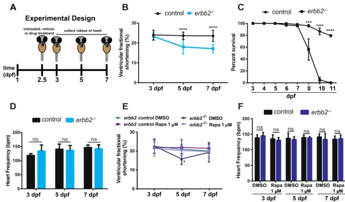
Chapter 2 was published in Scientific Reports on June 5, 2018. Figures and text have been reproduced and reformatted. Over the course of development, the topology of the cardiac chambers matures to optimize cardiac output. During his postdoctoral work, Dr. Liu reported that ErbB2 signaling is essential for cardiac trabeculation in a cell autonomous manner in zebrafish. Though it is evident that trabeculae are important, the exact function of trabeculae in the heart remains unclear. In this study, we examined the mechanisms of functional
compensation in trabeculae-deficient erbb2-/- zebrafish larvae. Our findings reveal that
Additionally, these findings reveal that trabeculae-deficient erbb2-/- zebrafish can be utilized for in-depth, yet efficient hypertrophic studies.
For this work, I designed and performed the majority of the experiments, analyzed the data and wrote the manuscript. Dr. Leigh Ann Samsa assisted in the experimental design and provided intellectual input. Dr. David Hassel and myself analyzed the fractional shortening data. Additionally, Drs. David Hassel, Li Qian, and Jiandong Liu provided intellectual input and
supervised the work. All authors provided feedback on the manuscript.
Chapter 3 is a potential manuscript in preparation, depending on the final contributions that I have made and the continuing contributions by another member in the lab. Previous work by van der Velden et al. (2013) revealed an essential role for the epigenetic factor, Ring1b, the core component of Polycomb Repressive Complex 1, in zebrafish pectoral fin and craniofacial development. However, the data did not highlight the cardiac defect that was also associated with ring1b deficiency in the zebrafish larvae hearts. Examining this heart defect was of particular interest to our lab because previous attempts to study Ring1b’s role in development had been hindered because mammalian knockout of Ring1B is embryonic lethal. In contrast, the zebrafish ring1b mutant can survive long enough for us to investigate the epigenetic
mechanisms underlying early heart development, specifically cardiac progenitor cell dynamics. In this chapter, we aimed to characterize the phenotypes of ring1b mutants to decipher which process of cardiogenesis is perturbed in the mutant hearts. Our findings suggest that Ring1b may be involved in second heart field (SHF)-mediated development. Future results from this study will be invaluable for the field, as it will be one of the first in vivo studies of epigenetic regulation of Ring1b in early cardiac progenitors.
TABLE OF CONTENTS
LIST OF TABLES ... xvii
LIST OF FIGURES ... xviii
LIST OF ABBREVIATIONS AND SYMBOLS ... xix
CHAPTER 1 GENERAL INTRODUCTION ... 1
1.1 Cardiac Development: Regulation of Cardiac Progenitor Cells and Chamber Maturation ... 1
Origin of first and second heart field progenitors ... 1
Zebrafish (Danio rerio) as a powerful model of heart development and disease ... 3
Transcriptional regulation of CPC specification, proliferation/migration, and differentiation ... 4
Polycomb repressive complexes play an essential role in cardiac development ...11
Chamber maturation ...13
Signaling pathways essential for trabeculation ...15
Biomechanical/hemodynamic regulators of cardiac maturation ...19
Cardiac remodeling during development ...20
Cardiac remodeling during disease...21
mTOR signaling in cardiac hypertrophy and the effects of rapamycin treatment ...23
Relevance to current research ...25
Early and late manifestations of CHDs ...25
Potential therapeutic interventions for CHDs through experimental approaches ...27
CHAPTER 2 RAPAMYCIN ATTENTUATES PATHOLOGICAL HYPERTROPHY
CAUSED BY AN ABSENCE OF TRABECULAR FORMATION ...34
2.1 Historical Context ...34
2.2 Rapamycin Attenuates Pathological Hypertrophy in the Absence of Trabecular Formation ...36
Introduction ...36
Results ...37
Discussion ...42
Materials and Methods ...45
Chapter 2.2 Figures ...50
2.3 Significance and Future Directions ...58
Significance ...58
Future Directions ...58
CHAPTER 3 CHARACTERIZING THE ROLE OF RING1B IN SECOND HEART FIELD DEVELOPMENT ...63
3.1 Historical Context ...63
3.2 Characterizing the Role of Ring1b in Second Heart Field Development ...64
Introduction ...64
Results ...68
Materials and Methods ...71
Chapter 3.2 Figures ...73
3.3 Significance and Future Directions ...77
Significance ...77
Future Directions and Additional Interpretations ...78
CHAPTER 4 CONCLUSIONS ...83
LIST OF TABLES
Table 1 Chapter 1 Summary of genes involved in human, mouse, and zebrafish
LIST OF FIGURES
Figure 1 Simplified schematic of zebrafish heart development ...29
Figure 2 Cardiac progenitor cell dynamics and transcriptional regulation: specification, proliferation/migration, and differentiation ...30
Figure 3 FHF and SHF-derived cells contribute to different compartments of the vertebrate heart ...31
Figure 4 Schematic of canonical Polycomb pathway ...32
Figure 5 erbb2 mutant develops HL phenotypes ...50
Figure 6 Inhibition of TOR signaling attenuates erbb2 mutant HL phenotypes ...51
Figure 7 Rapamycin treatment improves impaired heart function in erbb2 mutant ...52
Figure 8 erbb2 mutant HL phenotypes result from the absence of trabecular formation ...53
Figure 9 Schematic diagram illustrating that trabeculae-deficient erbb2 mutants undergo TOR-dependent pathological hypertrophy ...54
Figure 10 erbb2 mutant exhibits more pronounced HL phenotype over time ...55
Figure 11 Inhibition of TOR signaling with lower concentration of Rapamycin and Torin1 attenuates erbb2 mutant HL phenotypes ...56
Figure 12 ring1b mutant displays cardiac defects that are initially distinct at 48 hpf ...73
Figure 13 Small ventricle size observed in ring1b mutants is due to fewer ventricular CMs ...74
Figure 14 Inhibition of Ring1b activity between 24-72 hpf recapitulates “unlooped” heart phenotype observed in mutant embryos ...75
LIST OF ABBREVIATIONS AND SYMBOLS
A Atrium
ACTC1 Actin, alpha, cardiac muscle 1 ADAM17 Adam metallopeptidase domain 17
AKT Protein kinase B
ALPM Anterior lateral plate mesoderm AMHC Atrial myosin heavy-chain ANF Atrial natriuretic factor Anti-DIG-AP Anti-digoxigenin-AP
AV Atrio-ventricular
bHLH Basic helix-loop-helix
BMP10 Bone morphogenetic protein 10
bpm Beats per minute
Bry Brachyury
Cas9 CRISPR-associated protein 9
CBF1 Also known as RBPJK
CBX Chromodomain
Cdh2 Cadherin-2 (also known as N-cadherin) cDNA Complementary deoxyribonucleic acid
Cfk Cardiofunk
CHD Congenital heart disease ChIP Chromatin immunoprecipitation
ChIP-seq Chromatin immunoprecipitation coupled with sequencing
CM Cardiomyocyte
CRISPR Clusters of regulatory interspaced short palindromic repeats cTnT Cardiac muscle troponin T
DAPI 4′,6-diamidino-2-phenylindole
DGS DiGeorge syndrome
DMSO Dimethyl sulfoxide
DNA Deoxyribonucleic acid
DOX Doxorubicin
dpf Days post-fertilization E9.5/10.5 Embryonic day 9.5 or 10.5
EC Endothelial cell
EdU 5–ethynyl–2′–deoxyuridine Eed Embryonic ectoderm development
Eln2 Elastin 2
EPHB4 Ephrin-B4
erbb2 erb-b2 receptor tyrosine kinase 2 ErbB2/4 ERBB2 and ERBB4 heterodimer
ERK1/2 Extracellular signal-regulated kinases 1 or 2
ESC Embryonic stem cell
Ezh1/2 Enhancer of Zeste 1 or 2
FACS Fluorescent activated cell sorting
Fau Faust
FHF First heart field
FKBP12 FK506-binding protein of 12 kDa Flk1 Fetal liver kinase 1
FS Fractional shortening
GFP Green fluorescent protein
GO Gene ontology
H2AK119 Histone H2A at lysine-119 H3K27 Histone H3 at lysine-27
Hand1/2 Heart and neural crest derivatives-expressed 1 or 2
Hcn4 Hyperpolarization activated cyclic nucleotide gated potassium channel 4 Hey2 Hairy/enhancer-of-split related with YRPW motif protein 2
HL Hypertrophic-like
hpf Hours post-fertilization
Hst Heartstrings
IFT Inflow tract
Ift88 Intraflagellar transport protein 88 iPSCs Induced pluripotent stem cells
Isl1/2a/2b Insulin gene enhancer protein 1, 2a, or 2b Jarid2 Jumonji, AT rich interactive domain 2
KO Knockout
LA Left atrium
Ltbp3 Latent TGF-β binding protein 3
LV Left ventricle
LVNC Left ventricular non-compaction MEF2C Myocyte-specific enhancer factor 2C Mef2ca/cb Myocyte-specific enhancer factor 2ca or cb Mesp1 Mesoderm posterior bHLH transcription factor 1 Mespab Mesoderm posterior bHLH transcription factor ab
Mib1 Mindbomb1
mRNA Messenger ribonucleic acid
mTOR Mouse Target of Rapamycin
mTORC1/2 Mouse Target of Rapamycin Complex 1 or 2
MYH6 Myosin heavy chain 6
Myl7 Myosin light chain 7
Nb/Nb1NK Nkx2.5-Cre-mediated deletion of Numb/Numbl
NBT/BCIP Nitro-blue tetrazolium chloride/ 5-bromo-4-chloro-3'-indolyphosphate p-toluidine salt
Ncx1 Na+/Ca2+ exchanger
NICD NOTCH intracellular domain Nkx2.5 /2.7 Nkx2 homeobox 5 or 7
NOTCH NOTCH family receptors
Nppa/b Natriuretic peptide precursor A or B
Nrg Neuregulin
Numbl Numblike
OFT Outflow tract
PA Pulmonary artery
pAA Pharyngeal arch arteries PBS Phosphate buffered saline
PBST Phosphate buffered saline with tween
PcG Polycomb group
Pcgf4 Polycomb group RING finger protein 4 (also known as Bmi-1)
PCL Polycomblike
PCNA Proliferating cell nuclear antigen
PFA Paraformaldehyde
PRC1/2 Polycomb repressive complex 1 or 2 PRT4165 2-pyridine-3-yl-methylene-indan-1,3-dione PTU 1-phenyl 2-thiourea
qRT-PCR Quantitative reverse transcription polymerase chain reaction
RA Right atrium
RAPTOR Regulatory-associated protein of mTOR
RBPJ Recombination signal-binding protein for immunoglobulin kappa J region
Rapa Rapamycin
RFP Red fluorescent protein
Ring1A/B Ring finger protein 1 (also known as Rfn2)
RNAP II RNA polymerase II
RNAseq RNA sequencing
RV Right ventricle
S6K1 Ribosomal protein S6 kinase-1 scRNAseq Single-cell RNA sequencing SEM Standard error of the mean
SHF Second heart field
Six1 Sineoculis homeobox homolog 1
SMC Smooth muscle cell
Su(H) Suppressor of hairless Suz 12 Suppressor of zeste 12 TAC Transverse aortic constriction
Tbx1/5/5a/20 T-box transcription factor 1, 5, 5a, or 20 Tcf21 Transcription factor 21
Tnnt2a Troponin T type 2a, cardiac type
TOR Target of Rapamycin
TORC1 Target of Rapamycin Complex 1
UV Ultraviolet
V Ventricle
Vgo Van gogh
Vmhc Ventricular myosin heavy-chain
Wea Weak atrium
WT Wildtype
ZFN Zinc finger nuclease
ZsY ZsYellow
zTor Zebrafish Target of Rapamycin γ-secretase Gamma-secretase
-/- Homozygous mutant
% Percent
° Degree
Δ14 14 base pairs deletion
2D two-dimensional
3D three-dimensional
µM micromolar
µm microns
CHAPTER 1 GENERAL INTRODUCTION
1.1 Cardiac Development: Regulation of Cardiac Progenitor Cells and Chamber Maturation
Origin of first and second heart field progenitors
The heart is the first organ to form during embryogenesis and its function is vital for survival. Previous studies in Xenopus, chick, mouse, and, most recently, zebrafish have
provided invaluable insight into the cellular processes that take place during early cardiogenesis (Liu and Stainier, 2012; Olson and Srivastava, 1996; Dyer and Kirby, 2009). The developmental fate of cells is coordinated during gastrulation when the three germ layers, endoderm,
mesoderm, and ectoderm, are formed and the body plan is established (Warga et al., 1990; Ho, 1992; Dunwoodie, 2007; Yelon, 2001). The earliest events of heart development are conserved across vertebrates, in which naïve mesodermal cells begin to transiently express Mesoderm posterior 1 (Mesp1), a basic helix-loop-helix (bHLH) transcription factor, (Mespab- zebrafish homolog) and a subset of Mesp1-expressing cells receive precise extrinsic and intrinsic
within the bilateral areas of the anterior lateral plate mesoderm (ALPM) and begin to
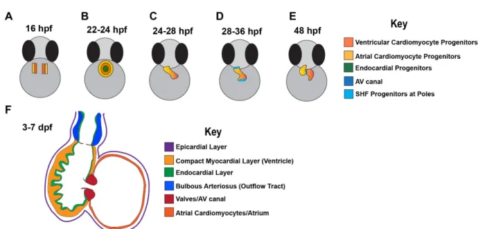
differentiate into mature cardiomyocytes (Fig. 1A). The differentiating cardiomyocytes, though not yet contractile, fuse to form the cardiac disk with developing endocardial cells. The cardiac disk begins to contract between 22 and 24 hpf (Fig. 1B) (Brown et al., 2016). The cardiac disk, then, elongates into a single heart tube from 24 to 28 hpf, primarily consisting of progenitors from the FHF (Figs. 1C and 2B). Following formation of the heart tube, from 28 to 36 hpf, relatively undifferentiated cells of the second heart field (SHF) progenitor pool begin to
proliferate and differentiate to contribute to the poles of the linear heart tube (Figs. 1D and 2C) (Brown et al., 2016). This addition of SHF cells is critical for proper looping morphogenesis and expansion of the heart (Kelly, 2012; Camarata et al., 2010). Maintenance of SHF cells in a progenitor-type state longer than FHF cells possibly occurs via signals that differentially affect SHF cells and not FHF cells, thus, suggesting that FHF and SHF segregation is coordinated through different genetic regulatory networks (Rochais et al., 2009; Black, 2007; Camarata et al., 2010). By 48 hpf, the simple heart tube of the zebrafish gradually transforms into a two-chambered organ (Fig. 1E) with a single compact myocardial layer overlaid by endocardial cells, and eventually the cardiac outflow tract, inflow tract, epicardium, and cardiac valves (Fig. 1F).
In mouse and zebrafish, FHF and SHF progenitors can give rise to all cell types in the mature heart: cardiomyocytes (CMs), smooth muscle cells (SMCs), arterial and venous endothelial cells (ECs), fibroblasts, and cells of the conduction system (Witman and Sahara, 2018) (Figs. 2B-C). Additionally, these progenitor-derived cells contribute to various
specific cardiac genes. Studies have revealed that FHF cells contribute to the atrium and half of the ventricle, while the SHF cells contribute to the other half of the ventricle, the inflow tract and the outflow tract (Fig. 3B) (Cavanaugh et al., 2015; de Pater et al., 2009; Zhou et al., 2011). Unlike the mammalian heart, the SHF in zebrafish is defined by latent TGF-β binding protein 3 (ltbp3) expression (Zhou et al., 2011), similar to isl1 in mice, and both FHF and SHF cells express nkx2.5 throughout development (Figs. 2B-C).
Zebrafish (Danio rerio) as a powerful model of heart development and disease Advantages of zebrafish as an experimental organism
Defining CPC dynamics during cardiac development and the key regulators entail the use of an appropriate model system and thorough experimental approaches. Though the zebrafish heart is morphologically distinct from its mammalian counterpart, the heart functions similarly and the genes responsible for critical steps of cardiac development are conserved throughout vertebrates (Howe et al., 2013; Moorman and Christoffels, 2003). Current
methodologies in most model organisms have limited resolution for understanding when and how single gene perturbations lead to whole organ deformities. Harnessing the power of zebrafish can circumvent this limitation. Due to their optical transparency, zebrafish allows for unprecedented access of developmental processes, with direct observation of early
defects that are embryonic lethal, because zebrafish embryos and adult zebrafish can survive days with cardiac abnormalities (Stainier et al., 1996; Chen et al., 1996).
Zebrafish as a genetic model of heart development
Historically, zebrafish have been used to study and understand the genetic
implications of several heart defects. In fact, one of the initial forward, genetic screens that identified a plethora of novel genes and mutations affecting various developmental processes was performed using male zebrafish. This large-scale genetic zebrafish screen led to the discovery of heart of glass, silent heart, bungee, and many other genes and mutations (Driever et al., 1996), which were later characterized and found to play vital roles in heart development and homeostasis (Mably et al., 2003; Sehnert et al., 2002; Just et al., 2011). Hence, the zebrafish is a powerful model for discovering novel genes and investigating the role of various genes in cardiac development, which in turn informs what occurs in cardiac disease.
Transcriptional regulation of CPC specification, proliferation/migration, and differentiation
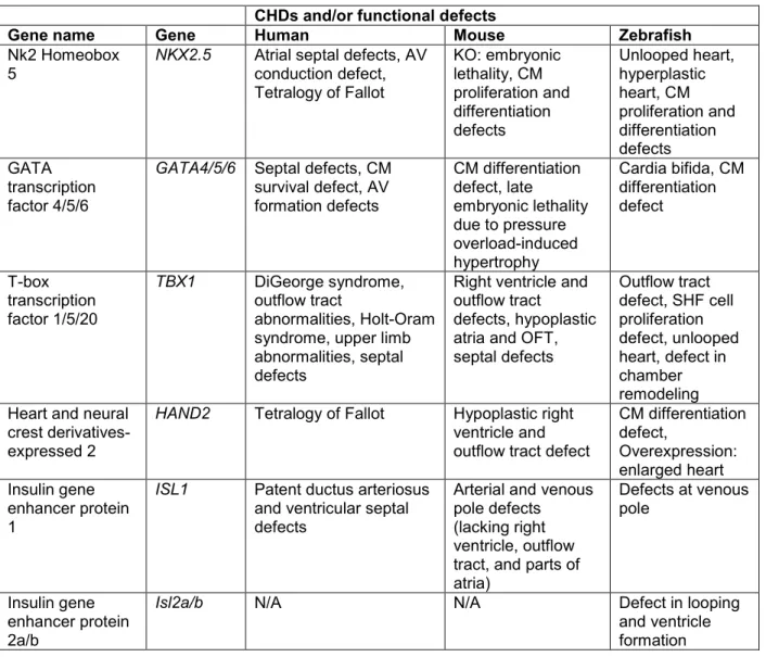
Coordinated transcriptional programs are essential in orchestrating the intricate cellular and molecular events that lead to a functioning heart. Cardiac cells are distinguished by a unique set of evolutionary conserved transcription factors, including Nkx2.5, T-box proteins, GATA proteins, HAND proteins, and ISL proteins, which are involved in various aspects of cardiogenesis (Table 1).
Nkx2.5
pharyngeal arches (Nagelberg et al., 2015). In humans, mutations in NKX2-5 are associated with numerous congenital heart diseases (CHDs) (Schott et al., 1998; McElhinney et al., 2003; Jay et al., 2003; Elliot et al., 2003). Recent studies in mice and zebrafish provide insights into the functions of Nkx2.5 at early stages of cardiogenesis and throughout embryogenesis into adulthood (George et al., 2015). In mice, Prall et al. (2007) have demonstrated that Nkx2.5 is involved in coordinating transitions of FHF and SHF progenitor specification, proliferation, and differentiation. In vivo co-transcription assays in fibroblasts also reveal that the functionality of Nkx2.5 requires interactions with other cardiac restricted factors. In fact, Nkx2.5 plays a key role in the induction of cardiogenesis by interacting with and activating the expression of Gata4, Mef2C, T-box, and Hand1. Knockout of Nkx2.5 in mice is embryonic lethal (Biben and Harvey, 1997; Tanaka et al., 1999; Lee et al., 1998; Gajewski et al., 1997). Embryos die around E9.5 due to severe cardiac malformations; prior to looping there is arrest of the developing heart and noticeable down-regulation of cardiac-specific genes (Lee et al., 1998; Gajewski et al., 1997). Though knockdown of nkx2.5 in zebrafish embryos does not result in any obvious heart defects at early embryonic stages, nkx2.5 morphant defects appear during the looping stage, as the heart fails to loop (George et al., 2015). Nkx2.5 has been shown to be necessary for limiting atrial cell number and maintaining ventricular identity, as loss of nkx2.5 and nkx2.7 (another tinman-related gene in zebrafish) leads to elimination of all ventricular cardiomyocytes (Targoff et al., 2008; Targoff et al., 2013; Tu et al., 2009). Furthermore, overexpression of nkx2.5 at the one-cell stage in zebrafish leads to an enlarged, hyperplastic heart and dorsoventral axial defects (Chen and Fishman, 1996). As a master regulator of CPC dynamics, Nkx2.5 can potentially be used as a molecular target and modifier in CHDs.
GATA 4/5/6
developing heart tissue (Laverriere et al., 1994; Morrisey et al., 1996; Kou et al., 1997). Together, GATA 4/5/6 share a high homologous amino acid sequence within the two adjacent zinc fingers that directs binding of target gene promoters. Additionally, GATA proteins have a conserved C-terminal zinc finger domain, which is the region found to physically interact with several transcription factors and co-activators in regulating cardiac gene promoters (Pikkarainen et al., 2004). Mutations in GATA genes also account for some of the CHDs observed in humans (Garg et al., 2003). In mice, Gata4 is not only expressed in the myocardium, but it is also
expressed in the endocardium and endocardial cushions. Constitutive, partial loss of Gata4 function was used to investigate the role of Gata4 in development. Gata4 knockout mice embryos died between E12.5-E.16.5 due to defects in the endocardial cushions and atrioventricular canal (Crispino et al., 2001; Pu et al., 2004; Rivera-Feliciano et al., 2009). Though NOTCH1 is the only gene that has been linked to cases of bicuspid aortic valve,
Laforest et al. (2011) sought to determine whether endocardial-specific Gata5 plays an essential role in heart morphogenesis, specifically, valve development. Targeted deletion of Gata5
(predominantly expressed in endocardial cells) in mice led to hypoplastic hearts and severe aortic valve defects. Furthermore, mice harboring SMC- or neural crest-restricted deletions of Gata6 exhibited defects in morphogenetic patterning, mainly in the outflow tract and great arteries (Lepore et al., 2006). In the zebrafish, gata genes are expressed in the ALPM and endoderm at approximately 10-24 hpf (Reiter et al., 2001; Reiter et al., 1999; Lu et al., 2016). The temporal requirement of these genes has been investigated using the zebrafish fau locus, which encodes Gata5. Reiter et al. (1999) demonstrates that gata5 (fau) mutants had almost a complete loss of CPCs during heart tube formation, which contributed to morphological heart defects such as cardia bifida. Additional experiments in zebrafish provide further evidence that gata5 is required for normal expression of nkx2.5 and various aspects of myocardial
rescues the cardiac fusion defect in the mutant and leads to ectopic expression of myocardial genes, including vmhc, nkx2.5, and cmlc2 (Reiter et al., 1999).
Tbx1/5/20
T-box transcription factor 1 (Tbx1) encodes a transcription factor belonging to a large family of T-box genes that have highly conserved DNA-binding domains and are essential for regulating embryonic development. TBX1 has been identified as a major genetic determinant of the 22q11.2 deletion syndrome in humans, DiGeorge syndrome, which is highly associated with defects in regions of the heart that are derived from SHF progenitor cells (Yamagishi et al., 2003). In addition to craniofacial defects, patients with 22q11.2 deletion syndrome also have cardiovascular defects that specifically affect the outflow tract region, such as tetralogy of Fallot and persistent truncus arteriosus (Yagi et al., 2003). Lineage-tracing experiments using Tbx1-Cre transgenic mice further demonstrate that Tbx1 plays an important role in promoting development of the SHF-derived right ventricle and the outflow tract (Maeda et al., 2006). In fact, Tbx1-knockout mice recapitulate the phenotype observed in DiGeorge syndrome patients, while partial loss of the gene results in a milder phenotype with cardiovascular but no
craniofacial malformations (Jerome and Papaioannou, 2001). Further, when Tbx1 is disrupted specifically in Nkx2.5- expressing cells, mice only had a single outflow tract with no aorto-pulmonary septation.
In addition to Tbx1, Tbx5 and Tbx20, have also been implicated in vertebrate cell-type specification, morphogenesis, and differentiation (Showell et al., 2004; Papaioannaou and Silver, 1998; Plageman and Yutzey, 2004). In humans, TBX5 mutations lead to Holt-Oram syndrome, a rare autosomal dominant disease that is characterized by upper limb abnormalities and heart malformations (i.e. atrial and ventricular septal defects, tetralogy of Fallot, or
hypoplastic left heart syndrome) (Basson et al., 1997; Liu et al., 1997). In fact, patients with Holt-Oram syndrome have a greater than 85% incidence of CHD (Bruneau et al., 2001).
Heterozygous null Tbx5 mice phenocopy the heart defects observed in patients with Holt-Oram syndrome, and homozygous null Tbx5 mice display hypoplastic atria and outflow tract and die by E10.5 (Bruneau et al., 2001; Newbury-Ecob et al., 1996). In zebrafish, tbx5 is expressed in the heart, specifically in the lateral plate mesoderm, the pectoral fins, and the eye (Begemann and Ingham, 2000). In zebrafish heartstrings (hst) mutants, where there is loss of tbx5 function, differentiation does appear to be arrested, affecting atrium and ventricle morphology. The zebrafish hst mutant heart fails to complete looping, displays a smaller ventricle, and eventually has a stretch or string-like morphology (Garrity et al., 2002). This is relative to the cardiac defects observed in mice or humans with Tbx5 deficiency (Basson et al., 1997; Bruneau et al., 2001; Newbury-Ecob et al., 1996).
Tbx20 also has distinct roles during cardiac development. In mice, Tbx20
overexpression leads to increased cardiomyocyte (CM) proliferation and thickening of the compact myocardium (Chakraborty and Yutzey, 2012). Furthermore, Tbx20 mutant mice display heart tubes that fail to loop and defects in CM proliferation (Cai et al., 2005; Singh et al., 2005; Stennard et al., 2005). The importance of Tbx20 for normal heart development is also evident as overexpression of tbx20 in zebrafish embryos, prior to cardiogenesis leads to CPC
Hand2
Heart and neural crest derivatives-expressed 2 (HAND2) is a basic helix-loop-helix transcription factor that is expressed in the heart chambers and plays a vital role in cardiac morphogenesis and limp development (Vincentz et al., 2011; Srivastava et al., 1997;
Thattaliyath et al., 2002; Morikawa and Cserjesi, 2008). In humans, HAND1 is expressed in the FHF and HAND2 is expressed in the SHF (Ottaviani and Buja, 2016). Recently, Lu et al. (2016) made a novel discovery in which a loss-of-function mutation of HAND2, p.L47P, was associated with increased vulnerability to tetralogy of Fallot in humans and reduction of co-activation
between HAND2 and GATA4 or NKX2.5. In mice, Hand2 has been shown to promote the development of SHF-derived cardiomyocytes, as Hand2 null mice display a hypoplastic right ventricle and outflow tract, mostly likely due to cardiomyocyte apoptosis (Thomas et al., 1998). These phenotypes are further worsened with partial or conditional knockout of Hand1 (Thomas et al., 1998).
In zebrafish, hand2 is expressed within the heart-forming region of the ALPM (Schindler et al., 2014). Hand2 has been implicated in regulating cardiomyocyte production during
zebrafish heart development. Schoenebek et al. (2007) reported that in hand2 zebrafish mutants, progenitor cells residing in the ALPM region of the embryos were unable to generate mature cardiomyocytes. Furthermore, Schindler et al. (2014) found that overexpression of hand2 in zebrafish embryos led to an enlarged heart, with a noticeable increase in the size of the outflow tract, via the promotion of cellular division by late-differentiating SHF-derived cells. Together, these findings suggest that hand2 is important for specifying FHF cardiomyocytes and promoting proliferation of SHF cells.
Isl1 and Isl2a/b
Insulin gene enhancer protein (Isl1) is another important transcription factor in
al., 2010). Isl1 has been identified as the best-established SHF marker in mice and in vitro because it is expressed in the entire second heart field (Cai et al., 2003). In mice, embryos devoid of Isl1 display defects in development at the arterial and venous poles of the heart. These mice lack all structures that are derived from the SHF progenitors, including the outflow tract, right ventricle, and parts of the atria. Additionally, Cai et al. (2003) demonstrated the requirement of Isl1 for proliferation, survival, and migration of heart-forming cells, along with coordinating transcriptional regulation of genetic networks involved in cardiac differentiation and SHF development.
Though isl1 expression is conserved in zebrafish (Hami et al., 2011), the differentiation defects observed in the heart are contained to only the venous pole (de Pater et al., 2009). Nevertheless, a recent study by Witzel et al. (2017) provides evidence that other Islet family members, isl2a and isl2b, are expressed in the developing zebrafish heart and have distinct functions during cardiogenesis. It is important to note that all members share the same
structure, which includes two N-terminal LIM domains and one C-terminal DNA-binding domain (Witzel et al., 2017). The expression patterns of the proteins vary in the zebrafish, with isl2a being expressed in the pericardial wall and endoderm, whereas isl2b is expressed in the arterial pole and inner curvature of the heart. Interestingly, isl2a mutant hearts showed defective
looping at 72 hpf, while isl2b mutants showed distinct looping defects at 48 hpf, which are characterized by a significantly smaller ventricle and eventually a stringy and collapsed heart tube (Witzel et al., 2017). In situ hybridization experiments provided additional evidence that is12b is required for anterior SHF development, as the expression of key regulators of
Polycomb repressive complexes play an essential role in cardiac development
Cellular programs required for CPC specification, proliferation, and differentiation are tightly orchestrated by cardiac-specific transcription factors and associated chromatin regulators that function to activate or suppress target gene expression (Waardenberg et al., 2014). Critical components of the Polycomb repressive complexes (PRC1 and PRC2) have been previously implicated in cardiac development and disease, specifically in self-renewal, cell identity, lineage specification, and cell cycle control (Collinson et al., 2016; He et al., 2012; Pasini and Di Croce, 2016; Pietersen et al., 2008; Surface et al., 2010).
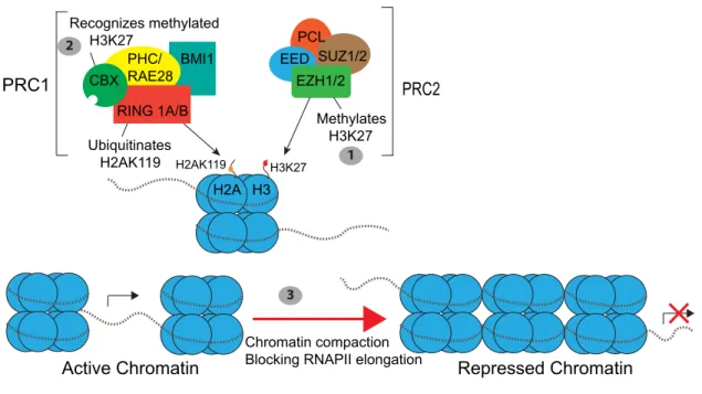
During cardiogenesis, epigenetic mechanisms influence the transcriptional activity of cardiac genes to enable proper CPC specification and promote differentiation. Mounting evidence has supported the existence of the co-operative relationship between transcription factors and epigenetic modifiers and has linked abnormalities in this interaction with congenital heart diseases (Huang et al., 2013). PRC1 and PRC2 repress gene expression primarily through histone modifications: histone methylation and histone ubiquitination (Fig. 4). These complexes have been shown to play a role in many aspects of cardiac development and homeostasis (Collinson et al., 2016). For example, Rae28 (also named PHC), a member of PRC1, is required for cardiac valve development and septation of the outflow tract through its role in maintaining Nkx2.5 expression. Rae28-null mice display many heart defects, including tetralogy of Fallot and double-outlet right ventricle (Shirai et al., 2002; Koga et al., 2002). A recent study in mice also reported that Ezh2, the enzymatic subunit of PRC2, stabilizes cardiac gene expression and prevents myocardial pathology through its role in repressing Six1, a transcriptional factor that functions in CPCs (Delagado-Olguin et al., 2012; San et al., 2016).
silencing, first, by PRC1 through Ezh2-mediated tri-methylation of H3K27 (Müller et al., 2002). The PcG (Polycomb group) protein in PRC1, CBX, recognizes this methylated H3K27 mark and recruitment of the PRC1 complex results in mono-ubiquitination of H2AK119 through the E3 ligase activity of Ring1B (Fig. 4) (Müller and Verrijzr, 2009; Wang et al., 2004). Ring1a and Ring1b, the two orthologs of the Drosophilia E3 ligase dRing, are found in mammals and amphibians (van der Velden et al., 2012). However, only a single homologous gene to Ring1b had been identified in zebrafish (van der Velden et al., 2012). Interestingly, investigating the role of Ring1B/Rfn2 in promoting proper cardiogenesis has been hindered by developmental arrest at the onset of gastrulation in Ring1b knockout mice (Voncken et al., 2003; O’Carroll et al., 2001). However, recent work has shown that in contrast to the mouse mutant, zebrafish embryos deficient of ring1b survive early embryogenesis but display a severe craniofacial phenotype (van der Velden et al., 2012). Interestingly, ring1b mutant zebrafish hearts also display cardiac phenotypes that have not yet been investigated. Thus, it will be important to study if and how Ring1b regulates key morphogenetic processes during cardiogenesis. In Chapter 3, I sought to address this gap in knowledge in the field by investigating what processes in early cardiac development is perturbed in ring1b mutants.
Summary: cooperativity between cardiac transcription factors and epigenetic regulators As depicted in the previous section, gene expression patterns reflect a major shift in the transcriptional profile of the CPC population between the earlier stages of cardiogenesis (i.e. cardiac crescent and heart tube stages) and later phases of heart development. Nevertheless, there is no single gene has been shown to be responsible for cardiac specification, proliferation, migration, and differentiation in vertebrate development, thereby signifying some functional redundancy and interconnectivity in cardiac genes that direct pathways to promote
structure, as Nkx2.5 also functions to maintain progenitor proliferation and differentiation by inhibiting bone morphogenetic protein (BMP) pathways (Lyons et al., 1995; Prall et al., 2007). Furthermore, in addition to regulating CPC dynamics, several cardiac transcription factors function to promote proper patterning and maturation of the early developing heart (Habets et al., 2002).
There is also a high degree of cooperation between epigenetic regulators and cardiac transcription factors. In numerous in vitro studies investigating chromatin localization of Nkx2.5, Mef2a, Gata4, and Tbx5, it has been consistently reported that these factors often bind together at genetic loci that are co-regulated during cardiogenesis (He et al., 2011). Still, the question of how these transcription factors are integrated at the level of chromatin to activate transcriptional programs still requires further investigation. Considering the importance of cardiac transcription factors and the need for epigenetic control, my work in Chapter 3 aims to explore whether key components of the Polycomb Repressive Complex 1 (i.e. Ring1b) can also be implicated in creating, inhibiting, or reinforcing molecular activation of cardiac or even non-cardiac genes during cardiovascular development.
Chamber maturation
Emergence, propagation of “sheet-like” networks, and compaction
Proper chamber maturation is essential for effective circulation of blood throughout the developing heart and organism (Samsa et al., 2015; Granados-Riveron et al., 2012). The zebrafish heart is morphologically more simplistic than the mammalian heart, and there are differences between species based on workload and circulatory demands. However, processes involved in patterning and regulating wall maturation are strikingly similar (Liu and Stainier, 2012; Brown et al., 2016; Samsa et al., 2013). Moreover, a myriad of studies has provided evidence that genes and signaling pathways involved in key steps of cardiovascular
that occur during cardiac chamber maturation is the emergence of myocardial, “sheet-like” projections, termed trabeculae, in the luminal layers of the ventricle. During embryogenesis, the lack of trabecular formation or perturbations in cardiac trabeculae causes congenital
cardiomyopathies. In fact, with the absence of the heart’s normal trabecular infrastructure, ventricular function substantially decreases, resulting in consequential effects to the organism (Liu and Stainier, 2012; Brown et al., 2016; Samsa et al., 2013; Liu et al., 2010).
Initially, the embryonic zebrafish heart tube is comprised of two layers: an outer myocardial layer and an inner endocardial layer. Later in development, the heart tube undergoes expansion and extensive growth. Cardiac trabeculae begin to develop after the looping stage: at Carnegie stage 12 in humans, at E9.5 in mice, and at approximately 60 hpf in zebrafish (Liu et al., 2010; Samsa et al., 2013; Peshkovsky et al., 2011; Moorman and
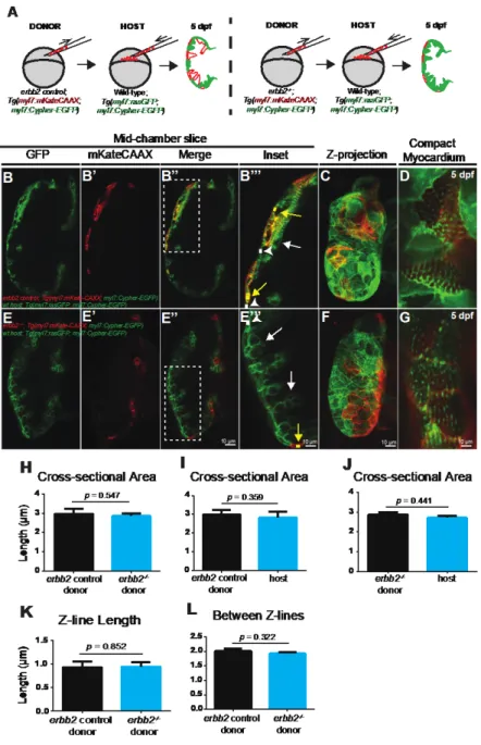
Christoffels, 2003; Sedmera et al., 2000). In zebrafish embryos, trabeculae begin to develop in the outer curvature of the ventricle and protrude into the ventricular lumen. More specifically, to form trabeculae, Liu et al. (2010) and others have demonstrated that cardiomyocytes
transformation of the spaces between trabeculae into capillaries for blood flow. Additionally, this remodeling process increases myocyte mass and eventually contributes to most of the working myocardium in the zebrafish adult heart (Samsa et al., 2013).
Ventricular trabeculation has been suggested to facilitate nutrient and oxygen uptake during development, prior to coronary vascularization, and to increase cardiac output or
hemodynamic demand in the developing embryos (Minot, 1901; Rychter and Ostádal, 1971; Liu et al., 2010). Nevertheless, direct evidence of how trabeculae help the heart to function
efficiently is lacking, and my work in Chapter 2 aims to address this gap in the field by using the erbb2 mutant zebrafish that do not form trabeculae to assess what occurs when the heart’s normal trabecular infrastructure is absent.
Signaling pathways essential for trabeculation Notch
Notch signaling has been shown to be crucial for ventricular trabeculation and chamber development (Grego-Bessa et al., 2007; MacGrogan et al., 2010). In fact, mutations in Notch signaling components are associated with human CHDs. Canonically, mammalian NOTCH receptors bind to their family of ligands, including Delta or Jagged. Upon binding, the
extracellular domain of NOTCH gets cleaved by ADAM metallopeptidase domain 17 (ADAM17), while the intracellular domain of NOTCH is cleaved by γ-secretase and released into the
compact myocardial wall, namely bone morphogenetic protein 10 (BMP10) signaling.
Interestingly, mammalian NOTCH receptors (Notch1, 2, 3, 4) and ligands (delta-like1, 3, 4 and jagged1, 2) show different expression patterns in the developing heart. For example, Notch1, Notch4, and Delta4 are expressed in the endocardial layer of the heart, while Notch2 and Jagged1 are expressed in the developing trabecular myocardium (Uyttendaele et al., 1996; Krebs et al., 2000). Endocardial-specific deletion of Notch1 and RBPJk mouse mutants show impaired trabeculation, decreased myocardial proliferation, and decreased expression of the following downstream signaling pathways: EphrinB2, Neuregulin1, and BMP10 (Grego-Bessa et al., 2007; Yang et al., 2012). Similarly, hypomorphic mutations of myocardial Notch2 in mice resulted in reduced trabeculation and a thin compact layer (Yang et al., 2012).
Ephrin B2/B4
Ephrin B2 is a transmembrane ligand that is essential for cardiovascular development. Ephrin B2 is also a direct target for endocardial NOTCH and is upstream of Neuregulin1 in the mammalian and zebrafish ventricle. In the vertebrate heart, Ephrin B2 and its receptor EPHB4 are expressed in endothelial cells lining the trabecular layer of the myocardium (Wang et al., 1998; Samsa et al., 2013). In mice deficient of Ephrin B2 or Ephrin B4, trabeculae do not form (Wang et al., 1998; Gerety et al., 1999). Similarly, in zebrafish larvae at 3 dpf efnb2a morphants lacked trabeculae in the outer curvature (Samsa et al., 2015), further signifying the importance of the endocardium in proper trabeculation.
BMP10
Bone morphogenetic protein 10 (BMP10) is a growth factor that belongs to the TGF-β superfamily and is transiently expressed in trabecular myocardium (Lowery and de Caestecker, 2010). Bmp10 has been shown to be involved in maintaining cardiomyocyte proliferation, as deletion of Bmp10 in mouse embryos is embryonic lethal; Bmp10-deficient embryos die at E10.5 (Chen et al., 2004). Further analysis of these Bmp10-deficient embryos revealed hypoplastic ventricular walls and an absence of trabeculae due to a marked reduction of proliferation of the cardiomyocytes (Chen et al., 2004).
Neuregulin/ErbB2/ErbB4
Numerous studies in mice highlight the importance of crosstalk between myocardial and endocardial cells within the heart that is facilitated through Neuregulin signaling (Liu et al., 2010; Peshkovsky et al., 2011; Samsa et al., 2013; Olson and Srivastava, 1996; Samsa et al., 2015; Yelon et al., 2001). In fact, the Neuregulin signaling pathway has been deemed essential for cardiac trabeculation. Neuregulins are growth factors that are expressed on endothelial cells and signal through ErB receptor tyrosine kinases, expressed on myocardial cells, to induce various cellular processes such as cell proliferation and delamination (Liu et al., 2010). Canonically, Neuregulin1 (Nrg1) gets cleaved and binds to its receptor, ErbB4. ErbB4
heterodimerizes with ErbB2, and ErbB2 gets phosphorylated. The kinetic activity of ErbB2 leads to downstream regulation of various genes involved in the formation of trabeculae (Meyer and Birchmeier, 1995; Lee et al., 1995). The importance of the Nrg/ErbB signaling pathway was first discovered by examination of ErbB2, ErbB4, and Neuregulin1 knockout mice, which either had reduced trabecular formation or failed to form trabeculae in the ventricle of the heart (Gassmann et al., 1995). These mutants eventually died at mid-gestation. Temporal and pharmacological inhibition of erbb2 revealed similar phenotypes in the zebrafish, which suggests that ErbB2 signaling has both an essential and direct role in the process of trabeculation (Liu et al., 2010).
expressed in the endocardium), and not nrg1, is a key regulator in zebrafish cardiac trabeculation (Rasouli and Stainier, 2017).
Biomechanical/hemodynamic regulators of cardiac maturation
Cardiac morphogenesis, particularly trabeculation, is dependent on biomechanical forces such as those generated by cardiac contractility and blood flow (Granados-Riveron and Brook, 2012; Staudt et al., 2014). During embryogenesis, cardiac contraction contributes to the cyclic force that is imposed on the organ as blood flows through the heart. This occurs in a bidirectional manner. Shear stress is induced by the force parallel to blood flow over the
endocardium, while a perpendicular force causes cyclic strain over the entire myocardial wall of the developing heart (Granados-Riveron and Brook, 2012; Staudt et al., 2014). In chick and zebrafish, several studies have reported that inhibition or reduction of blood flow in the cardiac chambers completely diminishes trabecular formation. Peshkovsky et al. (2011) used zebrafish weak atrium (wea) mutant embryos to further decipher the influences of blood flow on
trabeculation. Wea mutants display markedly weak blood flow, as the wea locus encodes an atrium-specific gene required for atrium contraction (Berdougo et al., 2003). In wea mutants, though some irregular thickening of the ventricular wall was initially observed, trabecular protrusions into the lumen did not progress toward creating myocardial ridges (Peshkovsky et al., 2011). These findings and others suggest that optimal blood flow through the ventricle is important for the progression of trabeculation.
One way that cells detect flow or contraction is through the bending of primary cilia. Cilia are microtubule-based organelles that protrude from the plasma membrane and are found on many cell types, including endocardial cells (Samsa et al., 2013; Samsa et al., 2015). In E11.5 Ift88-null mice, which do not form cilia, there is decreased trabeculation and ventricular dilation in the chambers, along with abnormal outflow tract development (Clement et al., 2009).
key role in flow detection even at low levels to upregulate notch1b and activate Notch1 in endocardial cells (Samsa et al., 2015). Notably, in addition to the impact of shear forces on endocardial cells, stretch forces “felt” by myocardial cells during normal ventricular loading can also induce intracellular signaling to impact trabeculation (Culver and Dickinson, 2010).
Recent studies have shown a relationship between Notch signaling in the endocardium and cardiac contraction in promoting trabeculation. By using a tnnt2a morpholino to eliminate cardiac contraction in zebrafish larvae, Samsa et al. (2015) showed that tnnt2a morphants lacked trabeculae and did not express endocardial Notch (Samsa et al., 2015). Moreover, through loss- and gain-of-function approaches, Rasouli and Stainier (2017) showed that contractility and blood flow are required for endocardial expression of nrg2a. Taken together, it is intriguing to consider that modified hemodynamics by mechanical manipulations or mutations in genes encoding proteins involved in responding to shear stress, proper cardiac contraction, and sensing can impact the initiation or advancement of trabeculation.
Cardiac remodeling during development
Chamber pressure is another major hemodynamic cue for trabeculation and as an organism grows, the heart has to make structural changes over time to increase cardiac output. An efficient way to compensate for increased hemodynamic burden is to increase cardiac mass and normalize wall tension either by hyperplasia or hypertrophy of existing cardiomyocytes. Therefore, it is likely that cell behaviors are key contributors to the process of chamber emergence and maturation.
Cardiac hypertrophy and hyperplasia are defined as cardiac remodeling processes that occur in response to a variety of intrinsic and extrinsic stimuli during normal development and stressful conditions (i.e. volume or pressure overload) (Lin et al., 2012). During normal
cardiomyocyte enlargement progresses throughout embryonic heart development, along with increasing maturation of myofibrils (Hirschy et al., 2006; Lin et al., 2012). During chamber formation in chick embryos, cardiomyocyte proliferation contributes to two-thirds of the overall chamber size, while hypertrophy of individual cardiomyocytes in the outer curvature accounts for the remaining one-third of the chamber (Soufan et al., 2006). Furthermore, an increase in cardiomyocyte size has also been observed in developing hearts of zebrafish, particularly at the outer curvature of the emerging ventricle (Auman et al., 2007).
Cardiac hypertrophy: features of concentric or eccentric hypertrophy
Cardiac hypertrophy can be characterized in two main forms, concentric or eccentric (Hou and Kang, 2012). Distinct features of concentric hypertrophy include an increase in wall thickness with little or no reduction in chamber volume. Additionally, during concentric
hypertrophy, the width of myocardial cells increases due to addition of sarcomeres in parallel within individual cardiomyocytes (Lips et al., 2003). In contrast, eccentric hypertrophy features minor changes to myocardial wall thickness and an increase in chamber volume (Müller and Dhalla, 2012; Mihl et al., 2008). On a cellular level, series addition of sarcomeres, which increases cardiomyocyte length, characterizes eccentric hypertrophy (Libonati, 2011).
Cardiac remodeling during disease
Cardiac hypertrophy can occur in a physiological or pathological manner, which can be difficult to differentiate. Along with anatomic features, physiological and pathological hypertrophy can also be distinguished by the functional alterations that develop. Physiological hypertrophy is characterized by normal or enhanced cardiac contractility, while pathological hypertrophy is associated with decreased cardiac function (Hou and Kang, 2012). Initially, the heart
hypertrophy include enlargement of individual cardiomyocytes, disarray of myofibrils
(sarcomeres that stack in parallel), re-activation of fetal transcriptional programs, fibrosis in the extracellular matrix, and decreased cardiac function (Carreño et al., 2006; Samak et al., 2016).
To investigate the pathogenesis of cardiac remodeling via hypertrophy and/or
hyperplasia in zebrafish, Sun et al. (2009) used tr265/tr265 zebrafish that display anemic stress due to a Band 3 mutation that disrupts erythrocyte formation. Data showed that many features of cardiomyopathy observed in mammals, including muscular disarray and re-activation of fetal genes (ex. ANF), and severe arrhythmia, were recapitulated in the adult zebrafish mutant. In addition to myocyte hypertrophy, Sun et al. (2009) showed that myocyte hyperplasia is involved in every stage of the remodeling process, with contribution from proliferating cardiomyocytes and differentiating cardiac progenitor cells. Interestingly, since the tr265/tr265 zebrafish survived the longest when only hyperplasia was prominent in the heart, it is suggested that myocyte hyperplasia might be a better remodeling strategy than cardiac hypertrophy (Sun et al., 2009). My work in Chapter 2 aims to undercover the specific cellular and molecular remodeling behaviors of cardiomyocytes that are in a trabeculae-deficient zebrafish ventricle.
As previously mentioned, in pathological cardiac remodeling, many fetal genes and other molecular factors are re-employed. Factors that are re-activated include atrial natriuretic factor (ANF), cardiac transcription factors (Mef2c, Nkx proteins, and Gata proteins), co-regulators, microRNAs, and/or those involved in epigenetic regulation (Dirkx et al., 2013). Though each of these factors act independently on a different level of genetic regulation, they also form a highly interconnected network with each other to promote transcriptional activation of various genes (Dirkx et al., 2013). Therefore, these genetic networks offer numerous therapeutic targets to reduce the aberrant expression of fetal genes, for example, and effectively address the adverse remodeling processes of the failing heart.
and other heart failure-related diseases in animal models. In human hearts of approximately 6 and 12 weeks of development, NPPA was expressed in the atrial chamber myocardium and the ventricular trabeculae (Christoffels et al., 2004). Furthermore, variants in the human NPPA gene are associated with hypertension, stroke, coronary artery disease, heart failure, and obesity (Song et al., 2015). To investigate the pattern and level of Nppa and Nppb expression living mice, Sergeeva et al. (2014) used Nppa/Nppb double reporter transgenic mice, in which the bioluminescent signal from Luciferase correlated with Nppa expression, and the intensity of red fluorescence was associated with expression of Katushka and Nppb. Sergeeva at al. (2014) performed transverse aortic constriction (TAC) as an experimental model for pressure-overload cardiac hypertrophy. Data showed that Nppa expression significantly increased in the TAC mouse ventricle compared to the control ventricle. In addition to being a functional marker of stress, Nppa is also a structural marker. In situ hybridization in mice showed that Nppa was restricted to the trabecular layer in the heart. Furthermore, in a Nppa-GFP knock-in mouse line, Tian et al. (2017) showed that endogenous Nppa expression was largely restricted to trabecular cardiomyocytes at E11.5 and E12.5, particularly in the left ventricle. Likewise, in zebrafish, nppa is also expressed in the heart and in the trabecular layer (Grassini et al., 2018; Aumen et al., 2007).
mTOR signaling in cardiac hypertrophy and the effects of rapamycin treatment
development and for maintaining cardiac homeostasis in postnatal life (Laplante and Sabatini, 2009; Sciarretta et al., 2018).
mTOR is a serine/threonine kinase, belonging to the phosphoinositide kinase-related kinase family. The mTOR pathway involves two functional complexes: mTOR complex 1 (mTORC1) and mTOR complex 2 (mTORC2). mTORC1 is more characterized (Laplante and Sabatini, 2009; Guertin and Sabatini, 2007). The core components of mTORC1 are regulatory-associated protein of mTOR (RAPTOR) and mammalian lethal with SEC13 protein 8 (mLST8), which are involved in substrate recruitment along with subcellular localization and kinase activity, respectively. mTORC1 is the main regulator of cellular growth by promoting various anabolic processes such as autophagy, protein synthesis, and lipid synthesis within the tissue (Laplante and Sabatini, 2009). Concerning protein synthesis, mTORC1 activates S6 kinase-1 (S6K1) by direct phosphorylation at Thr389, leading to initiation of translation through multiple mechanisms (Peterson et al., 2009; Sciarretta et al., 2018).
a 65% reduction in TOR expression, allowing the zebrafish to survive to adulthood. Myocyte enlargement was diminished with short-tern rapamycin treatment using DOX induction. When investigating long-term effects of mTOR inhibition, ztor fish displayed cardioprotective features characterized by improved cardiac function, reduced mortality, lessened apoptosis and
autophagy, and little pathological remodeling (i.e. parallel sarcomere addition) (Ding et al., 2011). Taken together, mTOR is a key therapeutic target for addressing many hypertrophic-associated cardiovascular diseases. More specifically, based on work presented in Chapter 2, rapamycin may be a promising therapy for patients with trabeculae-associated diseases.
Relevance to current research
Although several mice models of cardiac hypertrophy have been developed, it usually takes months for these mice to develop hypertrophic phenotypes. The advantage of studying the hypertrophic phenotype in the trabeculae-deficient erbb2 zebrafish mutant is that it only takes four days for these animals to show hypertrophic phenotypes (Lin et al., 2012; Fleming et al., 2018), thereby facilitating the study of the molecular and cellular mechanisms involved in compensatory growth of the myocardium. Likewise, PcG proteins, such as Ring1B, are also conserved in zebrafish as well as their accompanying epigenetic marks (San et al., 2016). Since the ring1b mutant zebrafish survives early embryogenesis, unlike Ring1b mutant mice, the zebrafish allows for detailed analysis of early cardiac phenotypes.
Early and late manifestations of CHDs
As previously noted, genetic mutations in genes that are essential during early
(McDonald-McGinn and Sullivan, 2011). Importantly, though genetics (i.e. genetic mutations) influence many of the structural anomalies, the subsequent morphogenetic defects that develop are what primarily lead to CHD symptoms, morbidity, and mortality. Individuals with DiGeorge syndrome have craniofacial defects, aortic arch malformations, and conotruncal heart defects. Several knockout experiments in mice and zebrafish have identified Tbx1 as a major factor in the onset of the malformations in DGS, particularly relating to the heart (Yamagishi et al., 2003; Yagi et al., 2008). Mutations in the same gene can also cause multiple phenotypes in the heart. For example, mutations in Nkx2.5 can lead to atrial or ventricular septal defects, tetralogy of Fallot
and conduction defects. Similarly, patients with mutations in GATA4 can display pulmonary
stenosis and double outlet right ventricle (Ottaviani and Buja, 2016).
In patients, CHDs also develop due to inappropriate execution of late cardiac
morphogenic processes. Presumably, CHDs that perturb early cardiac morphogenesis would produce severe phenotypes such that they would be embryonic lethal and thus not observable in the patient population. One of the many forms of CHDs is called left ventricular
non-compaction (LVNC), which is characterized by prominent trabeculae and large recesses between trabeculae (Morcos et al., 2015; Samsa et al., 2013). Ventricular non-compaction in patients with LVNC has been defined as a morphogenetic abnormality that arises from defects in trabeculation and compaction. Furthermore, various genetic mutations have been identified to be associated with this structural defect, particularly those found in sarcomere-encoding genes and calcium-handling genes (Samsa et al., 2013). Noteworthy, human patients with LVNC cardiomyopathy can survive to adulthood, but often suffer from heart failure, arrhythmias and/or sudden cardiac death (Ottaviiani and Buga, 2016; Jenni et al., 1999). In addition to structural defects, patients with LEOPARD syndrome or other CHDs also develop hypertrophic