GENETICALLY ENGINEERED MOUSE MODELS OF BREAST CARCINOMA: A TRANSLATIONAL RESOURCE FOR HIGHLIGHTING HUMAN BREAST SUBTYPE
ETIOLOGY AND DEVELOPING PERSONALIZED THERAPEUTIC APPROACHES
Adam David Pfefferle
A dissertation submitted to the faculty of the University of North Carolina at Chapel Hill in partial fulfillment of the requirements for the degree of Doctor of Philosophy in the Department
of Pathology and Laboratory Medicine
Chapel Hill 2015
Approved by: Charles M. Perou H. Shelton Earp III William K. Kaufmann Joel S. Parker
ii © 2015
iii ABSTRACT
Adam David Pfefferle: Genetically engineered mouse models of breast carcinoma: a translational resource for highlighting human breast subtype etiology and developing
personalized therapeutic approaches (Under the direction of Charles M. Perou)
Approximately one in eight women will be diagnosed with breast cancer during their lifetime. While increased public awareness has led to earlier detection of this common disease, a greater understanding of tumor biology has led to the development of many promising therapeutics. A difficult frontier, however, has been identifying the appropriate target population for new drugs as not all breast cancer patients will respond to a particular
therapeutic.Currently, approximately five percentof oncology drugs that enter clinical testing are ultimately approved by the US Food and Drug Administration for use. This low success rate reflects not only the difficulty of developing anticancer therapeutics, but also flaws in preclinical testing methodology for selecting the most appropriate cancer patient subset for early clinical testing. With so many patients either not responding or relapsing with the current standard of care, improved personalized therapeutic approaches are greatly needed.
iv
cancers in vivo under genetically controlled and immune competent conditions. Identifying murine models with conserved human tumor features will not only facilitate etiology determinations for the intrinsic subtypes, but also serve as a useful preclinical resource for testing the efficacy of new therapeutic approaches. These mice promise to be better predictors of clinical trial success because they resemble tumor biology more closely than other
v
I dedicate my research to all those who have been affected by breast cancer; either being diagnosed themselves, knowing a survivor, or celebrating the memory of someone who
vi
ACKNOWLEDGEMENTS
vii
TABLE OF CONTENTS
LIST OF TABLES ... xiii
LIST OF FIGURES ... xiv
LIST OF ABBREVIATIONS ... xvi
CHAPTER 1: INTRODUCTION ...1
A greater understanding of breast cancer biology will identify novel therapeutic targets ...1
Personalized drug regimens are the future of cancer treatment ...4
Murine models are excellent for translating biological discovery into clinical care ...5
References ...7
CHAPTER 2: TRANSCRIPTOMIC CLASSIFICATION OF GENETICALLY ENGINEERED MOUSE MODELS OF BREAST CANCER IDENTIFIES HUMAN SUBTYPE COUNTERPARTS ...12
Overview ...12
Background ...12
Results ...12
Conclusion ...13
Background ...13
Results ...14
viii
Comparison of murine class defining gene sets versus human
tumor subtypes ...23
Conserved tumorigenic pathway signatures identified between human-mouse counterparts ...29
Discussion ...33
Conclusion ...39
Materials and Methods ...39
Gene expression microarrays ...39
Murine intrinsic genes and subtypes ...41
Human and mouse intrinsic gene cocluster ...42
Comparison of murine and human expression subtypes...42
Conserved pathway gene signatures ...43
References ...44
CHAPTER 3: LUMINAL PROGENITOR AND FETAL MAMMARY STEM CELL EXPRESSION FEATURES PREDICT BREAST TUMOR RESPONSE TO NEOADJUVANT CHEMOTHERAPY ...50
Overview ...50
Background ...50
Methods...50
Results ...51
Conclusions ...51
Background ...51
Results ...53
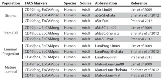
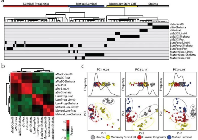
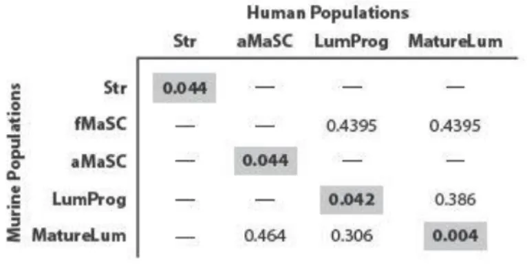
Comparison of human mammary subpopulation transcriptomic datasets ...53
ix
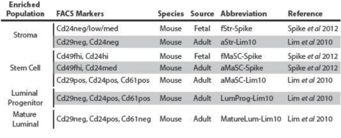
Murine mammary cell subpopulation enriched gene signatures ...61
LumProg and fMaSC features predict neoadjuvant chemotherapy response...65
Discussion ...70
Materials and Methods ...75
Mammary cell subpopulation gene signatures ...75
Comparison of human and murine normal mammary populations ...76
Mammary cell subpopulation centroids ...76
Chemotherapy response ...77
References ...79
CHAPTER 4: SECONDARY GENETIC ABERRATION PROFILING OF P53NULL MAMMARY TUMORS HIGHLIGHTS MET DNA AMPLIFICATION AS A GENETIC DRIVER OF MURINE BASAL-LIKE TUMORIGENESIS ...84
Overview ...84
Background ...84
Methods...84
Results ...85
Conclusions ...85
Background ...85
Results ...87
p53null transplant tumors are counterparts for human basal-like and claudin-low subtypes ...87
Secondary genetic aberration profiling highlights DNA copy number changes as drivers of tumorigenesis ...91
x
Discussion ...101
Materials and Methods ...104
Gene expression ...104
Flow cytometry ...105
DNA single nucleotide polymorphisms ...105
DNA structural variants ...106
DNA copy number ...106
Crizotinib treatment ...107
References ...108
CHAPTER 5: THE MMTV-WNT1 MURINE MODEL PRODUCES TWO PHENOTYPICALLY DISTINCT SUBTYPES OF MAMMARY TUMORS WITH UNIQUE CLINICAL OUTCOMES TO EGFR INHIBITORS ...112
Overview ...112
Background ...112
Methods...112
Results ...113
Conclusions ...113
Background ...113
Results ...116
Wnt1-EarlyEx and Wnt1-LateEx tumors have distinct gross pathology and histology traits ...116
Wnt1-EarlyEx tumors are enriched for canonical and non-canonical Wnt pathway signatures ...120
xi
Wnt1-EarlyEx and Wnt1-LateEx tumors have distinct mammary
subpopulation FACS profiles ...124
Both Wnt1-LateEx tumor FACS subpopulations have tumor initiating potential ...127
Both Wnt1-LateEx tumor FACS subpopulations have activating Hras1 mutations ...129
Discussion ...131
Materials and Methods ...134
Mouse husbandry ...134
Gene expression ...134
DNA copy number ...135
Immunofluorescence ...136
Drug treatment ...136
Flow cytometry ...137
Sanger Sequencing ...138
References ...139
CHAPTER 6: DISCUSSION ...143
References ...151
APPENDIX A: RELATED COAUTHORSHIPS ...153
Oncogenic PI3K mutations lead to NF-kB-dependent cytokine expression following growth factor deprivation ...153
LKB1/STK11 inactivation leads to expansion of a pro-metastatic tumor sub-population in melanoma ...154
Comparative oncogenomics implicates the Neurofibromin 1 gene (NF1) as a breast cancer driver ...155
xii
Conditional loss of ErbB3 delays mammary gland hyperplasia induced by mutant PIK3CA without affecting mammary tumor latency, gene expression
or signaling...157
Mutant PIK3CA accelerates HER2-driven transgenic mammary tumors and induces resistance to combinations of anti-HER2 therapies ...158
Endothelial-like properties of claudin-low breast cancer cells promote tumor vascular permeability and metastasis ...159
c-Myc and Her2 cooperate to drive stem-like phenotype with poor prognosis in breast cancer ...160
Expression of miR-200c in claudin-low breast cancer alters stem cell functionality, enhances chemosensitivity and reduces metastatic potential ...161
JNK2 prevents luminal cell commitment in normal mammary glands and tumors by inhibiting p53/NOTCH1 and BRCA1 expression ...162
APPENDIX B: CURRICULUM VITAE ...163
Education ...163
Research and Work Experience ...163
Publications ...164
Presentations ...166
Teaching and mentoring ...167
Certifications ...168
xiii
LIST OF TABLES
Table 1 – Summary of murine models studied ...15
Table 2 – Gene set analysis of murine classes ...24
Table 3 – Gene set analysis of murine classes and human subtypes ...27
Table 4 – Human FACS enriched normal mammary cell subpopulation studies ...54
Table 5 – Murine FACS enriched normal mammary cell subpopulation studies ...60
Table 6 – Gene set analysis of human and murine cell subpopulations ...62
Table 7 – Clinical characteristics of the neoadjuvant chemotherapy treated dataset ...66
Table 8 – Univariate logistic regression analysispredicting pathological complete response ...67
Table 9 – Multivariate logistic regression analysis predicting pathological complete response ...68
xiv
LIST OF FIGURES
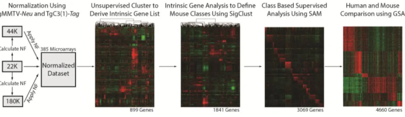
Figure 1 – Flowchart of murine expression data analysis...16
Figure 2 – Murine intrinsic class analysis...18
Figure 3 – Murine intrinsic tumor dendrogram by sample ...19
Figure 4 – Murine intrinsic tumor dendrogram by mouse model ...20
Figure 5 – Murine intrinsic cluster signature according to tumor subtype ...22
Figure 6 – Human and murine intrinsic co-cluster ...26
Figure 7 – Conserved signaling pathways between human-mouse counterparts ...31
Figure 8 – Flowchart of analysis ...55
Figure 9 – Comparison of mammary subpopulations across studies ...56
Figure 10 – Homo sapiens enriched gene signatures ...58
Figure 11 – Mus musculus enriched gene signatures ...63
Figure 12 – fMaSC enriched gene signatures ...69
Figure 13 – Human counterparts of p53null transplant tumors ...88
Figure 14 – Molecular pathway signatures and differentiation score ...89
Figure 15 – Murine p53null tumor datasets ...93
Figure 16 – Somatic non-silent mutation analysis ...94
Figure 17 – Chromosome structural variation analysis ...95
Figure 18 – DNA copy number analysis...97
Figure 19 – MET DNA amplification is a driver of 2250L (p53null-LuminalEx) tumors ...99
xv
Figure 21 – Wnt1-EarlyEx and Wnt1-LateEx have similar DNA copy
number landscapes ...118 Figure 22 – Wnt-EarlyEx tumors have expression of Wnt associated pathway
signatures ...121 Figure 23 – Wnt-EarlyEx tumors respond to EGFR inhibitors ...123 Figure 24 – Wnt-EarlyEx and Wnt1-LateEx tumors share features with different
xvi
LIST OF ABBREVIATIONS
aCGH array comparative genomic hybridization
Cy3 cyanine-3
Cy5 cyanine-5
DNA deoxyribonucleic acid D-Score differentiation score
EGFR epidermal growth factor receptor EMT epithelial-to-mesenchymal transition ER estrogen receptor
FACS fluorescence-activated cell sorting FDR false discovery rate
FET fisher’s exact test
GEMM genetically engineered mouse model GSA gene set analysis
H&E hematoxylin and eosin
HER2 human epidermal growth factor receptor 2 HsEnriched Homo sapiens enriched
IACUC institutional animal care and use committee LumProg luminal progenitor
MaSC mammary stem cell MatureLum mature luminal
xvii MVA multivariate analysis
PCA principle component analysis pCR pathologic complete response PR progesterone receptor
RNA ribonucleic acid
SAM significance analysis of microarrays SNP single nucleotide polymorphism SSP single sample predictor
Str stroma
1
CHAPTER 1: INTRODUCTION
About three quarters of a million American women were diagnosed with new cases of cancer last year [1]. With breast cancer accounting for about thirty percent of these incidences, one in eight women will be affected by this common disease in their lifetime [1]. Increased public awareness and a greater understanding of tumor biology has led to better patient survival rates since the early 1990s, yet this disease is still the second leading cause of cancer related deaths in American women [1]. With so many patients either not responding or relapsing with the current standard of care, the molecular mechanisms underlying breast cancer are under intense investigation to identify new, personalized drug targets that should improve patient outcomes [2, 3].
2
epidermal growth factor receptor 2 (HER2-). Luminal B tumors are distinguishable from the luminal A subtype by their faster, more aggressive proliferation rates. HER2-enriched tumors, on the other hand, are typically ER-, PR-, and HER2+, with similar high proliferation and low
survival rates as luminal B tumors when untreated [7]. The remaining two subtypes, basal-like and claudin-low, are broadly considered triple negative breast cancer (TNBC) (ER-, PR- and HER2-) [7]. Basal-like breast cancers have the highest proliferation rates and are the most genetically unstable of all the subtypes [11, 12]. Claudin-low tumors are characterized by low levels of cell adhesion molecules and high levels of inflammatory cells [6]. While targeted therapeutics exist for ER+ [13] (luminal A/B [14]) and HER2+ [15] (HER2-enriched [14]) breast cancer, targeted treatments for TNBC (basal-like and claudin-low [14]) remain an important unmet clinical need [16]. To address this need, a research emphasis has been placed on determining the molecular drivers of basal-like and claudin-low tumors to identify novel drug targets for these subtypes, which was an important aspect of my thesis work.
Even though clear distinctions between the intrinsic subtypes have been defined, the molecular mechanisms that give rise to breast tumors in general, and the individual intrinsic subtypes, are not fully known. For instance, it is unknown why some tumors of the same subtype respond differently to the same therapeutic treatment [3, 17]. A greater knowledge of the
molecular aberrations underlying breast tumors will identify these differences in genetic drivers, which we believe to be the key first step towards personalized drug therapies [3]. Segregating genetic drivers from passenger mutations is difficult due to the inherent heterogeneity of breast tumors and the large number of genetic aberrations seen within a given tumor. Several
3
intrinsic subtypes [18-21]. 2) Similarly, tumors develop through clonal expansion of evolving clones, resulting in intra-tumor pockets of cell colonies containing their own set of aberrations [22]. 3) The tumor microenvironment can interact with progressing cancer cells to affect the overall tumor phenotype [23, 24]. 4) External factors (e.g. environmental, lifestyle, and
comorbidity) influence tumor development [25]. All of these factors probably have some role in determining the ultimate phenotype of a progressing breast tumor, but more research is needed to validate the relative contributions of each.
Over the last fifteen years, genomics [12, 26] and transcriptomics [4] have fueled a greater understanding of breast tumor biology [8]. The cancer genome is broadly characterized as unstable. The inability to properly respond to and fix DNA damage leads to the accumulation of small scale mutations [26] (e.g. insertions, deletions) and large scale chromosomal
rearrangements [12] (e.g. translocations, aneuploidy). Microarray and sequencing techniques have been developed to identify genomic aberrations that can lead to decreased tumor suppressor function, increased oncogene signaling, or both. Transcriptomics (gene expression analysis) is a popular approach for characterizing tumors because it is easy to measure, provides a rough estimate of corresponding protein levels, and identifies overarching tumor phenotypes. Given the large number of transcriptomic studies, the Broad Institute has created the molecular signatures database (MSigDB), which compiles gene sets/modules from the literature into one place [27]. This database improves on the gene-gene comparison by allowing for the comparison of gene groups. While each of these approaches has improved our knowledge of cancer biology by themselves, studies integrating multiple 'omic approaches have an even greater power of
4
Personalized drug regimens are the future of cancer treatment
Developing clinical tests that predict drug response (i.e. companion diagnostics) is an important focus of cancer research. Two clinical trials have brought the use of molecular testing to the forefront of breast cancer research. In these trials, the Oncotype DXTM (TAILORx trial) [30] and MammaPrintTM (MINDACT trial) [31] assays are being used to determine which patients should receive chemotherapy. A recent comparison of Oncotype DXTM with the PAM50 intrinsic subtyping assay identified that the PAM50 risk of recurrence (ROR) as superior to the Oncotype DXTM recurrence score (RS) in endocrine-treated patients with ER-positive, node-negative disease [32]. While the use of molecular testing in the clinic has been groundbreaking, these assays do not identify which drug regimens to prescribe. Clinical assays that determine personalized drug regimens are greatly needed to improve patient survival.
Before personalized drug testing can begin in the clinic, the target population for molecularly targeted drugs needs to be identified. This is a difficult task, but is critical for
developing the best predictors of response. Targeted cancer drugs are designed to inhibit specific genetic aberrations, typically kinases or hormone receptors, but not all tumors with the aberration respond to treatment [17]. Additional factors (e.g. coexisting aberrations, cell of origin,
microenvironment) are hypothesized to confer sensitivity or resistance to drug treatment [33-36]. Even though studies have defined predictive signatures of response [37-41], most are not
5
mouse models of human breast cancer such that this heterogeneous set of models could be better linked to human breast tumors, which we predict would then help drug testing and development.
Murine models are excellent for translating biological discovery into clinical care
Currently, only ~5%of oncology drugs that enter clinical testing are approved by the FDA for use [45]. This dismal success rate not only reflects the difficulty of developing anticancer therapeutics, but also flaws in preclinical methodology for selecting the most promising drugs to use in clinical trials [43, 46]. Historically, preclinical drug testing has primarily involved a mix of in vitro cell line and in vivo cell line xenograft experiments. While these studies are a good first step, they do not represent true tumor biology [47]. For instance, the few cell lines available for use have limited biological diversity when compared to primary tumors, and they represent a subset of the original clones with the best growth advantages. These cell line based approaches are unable to accurately represent clinical heterogeneity or an intact microenvironment, and as a result, many of the drugs that pass cell line based preclinical testing fail during later stages.
6
Comparative studies between human and murine tumors provide an attractive approach for narrowing the genetic driver candidate list by highlighting conserved features between species [49] and thus focusing attention on the most likely driver genes. While dozens of murine models have been created to study the molecular mechanisms of breast cancer [50-56], the degree to which many of these models recapitulate the human subtypes is largely unknown. Before proper therapeutic comparative studies can be performed, it is essential that human-to-murine tumor counterparts are identified to ensure that the chosen model accurately replicates the genetic alterations and overall phenotypes observed in human tumors [49]. This is especially important for heterogeneous human diseases, such as breast cancer.
Given the advantages of murine models for studying tumorigenesis [47], the following four chapters utilize genetically engineered mouse model of breast carcinoma to simultaneously investigate human tumor etiology and as a tool for preclinical drug testing. Chapter 1, which was published in Genome Biology in 2013 [49], analyzes the transcriptomic profiles of 27 murine models to highlight the subset of GEMMs that mimic the human disease state. Chapter 2, which was published in Breast Cancer Research and Treatment in 2015 [57], analyzes the
transcriptomic profiles of normal mammary cell types to highlight conserved cell features between human-murine subtype counterparts. In addition, this study identifies several gene signatures that predict tumor pathologic complete response sensitivity to neoadjuvant
7
REFERENCES
1. AmericanCancerSociety: Cancer Facts and Figures. 2015.
2. Curigliano G, Goldhirsch A: The triple-negative subtype: new ideas for the poorest prognosis breast cancer.J Natl Cancer Inst Monogr 2011, 2011:108-110.
3. Curigliano G: New drugs for breast cancer subtypes: Targeting driver pathways to overcome resistance.Cancer Treat Rev 2011, 38:303-310.
4. Perou CM, Sorlie T, Eisen MB, van de Rijn M, Jeffrey SS, Rees CA, Pollack JR, Ross DT, Johnsen H, Akslen LA, et al: Molecular portraits of human breast tumours.
Nature 2000, 406:747-752.
5. Sorlie T, Perou CM, Tibshirani R, Aas T, Geisler S, Johnsen H, Hastie T, Eisen MB, van de Rijn M, Jeffrey SS, et al: Gene expression patterns of breast carcinomas
distinguish tumor subclasses with clinical implications.Proc Natl Acad Sci U S A
2001, 98:10869-10874.
6. Prat A, Parker JS, Karginova O, Fan C, Livasy C, Herschkowitz JI, He X, Perou CM: Phenotypic and molecular characterization of the claudin-low intrinsic subtype of breast cancer.Breast Cancer Res 2010, 12:R68.
7. Prat A, Perou CM: Deconstructing the molecular portraits of breast cancer.Mol Oncol 2010.
8. CancerGenomeAtlasNetwork: Comprehensive molecular portraits of human breast tumours.Nature 2012, 490:61-70.
9. Parker JS, Mullins M, Cheang MC, Leung S, Voduc D, Vickery T, Davies S, Fauron C, He X, Hu Z, et al: Supervised risk predictor of breast cancer based on intrinsic subtypes.J Clin Oncol 2009, 27:1160-1167.
10. Carey LA, Dees EC, Sawyer L, Gatti L, Moore DT, Collichio F, Ollila DW, Sartor CI, Graham ML, Perou CM: The triple negative paradox: primary tumor
chemosensitivity of breast cancer subtypes.Clin Cancer Res 2007, 13:2329-2334. 11. Rakha EA, Reis-Filho JS, Ellis IO: Basal-like breast cancer: a critical review.J Clin
Oncol 2008, 26:2568-2581.
8
13. Jordan VC: Tamoxifen: a most unlikely pioneering medicine.Nat Rev Drug Discov
2003, 2:205-213.
14. Prat A, Perou CM: Deconstructing the molecular portraits of breast cancer.Mol Oncol 2011, 5:5-23.
15. Hynes NE, Lane HA: ERBB receptors and cancer: the complexity of targeted inhibitors.Nat Rev Cancer 2005, 5:341-354.
16. Carey L, Winer E, Viale G, Cameron D, Gianni L: Triple-negative breast cancer: disease entity or title of convenience?Nat Rev Clin Oncol 2010, 7:683-692. 17. Bates M, Sperinde J, Kostler WJ, Ali SM, Leitzel K, Fuchs EM, Paquet A, Lie Y,
Sherwood T, Horvat R, et al: Identification of a subpopulation of metastatic breast cancer patients with very high HER2 expression levels and possible resistance to trastuzumab.Ann Oncol 2010, 22:2014-2020.
18. Lim E, Vaillant F, Wu D, Forrest NC, Pal B, Hart AH, Asselin-Labat ML, Gyorki DE, Ward T, Partanen A, et al: Aberrant luminal progenitors as the candidate target population for basal tumor development in BRCA1 mutation carriers.Nat Med
2009, 15:907-913.
19. Raouf A, Zhao Y, To K, Stingl J, Delaney A, Barbara M, Iscove N, Jones S, McKinney S, Emerman J, et al: Transcriptome analysis of the normal human mammary cell commitment and differentiation process.Cell Stem Cell 2008, 3:109-118.
20. Spike BT, Engle DD, Lin JC, Cheung SK, La J, Wahl GM: A mammary stem cell population identified and characterized in late embryogenesis reveals similarities to human breast cancer.Cell Stem Cell 2012, 10:183-197.
21. Stingl J, Eirew P, Ricketson I, Shackleton M, Vaillant F, Choi D, Li HI, Eaves CJ: Purification and unique properties of mammary epithelial stem cells.Nature 2006, 439:993-997.
22. Siegmund KD, Marjoram P, Woo YJ, Tavare S, Shibata D: Inferring clonal expansion and cancer stem cell dynamics from DNA methylation patterns in colorectal
cancers.Proc Natl Acad Sci U S A 2009, 106:4828-4833.
23. Hatiboglu MA, Kong LY, Wei J, Wang Y, McEnery KA, Fuller GN, Qiao W, Davies MA, Priebe W, Heimberger AB: The tumor microenvironment expression of p-STAT3 influences the efficacy of cyclophosphamide with WP1066 in murine melanoma models.Int J Cancer 2012, 131:8-17.
24. Bissell MJ, Radisky DC, Rizki A, Weaver VM, Petersen OW: The organizing principle: microenvironmental influences in the normal and malignant breast.Differentiation
9
25. Hurria A: Embracing the complexity of comorbidity.J Clin Oncol 2011, 29: 4217-4218.
26. Sjoblom T, Jones S, Wood LD, Parsons DW, Lin J, Barber TD, Mandelker D, Leary RJ, Ptak J, Silliman N, et al: The consensus coding sequences of human breast and colorectal cancers.Science 2006, 314:268-274.
27. Subramanian A, Tamayo P, Mootha VK, Mukherjee S, Ebert BL, Gillette MA, Paulovich A, Pomeroy SL, Golub TR, Lander ES, Mesirov JP: Gene set enrichment analysis: a knowledge-based approach for interpreting genome-wide expression profiles.Proc Natl Acad Sci U S A 2005, 102:15545-15550.
28. Consortium TCGA: Integrated genomic analyses of ovarian carcinoma.Nature 2011, 474:609-615.
29. Consortium TCGA: Comprehensive genomic characterization defines human glioblastoma genes and core pathways.Nature 2008, 455:1061-1068.
30. Zujewski JA, Kamin L: Trial assessing individualized options for treatment for breast cancer: the TAILORx trial.Future Oncol 2008, 4:603-610.
31. Cardoso F, Van't Veer L, Rutgers E, Loi S, Mook S, Piccart-Gebhart MJ: Clinical application of the 70-gene profile: the MINDACT trial.J Clin Oncol 2008, 26: 729-735.
32. Dowsett M, Sestak I, Lopez-Knowles E, Sidhu K, Dunbier AK, Cowens JW, Ferree S, Storhoff J, Schaper C, Cuzick J: Comparison of PAM50 risk of recurrence score with oncotype DX and IHC4 for predicting risk of distant recurrence after endocrine therapy.J Clin Oncol 2013, 31:2783-2790.
33. Chaft JE, Arcila ME, Paik PK, Lau C, Riely GJ, Pietanza MC, Zakowski MF, Rusch V, Sima CS, Ladanyi M, Kris MG: Coexistence of PIK3CA and other oncogene
mutations in lung adenocarcinoma-rationale for comprehensive mutation profiling.
Mol Cancer Ther 2011, 11:485-491.
34. Yuan TL, Cantley LC: PI3K pathway alterations in cancer: variations on a theme.
Oncogene 2008, 27:5497-5510.
35. Kuo YW, Wu SG, Ho CC, Shih JY: Good response to gefitinib in lung
adenocarcinoma harboring coexisting EML4-ALK fusion gene and EGFR mutation.
J Thorac Oncol 2010, 5:2039-2040.
10
37. Augustine CK, Jung SH, Sohn I, Yoo JS, Yoshimoto Y, Olson JA, Jr., Friedman HS, Ali-Osman F, Tyler DS: Gene expression signatures as a guide to treatment strategies for in-transit metastatic melanoma.Mol Cancer Ther 2010, 9:779-790.
38. Bild AH, Parker JS, Gustafson AM, Acharya CR, Hoadley KA, Anders C, Marcom PK, Carey LA, Potti A, Nevins JR, Perou CM: An integration of complementary strategies for gene-expression analysis to reveal novel therapeutic opportunities for breast cancer.Breast Cancer Res 2009, 11:R55.
39. Bild AH, Yao G, Chang JT, Wang Q, Potti A, Chasse D, Joshi MB, Harpole D, Lancaster JM, Berchuck A, et al: Oncogenic pathway signatures in human cancers as a guide to targeted therapies.Nature 2006, 439:353-357.
40. Ooi CH, Ivanova T, Wu J, Lee M, Tan IB, Tao J, Ward L, Koo JH, Gopalakrishnan V, Zhu Y, et al: Oncogenic pathway combinations predict clinical prognosis in gastric cancer.PLoS Genet 2009, 5:e1000676.
41. Wu CJ, Cai T, Rikova K, Merberg D, Kasif S, Steffen M: A predictive phosphorylation signature of lung cancer.PLoS One 2009, 4:e7994.
42. Borst P, Wessels L: Do predictive signatures really predict response to cancer chemotherapy?Cell Cycle 2010, 9.
43. Begley CG, Ellis LM: Drug development: Raise standards for preclinical cancer research.Nature 2012, 483:531-533.
44. Weigelt B, Pusztai L, Ashworth A, Reis-Filho JS: Challenges translating breast cancer gene signatures into the clinic.Nat Rev Clin Oncol 2011, 9:58-64.
45. Kola I, Landis J: Can the pharmaceutical industry reduce attrition rates?Nat Rev Drug Discov 2004, 3:711-715.
46. Hutchinson L, Kirk R: High drug attrition rates--where are we going wrong?Nat Rev Clin Oncol 2011, 8:189-190.
47. Sharpless NE, Depinho RA: The mighty mouse: genetically engineered mouse models in cancer drug development.Nat Rev Drug Discov 2006, 5:741-754.
48. Usary J, Zhao W, Darr D, Roberts PJ, Liu M, Balletta L, Karginova O, Jordan J, Combest A, Bridges A, et al: Predicting drug responsiveness in human cancers using
genetically engineered mice.Clin Cancer Res 2013, 19:4889-4899.
11
50. Herschkowitz JI, Simin K, Weigman VJ, Mikaelian I, Usary J, Hu Z, Rasmussen KE, Jones LP, Assefnia S, Chandrasekharan S, et al: Identification of conserved gene expression features between murine mammary carcinoma models and human breast tumors.Genome Biol 2007, 8:R76.
51. Chan SR, Vermi W, Luo J, Lucini L, Rickert C, Fowler AM, Lonardi S, Arthur C, Young LJ, Levy DE, et al: STAT1-deficient mice spontaneously develop estrogen receptor alpha-positive luminal mammary carcinomas.Breast Cancer Res 2012, 14:R16. 52. Guy CT, Cardiff RD, Muller WJ: Activated neu induces rapid tumor progression.J
Biol Chem 1996, 271:7673-7678.
53. Husler MR, Kotopoulis KA, Sundberg JP, Tennent BJ, Kunig SV, Knowles BB:
Lactation-induced WAP-SV40 Tag transgene expression in C57BL/6J mice leads to mammary carcinoma.Transgenic Res 1998, 7:253-263.
54. Pond AC, Herschkowitz JI, Schwertfeger KL, Welm B, Zhang Y, York B, Cardiff RD, Hilsenbeck S, Perou CM, Creighton CJ, et al: Fibroblast growth factor receptor signaling dramatically accelerates tumorigenesis and enhances oncoprotein translation in the mouse mammary tumor virus-Wnt-1 mouse model of breast cancer.Cancer Res 2010, 70:4868-4879.
55. Zhang X, Podsypanina K, Huang S, Mohsin SK, Chamness GC, Hatsell S, Cowin P, Schiff R, Li Y: Estrogen receptor positivity in mammary tumors of Wnt-1 transgenic mice is influenced by collaborating oncogenic mutations.Oncogene 2005, 24: 4220-4231.
56. Herschkowitz JI, Zhao W, Zhang M, Usary J, Murrow G, Edwards D, Knezevic J, Greene SB, Darr D, Troester MA, et al: Comparative oncogenomics identifies breast tumors enriched in functional tumor-initiating cells.Proc Natl Acad Sci U S A 2011, 109:2778-2783.
1
This chapter previously appeared as an article in Genome Biology. The original citation is as follows: Pfefferle AD et al, “Transcriptomic classification of genetically engineered mouse
models of breast cancer identifies human subtype counterparts”, Genome Biology 2013
12
CHAPTER 2: TRANSCRIPTOMIC CLASSIFICATION OF GENETICALLY ENGINEERED MOUSE MODELS OF BREAST CANCER IDENTIFIES HUMAN
SUBTYPE COUNTERPARTS1
OVERVIEW Background
Human breast cancer is a heterogeneous disease consisting of multiple molecular subtypes. Genetically engineered mouse models are a useful resource for studying mammary cancers in vivo under genetically controlled and immune competent conditions. Identifying murine models with conserved human tumor features will facilitate etiology determinations, highlight the effects of mutations on pathway activation, and should improve preclinical drug testing.
Results
Transcriptomic profiles of 27 murine models of mammary carcinoma and normal mammary tissue were determined using gene expression microarrays. Hierarchical clustering analysis identified 17 distinct murine subtypes. Cross-species analyses using three
13
TgWAPCre-Etv6 model mimicked the HER2-enriched subtype, a group of human tumors without a murine counterpart in previous comparative studies. Gene signature analysis identified hundreds of commonly expressed pathway signatures between linked mouse and human subtypes, highlighting potentially common genetic drivers of tumorigenesis.
Conclusions
This study of murine models of breast carcinoma encompasses the largest comprehensive genomic dataset to date to identify human-to-mouse disease subtype counterparts. Our approach illustrates the value of comparisons between species to identify murine models that faithfully mimic the human condition and indicates that multiple mouse models are needed to represent the diversity of human breast cancers. The reported trans -species associations should guide model selection during preclinical study design to ensure appropriate representatives of human disease subtypes are used.
BACKGROUND
Breast cancer is the second leading cause of cancer related deaths in American women [1]. While increased public awareness has led to earlier detection, a greater
understanding of tumor biology has led to the development of many promising therapeutics [2, 3]. A difficult frontier, however, has been identifying the appropriate target population for new drug(s) as not all breast cancer patients will respond to a particular therapeutic.
14
flaws in preclinical testing methodology for selecting the most appropriate cancer patient subset for early clinical testing [5, 6].
Numerous murine models of breast cancer have been created to mimic the genetic aberrations found in human tumors [7-30]. Historically, each model has been analyzed independent of other models, which complicates effective comparisons with human tumors. However, when multiple models are consolidated into a single dataset, there is increased sensitivity to detect features that are conserved with the human disease state [31, 32]. Identifying murine models that faithfully mimic specific human breast cancer subtypes [33-35] is an important need for the proper interpretation of mouse model results, and thus, for translating preclinical findings into effective human clinical trials [36].To address this need, we useda transcriptomic approach to profile tumors from 27 different genetically engineered mouse models (GEMMs). We define and characterize 17 distinct murine subtypes of
mammary carcinoma (referred to as classes herein to distinguish them from the human subtypes), which we compare to three human breast tumor datasets comprising over 1700 patients to determine which GEMM classes resemble specific human breast cancer subtypes.
RESULTS
Expression classes of genetically engineered mouse models
15 Table 1: Summary of murine models studied
16
Figure 1: Flowchart of murine expression data analysis
17
global gene expression measurements from 356 unique murine tumors and 16 normal murine mammary samples were analyzed using Agilent microarrays (see Table 1A, Figure 1). Using this larger and more diverse murine dataset, a new mouse ‘intrinsic gene list’ was derived to identify genes associated with all 27 models. As expected, many of the genes from the previous intrinsic gene list were also present in the updated list. After filtering for genes found in both datasets, 76.5% (500/654) of the intrinsic probes from Herschkowitz et al 2007 were again included within the new intrinsic list of 1855 probes, which represents 1841 genes.
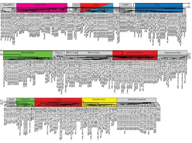
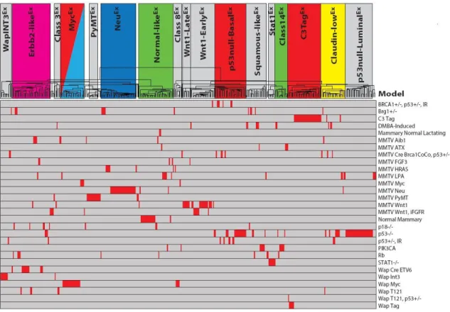
To determine if new murine subtypes/classes exist in this expanded dataset, SigClust analysis [37] was performed using supervised hierarchical clustering of the 385 murine microarrays and the intrinsic 1855 probe list (Figure 2). Murine ‘classes’ were defined as having at least five tumors with a SigClust p-value ≤ 0.01. Using these criteria, 17 murine classes were identified with 94% (363/385) of tumors being included within one of these classes (Figure 2B and Figure 3). The name for each class was determined based upon the major model contributor (e.g. MycEx), the major biological feature (e.g. Squamous-likeEx), or both (e.g. p53null-BasalEx), with the ‘Ex’ designation used to denote that this is an expression-based class. As previously observed [31], the Brca1+/-Trp53+/- irradiated, TgC3(1)-Tag, TgMMTV-Neu, TgWAP-Int3, TgWAP-Myc, and TgWAP-Tag murine models have ‘homogeneous’ gene expression patterns in this dataset; here, a model was considered ‘homogenous’ if ≥ 80% of tumors from that GEMM were found within a single expression-defined class (Table 1B and Figure 4). Many of the newest models also showed
homogeneous gene expression patterns including Stat1-/-, TgMMTV-Myc,
18 Figure 2: Murine intrinsic class analysis
19
20
Figure 4: Murine intrinsic tumor dendrogram by mouse model.
21
Other models showed a ‘semi-homogeneous’ gene expression pattern, defined as ≥ 80% of tumors from a single GEMM being found within two classes. These included Pik3ca- H1047R, TgMMTV-Atx, TgMMTV-Fgf3, TgMMTV-Hras, TgWap-T121, and
TgMMTV-Wnt1. Interestingly while maintaining the TgMMTV-Wnt1 mouse colony, it was observed that there might be two types of tumors based on latency, namely early and late arising tumors. This observation was also reflected in the two TgMMTV-Wnt1 expression classes that also differed by median tumor latency: Wnt1-EarlyEx (8.8 weeks) and Wnt1-LateEx (22.2 weeks) (Wilcoxon Rank Sum p-value < 0.001). Lastly, about 40% of MMTV provirus driven Wnt1 tumors have cooperative activation of FGF signaling [38], a phenotype that is known to decrease tumor latency [16], and consistent with this, 88% (7/8) of TgMMTV-Wnt1/iFgfr2
tumors in our dataset were also classified as Wnt1-EarlyEx.
The remaining models had ‘heterogeneous’ gene expression patterns, which were defined as no two classes containing at least 80% of the tumors analyzed: Brg1+/- (five classes), DMBA-induced (five), p18-/- (three), Rb1-/- (five), TgMMTV-Aib1 (four), TgMMTV-Cre BrcaCo/CoTrp53+/- (three), TgMMTV-Lpa (four), Trp53-/- (seven), and
Trp53+/- irradiated (four). Similar to recent reports [32], the Trp53-/- model (which is distinct from the Trp53+/- irradiated model) was primarily defined by three murine classes in this analysis: p53null-luminalEx (27/58), p53null-basalEx (15/58), and Claudin-lowEx (7/58).
To begin investigating the defining features of these classes, a comparison of selected cell lineage markers was performed (Figure 2C). Several mouse classes highly expressed luminal cell markers (e.g. Erbb2, Esr1, Krt18, and/or Krt19), including Erbb2-likeEx, PyMTEx, NeuEx, MycEx, and Stat1Ex. Other classes expressed basal cell cytokeratins (e.g.
22
Figure 5: Murine intrinsic cluster signatures according to tumor subtype
23
Squamous-likeEx, Class14Ex, and C3TagEx. As identified previously [31], a murine Claudin-lowEx class was observed to be characterized by low expression of multiple cell adhesion genes (Cldn3, Cldn4, and Cldn7) and high expression of epithelial-to-mesenchymal transition (EMT) genes (Snai1 and Zeb2), similar to the human claudin-low subtype [34].
Comparison of murine class defining gene sets versus human tumor subtypes
To specifically compare murine classes to human breast cancer subtype features, each murine class defining signature (Figure 2i-v) was tested for differential expression across the human subtypes using the UNC308 dataset (Figure 5A-E) [34]. For example, the high expression signature that defines the murine Claudin-lowEx class (Figure 2i, including Hic1,
Il6st, Klf2, Maf, Pdgfra, Prrx1, Snai1) was also the most highly expressed in human claudin-low tumors (Figure 5A).
Figure 2ii shows genes that are highly expressed in the newly identified Stat1Ex and Class14Ex murine classes, which show luminal characteristics (e.g. Foxa1, Esrrb) and are the most highly expressed in human luminal A tumors (Figure 5B). While most of the GEMMs in this dataset are considered estrogen receptor (ER) negative, murine models comprising these two classes (Stat1-/- and Pik3ca-H1047R, respectively) were often ERα+ [9, 11], and these data suggest that they overall have a ‘luminal’ expression profile. Interestingly, these classes cluster independent from the previously defined murine luminal models,
TgMMTV-Neu and TgMMTV-PyMT. Consistent with the individual cell lineage marker analysis, the Wnt1-LateEx, Wnt1-EarlyEx, p53null-BasalEx, Squamous-likeEx, and Class14Ex murine classes express a basal-like gene signature (Figure 2iii). As in human tumors, a proliferation
24 Table 2: Gene set analysis of murine classes
Displayed are the p-values for the gene set analysis comparison of each murine class versus each murine class described in Herschkowitz et al 2007. Empty boxes are trending
25
murine C3TagEx and human basal-like tumors, and lowest expression in normal tissues from both species. This finding is likely due to the loss of RB1 function in both human basal-like [39, 40] and TgC3(1)-Tag murine tumors (due to T-antigen expression). Lastly, Figure 2v highlights a gene cluster that is highly expressed in several murine classes including Erbb2-likeEx, PyMTEx, and NeuEx; this signature was lower in normal mammary tissue, but highly expressed in the two lactating mammary samples (Figure 5E). Consistent with this
observation, many of the genes in this signature are involved in alveolar function (e.g. Abcg2,
Folr1, and Lalba).
For the dual purpose of validating our new classification system and for investigating the degree of diversity in our expanded dataset, the murine classes defined here were
compared to those from Herschkowitz et al 2007 [31] using gene set analysis (GSA) (Table 2). The majority of the Herschkowitz et al 2007 classes had one-to-one matching
counterparts to those described here; however, two previous groups (IWapTag and X-C3Tag) were combined into a single class in our dataset (C3TagEx). Importantly, several of the 17 murine classes defined here were not present within the ten classes of Herschkowitz et al 2007 (Erbb2-likeEx, Class3Ex, Class8Ex, and Stat1Ex), almost all of which were populated by GEMMs that were new to this study.
Given the discovery of novel murine classes, it was of great interest to determine the degree to which this expanded murine dataset might better encompass the molecular diversity of the human subtypes. To directly compare tumors across species, this mouse and the
26 Figure 6: Human and murine intrinsic co-cluster
27
Table 3: Gene set analysis of murine classes and human subtypes
Displayed are the p-values for the gene set analysis comparison of each murine class versus each human subtype. Empty boxes are trending associations, while filled boxes are
28
platforms, different common references) may limit interspecies clustering, several across species dendrogram nodes were observed (Figure 6A). Interestingly, all major nodes contained a combination of human and mouse subtypes (Figure 6B), indicating a degree of similarity not only between specific corresponding tumor subtypes, but also globally across species. Most of the major intrinsic gene sets driving the nodes are highlighted below the dendrogram, including the basal (2.4C), proliferation (2.4D), normal breast (2.4E), claudin-low subtype high expression (2.4F), and luminal (2.4G) signatures. These clusters highlight the broad conserved intrinsic features between mouse and human tumors. For instance, most C3TagEx tumors cluster with the basal-like subtype, an association that is driven in part by the high expression of the proliferation gene set [31], which is known to contain many E2F-regulated genes.
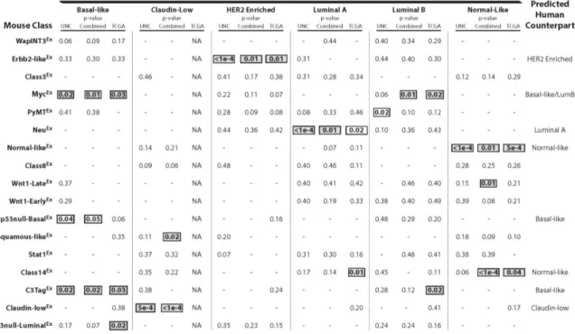
To more objectively validate the trans-species associations observed in Figure 6, similarity between specific human and mouse subtypes was measured GSA (Table 3) [42]. Using this approach, a murine class was judged to be a strong human subtype counterpart if the human-to-mouse comparison was statistically significant (p ≤ 0.05) in at least two of the three human datasets analyzed (UNC308 [34], Combined855 [43], and TCGA547 [39]).
As previously observed [31], the murine Normal-likeEx, C3TagEx, and Claudin-lowEx classes associate with the human normal-like, basal-like, and claudin-low subtypes,
29
breast cancer, which is consistent with multiple human studies linking basal-like breast cancers with cMYC amplification and expression signatures [39, 44]. Interestingly, a connection between the MycEx class and human luminal B tumors was also identified, highlighting Myc activation as a potentially important etiological mechanism that is shared between these two aggressive human subtypes.
Previously defined as a ‘luminal’ model [31], the NeuEx
murine class associated with the human luminal A subtype in this newest analysis; this correlation was somewhat
surprising given the lack of ERα and ERα-regulated gene expression in the murine NeuEx class, but does suggest that human Luminal A tumors have many ERα independent features. Although the murine p53null-BasalEx versus human comparisons were not significant after controlling for multiple comparisons, an almost consistent significant association was seen with human basal-like tumors (p=0.04, 0.05, and 0.06) in all three human datasets. Lastly, Class14Ex tumors were identified as a counterpart for normal-like human tumors, and of the 13 murine tumors comprising this class, 38% (5/13) are from the Pik3ca-H1047R model. This class clusters independent of normal mammary tissue samples (which are all classified as Normal-likeEx), indicating that this association is possibly not driven by contamination of normal tissue in the tumor biopsies.
Conserved tumorigenic pathway signatures identified between human-mouse counterparts
Many researchers have hypothesized that gene expression signatures may be a more robust means of utilizing gene expression data for discovery and pathway-based
30
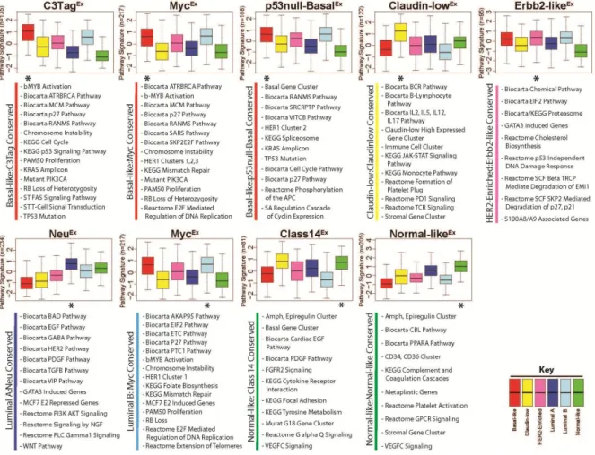
take advantage of this approach, the median expression values for 963 publicly available pathway gene-signatures were calculated separately for the mouse and human datasets, and a two-class (Class X versus all others) Significance Analysis of Microarrays (SAM) was used to identify pathways that were highly expressed by each class/subtype with a false discovery rate (FDR) of 0%. To visualize pathway similarities across species, gene signatures highly expressed within each mouse class were first grouped into ‘pathway meta-signatures’, similar to the way coordinately expressed genes can be grouped into ‘gene signatures’. The average value of these ‘pathway meta-signatures’ was then calculated for each human tumor and displayed as standardized boxplots based on their human breast cancer subtype for the eight mouse classes with human counterparts (Figure 7). These boxplots allow for broad trends to be observed between the pathways highly expressed within each mouse class relative to human tumors, and in all instances, identified tens of pathway signatures that were
commonly expressed across species. For instance, the average expression of the 135 pathway signatures highly expressed in C3-TagEx tumors were also very highly expressed in human basal-like tumors (Figure 7, top left panel), consistent with the gene level analysis. While these trends are informative, it was of most importance to identify the specific pathways that were highly expressed in both mouse and their human counterparts; it is likely that these shared pathways provide etiological insight and highlight potentially important cancer
driving pathways. A subset of the pathways identified as highly expressed in both human and mouse counterparts are displayed below each graph.
31
Figure 7: Conserved signaling pathways between human-mouse counterparts
A two-class SAM (Class X versus all others) was used to identify pathways highly expressed in each murine class. Pathways highly expressed with a FDR of 0% were grouped together to define a ‘pathway meta-signature’ for each murine class (with the total number of pathway signatures included shown on the left axis). The standardized, average expression values of each ‘pathway meta-signature’ were calculated in the UNC308, Combined855, and
32
apparent in C3-TagEx and p53null-BasalEx murine tumors on both a genetic and expression level. The second cardinal feature of human basal-like tumors is high proliferation, primarily resulting from RB loss [39, 40]. Consistent with this finding, all three basal-like mouse classes highly expressed cell cycle and/or RB-pathway related signatures. In addition, C3TagEx tumors were enriched for KRAS amplicon genes, b-MYB activation, mutant
PIK3CA, and FAS signaling. Murine MycEx tumors were also enriched for b-MYB activation and mutant PIK3CA signaling, in addition to a HER1-pathway signature and E2F signaling. Lastly, the p53null-BasalEx class was enriched for a SRC activation signature, a HER1-pathway signature, and the KRAS amplicon. These findings are relevant since it has been shown that human basal-like tumors also highly express the b-MYB signature [45], are often KRAS [46] and cMYC amplified [39], and show a PIK3CA-activation signature [39, 47]. Thus for human and murine basal-like cancers, both the underlying molecular genetics and their expression profiles are very similar across species.
33
MycEx class was also a counterpart for the luminal B subtype. Interestingly, many of the same pathways that were common with basal-like tumors are also shared with luminal B tumors, highlighting potentially important etiological events that are shared between these two aggressive intrinsic subtypes; these features include proliferation/RB related pathways, increased chromosome instability, and altered DNA damage repair mechanisms.
DISCUSSION
Human breast cancer is a genetically complex disease consisting of well characterized molecular subtypes [33, 35]. Mouse models can provide an excellent resource to study
human disease, but it is essential to ensure the chosen models accurately replicate genetic alterations and overall phenotypes observed in human tumors. Thus, a number of
considerations must be kept in mind when designing and/or selecting GEMMs to mimic the human disease state; these features should include intramodel tumor diversity, the degree of genetic similarity, the degree of transcriptomic similarity, and histological similarity (a topic not addressed here). By consolidating mouse models of breast carcinoma into a single dataset, this study was able to investigate the first three of these issues, in which we identified murine models for all of the major human expression subtypes.
To address intramodel tumor diversity, three types of models were identified based on hierarchical clustering analysis: ‘homogenous’, ‘semi-homogeneous’, and ‘heterogeneous’. ‘Homogeneous’ GEMMs were associated with a single murine expression class and were generally created through the expression of oncogenes, possibly relying less on secondary or tertiary mutations that arise during tumor progression. These GEMMs make good
34
‘Semi-homogeneous’ models, such as TgMMTV-Wnt1, were associated with two murine classes. We hypothesize that unknown secondary events after the initial transgene lesion determine the class fate of these developing tumors. These varying combinations of
secondary lesions may cooperate with aberrant Wnt1 signaling to target different mammary cell populations, contributing to model complexity. The last type of model comprises tumors with ‘heterogeneous’ gene expression patterns (i.e. models showing three or more distinct phenotypes). In contrast to ‘homogeneous’ models, the majority of the ‘heterogeneous’ models were based on disrupting the function of tumor suppressor genes. Again, we
hypothesize that secondary events after the initial transgene lesion are involved in the class fate determination of these tumors. For example, the Trp53-/- model shows specific DNA copy number changes associated with each expression class [32]. From an experimental perspective, special considerations (i.e. phenotyping each individual tumor) must be made to account for this heterogeneity, especially when these models will be utilized for therapeutic efficacy testing.
Despite the diversity of the models tested here, we found that these mouse models collapse into distinct murine classes which recapitulate specific human subtypes on a gene expression-based level. These results are important as they allow for the identification of shared characteristics/lesions between murine and human tumors, and they direct researchers toward appropriate in vivo models of specific human subtypes for future experimental
35
These murine classes share these hallmarks as evident by high expression of the proliferation gene cluster, cell cycle pathways, and chromosome instability gene-signatures; thus there are clear GEMMs of human basal-like tumors that share both common genetic drivers and expression features.
Murine Claudin-lowEx tumors were identified that significantly mimic the human claudin-low subtype; however, no homogeneous murine model was specific to this
class/subtype. Instead, rare tumors from multiple heterogeneous models coalesced into the murine claudin-low group. As an experimental solution to this heterogeneous GEMM complication, the T11 orthotopic, transplantable syngeneic model was derived from a Claudin-lowEx BALB/c Trp53-/- tumor (753R), which maintains its claudin-low expression features even after multiple transplant passages [32]. This transplantable model has been used for extensive therapeutic testing [48], thus suggesting that one method of ‘capturing’ a heterogeneous model in a single state can be accomplished via the serial transplantation of a phenotypically characterized individual tumor. As in the human claudin-low subtype, Trp53
mutation/loss was a common genetic event in mouse Claudin-lowEx tumors. Similarly, both species highly express EMT related genes, inflammatory gene-signatures, and have low expression of many epithelial cell adhesion genes including E-cadherin [34].
Discovered here was the Erbb2-likeEx murine class, which associated with human HER2-enriched tumors even without highly expressing the Erbb2 gene; no mouse model from our previous studies mimicked this aggressive human tumor subtype. One
36
secretory breast cancers [50]. Consistent with this, we observed that murine Erbb2-likeEx tumors highly express a gene signature in common with lactating normal mammary tissue.
For the human luminal breast cancer subtypes, our previous study identified that the TgMMTV-Neu model represents the luminal subtypes more than it resembles HER2-enriched tumors [31]. We provide further evidence here that the murine NeuEx class
specifically associates with human luminal A tumors. Conserved with humans, murine NeuEx tumors highly express several tyrosine kinase pathway related gene-signatures, namely EGFR and HER2, which would be expected based upon the nature of the Neu/ERBB2 transgene. It has been shown that TgMMTV-Neu tumors regress with lapatinib treatment [48], giving credence to our approach for identifying drug targetable driver/maintenance pathways in these tumors using a computational pathway-based approach. Interestingly, only the murine MycEx class was shown to consistently associate with luminal B tumors. Since the MycEx class was also identified as a basal-like model, aberrant Myc activation may be a common hallmark of these two aggressive subtypes.
While our main focus was to identify human-to-mouse disease counterparts, about half of the mouse classes did not statistically associate with specific human subtypes by our broad analysis. Several of these mouse specific classes, however, had clear basal-like tumor expression features including WapINT3Ex, Wnt1-LateEx, Wnt1-EarlyEx, and Squamous-likeEx. Unlike the other three, the Squamous-likeEx class consisted of a variety models (e.g. Pik3ca -H1047R, Brg1+/-, and DMBA-induced) and trended toward an association with human claudin-low tumors. Similarly, several classes had luminal expression features, highlighted by PyMTEx and Stat1Ex. Although the PyMTEx class had a relatively small number of
37
class also had several strong luminal features, consistent with prior characterization of this model [11]. Given the expression of ERα in these STAT1-defecient tumors [11], the lack of an association with either the luminal A or luminal B human subtypes was unexpected.
An unanswered question concerning these human-to-mouse associations is the finding that murine classes like Erbb2-likeEx, and NeuEx, associate with specific human subtypes despite the fact that they apparently do not show expression of one of these human subtype defining genes (HER2/ERBB2 in the case of Erbb2-likeEx and ESR1 in the case of NeuEx). Three hypotheses that could explain this finding are: 1) the cell type of origin of the tumor (but not a genetic driver) is the same across species and this is the major linking phenotype, 2) additional unknown genetic driver(s) are responsible for the common phenotype across species, or 3) some combination of hypothesis 1 and 2. We favor the
common cell type of origin hypothesis, but additional experiments like lineage tracing will be required to unequivocally determine this.
38
adequately address these two confounding features, but future experiments/models could be designed to address these questions.
While some of the mouse classes were identified as good counterparts for specific human subtypes, many were not. There are several possibilities to explain this lack of
association. The first is that these classes are specific to murine mammary carcinomas and do not have a matching counterpart in humans. The second might be that these murine classes model rare phenotypes that exist in only a small subset of human breast cancer patients, and that these rare human subtypes were not present in the datasets used here. Similarly, more mouse tumors for classes with small numbers may be required to increase statistical power to detect an association; for example, we hypothesize this to be the case for the PyMTEx class. The third possibility is that these novel murine classes share phenotypes with multiple human subtypes, and thus may never be classified as being similar to a single human subtype. Some murine tumor features were shared across multiple human subtypes (e.g. MycEx with human basal-like and luminal B), which our presented analysis is more
likely to undervalue.
39
basal-like TgC3(1)-Tag tumor were not; these studies are consistent with findings coming from human clinical trials of luminal/ER+ breast cancers, which were generally noted to be sensitive to a CDK4/6 inhibitor [54]. Similarly, a trans-species genetic screen by Bennett et al. [53] identified two ribonucleotide reductase genes (RRM1 and RRM2) and a checkpoint kinase (CHK1) as potential targets for triple-negative breast cancer patients, which they validated in both species with drug treatment experiments using TgC3(1)-Tag and human xenograft tumors.
CONCLUSION
In summary, we consolidate 27 murine models of breast carcinoma into the largest comprehensive genomic dataset to date, and we provide a detailed characterization of each to better understand how these GEMMs recapitulate phenotypes of the human subtypes. The data presented here provide insight into the molecular pathways involved in specific breast cancer subtypes and should serve as a useful resource when designing preclinical studies and interpreting their results.
MATERIALS AND METHODS Gene expression microarrays
40
samples from multiple participating investigators using methods approved by international animal husbandry guidelines. Total ribonucleic acid (RNA) was purified from 20-30mg of mouse mammary tumor using Qiagen's RNeasy Mini Kit following manufacture protocols. RNA quantity and quality were determined using the Nanodrop spectrophotometer and Agilent Bioanalyzer, respectively. Total RNA was reverse transcribed and labeled with cyanine-5 (Cy5) dye for experimental samples and cyanine-3 (Cy3) dye for mouse reference samples [31] using the Agilent Low RNA Input Fluorescent Linear Amplification Kit. Equal quantities of labeled mouse reference RNA and tumor RNA were co-hybridized overnight to Agilent microarrays, washed, scanned and signal intensities were determined.
All tumor samples were co-hybridized to one of three Agilent Technology gene expression microarray types: 22K, 4X44K, or 4X180K (Figure 1). Two ‘homogeneous expression’ murine models [31], namely TgMMTV-Neu and TgC3(I)-Tag, were analyzed on all three array types. Therefore, we used both of these models to normalize expression between microarray types [32]. Ten microarrays (five TgMMTV-Neu and five TgC3(I)-Tag) from each array type were used for normalization (30 microarrays total). All microarray data was independently extracted from the UNC Microarray Database for each array type as log2 Cy5/Cy3 ratios, filtering for probes with Lowess normalized intensity values greater than ten in both channels and for probes with data on greater than 70% of the microarrays [31, 34]. Before normalization, each data set was imputed (via the ten-nearest neighbor gene values) and then reduced to the probes that were present on all three array type datasets (11690 probes, 11167 genes). Using the ten normalization arrays per three array platforms, the median expression value was calculated for each probe, on each array type, and a
41
each array type. Probe expression values were ‘median centered’ to obtain the final normalized dataset. A principle component analysis (PCA) was performed to verify the normalization.
Murine intrinsic genes and subtypes
After removing technical replicates, the dataset was filtered to probes with at least three observations with an absolute log2 expression value greater than three using Gene Cluster 3.0 [56], which included 908 probes (899 genes). Hierarchical clustering was
performed with this unsupervised probe list using centroid linkage and was viewed with Java Treeview v1.1.5r2 [57]. Potential ‘intrinsic groups’ of murine samples were defined as any set of samples/arrays within this hierarchical cluster that had a Pearson correlation value of 0.65 or greater [31]. Using these defined groups (42 total), an ‘intrinsic gene list’ of 1855 probes (1841 genes) was identified with Intrinsic Gene Identifier v1.0 (Max Diehn/Stanford University) by using a cutoff of one standard deviation below the mean intrinsic gene value [31].
To identify significant murine ‘intrinsic subtypes’, the 385 sample dataset was clustered again using the 1855 intrinsic probe list and SigClust [37] was used to identify groups of samples with a significant association to one another (p<0.01) [32]. GEMM classes were defined as having at least five tumors and a SigClust p-value ≤ 0.01, yielding 17
42 Human and mouse intrinsic gene cocluster
Prior to combining the two datasets, probes corresponding to orthologous gene IDs (as determined by the Mouse Genome Informatics of the Jackson Laboratory) were averaged for both the mouse and UNC308 human datasets. Using only orthologous genes found in both datasets (8034 genes), each tumor and gene was standardized to have an average
expression of zero and a standard deviation of one (N(0,1)) separately for each species. Then, the datasets were merged and each gene was median centered to obtain the final, normalized combined dataset. A merged intrinsic gene list was created by combining the 1841 mouse intrinsic genes defined here and the 1918 human intrinsic genes from Parker et al [41] (3310 unique genes in the combined gene set). An intrinsic gene set hierarchical co-cluster was performed using centroid linkage in Gene Cluster 3.0.
Comparison of murine and human expression subtypes
43
These same methods were used to identify significant overlap between our 17 newly derived murine classes and the 10 previously defined GEMM classes from Herschkowitz et al 2007 [31], noting that all 122 arrays used for the Herschkowitz et al study were also present within the 385 sample dataset used here.
Conserved pathway gene signatures
44 REFERENCES
1. Society AC: Cancer Facts & Figures 2011.Cancer Facts & Figures 2011.
2. Toft DJ, Cryns VL: Minireview: Basal-like breast cancer: from molecular profiles to targeted therapies.Mol Endocrinol 2011, 25:199-211.
3. Schlotter CM, Vogt U, Allgayer H, Brandt B: Molecular targeted therapies for breast cancer treatment.Breast Cancer Res 2008, 10:211.
4. Kola I, Landis J: Can the pharmaceutical industry reduce attrition rates?Nat Rev Drug Discov 2004, 3:711-715.
5. Begley CG, Ellis LM: Drug development: Raise standards for preclinical cancer research.Nature 2012, 483:531-533.
6. Hutchinson L, Kirk R: High drug attrition rates--where are we going wrong?Nat Rev Clin Oncol 2011, 8:189-190.
7. Bultman SJ, Herschkowitz JI, Godfrey V, Gebuhr TC, Yaniv M, Perou CM, Magnuson T: Characterization of mammary tumors from Brg1 heterozygous mice.Oncogene 2008, 27:460-468.
8. Pei XH, Bai F, Smith MD, Usary J, Fan C, Pai SY, Ho IC, Perou CM, Xiong Y: CDK inhibitor p18(INK4c) is a downstream target of GATA3 and restrains mammary luminal progenitor cell proliferation and tumorigenesis.Cancer Cell
2009, 15:389-401.
9. Adams JR, Xu K, Liu JC, Agamez NM, Loch AJ, Wong RG, Wang W, Wright KL, Lane TF, Zacksenhaus E, Egan SE: Cooperation between Pik3ca and p53
mutations in mouse mammary tumor formation.Cancer Res 2011, 71:2706-2717. 10. Jiang Z, Deng T, Jones R, Li H, Herschkowitz JI, Liu JC, Weigman VJ, Tsao MS,
Lane TF, Perou CM, Zacksenhaus E: Rb deletion in mouse mammary progenitors induces luminal-B or basal-like/EMT tumor subtypes depending on p53 status.J Clin Invest 2010, 120:3296-3309.
11. Chan SR, Vermi W, Luo J, Lucini L, Rickert C, Fowler AM, Lonardi S, Arthur C, Young LJ, Levy DE, et al: STAT1-deficient mice spontaneously develop estrogen receptor alpha-positive luminal mammary carcinomas.Breast Cancer Res 2012, 14:R16.