TELOMERE MAINTENANCE AS A THERAPEUTIC TARGET
Rachel Henderson
A thesis submitted to the faculty at the University of North Carolina at Chapel Hill in partial fulfillment of the requirements for the degree of Master of Science in the Chemical Biology and
Medicinal Chemistry division in the Eshelman School of Pharmacy.
Chapel Hill 2014
Approved by:
Michael Jarstfer
Rihe Liu
ii ABSTRACT
Rachel Henderson; Telomere Maintenance as a Therapeutic Target (Under the direction of Michael Jarstfer)
Telomere maintenance is essential for long term cell survival, with two mechanisms
contributing to telomere maintenance: telomeric DNA elongation by telomerase and a capping
mechanism contributing to telomere stability. Several therapeutic approaches targeting telomeres
have been explored but many rely on a lag period of telomere degradation before
anti-proliferation occurs. Two strategies presented here aim to disrupt telomere maintenance while
eliminating the lag period. First, telomerase was inhibited using antisense oligonucleotides
targeting its RNA subunit (hTR). The goal of this study was to both prevent active holoenzyme
assembly and induce degradation of the protein subunit (hTERT) thought to be associated with
anti-apoptotic activity. Additionally, a fluorescence polarization assay was designed for the
identification of small molecule inhibitors of telomeric repeat binding factor 2 (TRF2), a key
capping protein involved in prevention of chromosomal end fusions and ultimately cellular
iii
ACKNOWLEDGEMENTS
Dr. Mike Jarstfer for his guidance, encouragement, and mentorship throughout my
graduate career. He is always willing to offer help and knowledge and I am grateful for his many
contributions to my advancement as a scientist. His genuine concern and valuable guidance as a
mentor helped me to achieve my goals throughout this personal journey.
My committee, Dr. Rihe Liu and Dr. Qisheng Zhang for their participation and
contribution.
The Jarstfer lab, especially Carrie Ann Gordon for her support, friendship, and
willingness to help.
Brandi Baughman for kindly sharing her expertise with me.
My family and friends for their encouragement and support, especially my parents who I
cannot express enough gratitude to. They have always supported my goals and aspirations and
helped me become who I am today. Additionally, the graduate students and faculty of CBMC
who were welcoming, considerate, and supportive from day one.
iv
TABLE OF CONTENTS
LIST OF FIGURES ... iv
LIST OF TABLES ... viii
LIST OF ABBREVIATIONS ... ix
CHAPTER 1: INTRODUCTION ...1
I. Overview of Telomeres, Telomerase, and Cancer ... 1
II. Telomere Replication and Telomerase Function ...2
III. Implications in Telomere Shortening... 4
IV. Telomere Capping ... 7
V. Research Projects ...10
CHAPTER 2: TELOMERASE INHIBITION THROUGH OLIGONUCLETOIDE TRANSFECTION ...15
I. Introduction ... 15
II. P6.1 Loop Target and hTRas012 Development ...16
III. Materials and Methods ...21
IV. Results ... 23
V. Discussion ... 28
CHAPTER 3: A FLUORESCENCE POLARIZATION ASSAY TO IDENTIFY TRF2 SMALL MOLECULE INHIBITORS ... 31
I. Introduction ... 31
v
III. Results ... 36
IV. Discussion ... 40
CHAPTER 4: CLOSING REMARKS... 42
vi
LIST OF FIGURES
Figure 1.1 DNA Packaging ... 2
Figure 1.2 The End Replication Problem ...3
Figure 1.3 Telomerase-Mediated Telomere Extension ... 4
Figure 1.4 Mechanism of Cellular Immortalization ...5
Figure 1.5 Cellular Senescence Induced by Telomere Shortening ... 6
Figure 1.6 Shelterin Complex and T-loop Formation ... 7
Figure 1.7 Telomere Capping Two State Model ... 9
Figure 1.8 Therapeutic Lag Period ... 11
Figure 2.1A hTR RNA Subunit Structure... 17
Figure 2.1B hTR P6.1 Loop ...17
Figure 2.2 Oligonucleotide Backbone Modifications ... 19
Figure 2.3 TRAPeze Assay Scheme ... 21
Figure 2.4TRAP Assay Gel for 24 hr. Transfection ... 25
Figure 2.5 hTRas012 Inhibition in 24 hr. Transfection ... 25
Figure 2.6 hTRas012 Inhibition in 48 hr. x 2 Transfection ... 26
Figure 2.7A hTRas012 Inhibition in 72 hr. x 3 Transfection ... 27
vii
Figure 3.1Basic Principle of Fluorescence Polarization ...32
Figure 3.2 Physical Basis of Fluorescence Polarization ... 33
Figure 3.3 Mobility Shift Assay for TRF2 Binding Cy5 dsDNA ...36
Figure 3.4 TRF2 Titration Experiment ...38
Figure 3.5 Displacement of Cy5 DNA with Unlabeled DNA ... 38
viii
LIST OF TABLES
ix
LIST OF ABBREVIATIONS
ATM Ataxia telangiectasia mutated
DNA Deoxyribonucleic acid
dsDNA Double stranded deoxyribonucleic acid
EC80 Effective concentration (80%) EMSA Electrophoretic mobility shift assay
FBS Fetal bovine serum
FP Fluorescence polarization
hTERT Human telomerase reverse transcriptase
hTR Human telomerase RNA
HTS High throughput screening
IC50 Inhibitory concentration (50%)
Kd Disassociation constant
mP Millipolarization
mRNA Messenger ribonucleic acid
PAGE Polyacrylamide gel electrophoresis
PCR Polymerase chain reaction
PNA Peptide nucleic acid
x
Rap1 Ras-related protein 1
RNA Ribonucleic acid
SPA Scintillation proximity assay
TIN2 TERF1-interacting nuclear factor 2
TPP1 Tripeptidyl peptidase 1
TRAP Telomeric repeat amplification protocol
TRF1 Telomeric repeat binding factor 1
1
CHAPTER 1: INTRODUCTION
I. Overview of Telomeres, Telomerase, and Cancer
Since its discovery, deoxyribonucleic acid (DNA) has been considered the primary
carrier of the ‘blue print’ of life utilizing four nucleotide bases in an alpha helix. With the
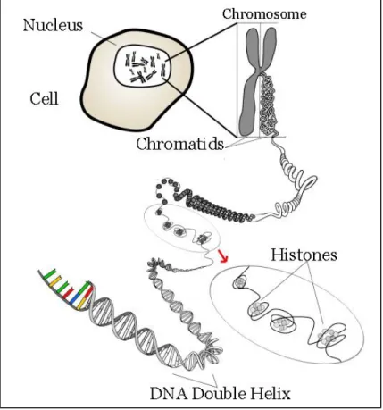
average length of a single DNA strand being ~6 feet, high orders of structure and unique
mechanisms are required to both effectively package the DNA into the nucleus of a single cell
and to ensure that genetic data is protected and maintained. This abundance of DNA is tightly
coiled around histone proteins into structures called chromosomes. The ends of eukaryotic
chromosomes are capped with tandem, G-rich TTAGGG DNA sequences called telomeres.
Repeating telomeric sequences are oriented 5’ to 3’ towards the end of the chromosome leading
to a single stranded 3’ overhang (Figure 1.1).1
Telomeres provide stability and protection from
DNA degradation and chromosomal end fusions to prevent the loss of essential genes and ensure
information stored in DNA is properly replicated during mitosis.2,3
Maintenance of telomere ends is essential for long term survival. The enzyme responsible
for maintaining telomeric DNA is telomerase, a specialized RNA-dependent DNA polymerase.
Telomerase functions to lengthen telomeres protecting them from erosion. It is clear that
telomerase activity is important for the survival of the cell and protection of its genetic data.
However, catalytic telomerase is not expressed in differentiated human cells. Interestingly, it is
found almost ubiquitously in cancer cells, nearly 90% are telomerase positive, making the
2
Cancer remains one of the leading causes of death in the U.S. Because telomerase is
active in the majority of cancer cells, it is an almost universal marker for human cancer.2,3 While standard chemotherapeutic regimens are often associated with toxic side effects, telomerase is a
therapeutic target that provides a means of reducing toxicity to healthy cells due to its specificity
to tumor cells.4
II. Telomere Replication and Telomerase Function
During telomere replication, DNA polymerase duplicates template strands of DNA from
the origin of replication to the chromosome termini. The leading strand is continuously replicated
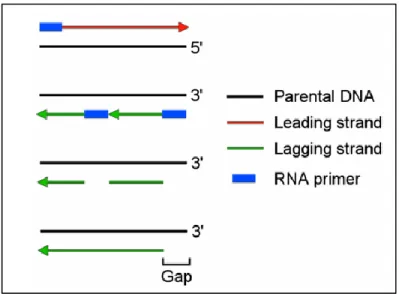
in the 5’ to 3’ direction to the end of the template. In comparison, the lagging strand requires a Figure 1.1 DNA Packaging. Telomeric DNA caps the ends of
3
backstitching mechanism leaving a gap in the lagging strand DNA copy in what is known as “the
end replication problem” (Figure 1.2). This mechanism involves the introduction of RNA
primers at regular intervals. DNA polymerase extends these primers to synthesize DNA
fragments termed Okazaki fragments that make up the lagging strand. The primers are eventually
removed and the gaps left behind are filled in with DNA and the fragments are ligated together.
However, when the last RNA primer is removed DNA polymerase cannot bind to fill in this gap,
leaving a shortened lagging strand. If not corrected, chromosomes shorten with each successive
replication cycle. Telomerase is the human enzyme that counteracts this end replication problem.
Telomerase is a multiunit complex consisting minimally of two essential components, a 451
nucleotide long RNA subunit (hTR) and a protein subunit (hTERT). hTR contains several
functional domains and a templating sequence serving as a template for synthesis of telomeric
repeats while hTERT is a reverse transcriptase. Although hTERT is repressed during
differentiation, hTR is expressed constitutively. Telomerase stabilizes telomere length by adding
telomeric repeat (TTAGGG)n to the 3’ end of chromosomes. To accomplish this, the RNA Figure 1.2 The End Replication Problem. Inefficient
4
subunit hybridizes to the 3’ overhang and the catalytic subunit repetitively reverse transcribes the
template region of the RNA moiety. DNA polymerase then fills in the region on the opposite
strand (Figure 1.3).5
III. Implications of Telomere Shortening
In the absence of telomerase, linear chromosomes progressively shorten in proliferative
cells, resulting in a finite number of possible cell divisions.6 Once the telomeres in a cell reach a critically short length, division ceases and cells enter replicative senescence or mortality stage
(M1).7 Occasionally, cells bypass senescence due to mutations in the p53 tumor suppressor protein. p53 is an essential cell-cycle checkpoint protein that serves to halt cell cycle progression
5
and has an important role in the initiation and maintenance of the senescence state. Cancer
progression involves both the enhancement of cell growth factors and the repression of cancer
suppressors, therefore it is not surprising that when wild type p53 was introduced into SiHa cells
it was shown to negatively regulate hTERT mRNA expression.8 Cells with mutated p53 continue to divide despite short telomeres until they reach a crisis stage (M2) in which massive cell death
occurs. In rare occasions cells are able to escape crisis by up regulating the catalytic hTERT
subunit and activating telomerase, thus leading to immortal cancer cells (Figure 1.4 and 1.5).1,9,10 As mentioned previously, telomerase is expressed in the majority of cancers, however in the
remaining 10% of telomerase negative tumor cancer cells, an alternative recombination
mechanism is employed to maintain telomere length, suggesting the importance of telomere
maintenance in cell survival and immortality.
Figure 1.4 Mechanism of Cellular Immortalization. Cellular senescence occurs after telomere shortening which can lead to the crisis stage. In rare events cells escape crisis and become immortal. Adapted from Shay and Wright,
6
Cancer cells undergo robust proliferation, and telomere maintenance provided by
telomerase is key for their survival. Tumor cells tend to have shorter telomeres than healthy cells
but show no net loss of average telomere length with each successive cell division, again
implying telomere stability is needed for continuous proliferation.2 Because it is up-regulated in tumor cells and is essential for the survival of many cancer cells, telomerase has been explored
as an anticancer drug target. Inhibition of telomerase activity results in a gradual loss of
telomere length, thus causing cancer cells to enter into a crisis stage leading to senescence and/or
cell death.
7 IV. Telomere Capping
Telomere DNA maintenance is important because short telomeres cannot form the proper
protective cap.In addition to targeting cancer cells via telomerase inhibition, one could envision
targeting the DNA binding proteins involved in telomere capping. These proteins contribute to
the stabilization and maturation of the telomerase shelterin complex. The shelterin complex is
composed of several telomere specific proteins including POT1, TIN2, TPP1, Rap1, and
homodimers of TRF1 and TRF2 that bind single and double stranded telomere regions to form a
complex that caps the ends of chromosomes. Shelterin functions to protect telomeres by
establishing the structure of the telomere terminus and controlling synthesis of telomeric DNA
by telomerase. In part, protection appears related to the generation of a telomeric loop structure
known as a t-loop (Figure 1.6).3,12 T-loops are formed when the single stranded 3’ overhang tucks into the duplex part of the telomeric repeat array protecting telomeres from degradation,
8
recombination, and end-joining reactions.2,13 Additionally, it is important to note that shelterin associated proteins are essential in assemblage of additional protein components involved in the
formation of a higher order nucleoprotein complex present at telomeres.1,11
Telomeres can be considered as either capped or uncapped. Capped telomeres are
telomerase inaccessible and allow cell division to proceed. However, telomere shortening
increases the probability of telomeres switching to their uncapped state where elongation by
telomerase can occur. Consequently, the shortest telomeres are preferentially targeted for
elongation allowing maintenance of a steady-state telomere length.14 The uncapped state can lead to irreversible cell cycle arrest and death depending on the functionality of the
telomere/telomerase complex including the loss of active telomerase and prevention of t-loop
formation.11 If left uncapped, the telomere is recognized as DNA damage and the cell cycle arrests. Cell machinery works to remove the damage signal by fixing DNA breaks through
end-to-end fusion of telomeres, leading to telomere instability upon resuming cell division (Figure
1.7).6 These defective chromosomes break during mitosis again activating damage signaling.15 However, it is evident that functional telomeres are capable of avoiding the DNA damage
response as the telomeric complex grants cells the ability to distinguish chromosome ends from
random DNA breaks.6,13,16 It has been shown that telomere repeat binding factor 2 (TRF2) may play a major role in the protection of human chromosome ends by preventing the damage
response. TRF2 coats human telomeres during all stages of the cell cycle by binding directly to
the tandem TTAGGG repeats.13 In vitro data indicates that the addition of TRF2 protein to a linear DNA telomeric model promotes t-loop formation.16 Furthermore, inhibition of TRF2 and POT1, a similar single stranded telomeric DNA binding protein, by dominant negative alleles
9
apoptosis in many cell types as well as a substantial fraction of fusing between telomeres.
Similar results were seen in TRF2 knock out mouse models.17 This is consistent with the model that TRF2 depleted telomeres are perceived as sites of DNA damage. It is thought that telomere
dysfunction may be caused by the loss of the 3’ overhang leading to the failure to reform the
t-loop.13,16 From these observations it is clear that targeting TRF2 would be an appealing approach for drug discovery. However, it is possible this mechanism may be associated with
reduced specificity because proper telomere formation is required in all healthy somatic cells.
Studies using a mouse model suggest that telomere disrupters are highly tolerated in normal cells
for finite periods of time. Like TRF2, POT1 is essential for telomere protection. In one study a
ligand stabilized version of POT1 allowed POT1 to be inhibited transiently and reversibly.
Remarkably, POT1 inhibition resulted in cancer cell death, but normal cells underwent arrest that Figure 1.7 Telomere Capping Two State Model. Telomeres
switch between a capped and uncapped state in response to telomere length and telomerase status. Adapted from Blackburn,
10
was reversed by POT1 reactivation.18 This suggests that small molecule inhibition of telomere binding proteins may provide a high therapeutic index as anti-cancer agents.
V. Research Projects
Several strategies to target telomere maintenance have been explored. These include the
use of antisense oligonucleotides and hammerhead ribozymes to target the mRNA of hTERT,
immunotherapies, and gene therapy approaches.19 Although complex in their modes of action these methods are associated with immediate anti-proliferative effects. Alternatively, there are
antisense oligonucleotides targeting the hTR template region, small molecules telomerase
inhibitors, and G-quadraplex stabilizers.9,15,19 These therapies directly interfere with telomerase enzymatic function, relying on a lag period of telomeric erosion before proliferative effects
occur. As a whole these strategies encompass a broad area of research, differing considerably in
their anti-cancer mechanisms. We will first focus on the latter group whose therapeutic effects
are controlled by a lag period, and then introduce the two approaches presented in this thesis
aiming to eliminate the lag period.
Much progress has been made with telomerase based therapeutics, but as with any
telomerase inhibitor it may require several cell divisions before proliferation effects becomes
apparent. The initial lengths of telomeric DNA in cancer cells are hundreds or thousands of base
pairs long. In the absence of telomerase, proliferating cells lose only 50-200 base pairs of
telomeric DNA with each cell cycle. Once telomerase is inhibited, telomere length progressively
shortens until cellular senescence is attained. As shown in Figure 1.8, traditional
11
inhibitor’s is delayed. Not only does this lag period depend on telomere length but also rate of
erosion.20,21 One study using oligonucleotides complementary to the hTR template region showed once telomerase activity was inhibited cell lines did not display anti-proliferative effects
until thirty days after treatment.22
It is accepted that hTR is highly expressed in all normal tissue whereas hTERT is present
only in immortal cells. Tumor growth requires reactivation of hTERT and its introduction into
telomerase silent cells is sufficient to reactivate telomerase leading to cell immortalization.2,9,22 For example when oligonucleotides are used to inhibit telomerase, upon discontinuing
oligonucleotide treatment telomerase is reactivated and telomeres return to their initial length. Figure 1.8 Therapeutic Lag Period. Telomerase inhibitors rely on telomere
erosion resulting in an anti-proliferative lag period. Adapted from Corey,
12
Therefore, one challenge in designing telomerase targeted treatment strategies is the need to
continuously treat patients during multiple tumor cell population doublings.15 A valuable alternative strategy would be to evade telomerase reactivation by hTERT. Using antisense
oligonucleotides as a therapeutic approach, hTR was selected as the target for my first project
based on our hypothesis that a misassembled holoenzyme may lead to the degradation of hTERT
possibly allowing treatment without continuous dosing. Furthermore, it is widely known that the
classical role of telomerase is to elongate telomeric DNA, but there is emerging evidence that
hTERT is involved in other functions including apoptotic activity.21 Previous studies
demonstrated that overexpression of hTERT renders cells more resistant to apoptosis.23 Likewise, there have been several studies showing that inhibiting hTERT expression can cause an
immediate apoptotic response.24,25,26 One study using modified antisense oligonucleotides to target both hTERT and hTR mRNA in DU145 human prostate cancer cells found that while both
targets caused complete inhibition of telomerase activity, hTERT down regulation showed an
early decline in cell growth and an induction of apoptotic cell death, whereas hTR down
regulation failed to interfere with cell proliferation prior to telomeric DNA erosion.27 Additionally, studies using cell culture and a transgenic mouse model show that hTERT
promotes cellular and organismal survival independent of telomerase activity.28 Expression level of hTERT positively correlated with cell survival after exposure to several lethal stresses,
whereas expression level of hTR had no effect on sensitivity.27 The mechanism behind hTERT induced apoptosis is unclear, however these results are significant because not only would we
like to use antisense oligonucleotides to achieve hTERT degradation and prevent telomerase
function and reactivation, but also consequently induce apoptosis of cancer cells to eliminate the
13
hybridizing oligonucleotides to the hTR structural subunit. This strategy can be used to examine
what happens when telomerase assemblage is disrupted and may serve as a model for the
development of future therapeutics, for example small molecule telomerase inhibitors.
Direct telomerase inhibition through oligonucleotide hybridization is a multifaceted
therapeutic approach with many considerations. Nonetheless it has shown to be promising as an
anticancer strategy. GRN163L is a lipid-conjugated N3′→P5′ thio-phosphoramidate
oligonucleotide that blocks the template region of telomerase. Various studies have demonstrated
that GRN163L treatment leads to significantly reduced telomerase activity, promoting telomere
loss and apoptosis in several cancer lines.29,30 The high potency and specificity of GRN163L as well as modifications to improve its bioavailability has led to its testing in ongoing clinical trials.
Although not effective within hours or days as most primary anti-proliferative cancer therapies,
telomerase inhibition may weaken cells making them more susceptible to other anti-proliferative
agents, suggesting that use in combination treatments may be an effective route.4,20 Again, we would like to discover a novel approach, eliminating the lag period and need for combination
therapy, and instead offer telomerase inhibition as a primary anti-cancer treatment.
In addition to telomerase inhibition induced by oligonucleotide hybridization, disruption
of telomere maintenance can be achieved through the targeting of DNA binding proteins
involved in the regulation of telomeres. As mentioned previously, TRF2 is critical to the
protection of telomeric DNA through its function in telomere capping and elimination of DNA
damage signals. Impaired telomere capping leads to cell cycle arrest, meaning that by disrupting
the function of TRF2 cellular senescence can be obtained without relying on telomere
shortening. This is another approach that eliminates the delay in anti-proliferative effects that is
14
Current treatments targeting telomerase activity exhibit a lag period and will have to be
used in combination therapies. As an alternative approach to remove the lag period, my thesis
research involves inducing the dysfunction of telomeres, specifically examining how both
telomere length and telomere capping status contributes to telomere function. The first part of
my project involves oligonucleotide based inhibition of telomerase activity using a novel
approach to inhibit telomerase that may block noncanonical hTERT activity and induce
apoptosis. My second project involves the disruption of the essential capping protein TRF2 using
small molecule inhibitors to test the hypothesis that the uncapping of telomeres leads to rapid
15
CHAPTER 2: TELOMERASE INHIBITION THROUGH OLIGONUCLEOTIDE TRANSFECTION
I. Introduction
Transfection, the process of introducing nucleic acids into mammalian cells, is commonly
used in drug discovery to study the effects of a modified biological activity. In this project,
oligonucleotides were introduced into PC-3 cancer cells to investigate the inhibition of
telomerase by disrupting specific protein-RNA interactions. Oligonucleotides are short, single
stranded modified DNA or RNA molecules that can be designed to be complementary to a
specific target allowing for hybridization and inhibition of a desired biological function.
Antisense oligonucleotides are generally used to prevent protein translation by targeting mRNA.
In this project, a RNA antisense oligonucleotide with modified bases was used to inhibit
telomerase, specifically through hybridization with the structural hTR subunit to sterically
prevent proper holoenzyme association with hTERT. A major challenge in this therapeutic
approach is getting oligonucleotides into the cell and to telomerase without being degraded.2 Fortunately, cationic lipid transporters can be used to facilitate uptake. Cationic lipids are
positively charged having the ability to interact with negatively charged DNA and cell
membranes. Lipid and oligonucleotides spontaneously complex during an incubation period and
then fuse the cell membrane to deposit the oligonucleotide inside the cell.31 There are several advantages to using oligonucleotides for hTR targeting. For one, the necessity of hTR for
16
oligonucleotides are commercially available and because they are complementary to the known
hTR sequence they are both highly specific and easily designed.20
In standard studies, to conclude that oligonucleotides are inhibiting telomerase through
complementary binding rather than off target effects, one must consider the following; due to the
lag period, inhibitors should reduce telomerase activity but should not affect cell growth rates
initially. Hence, cells should eventually undergo growth arrest and apoptosis but again this time
is dependent on initial telomere length.22 However with our approach, holoenzyme misassembly and hTERT degradation could lead to immediate cellular consequences by interruption of an
anti-apoptotic pathway.
II. P6.1 Loop Target and hTRas012 Development
The specific target sequence I focused on was the P6.1 loop of telomerase hTR, a RNA
sequence critical for telomerase subunit assembly (Figure 2.1A).32 By targeting hTR/hTERT binding, proper assemblage of the active holoenzyme complex is prevented causing inhibition of
enzyme function. As mentioned previously a misassembled holoenzyme may lead to the
degradation of hTERT. This is significant because this subunit correlates to activation of
telomerase dependent telomere lengthening and cancer cell progression. It is also important to
note that although hTR is highly expressed in normal tissue, there is no evidence that it has any
17
Previous work in our laboratory showed that targeting certain hTR regions using
antisense oligonucleotides results in decreased telomerase activity(Table 2.1).35 To determine inhibitory effects on the enzyme, a telomerase assemblage assay was employed in which hTR
and hTERT were assembled in reticulocyte lysates. Telomerase activity was measured using a
direct telomerase assay. In the regions targeted, the greatest inhibitory effect was observed in the
pseudoknot (P3/P1) and CR4-CR5 (P6.1) domains demonstrating that these areas are essential
for telomerase activity and subunit binding. Accordingly, both of these regions are conserved in
vertebrate telomerase RNAs, and neither are exposed after holoenzyme assembly.36 In a telomerase assemblage assay oligonucleotides hTRas009 and hTRas010 targeting the P3/P1
18
pairing region and the P6.1 loop within the CR4-CR5 domain respectively, inhibited telomerase
with IC50s in the nanomolar range . Co-immunoprecipitation assays confirmed the
oligonucleotides ability to prevent binding of targeted hTR regions to hTERT. However no
significant inhibition was seen when added to preassembled telomerase in a direct telomerase
assay. Furthermore, full length hTR still had the ability to bind hTERT in the presence of
oligonucleotides suggesting binding of hTRas009 and hTRas010 prevents proper holoenzyme
assembly and sequesters telomerase subunits in an inactive state. Preference for which
oligonucleotide to further examine was influenced by the observation that hTRas010 showed a
greater ability to prevent binding between its targeted region and hTERT than hTRas009 (~92%
compared to ~50%). In accordance with these results previous studies had demonstrated that the
presence of the P6.1 loop was necessary for interaction between the CR4-CR5 region and
hTERT as well as enzymatic activity of the mammalian telomerase complex.32 Based on this
Name Sequence hTR region targeted
nucleotide targeted
% activity
hTRas001 5'-ATGGCAAGTCCGAATCGATCGT-3' none N/A 804
hTRas002 5'-TAGGGTTAGACAA-3' template (CR1) 42-54 97
hTRas003 5'-AAAGTCAGCGAGAAAAACAGCG-3'
pseudoknot domain
(CR2/CR3) 94-115 97
hTRas004 5'-AACGGGCCAGCAGCTGACATTT-3' P3/P1 pairing region 174-195 37
hTRas005 5'-TGGGTGCCTCCGGAGAAGCCCC-3' L6 loop 268-289 100
hTRas006 5'-CGGCTGACAGAGCCCAACTCTT-3' CR4-CR5 domain 301-322 54
hTRas007 5'-GCCTGAAAGGCCTGAACCTCGC-3'
hypervariable paired
region 343-364 115
hTRas008 5'-ACAGCTCAGGGAATCGCGCCGC-3' CR7 domain 397-418 74
hTRas009 5'-AACGGGCCAGCAGCUGACAUUU-3' P3/P1 pairing region 174-195 12 hTRas0010 5'-CGGCUGACAGAGCCCAACUCUU-3' CR4-CR5 domain 301-322 18
Table 2.1 Summary of Inhibition Data with hTR-Targeted Oligonucleotides. DNA oligonucleotides were added to hTERT and hTR prior to assemblage. Telomerase activity was determined using a direct telomerase assay. “% Activity” indicates the amount of residual telomerase activity at a 1 uM concentration compared to the primer-only control.hTRas009 and hTRas010 are 2-O-methyl oligonucleotides, and underlined
19
consideration, oligonucleotide hTRas012 was designed by lab members with the capability of
interacting with the P6.1 stem loop, specifically targeting nucleotides 298-310 (Figure 2.1B).
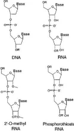
The hTRas012 sequence is 5’- CCCAACTCTTCGC-3’ with all RNA bases 2’-O-methyl
modified and underlined bases indicating phosphorothioate linkages (Figure 2.2). Although DNA
is the native substrate of telomerase, the 2’-O methyl RNA modifications are used to increase
binding affinity to prevent nonspecific interactions, while phosphorothioate linkages enhance
stability against nuclease digestion.20 Fully modified phosphorothioate linkages have been shown to have poor sequence selectivity possibly due to nonspecific protein interactions. Therefore, 2’-
Figure 2.2 Oligonucleotide Backbone Modifications.
20
O- methyl modified RNA oligomers with terminal phosphorothioate linkages were selected to
test this inhibition platform. These modifications also increase the serum half-life of the
oligonucleotide, increasing its pharmacokinetic properties. Additionally, this design is both
chemically and sterically similar to DNA allowing for favorable electrostatic contacts with the
protein component of telomerase.37
As expected hTRas012 showed complete telomerase inhibition with IC50 ranges in the low nM range when tested in a telomerase assemblage assay (~21 nM). Results from a
scintillation proximity assay (SPA), performed to determine if the oligonucleotide prevented
interaction between the protein and RNA subunits, showed that the addition of hTRas012
prevented CR4-CR5/hTERT interaction in a concentration dependent manner yielding an IC50 value of 96 nM. Because hTRas012 successfully inhibited telomerase in our previous studies it
was chosen for further investigation in biochemical inhibition studies using cultured cells.
Oligonucleotide transfection of hTRas012 was performed in PC-3 human cancer cells. A
scrambled oligonucleotide with the sequence 5’-AGACUACUGAACU-3’ was used as a
negative control for nonspecific effects. Again, both were 2’-O-methyl modified with underlined
bases indicating phosphorothioate linkages. An Oligofectamine only negative control was also
evaluated. Telomerase activity in treated cells was detected using the Telomeric Repeat
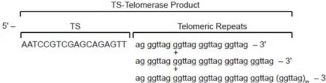
Amplification Protocol (TRAP) assay. TRAP is a two-step PCR-based telomerase detection
method (Figure 2.3). In the first step of the assay lysate telomerase adds telomeric repeats onto
the 3’ end of substrate oligonucleotide (TS). This reaction mixture contains the reaction buffer
provided with the kit, dNTPs, TS primer, and the desired cell lysate sample. In the second step,
extended products are amplified by PCR using a primer mixture to produce a ladder of products
21
polymerase. There are several control samples included in the assay. The positive control is a
telomerase active pellet supplied in the kit. There are two negative controls including a lysis only
sample to monitor contamination and a negative telomerase heat treated control corresponding to
each lysate sample. Additionally, there is an internal control for each sample producing a 36 bp
band in every lane to ensure no Taq polymerase inhibitors are present in the sample and to help
monitor the amplification process.
III. Materials and Methods
PC-3 human cancer cells were obtained from UNC Lineberger Comprehensive Cancer
Center. Cells were maintained in F-12K media with 10% FBS and a 1% concoction of penicillin,
22
oligonucleotides were purchased from Integrated DNA Technologies. For all transfections cells
were seeded in 10% FBS, antibiotic free F-12K media in 12 well plates at 78,000 cells per well
in order to obtain 40% confluence 24 hours after seeding. The transfection protocol supplied
with the cationic lipid carrier Oligofectamine, provided by Invitrogen, was followed for all
transfections. Twenty four hours after seeding, oligonucleotides were incubated with
Oligofectamine in Opti-Mem media before adding to cells. Cells were washed and then
transfected with oligonucleotides in serum free, antibiotic free F12-K media at a total volume of
500 L for 4 hours before either adding 30% FBS, antibiotic free F12-K media to a final FBS
concentration of 10% or alternatively removing transfection media and refreshing cells with 10%
FBS, antibiotic free F12-K media. For longer transfections periods, when cells reached 100%
confluence, populations were split to 40% confluence 2 hours prior to the next transfection. After
the appropriate incubation period, cells were harvested by trypsinization and washed twice with
PBS before spinning into pellets at 13,000 for 3 min at 4°C. Pellets were lysed using a CHAPS
cell lysis buffer with added RNAase inhibitor at 200 units/mL. Lysate protein concentration was
determined using a Bradford assay before separating lysates into 10 L aliquots and flash
freezing for storage at -80°C. A TRAP assay was employed to assess telomerase activity.
Components for TRAP were provided by the TRAPeze telomerase detection kits purchased from
Millipore. A Master mix was prepared by mixing 1.25 L of 10X TRAP reaction buffer, 0.25 L
50X dNTP, 50 ng/L TS primer, and 8.50 L PCR grade water per sample. Each sample
required 10 L of Master mix and 3 L of lysate at a protein concentration of 200 ng/L.
Samples were incubated at 30°C for 30 minutes and 95°C for 5 minutes. Next, 12 L of a PCR
mix was added containing 0.50 L primer mix, 10.40 L PCR grade water, 1.25 L Taq buffer,
23
was 25 L. Samples underwent 33 PCR cycles at 94°C for 30 seconds, 60°C for 30 seconds, and
72°C for 1 minute. Both the Master mix and PCR mix as well as controls samples were prepared
before preparing cell lysate samples. Here we used the gel based non-isotopic detection method
with a 12.5% non-denaturing polyacrylamide gel (10% APS, 1% TEMED). TRAP dye
composed of glycerol, 1.25% bromophenol blue, 1.25% xylene cyanol, and 0.05 M EDTA was
added to PCR samples before gel loading and gels were run in 0.5X TBE buffer at 400 V for 45
minutes. Gels were stained with SYBR Green DNA dye and visualized using a Storm scanner at
450 nM. ImageQuant software was used to quantify the amount of telomeric product obtained
from each cell sample lysate. Values were calculated by quantifying the amount of product in the
non-heat treated sample (x), the corresponding heat treated sample (xo), the lysis buffer only control (c), and the internal standard (cr). Relative activity was calculated with the equation (x- xo / c) / (cr) and then graphed to compare the effects of different oligonucleotide concentrations.
IV. Results
To study the effect of inhibiting telomerase by blocking the interaction between P6.1 of
hTR and hTERT, I determined conditions for transfecting PC-3 cells with hTRas012. It was
expected that an increase in oligonucleotide concentration would reflect an increase in
telomerase inhibition and less telomeric product. As mentioned previously, hTRas012 does not
inhibit preassembled hTR-hTERT holoenzyme because the CR4-CR5 regions are not exposed
after assemblage, meaning a telomerase turnover must occur before binding of the
oligonucleotide to hTR. However, cancer cells are rapidly dividing and therefore rapidly
transcribing hTR for the generation of more telomerase thus providing an opportunity for
24
needed to ensure binding, transfected cells were allowed to incubate for different time intervals
before harvesting.
In initial experiments cells were transfected with increasing oligonucleotide
concentrations and incubated for 24 hours before harvesting and assessing telomerase activity.
Four hours after transfection 30% FBS, antibiotic free F-12K media was added to transfection
media to reach a serum concentration of 10%. When assessed for activity a concentration
dependent inhibition of telomerase activity was observed (Figures 2.4 and 2.5). Repeating the
transfection with increased concentrations (200 nm- 1 M) reflected similar results, however
there was only about 45% inhibition associated with the maximum concentration of 1 M.
In an attempt to completely inhibit telomerase activity, 48 hour single transfections and
48 hour double transfections (2 transfections 24 hours apart) were performed with
oligonucleotide concentrations ranging from 200 nM-1 M. When preparing for harvesting 48
hours later the cells were clumped and floating including those transfected with the negative
scrambled control. Transfection agents can cause toxicity to cells when introduced at too low of
a cell confluency. To determine if toxicity was the problem, cell confluence was varied (30%,
50%, 80%) before transfection while keeping the oligonucleotide concentration the same (200
nM). Additionally, while keeping confluency at 40%, the oligonucleotide concentration was
decreased (20-200 nM). After 24 hours all the cells were still alive, however cells were already
completely confluent in all the wells except the initial 30% well. After 48 hours all the cells had
died. It was concluded that the transfection conditions were toxic to the cells and media must be
removed and refreshed after each 4 hour transfection period. Forty-eight hour transfections were
25
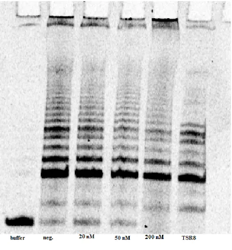
Figure 2.5 hTRas012 Inhibition in 24 hr. Transfection. Telomerase activity based on
quantification of telomeric product from the
TRAPeze assay gel in Figure 2.4.
Figure 2.4 TRAP Assay Gel for 24 hr. Transfection. A dose dependent telomerase inhibition is observed (20 nM-200nM). As hTRas012
oligonucleotide
26
with fresh 10% FBS, antibiotic free F-12k media. Cells survived but results showed inconsistent
telomerase activity with no dose dependent inhibition. The experiment was repeated, but this
time after 24 hours cells were split to achieve 40% confluence before the second transfection.
Still adequate inhibition of telomerase activity was not observed in either the double or single 48
hour transfections and additionally there was no dose dependent decrease in the 48 hour single
transfection assay. However, the 48 hour double transfection did show a concentration dependent
decrease in activity (Fig 2.6).
Due to these results, a 72 hour (3 transfections 24 hours apart) incubation period of cells
post initial transfection was attempted. Again, cells were split prior to each transfection to
produce 40% confluence. After performing the TRAP assay on samples, results continued to be Figure 2.6 hTRas012 Inhibition in 48 hr. x 2 Transfection.
27
inconsistent, no dose dependent results were observed (Figure 2.7A). Transfections were
repeated but this time without replacing media after the 4 hour transfection period and instead
28
adding 30% FBS to a total of 10% serum concentration as done in initial experiments. This time
cells were split prior to each transfection and survived the 72 hour incubation period without
toxicity. Moreover a dose dependent inhibition was observed with 45% inhibition of the negative
control in the maximum dose (Figure 2.7B). Unfortunately, in all the experiments telomerase
inhibition never reached more than about 40% of negative controls.
V. Discussion
Twenty four hour incubations consistently showed an hTRas012 dose dependent decrease
in telomeric product, however complete inhibition of telomerase was never achieved. It is
questionable whether discrepancies seen in longer than 24 hour experiments were due to
complications arising from altered experimental conditions including longer incubation time
intervals, cell splitting, and refreshment of transfection media after 4 hours. Despite the
successful 48 hour double transfection, results from the 72 hour experiments suggest
inconsistencies arouse from refreshing transfection media. Still these results are promising and
suggest modifications to the current method may provide more satisfactory results. Additional
studies may include testing higher concentrations of hTRas012 oligonucleotide or increasing
transfection incubation times, however further optimization of transfection conditions is likely a
beneficial approach to improving experimental results. There are several barriers to efficient
transfection including the formation of oligonucleotide/cationic lipid complexes, entry of
complexes into cells, oligonucleotide disassociation, and transport to the nucleus.39
Consequently, the ratio of cationic lipid reagent to DNA concentration is a key transfection
parameter and special attention should be focused on determining ideal proportions. Although it
29
a positive control is beneficial in optimizing transfection efficiency. There are several controls
commercially available to help monitor whether oligonucleotides are entering the nucleus and to
access toxicity of transfection conditions on cells. These methods commonly involve
fluorescently tagged oligonucleotides and stains for cellular viability.
Although efficient transfection is important to the success of oligonucleotide dependent
telomerase inhibition, it is still possible a major drawback to this project involved inconsistencies
associated with the TRAP assay. Results were difficult to reproduce and variable amongst
duplicate experimental samples. Although one cannot rule out discrepancies in methodology,
there are several limitations to TRAPeze involving factors affecting quantitative determination.40 It is a multi-step assay requiring several post PCR steps, thus allowing more opportunities for the
introduction of error and contamination. In one study ten parallel TRAP reactions were
performed using the TRAPeze kit and resulting telomeric product was quantified. A coefficient
of variation from experiments was calculated to be 12%.40 Distributions with a coefficient of variation greater than one are considered to be of high variance, suggesting the TRAPeze assay is
associated with high variability. Additionally the assay has a linear range of 250-5000 cells and
is sensitive to sample concentration. The TRAP protocol suggests sample protein concentrations
ranging from 10-750 ng/uL, but where within in this range is difficult to predict and once
established for one experiment it may not produce optimal results for similar experiments run
under the same conditions. Additionally, too low or too high protein concentrations can lead to
PCR artifacts. Another common problem that existed throughout this project was negative
controls containing telomerase positive cells resulted in low telomerase activity. However this
data is questionable because as the protocol states, positive telomerase activity sometimes cannot
30
maintain corresponding initial concentrations when comparing dose dependent samples. Finally,
samples with inhibited telomerase may show an enhanced amount of telomerase activity because
there are too many PCR cycles. In this case dramatically inhibited samples show telomeric
product due to saturation of the PCR.
Again, optimization of the current method may allow for improved results. Attention
should first be focused on establishing optimal transfection conditions to improve transfection
efficiency. Although TRAPeze is a widely used protocol for measuring telomerase activity, it
may be beneficial to test samples using alternative quantification methods. For example, there
are several modifications to the standard TRAP protocol including the incorporation of
fluorescently labeled primers and real time PCR methods. There are also direct telomerase assays
using radiolabeled dNTPs or primers that omit the PCR amplification steps, but these require
large sample sizes to achieve enough telomerase activity.41
Despite the challenges associated with this project, the dose dependent telomerase
inhibition results are promising. Further efforts may uncover exciting knowledge contributing to
31
CHAPTER 3: A FLOURESCENCE POLARIZATION ASSAY TO IDENTIFY TRF2 SMALL MOLECULE INHIBITORS
I. Introduction
In addition to telomerase inhibition, disruption of telomere maintenance can also be
achieved through targeting of DNA binding proteins involved in the formation of the telomere
shelterin complex. As discussed above, TRF2 is critical to the protection of telomeric DNA
through its function in telomere capping and inhibition of DNA damage signals. Disrupting the
function of TRF2 leads to cellular senescence or death without telomere shortening, eliminating
the lag period that exists with telomerase inhibition. There is also promising evidence that
targeting telomere binding proteins may be well tolerated by normal healthy cells. The goal of
this project was to develop a high throughput screen to identify small molecule inhibitors of
TRF2 by direct binding to the TRF2 protein.
High throughput screening (HTS) is a method used in drug discovery for rapid
identification of active compounds. It requires miniaturization and automation of bioassays to
simultaneously test libraries of drug-like compounds. Typically assays are carried out on
microplates and are assessed in a relatively short time period, however before large screenings
can occur assays need to first be designed and optimized. Here we utilize fluorescence
polarization (FP), a technique providing fast and accurate quantitative measurements, for the
identification of TRF2 binding small molecules. Polarized light waves are characterized as
32
light emitted from fluorescent molecules that can be characterized based on their fixed light
excitation and emission properties. In this technique, a biological sample labelled with
fluorophore is illuminated with linearly polarized u.v.-visible light at the wavelength of
fluorophore absorption. The fluorophore absorbs a photon, briefly exciting it to a higher energy
state before emitting it at a specific wavelength. The light photon emission passes through a
rotatable linearly polarizing filter before detection (Figure 3.1). Instead of detecting the degree of
polarization, change in fluorescence intensity is used to indirectly measure polarization. Change
in fluorescence intensity is described by a ratio of two measurements, the emission intensity
parallel and perpendicular to the plane of linearly polarized illumination light.28 The polarization value, being the ratio of the two fluorescence intensities, is a dimensionless number expressed in
millipolarization units (mP).
33
This concept is illustrated in Figure 3.2. Light has an electric field. When fluorophore
adsorption vectors align parallel with the electric vector of linearly polarized excitation light they
are selectively excited, whereas those perpendicular elude excitation. Small rapidly rotating
molecules orient randomly during emission, resulting in low fluorescence. Larger, slowly
rotating molecules align in the same plane as the excitation energy, resulting in higher
fluorescence.43,44
I attempted to optimize a homogenous FP assay to identify TRF2 inhibitors. In this assay
Cy5 fluorescent dye was linked to double stranded telomeric DNA (TTAGGG)3 to observe its interaction with TRF2. Cy5 labeled DNA rotates quickly resulting in decreased polarization, but
after incubation with TRF2 binding occurs resulting in a larger complex and slower rotation of
the fluorescent dye, therefore increasing FP. The low and high fluorescence measurements were
used as references for unbound TRF2 and full complex formation in experiments. The idea was
that when a TRF2 binding small molecule was introduced, displacement of TRF2 from DNA or Figure 3.2 Physical Basis of Fluorescence Polarization. Larger rotating fluorophore
34
inhibition of DNA binding would result in rapidly rotating Cy5 fluorophores, decreasing
fluorescence measurements. In theory these small molecules would be acting to inhibit TRF2
function by preventing binding with DNA. Without telomeric DNA binding by TRF2, telomere
capping cannot occur leading to telomere dysfunction. Using HTS we aimed to identify small
molecules that lead to a decrease in FP of Cy5 using an automated plate reader. In preliminary
assay experiments we used an excess of unlabeled telomeric double stranded DNA (dsDNA) to
behave as a TRF2 binding small molecule by displacing TRF2 from Cy5 labelled DNA.
II. Materials and Methods
Active TRF2 protein was obtained from Brian Bower of the Griffith laboratory at UNC-
Lineberger Cancer Center. The protein was provided in 15 g/ 25 L aliquots at a concentration
of 9.97 M. The TRF2 oligonucleotides (TTAGGGTTAGGGTTAGGG) were ordered from
Integrated DNA Technologies. The G rich strands were covalently labelled on their 5’ end with
Cy5 fluorophore. dsDNA was made from annealing Cy5 labelled G rich strands with the
unlabeled C rich strands.
Electrophoretic Mobility Shift Assay
A master mix composed of binding buffer (50 mM HEPES, 50 mM KCl, 1 mM MgCl2,
0.1 mM EDTA), 1.8 mg/mL BSA, and 3.33 nM Cy5 was prepared. TRF2 protein (0-3600 ng)
was titrated into 16 L master mix samples and water was added to a final volume of 28 L.
Samples were left to incubate in the dark for 30 minutes at room temperature. Before loading
samples, 7.5% polyacrylamide gels (30% APS, 1% TEMED) were pre-run for 30 minutes at 100
35
pre-run gels and run in 0.5X TBE buffer at 220 V for 25 minutes in a dark cold room (8°C). Gels
were viewed using a Typhoon scanner at 650 nm.
FP Assays
All experiments were run in triplicates. Background measurements containing binding
buffer (50 mM HEPES, 50 mM KCl, 1 mM MgCl2, 0.1 mM EDTA) and BSA only were
subtracted from fluorescence measurements. Fifteen microliter samples were prepared in black
384-well polypropylene plates using binding buffer, 1.8 mg/mL BSA and varying concentrations
of Cy5 DNA and TRF2 protein. The TRF2 titration used 5 nM Cy5 DNA and TRF2
concentrations ranging from 0.05 nM to 1.665 M. Two displacement assays were employed
using 5 nM Cy5 DNA, 300 nM TRF2, and unlabeled DNA with concentrations ranging from
0.76 nM to 25 M. In the first assay, Cy5 DNA and TRF2 were premixed and incubated for 20
minutes before adding unlabeled DNA. In the second assay, TRF2 and unlabeled DNA were
premixed and incubated for 20 minutes before adding Cy5 DNA. Plates were spun down and left
to incubate in the dark for 60 minutes at room temperature. Plates were read using an EnVision
multilabel plate reader with excitation at 650 nm and emission at 670 nm.
Z factor Calculation
To calculate the Z factor, 5 nM Cy5 DNA and 300 nM TRF2 were premixed with
binding buffer and 1.8 mg/mL BSA and incubated for 20 minutes in black 384-well
polypropylene plates. Unlabeled DNA was used as a positive control and added to wells to a
final concentration of 500 M. DMSO was used as a negative control. Plates were spun down
and left to incubate in the dark for 60 minutes at room temperature. Plates were read using an
36 III. Results
An electrophoretic mobility shift assay (EMSA) was used to detect sequence specific
DNA binding properties of TRF2. This method takes advantage of the concept that free DNA
will travel farther through a gel than DNA bound to protein because larger complexes experience
a hindrance in mobility, thus resulting in slower gel migration. Distinct bands visible in the gel
correspond to protein-DNA complexes providing information on how far the DNA traveled and
more importantly its extent of binding to a compound of interest. Here the assay was performed
to ensure proper binding between TRF2 protein and prepared Cy5 labeled DNA. Increasing
concentrations of TRF2 (0-3600 ng) were titrated into Cy5 DNA and left to incubate before
37
employing PAGE. While free Cy5 DNA travels farthest down the gel, a major shift upward in
Cy5 location is observed at about 1200 ng of TRF2, confirming protein-DNA binding (Figure
3.3).
Once DNA binding was verified by EMSA, a FP assay was designed using an Envision
multilabel plate reader for detection. After determining the lowest concentration of Cy5 DNA
sufficient enough to give a consistent, readable signal, a TRF2 titration experiment was
performed with 5 nM Cy5 DNA and varying amounts of TRF2 (0.05 nM- 1.665 uM). Here we
aimed to verify results from the EMSA. The resulting TRF2-DNA binding curve again
confirmed assay components were behaving properly (Figure 3.4). The Kd of TRF2-DNA binding in our assay was calculated to be 182 nM. The literature value Kd was found to be 180 nM using surface plasma resonance.47 An EC80 of 300 nM was used for TRF2 concentrations in displacement experiments to ensure that a significant amount of TRF2 remained bound to DNA
without saturating it. In the displacement assay an excess of unlabeled telomeric DNA was
titrated into Cy5 DNA-TRF2 complexes to displace Cy5 DNA from protein. Two experiments
were performed to determine order of addition effects (Figure 3.5). In Experiment #1, Cy5 DNA
and TRF2 were premixed and incubated before the addition of unlabeled DNA, whereas in
Experiment #2 TRF2 and unlabeled DNA were premixed and incubated before Cy5 DNA
addition. As concentrations of unlabeled DNA increase, the FP signal should decrease because
Cy5 labelled DNA becomes increasingly unbound and as a consequence rotates at a quicker
speed. In theory, an inhibitor screen should produce similar results as unlabeled TRF2 binding
DNA. The displacement assay data from Experiment #1 produced an acceptable curve however
38
Figure 3.4 TRF2 titration experiment. Cy5 DNA binding to
TRF2 at varying concentrations
39
To determine if the assay could be used in HTS, a Z factor was calculated. The Z factor is
a dimensionless statistical score used in HTS analysis to evaluate the quality and efficacy of an
assay.45 It helps decide if the screen has the ability to accurately predict if a compound is active. Calculations comprise testing positive and negative controls in replicate, finding the average and
standard deviation for each control and plugging these values into the equation 1-((3σ- + 3σ+)/(l
μ- + μ+ l)) . The unlabeled TRF2 DNA was used as the positive control and a random 21
nucleotide long primer was used as the negative control. Unexpectedly the random primer
resulted in about 50% binding to TRF2 DNA as compared to the unlabeled positive control
DNA. The experiment was repeated with DMSO as the negative control (Figure 3.6). An
excellent Z factor is in the range of 0.5-1 and a marginal assay is between 0 and 0.5. The Z factor
175 185 195 205 215 225 235 245 255 265
0 20 40 60 80 100
mP
Sample well #
dsDNA
DMSO
40
was calculated to be 0.12. Based on this calculation, it was concluded that assay efficacy was
inadequate and further optimization would be required for HTS.
IV. Discussion
There are various limitations to using FP approaches, however the low dynamic range
proved to be a major reason for the failure of this assay. There are many possible causes for a
low dynamic range. First, FP requires a large change in molecular volume for maximum change
in mP value. Our TRF2 protein has molecular weight of 60.26 kDa making it a 120.52 kDa
homodimer, therefore it is unlikely our TRF2 protein was not large enough to yield a substantial
mP change. Also, FP measurements increase with molecular weight of the attached compound
but tend to plateau dependent on the fluorescent lifetime of the fluorophore. This means that the
fluorescent lifetime of the excited fluorophore must be longer than the rotational correlation time
of the bound DNA molecule.42 This allows the free Cy5 DNA time to randomize its orientation during the process of emission for a depolarized effect and lower mP readings. Cy5 has a
relatively short lifetime of about 1 nanosecond which may not be long enough to allow
randomization of the labeled DNA. In order to fix this problem it may be advantageous to test a
fluorophore with a longer lifetime like Flourescien or Alexa Flour 488 both with lifetimes of
about 4 nanoseconds, or try using a smaller DNA fragment. However, DNA must be long
enough to ensure attached Cy5 does not affect its binding affinity to TRF2. Additionally
sometimes DNA will autofluoresce causing significant distortions in the background signal and a
41
meaning there was no autofluorescence by DNA. Again, there are various other causes for a low
dynamic range. This assay will need to be optimized before moving to HTS. Factors to consider
include fluorophore selection, such as lifetime, stability, and concentration, linker length and
42
CHAPTER 4: CLOSING REMARKS
In this thesis two strategies to advance telomere disrupting therapeutics were explored.
While both had promising findings neither project produced completely ideal results and further
efforts should be focused on improving experimental methods. The use of antisense
oligonucleotides to target the hTR subunit of telomerase showed a decrease in enzymatic activity
suggesting binding was indeed occurring. However, complete inhibition of telomerase was never
achieved. While optimizing transfection conditions would likely be a valuable endeavor to
improve experimental results, it is possible that complete inhibition was difficult to achieve
because hTRas012 is unable to bind preassembled telomerase present in initial cancer cell
populations. A telomerase complex that is stable with slow turnover will have active telomerase
that exists throughout multiple hTRas012 dosings. However, telomerase is thought to have a
half-life of twenty four hours meaning longer transfections should show complete inhibition of
activity.48 Alternatively, the P6.1 loop of telomerase may not be easily accessible to hTRas012 even prior to holoenzyme assembly. Conserved regions of hTR are predicted to be recognition
sites for hTR-associated proteins.9 These binding proteins may block interactions between htRas012 and the hTR P6.1 loop. Fortunately, a variety of oligonucleotides with chemically
modified backbones have been designed to enhance the therapeutic potential of antisense
strategies. For example, peptide nucleic acid (PNA) modifications have been shown to be potent
43
considerably stable PNA-RNA duplexes in vitro. Therefore it may be favorable to test the
inhibitory ability of oligonucleotides with PNA backbone modifications.
Additionally, the FP assay used to observe TRF2 and telomeric DNA interaction
produced an acceptable binding curve and confirmed components in the assay were behaving
properly. Unfortunately the low dynamic range of the assay made it impractical for use in high
throughput screening. Again, optimizing assay conditions may increase the dynamic range of the
assay, however it may be more desirable to try a different approach. For example, there are many
alternative methods for studying protein binding interactions including thermal shift assays and
AlphaScreens.49,50
Thermal shift assays use thermal-denaturation to evaluate the stability of a target protein
based on the knowledge that ligands induce conformational changes in proteins, providing
enhanced stability upon binding. Observations of ligand-dependent changes in the melting
transition temperatures of ligand-protein complexes relative to the uncomplexed protein are used
to evaluate ligand binding affinity. In this technique, fluorescent dyes binding hydrophobic
regions of the target protein are used to monitor protein denaturation. As the protein gets
denatured in solution, hydrophobic surfaces become increasingly exposed activating fluorescent
dyes. Using this approach, TRF2 and fluorescent dye are dispensed into microplate wells
followed by the addition of test compounds to the solution. Plates are then heated and thermal
melting of TRF2 is monitored by detecting changes in fluorescence. Addition of a stabilizing
small molecule should shift the midpoint of the melting curves toward a higher temperature. By
comparing the thermal melting curves of TRF2 in the presence of a small molecule with TRF2
alone and TRF2 bound to dsDNA, we can determine the extent of small molecule binding to
44
Alternatively AlphaScreen, a bead based proximity assay, can be used to monitor
protein-ligand binding using a histidine/nickel chelate detection kit. In this technique streptavidin coated
donor beads bind biotin labeled telomeric DNA, while nickel chelated acceptor beads are used to
immobilize histidine tagged TRF2. Donor beads contain a photosensitizer phylthalocyanine,
which upon illumination converts ambient oxygen to an excited and reactive singlet oxygen
having a 4 μsec half-life. If the acceptor bead is within 200 nm of the donor bead, energy is
transferred from the singlet oxygen to thioxene derivatives within the acceptor bead producing
detectable luminescent/fluorescent light. If the donor bead is not in proximity of the acceptor
bead then no signal is produced. When TRF2 and substrate DNA bind, a resulting signal is
detected. However when a small molecule binds TRF2, interfering with DNA binding, a
decrease in signal is observed.
While a FP assay alone could not accurately predict inhibitors of TRF2 there are
numerous other applications for this technique focusing on protein-ligand interactions, with only
two presented here. Both of these methods allow for the use of a fully automated, miniaturized
fluorescence based assay for HTS of small molecule libraries. Similarly, there are countless
variations to oligonucleotide transfection methods that may be beneficial to consider for this
study. Further exploration in these areas will not only advance knowledge of mechanisms
behind telomere maintenance but assist in the identification and development of future telomere
45
REFERENCES
1. Bailey, S. and Murnane, J. Telomeres, chromosome instability, and cancer. Nucleic Acids Research 34, 2408-2417 (2006).
2. Shay, JW. and Wright, WE. Telomerase therapeutics for cancer: challenges and new directions. Nat Rev Drug Discov.7, 577-584 (2006).
3. Shay, JW. and Keith, WN. Targeting telomerase for cancer therapeutics. British Journal of Cancer 98, 677 – 683 (2008).
4. Gellert, GC. et al. Telomerase as a therapeutic target in cancer. Drug Discov. Today 2, 159-164 (2005).
5. Gilson, E. and Geli, V. How telomeres are replicated. Nature Reviews 8, 825-838 (2007).
6. Blackburn, E. H. Telomerase states and cell fates. Nature 408, 53-56 (2000).
7. Bodnar, A. G. et al. Extension of life-span by introduction of telomerase into normal human cells. Science 279, 349-352 (1998).
8. Kanaya, T. et al. Adenoviral expression of p53 represses telomerase activation through down-regulation of human telomerase reverse transcriptase transcription. Clin Cancer Res. 6, 1239-1247 (2000).
9. Cong, YS. et al. Human telomerase and its regulation. Microbiology and Molecular Biology Reviews 66, 407–425 (2002).
10.Shay, JW. and Wright, WE. Senescence and immortalization: role of telomeres and telomerase. Carcinogenesis 26, 867-874 (2005).
11.Blackburn, E. H. Switching and signaling at the telomere. Cell 106, 661-673 (2001).
12.de Lange, T. Shelterin: the protein complex that shapes and safeguards human telomeres.
46
13.de Lange, T. Protection of mammalian telomeres. Oncogene 21, 532-540 (2002).
14.Smogorzewska, A. Control of human telomerase length by TRF1 and TRF2. Molec. and
Cell. Biol. 20, 1659-1668 (2000).
15.Rezler, EM. et al. Telomere inhibition and disruption as a means for drug targeting. Annu. Rev. Pharmacol. Toxicol. 43, 359–379 (2003).
16.Griffith, J. D. et al. Mammalian telomeres end in a t-loop. Cell 97, 503-514 (1999).
17.Celli GB, de Lange T. DNA processing is not required for ATM-mediated telomere
damage response after TRF2 deletion. Nat Cell Biol. 7, 712-718 (2005).
18.Gong, Y. and de Lange, T. A ShlD1-controlled POT1a provides support for repression of ATR signaling at telomeres through RPA exclusion. Mol Cell 40, 377-387 (2010).
19.Sekaran, V. et al. Telomere maintenance as a target for drug discovery. J Med Chem. 57, 521-538 (2014).
20.Corey, D.R. Telomerase inhibition, oligonucleotides, and clinical trials. Oncogene 21, 631-637 (2002).
21.Cong, Y.; Shay, J. W. Actions of human telomerase beyond telomeres. Cell Res. 18, 725-732 (2008).
22.Herbert, B. S. et al. Inhibition of human telomerase in immortal human cells leads to progressive telomere and cell death. Proc. Natl. Acad. Sci. USA 96, 14276-14281 (1999).
23.Gorbunova V, Seluanov A, Pereira-Smith OM. Expression of human telomerase (hTERT) does not prevent stress-induced senescence in normal human fibroblasts but protects the cells from stress-induced apoptosis and necrosis. J Biol Chem 277, 38540-38549 (2002).
24.Saretzki G, Ludwig A, von Zglinicki T, et al. Ribozyme-mediated telomerase inhibition induces immediate cell loss but not telomere shortening in ovarian cancer cells. Cancer Gene Ther 8, 827–834 (2001).
25.Kraemer K, Fuessel S, Schmidt U, et al. Antisense-mediated hTERT inhibition