EROSIVE TOOTH WEAR: PREVALENCE IN GERD PATIENTS AND EDUCATION IN US AND CANADIAN DENTAL SCHOOLS
Caroline Nguyen Ngoc
A thesis submitted to the faculty at the University of North Carolina at Chapel Hill in partial fulfillment of the requirements for the degree of Master of Science in the School of Dentistry
(Operative)
Chapel Hill 2017
Approved by:
Terry Donovan
Sumitha Ahmed
Nicholas Shaheen
© 2017
iii
ABSTRACTCaroline Nguyen Ngoc: Erosive Tooth Wear: Prevalence in GERD Patients and Education in US and Canadian Dental Schools
(Under the direction of Terry Donovan)
Specific aims were to determine the prevalence of erosive tooth wear (ETW) in
gastroesophageal reflux disease (GERD) and control populations after controlling for other
known risk factors (diet, medications, salivary flow rate and buffering capacity) and to
survey US and Canadian dental schools regarding their teaching of ETW.
ETW was determined for subjects recruited (GERD/Control) using the Basic Erosive
Wear Examination (BEWE) index. A survey was also sent to all US and Canadian dental
education programs.
Results showed that prevalence of ETW in GERD subjects was 51.7% versus 18.2%
for controls. ETW worsen with age and was the only co-factor investigated that significantly
affected ETW. Although 100% of dental schools in the US and Canada taught dental erosion,
only 15.3% of respondents could correctly identify clinical signs of dental erosion, showing
that diagnosis of this condition remains unclear. Tooth wear index and diet counselling are
To my wonderful parents, Nguyen Ngoc Tan & Thi Thu Tran.
Thank you for your unconditional love and support. Words are powerless to express my gratitude for all your hard work and sacrifices.
Your bravery and perseverance have given Bi, Chi and myself a life full of opportunities, and I will strive every day to be the daughter that you deserve.
v
ACKNOWLEDGEMENTS
Thank you to my thesis committee for your invaluable time and input. Dr. Donovan,
your passion is inspiring and your stories, captivating. Thank you for your wisdom and for
putting up with me and my signature gyrating hand movements. It has been the greatest
honor to be mentored by you. Dr. Shaheen and Dr. Dellon, thank you for all your guidance
and generosity. Thank you for sharing your knowledge and it has been such a pleasure
working with you and all of your team, including (but not limited to) Dr. Shifali, Dr. Wolf,
Dr. Madanick, Melissa Spacek and Kathleen Ferrell. Thank you all for making me feel
welcome into your clinic.
Many thanks to everyone at UNC-SOD who have helped me along the way to make
this research project possible. Dr. Phillips, Kate McGraw, Teresa Etscovitz, Teresa Edwards
and the Odum Institute, thank you for your patience, your kindness and your attention to
detail. Dr. Arnold and Eric Simmons, thank you for your expertise and your kind generosity.
The amount of support has been incredible.
Special thanks to Dr. Kristi Erickson, my ghost committee member, without whom I
would be lost at sea. Thank you for being my compass from a distance and getting me
through this research project.
To all my friends, co-residents, faculty and staff in the Operative department, thank
you for making my experience in Chapel Hill such a (Chapel) Thrill and becoming my family
Finally, I would like to thank Université de Montréal and the restorative department,
especially Dr. Annie St-Georges for this once in a lifetime opportunity. I wouldn’t be here if
it wasn’t for the legacy you have built during your time at UNC. Thank you for believing in
me and for putting your trust in me. I’m honored to call you my colleague and hope to
vii
TABLE OF CONTENTS
LIST OF TABLES ... ix
LIST OF FIGURES ... x
LIST OF ABBREVIATIONS ... xi
INTRODUCTION... 1
Definitions ... 1
Prevalence ... 2
Erosive Process and Clinical Signs ... 3
Etiology and Risk Factors ... 5
External Factors ... 5
Internal Factors ... 7
Preventive and Restorative Management ... 10
Monitoring and Tooth Wear Indices ... 12
PART 1: PREVALENCE OF EROSIVE TOOTH WEAR IN GERD PATIENTS ... 25
Introduction ... 25
Materials and Method ... 27
Subject Selection ... 27
Procedures ... 28
Dental examination ... 28
Stimulated Salivary Sample ... 29
Diet Diary Analysis ... 29
Statistical Analysis ... 30
Results ... 30
Combined data ... 32
Discussion ... 33
Conclusion ... 38
PART 2: EDUCATION OF DENTAL EROSION IN US AND CANADIAN DENTAL SCHOOLS ... 51
Introduction ... 51
Materials and Methods ... 53
Survey development ... 53
Sample and Survey Distribution ... 53
Statistical Analysis ... 54
Results ... 54
Sample Distribution ... 54
Dental Erosion in the Curriculum ... 55
Indicators and Etiology ... 56
Preventive and Restorative Management ... 57
Tooth Wear Indices ... 57
Diet Analysis ... 57
Competency and Continuing Education ... 58
Discussion ... 58
Conclusion ... 61
APPENDIX A: DIET DIARY ... 73
APPENDIX B: DENTAL EROSION SURVEY ... 79
ix
LIST OF TABLES
Table 1: Types of Tooth Wear ... 16
Table 2: The Erosion WATCH Strategy for Diet Analysis and Advice for Patients with TW ... 18
Table 3: Eccles Classification of Dental Erosion ... 19
Table 4: Smith and Knight Tooth Wear Index (TWI) ... 20
Table 5: Simplified Tooth Wear Index - Bardsley ... 21
Table 6: Basic Erosive Wera Examination (BEWE) - Criteria for Grading Erosive Wear ... 22
Table 7: Basic Erosive Wear Examination (BEWE) - Risk Levels as a Guide to Clinical Management ... 22
Table 8: Visual Erosion Dental Examination (VEDE) ... 23
Table 9: The Exact Tooth Wear Index ... 24
Table 10: Prevalence of ETW in Adults with GERD ... 40
Table 11: BEWE Criteria for Grading Erosive Wear ... 42
Table 12: BEWE Categories of ETW Severity ... 42
Table 13: Salivary Risk Categories ... 43
Table 14: Demographics and Covariates for 2015-2016 Data ... 44
Table 15: Association of Covariates and ETW for 2015-2016 Data ... 45
Table 16: Logistic Regression of Factors Associated with ETW for 2015-2016 Data ... 46
Table 17: Demographics and Covariates for Combined Data ... 47
Table 18: Association of Covariates and ETW for Combined Data ... 49
Table 19: Logistic Regression of Factors Associated with ETW for Combined Data ... 50
Table 20: Sample Distribution ... 63
LIST OF FIGURES
Figure 1: Interactions of the Different Factors for the Development of ETW ... 17
Figure 2: The Overall Definition of GERD and its Constituent Syndromes ... 39
Figure 3: Sample Distribution of Combined Data ... 48
Figure 4: Average Didactic Time Dedicated to Teaching Dental Erosion ... 64
Figure 5: Devoted Time to Teaching DE in 1st Year ... 65
Figure 6: Devoted Time to Teaching DE in 2nd Year ... 65
Figure 7: Devoted Time to Teaching DE in 3rd Year ... 66
Figure 8: Devoted Time to Teaching DE in 4th Year ... 66
Figure 9: Correct Identification of DE as Respondents Needed to Select ... 67
Figure 10: Frequency of Clinical Signs Chosen as Indicators for DE ... 67
Figure 11: Positive Etiologic Factors Selected by Respondents as Positive Factors ... 68
Figure 12: Negative Etiologic Factors Selected by Respondents as Positive Factors ... 68
Figure 13: Preventive Measures Taught for Mild DE ... 69
Figure 14: Advocated Restorative Treatments for DE ... 69
Figure 15: Tooth Wear Indices Taught to Assess and Monitor DE ... 70
Figure 16: Inclusion of Diet Analysis in the Curriculum ... 71
xi
LIST OF ABBREVIATIONS ADA American Dental Association
BEWE Basic Erosive Wear Examination
CDA California Dental Association
CE Continuing Education
CEJ Cemento-Enamel Junction
CODA Commission on Dental Accreditation
CPAP Continuous Positive Airway Pressure
DDS Doctor of Dental Surgery
DE Dental Erosion
DMD Doctor of Medicine in Dentistry
EFCD European Federation of Conservative Dentistry
ETW Erosive Tooth Wear
GERD Gastroesophageal Reflux Disease
GI Gastrointestinal
IRB Institutional Review Board
NC North Carolina
OSA Obstructive Sleep Apnea
PPI Proton Pump Inhibitor
SOD School of Dentistry
TW Tooth Wear
TWI Tooth Wear Index
UK United Kingdom
UNC University of North Carolina
US United States
USA United States of America
1
INTRODUCTION
Dental care providers are well-versed when it comes to prevention and management
of dental caries. With deeper knowledge and efforts from the profession, and better
awareness in the general population, caries rate continues to decrease. Since people are live
longer and consequently keep their teeth longer, another notable problem arises in the form
of non-carious tooth loss or tooth wear, that will surely require further preventive and
restorative skills from the dental profession.1, 2
Definitions
Tooth wear can be defined as a chemical-mechanical cumulative loss of hard tooth
structure unrelated to bacterial disease.3, 4 This clinical observation may involve
combinations of various etiologies, usually confluent and not mutually exclusive. These
include attrition, abrasion, abfraction and erosion (table 1). In addition to accelerating other
causes of tooth wear, acid is usually the main contributor and the term of choice to describe
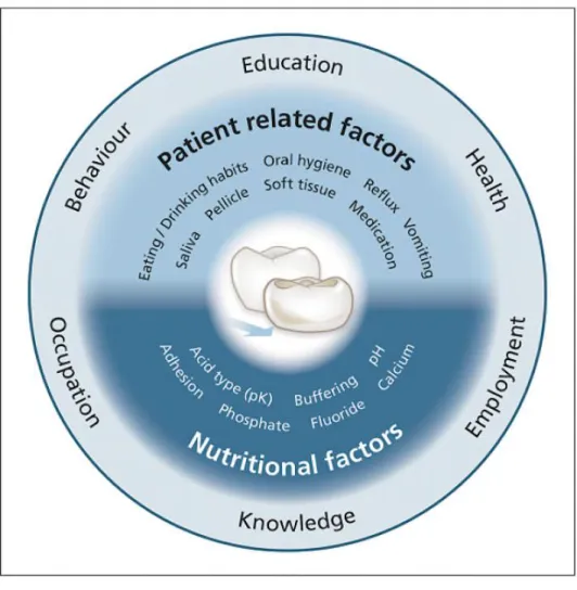
clinical manifestations should be erosive tooth wear (ETW) as it encompasses more
accurately the multifactorial process involved (figure 1).1 However, dental erosion (DE) and
ETW is often used interchangeability in the literature. It has been suggested that DE should
refer to surface loss caused exclusively by exposure to acid, whereas ETW should include
Prevalence
The prevalence of ETW is significant in general, both in primary and permanent
teeth. Systematic reviews report that while a good amount of data exists on tooth wear in
children and adolescent, studies are more scattered in adults because of the heterogeneity
of the methodology used. However, there is a general consensus that the severity of ETW
increases with age.6, 7 This trend can be found in different parts of the world. In more recent
studies, the prevalence of ETW found in Israel increased from 36.6% between the ages of
15-18 to 61.9% between the ages of 55-60.2 Similarly, prevalence in Chinese adults was
67.5% among 35-49 year-olds and 100% among 50-74 year-olds.8 The range of prevalence
found in the literature is very wide. In adults (18-88 years-old), prevalence can vary as
much as 4 to 100%.9 Aside from the diverse methodologies used among existing studies
making comparison difficult, major differences would be expected between countries solely
based on cultural and environmental factors. A pan-European study including countries such
as Estonia, Latvia, Finland, France, Italy, Spain and UK, found that an average of 29% of
young adults (18-35 years old) had ETW with 3% showing severe signs of erosion.
Differences between countries were significant.10 It is interesting to note, however, that
some authors have observed that the adoption of a more Westernized diet and lifestyle in
Asia, is likely to have an effect on ETW in all age groups.11 Numbers appear to be similar in
an American and Japanese study where prevalence was 25% and 26.1%, respectively.11, 12
A rise in prevalence and severity has also been observed especially in adolescents and
young adults in many European countries and the USA.2, 10, 13-15It may be hypothesized that
dietary habits have changed in recent years, and more importantly in those age groups,
where higher frequency of consumption of newly marketed acidic foods and drinks may
affect ETW.16, 17 Because ETW is an irreversible cumulative process during a lifetime,
prevalence is expected to increase in the future if acidic dietary trends continue in the same
3
Erosive Process and Clinical Signs
When searching the literature, there seems to be a greater awareness of ETW in
Europe compared to North America, where it is often dismissed as attrition and erroneously
treated as such.18 In fact, a recent survey of general practitioners in the US showed that
only 30.5% could correctly identify all the clinical signs of dental erosion, although 86% felt
competent to do so.19 Because of the growing interest, the European Federation of
Conservative Dentistry (EFCD) recently published a consensus report to help dental
practitioners with diagnosis and management.3 There are no specific diagnostic tools for
ETW. Thus, diagnosis, prevention and management rely heavily on the dentist’s ability to
accurately identify clinical signs and relevant etiologies to assist in developing adequate
management strategies.
Progression of ETW may be accelerated by erosion in combination with attrition,
abrasion and abfraction. The erosive process begins with demineralization of hydroxyapatite
or fluorapatite crystals in enamel, softening the outer surface. This initial stage is still
somewhat reversible because of possible remineralization, but undetectable clinically
making early diagnosis difficult.20 Disorganized and defective apatite crystals are
predisposed to further dissolution by non-bacterial acidic challenges and further attacks will
eventually lead to permanent and clinically detectable loss of dental hard tissue. This
process appears to progress at a much faster rate than caries as a surface lesion.21 Dentin
is even more susceptible to acid attacks than enamel because of its composition. Once the
dentin is exposed, there is demineralization of apatite crystals at the interface between
intertubular and peritubular dentin, and dentinal tubules may become significantly
expanded.22, 23 Collagen in exposed dentin is also vulnerable to attacks by the gastric
enzymes pepsin and trypsin. Thus, clinical signs of rapid progression may be dentin
hypersensitivity and absence of staining of the lesion. However, most patients do not
to obliterate tubules. Sometimes, even the most severe erosion cases leave the patients
asymptomatic, emphasizing once again the importance of dental practitioner’s awareness
and early detection.3, 24
Early clinical signs of ETW is characterized by loss of enamel texture, a silky glossy
appearance and sometimes a dulling of the surface gloss, referred to as the “whipped clay
effect”.4, 25 Other characteristic signs include cupping of cusps on the occlusal surfaces and
flattening of the occlusal structures. In later stages, occlusal morphology can completely
disappear with hollowed out surfaces and restorations “standing proud” above adjacent
tooth structures.3, 4, 26-28 On smooth surfaces, convex areas flatten or concavities appear
with the width usually exceeding the depth. Lesions are located coronal from the CEJ with
an intact rim of enamel along the gingival margin, possibly due to plaque remnant acting as
a diffusion barrier for acids or the neutralization effect of slightly alkaline sulcular fluid.
Progression can result in pseudo-chamfers at the margin of the eroded surface.29
Initial enamel and dentin lesions are often difficult to differentiate from abrasive
lesions.4, 30 However, wedge-shaped defects from abrasion or abfraction usually have sharp
margins coronally with cuts at right angles into the enamel surface, and the depth usually
exceeds the width.29 Abrasion is caused by an abnormal mechanical process, and aggressive
oral hygiene habits (e.g. traumatic brushing or abrasive toothpaste ) is most often at
fault.27, 31 It is also important to distinguish defects caused by attrition, where action of
opposing teeth produces matching polished wear facets on the occlusal or incisal surfaces.27
Lesions are typically flat, sharp bordered and glossy.3, 29 However, thorough information
gathering about medical and dental history is unequivocally necessary to confirm causative
5
Etiology and Risk Factors
Acid that contributes to ETW can be of extrinsic or intrinsic origins, or a combination
of both. Extrinsic acids are usually related to dietary habits, occupational hazards or acidic
medications and other drugs, whereas intrinsic acids will involve rumination, vomiting or
regurgitation that allow gastric acid to reach the oral cavity, such as in gastro-esophageal
reflux disease (GERD), eating disorders and alcoholism.32, 33 To assist clinicians identify the
source of acid for proper management, locations of erosive lesions may be used as an
indicator, but should not be the sole factor in determination.34 Extrinsic erosion typically
presents on labial surfaces of anterior teeth, buccal surfaces of posterior teeth and occlusal
surfaces posterior mandibular teeth.27 On the other hand, intrinsic erosion tends to occur on
the anterior maxillary palatal surfaces, posterior maxillary and mandibular occlusal surfaces,
and posterior mandibular buccal surfaces.18, 27, 28, 31
Much like dental caries, not everyone is at the same risk for ETW and various
external and internal factors play an important role in susceptibility.20
External Factors
Dietary Habits
Dietary acids are largely responsible for ETW. The amount and more importantly the
frequency of daily consumption of acidic foods and drinks increases risk significantly.3 This
applies particularly to teenagers and young adults who may regularly consume acidic
beverages, like sports drinks, following intense physical exercise where dehydration and
decrease in salivary flow may occur as well. Furthermore, the availability of heavily
marketed flavored and energy drinks has been on the rise.20, 28 In fact, a recent study of
beverages in the United-States alone tested 379 beverages, 93% of which had a pH of less
drinks, sports drinks and carbonated beverages in general is even thought to be the leading
cause of dental erosion observed among children and adolescents.35, 36 However, the erosive
potential of food, beverages and medicines should not be determined solely based on pH.
Other factors must be taken into account, including buffering capacity, calcium, phosphate
and fluoride concentrations, chelating properties, adherence to enamel, ability to stimulate
salivary and temperature.3, 21, 37 Another high risk group include populations on special
diets, such as vegetarian or raw food diets, whose consumption of fruits can consist of up to
96% of their diet.33, 38 It has been shown that consumption of citrus fruits more than twice
daily increases ETW risk about 37 times compared to subjects who eat fruit less often.39
Occupational hazards
A few professional occupations put personnel at risk for ETW, such as workers in
battery, galvanizing or plating factories, or in chemical, pharmaceutical or biotechnological
labs or enterprises where they might be regularly exposed to acidic vapors without proper
safety measures. Wine tasters are another group at risk for ETW as a result of swishing and
swilling each mouthful of wine abundant in tartaric and malic acids for many seconds.
Moreover, tasting sessions can last for hours, resulting in higher prevalence and severity of
erosive lesions.33, 40 Finally, competitive swimmers have also been reported to be
susceptible to ETW, especially if the pH of the pool water is incorrectly monitored.41, 42
Medication and Other Drugs
Some medications (e.g. acidic saliva stimulants or acetylsalicylic acid) and food
supplements (e.g. vitamin C) in chewable tablets, syrup or effervescent drinks are
potentially erosive.3, 33 Other medications, such as antihistamines, antiemetic and
antiparkinson medicines can decrease salivary flow as a side effect, which can impact
7
antagonists and cancer chemotherapeutic agents can cause vomiting or emetic effects.
Medications, such as aspirin and diuretics, can also irritate the stomach causing vomiting,
which in return can cause dental erosion.43, 44 Elderly populations are particularly affected
since they usually are on multiple medications.21, 28 Albuteral sulfate taken for asthma is
acidic and significantly reduces salivary flow rates.
Internal Factors
Eating Disorders and GERD
Reported pH of gastric acids can be as low as 1 and can travel up to the oral cavity
by vomiting (e.g. eating disorders) or regurgitation (e.g. GERD).43 Populations at risk
usually include young teens with anorexia or bulimia nervosa. These disorders are highly
prevalent in females with body image issues, who self-induce vomiting.28, 45 In contrast to
vomiting, regurgitation is an involuntary condition that doesn’t involve nausea, retching or
abdominal contractions and is prevalent in GERD populations.43, 46 GERD is defined as a
condition which develops when the reflux of gastric content causes troublesome symptoms
or complications, which includes possible repercussions in the oral cavity, especially when
esophageal sphincters are weakened.43, 47, 48 This chronic condition is usually diagnosed
based on symptoms that motivate patients to consult their physicians, such as bitter or sour
taste and burning sensation in the chest also known as heartburn, and is prevalent in about
10-20% of the general population.33, 49 However, it has been reported that in 25% of
confirmed GERD cases showing tooth erosions, condition is asymptomatic and may be left
undiagnosed, which can lead to critical consequences including esophageal
adenocarcinoma.50, 51 Diagnosis of unexplained ETW by both dental and medical general
Saliva
Saliva is considered to be a biologic protective factor against ETW through43, 52
1) Formation of acquired pellicle that act as a semi-permanent membrane
covering tooth surfaces
2) Dilution, clearance and neutralization of acid by mechanical cleansing
(swallowing) or dependent on flow rate and buffering capacity
3) Prevention of demineralization by remineralization per its mineral content
Composition
Both saliva and teeth contain minerals such as calcium, phosphate and fluoride and
ion exchange is possible.43 Fluoride has been used in dentistry for many years because it
has been shown that in high doses, fluoride has the ability to increase remineralization and
prevent demineralization.43 Calcium and phosphate are believed to also play similar roles,
depending on their degree of saturation in teeth, saliva and ingested solutions.1, 43, 53 In
fact, classic concept of critical pH, taught for enamel and dentin of 5.2 and 6.7, respectively,
concerns only dental caries as it refers to the average concentrations of minerals in plaque
fluid.1, 43, 54 Therefore, even if a solution has a pH below 5.2, it is possible that enamel
erosion will not ensue.20 The process of erosion is independent of plaque, this is why when it
comes to erosion, a fixed critical pH does not exist and will vary depending on the
concentration of calcium, phosphate and fluoride of the solution. Critical pH can be defined
as the value at which a solution is saturated with respect to a specific solid, in this case,
tooth minerals. At critical pH, there is an equilibrium where no dissolution or precipitation
occur. Below critical pH however, fluid is under-saturated with tooth minerals and that’s
when dissolution of tooth surface can occur.1, 20, 43, 52 After an acid attack, salivary calcium
and phosphate may remineralize enamel in conjunction with fluoride ions, but the protective
9
Flow RateFlow rate is considered to be the best clinical indicator of protective properties of
saliva since other salivary parameters, such as mineral content described above and amount
of bicarbonate involved in buffering acids, are directly correlated to flow rate.52 Swallowing
also increases with increased flow rate, which permits dilution and clearance of acid more
quickly.43 Average unstimulated salivary flow rate is reported to be >0.3 mL/min with
normal daily production between 0.5 and 1.5 L.52, 55 If unstimulated and stimulated salivary
flow reaches rates of 0.1 and 0.7 mL/min respectively, this is considered to be
hyposalivation.20, 55 Several activities, conditions and medications can decrease salivary flow
resulting in hyposalivation or xerostomia, which puts populations concerned at higher risk
for erosive damage. This includes most hypertension medications and antidepressants,
dehydration from exercising, and reduction/loss of function of salivary glands from head and
neck radiation in cancer therapy or Sjögren syndrome.20, 43, 52 It has also been shown that
patients taking more than 3 medications daily experience xerostomia, regardless if it is
listed as side-effect or not.19, 56
Buffering capacity
The main buffering component of saliva is bicarbonate, which neutralizes acids in the
mouth and shortens erosive episodes.43 Its concentration in stimulated saliva is much more
significant, and is about 12 times higher than in unstimulated saliva.43, 57 Thus, stimulated
saliva has high buffering capacity that plays an important function in protecting teeth from
acidic challenges.58 With good salivary flow rates, saliva can buffer acids with a pH of 3.5 to
Acquired Pellicle
Acquired pellicle is a semi-permanent organic barrier devoid of bacteria that naturally
coats tooth surfaces.20, 59 It is formed by the adsorption of proteins, peptides, lipids and
other macromolecules present in saliva, and provides protection since acid must diffuse
through it to come into contact with teeth.59 In addition to slowing down acid attacks, it also
reduces calcium and phosphate release from enamel and dentin.59 Composition and
thickness varies between individuals and can be influenced by age and degeneration of
salivary glands, which may influence its permeability.19-21 It has been reported that patients
with erosion appear to have less pellicle compared to a control population, while another
study showed that its thickness varies within dental arches, with the thinnest found at the
upper anterior palatal surface.20, 60, 61
Although saliva provides several protective properties, they are very limited when
confronted with frequent and large amounts of strong acids over a long period of time,
which can also easily displace acquired salivary pellicle.43
It is useful to take into consideration these external and internal factors when
treating patients to determine, first of all, high risk populations and monitoring them
accordingly. Secondly, when diagnosing patients with ETW, these factors can help providers
better understand possible etiologies and therefore develop appropriate management
strategies.
Preventive and Restorative Management
Prevention and early detection of ETW should be primary goals for practitioners, as
11
A multidisciplinary approach should be considered, especially if etiology of ETW is
determined to be intrinsic. For example, psychological counseling referrals should be made
in eating disorders patients, and referrals to a primary care physician and gastroenterologist
should be done in suspected GERD patients. If medications significantly affect the quality
and quantity of saliva, discussions should take place with medical providers to assess
different strategies to decrease risk, whether it be changing medications, dosage or
frequency. To increase salivary flow, sugar free or xylitol mints and gums may also be used
in addition to pilocarpine.28 As for extrinsic sources, referral to a registered dietician may be
recommended, but more importantly, a written diet diary should be prescribed to patients
at risk for the dental team to analyze. Dietary counseling can then be personalized
efficiently. It is suggested that two weekdays and a weekend be recorded to reflect as much
as possible patient’s dietary habits, according to which, diet modifications can be
recommended.19, 27, 28, 62 Frequent consumption of acidic foods and drinks, and some oral
habits such as swishing or holding drinks in the mouth, may exacerbate erosive
potentials.28, 31, 37, 62, 63 Hence, behavioral management should also include the manner in
which food is consumed (chewed, sucked, dissolved), eliminating certain foods or
decreasing contact time (e.g. use of a straw).19, 27, 64 The WATCH strategy was developed to
offer straightforward understanding and advice regarding diet.63 (Table 2) Products
containing fluoride can also be used as adjuncts, such as fluoride varnish or prescription
toothpastes.3, 37 Ultimately, the objective of controlling risk factors is to stop progression of
ETW, assuming that the patient is compliant.
Once etiologies have been identified and risk factors controlled, restorative
management can be considered. Resin sealants or bonding agents can be applied over
dentin when the erosive lesion does not compromise the existing tooth structure. This may
reduce ETW progression and sensitivity for a limited time period.3, 28 Restorations should be
advanced lesions, more aggressive therapies to restore esthetic and function may be
indicated, especially if loss of vertical dimension has occurred due to severe loss of tooth
structure.65 Regular monitoring and evaluation of ETW management should be done during
recall visits.3
Monitoring and Tooth Wear Indices
In order to monitor progression and management of ETW, photographs and
diagnostic casts should be made periodically.1, 25 To further raise awareness and aid
practitioners in screening and monitoring progression and severity of ETW, wear indices
have also been developed. These were designed to be used both in private practice and for
research purposes.66 Many research groups have developed their own tooth wear index,
however, making research in this field challenging to compare. They are modified for each
specific study according to study aims, and may vary in their manner of assessment, scale,
choice of teeth, and other differing modalities.2 In fact, World Health Organization (WHO)
has stated that there is a need for more systematic population-based studies worldwide on
the prevalence of dental erosion using a standard index of measurement.67
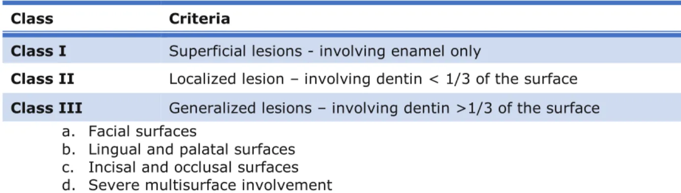
Many indices are largely based on the work by Eccles and Jenkins, which is a
classification for assessment of dental erosion of non-industrial origin with three classes of
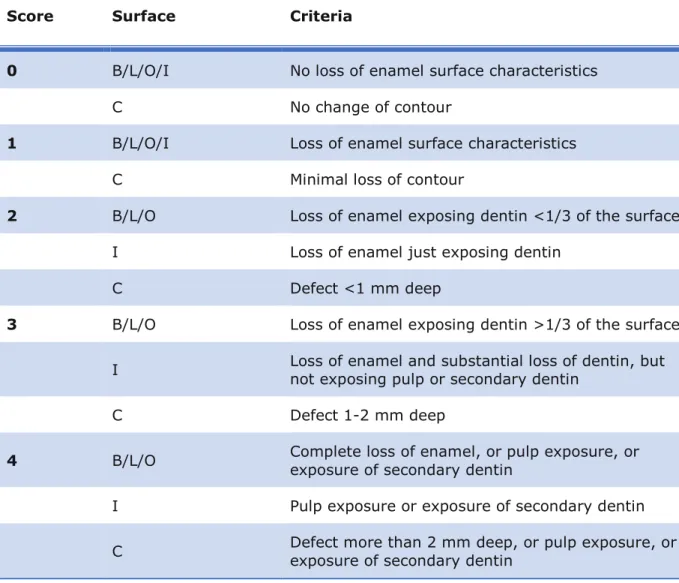
lesions assigned to four tooth surfaces (table 3).68 Smith and Knight later introduced the
Tooth Wear Index (TWI), a comprehensive system where all four visible surfaces of all teeth
are scored (table 4).69 Although very reliable, this index was not suitable for use in
day-to-day practice.70 This index was then modified by Bardsley in 2004 to the Simplified Tooth
Wear Index (table 5).71 In addition to difficult study comparisons in meta-analyses, inter
and intra-examiner reliability of many indices is an area of concern.30 The accurate
13
workshop was conducted in Switzerland to discuss the various dental erosion indices
available, and it was reiterated that a simple and standardized index is necessary. The
workshop proposed the Basic Erosive Wear Examination (BEWE) be used for both the
research field and dental clinicians.30 In their opinion, and ideal index should be:
1. Easily applicable in general dental practice
2. Adaptable for epidemiological prevalence studies
3. Suitable for monitoring erosive lesion activities such as progression or arrestment
of lesions
4. Easily reproducible under varying conditions for examination such as with/without
magnification devices, ambient light, and hydration state of the tooth surface
(dry/wet)
5. Capable of reflecting net exposure of an affected individual to the erosive
challenge
6. Capable of indicating the need for treatment
7. Suitable for both children and adult, as well as permanent and primary teeth
BEWE, developed and recommended in 2008 by Bartlett et al., is a simple,
reproducible and transferable scoring system for recording clinical findings and assisting in
the decision-making process for the management of ETW.66 A sextant based exam is
conducted, where the surface of the tooth with the worst wear is graded in each sextant,
resulting in a calculated cumulative score which allows risk and guidelines for management
to be determined (table 6 and 7). A clinical study aiming to assess reliability of BEWE found
it to be similar in distribution to TWI, and although it slightly underscored moderate to
severe wear, the examination gave very few false positives, predicting severe wear with a
sensitivity of 90.9% and a specificity of 91.5%.70, 73 When comparing scores between 2
examiners, reliability showed moderate agreement and it was concluded by the authors that
simple, scores should be interpreted with some caution.73 Similarly, when BEWE was
compared to another index, the visual erosion dental examination (VEDE) widely used in
Norway where it originated, examiner reliability was acceptable for both, and highest
agreement was found for sound and more severely affected surfaces involving dentin, while
smallest agreement was found for initial and mild enamel lesions. However, no direct
statistical comparison was made between both systems.74 It can be argued that
differentiation between enamel and dentin is an important factor for recording progression
of ETW, hence, supporting the use of recent indices such as VEDE and the exact tooth wear
index that distinguish these variables (table 8 and 9).74, 75 However, these detailed scales
not only impact the reproducibility of scores, but also may discourage its use in a clinical
setting since it is not as easy and straightforward. To avoid diagnostic uncertainties, BEWE
does not distinguish between enamel loss and exposed dentin.66 A recent study further
validated BEWE as a screening tool by showing that sextant cumulative score provided a
good representation of tooth wear when compared to scores of all tooth surfaces.76
Although wear indices are still advocated, technology has expanded quickly in the
field of 3D scanning. However, the absence of stable reproducible reference points make
longitudinal assessment challenging.77, 78 Current research in this area is promising, and
further development is, without a doubt, warranted.
The purpose of this thesis was to add to the existing literature regarding prevalence
of ETW, specifically in an American adult population and to assess education on the subject
in U.S. and Canadian dental schools.
In part I, primary aim was to determine if subjects diagnosed with GERD had
increased risk for ETW compared to a control population, therefore assessing prevalence of
15
other potential risk factors, including age, sex, number of years since GERD diagnosis, diet,
medication and quantity and quality of saliva.
In part 2, the aim was to investigate what is taught in dental schools regarding
diagnosis, and both preventive and restorative management of dental erosion. Furthermore,
respondents were asked about their impression of students’ competence on the subject at
Table 1: Types of Tooth Wear24, 79
Type Definition Examples
Attrition
Physical wear as a result of the action of antagonistic teeth with no foreign substance intervening (two-body wear)
Bruxism
Abrasion
Physical wear as a result of mechanical processes involving foreign substances or objects (three-body wear)
Excessive forces used in tooth brushing, effect of abrasives in toothpaste, habits such as pen chewing, coarse or sandy food
Abfraction
Physical wear as a result of tensile or shear stress in the cemento-enamel region provoking microfractures in enamel and dentin (fatigue wear)
Tooth flexure
Extrinsic Erosion Chemical wear as a result of extrinsic
factors
Acidic diet (citrus fruits, fruit-based drinks, some
carbonated drinks, herbal teas), occupational hazards (sommeliers, factory
workers, competitive swimmers)
17
Table 2: The Erosion WATCH Strategy for Diet Analysis and Advice for Patients with TW63
Analysis Advice
Water Do you drink enough water? Drink 1.5L of pure water/day. 2L, 2 hours before a game or 1L 1 hour before a game
Acids
Do you drink excess soft or
sports drinks containing
ascorbic, citric or phosphoric acid?
Avoid acid drinks when dehydrated in sports, work, or when drugs shut off salivary protection
Taste Do you taste enough fresh fruit daily?
Eat a piece of fruit with every breakfast to stimulate saliva
Calcium Are you getting enough calcium in your diet? Milk, cheese and yogurt contain calcium and protect teeth against acids
Health
Do you have a healthy lifestyle and diet?
Healthy lifestyles can be dehydrating. Excess alcohol is dehydrating and causes gastric reflux
19
Table 3: Eccles Classification of Dental Erosion68Class Criteria
Class I Superficial lesions - involving enamel only
Class II Localized lesion – involving dentin < 1/3 of the surface
Class III Generalized lesions – involving dentin >1/3 of the surface
a. Facial surfaces
b. Lingual and palatal surfaces
c. Incisal and occlusal surfaces
Table 4: Smith and Knight Tooth Wear Index (TWI)69
Score Surface Criteria
0 B/L/O/I No loss of enamel surface characteristics
C No change of contour
1 B/L/O/I Loss of enamel surface characteristics
C Minimal loss of contour
2 B/L/O Loss of enamel exposing dentin <1/3 of the surface
I Loss of enamel just exposing dentin
C Defect <1 mm deep
3 B/L/O Loss of enamel exposing dentin >1/3 of the surface
I Loss of enamel and substantial loss of dentin, but not exposing pulp or secondary dentin
C Defect 1-2 mm deep
4 B/L/O Complete loss of enamel, or pulp exposure, or
exposure of secondary dentin
I Pulp exposure or exposure of secondary dentin
21
Table 5: Simplified Tooth Wear Index - Bardsley71Score Criteria
0 No wear into dentin
1 Dentin just visible (including cupping) or dentin exposed <1/3 of surface
2 Dentin exposure >1/3 of surface
Table 6: Basic Erosive Wera Examination (BEWE) - Criteria for Grading Erosive Wear66
Score Criteria
0 No erosive tooth wear
1 Initial loss of surface texture
2 Distinct defect, hard tissue loss <50% of the surface area
3 Hard tissue loss ≥ 50% of the surface area
Table 7: Basic Erosive Wear Examination (BEWE) - Risk Levels as a Guide to Clinical Management66
Risk
Level Cumulative score of all sextants Management
None Less than or equal to 2 Routine maintenance and observation Repeat at 3-year intervals
Low Between 3 and 8 Oral hygiene and dietary assessment, an advice, routine maintenance and observation
Repeat at 2-year intervals
Medium Between 9 and 13
Oral hygiene and dietary assessment, and advice, identify the main etiological factor(s) for tissue loss and develop strategies to eliminate respective impacts
Consider fluoridation measures or other strategies to increase the resistance of tooth surfaces
Ideally, avoid the placement of restorations and monitor erosive wear with study casts, photographs, or silicone impression
Repeat at 6-12 months intervals
High 14 and over
Oral hygiene and dietary assessment, and advice, identify the main etiological factor(s) for tissue loss and develop strategies to eliminate respective impacts
Consider fluoridation measures or other strategies to increase the resistance of tooth surfaces
Ideally, avoid restorations and monitor tooth wear with study casts, photographs, or silicone impressions
Especially in cases of severe progression consider special care that may involve restorations
23
Table 8: Visual Erosion Dental Examination (VEDE)74Score Definition
0 No erosion
1 Initial loss of enamel, no dentin exposed
2 Pronounced loss of enamel, no dentin exposed on the surface area
3 Exposure of dentin, <1/3 of the surface involved
4 1/2 - 2/3 of dentin exposed
Table 9: The Exact Tooth Wear Index75
Exact Tooth Wear Index for Enamel
0 No tooth wear: no loss of enamel characteristics or change in contour 1 Loss of enamel affecting <10% of the scored surface
2 Enamel loss affecting between 10% and 1/3 of the scored surface 3 Enamel loss affecting at least 1/3 but <2/3 of the scored surface 4 Enamel loss affecting 2/3 or more of the scored surface
Exact Tooth Wear Index for Dentin
0 No dentinal tooth wear: no loss of dentin
1 Loss of dentin affecting <10% of the scored surface
2 Dentin loss affecting between 10% and 1/3 of the scored surface 3 Dentin loss affecting at least 1/3 but <2/3 of the scored surface
4 Dentin loss affecting 2/3 or more of the scored surface, no pulpal exposure
25
PART 1
PREVALENCE OF EROSIVE TOOTH WEAR IN GERD PATIENTS
Introduction
There had long been a need for general consensus over the blue-ribbon definition of
gastro-esophageal reflux disease (GERD). Hence, the Montreal definition and classification
of GERD was developed in 2006 and defines this disease as a condition which develops
when the reflux of gastric content causes troublesome symptoms or complications.47(figure
2) According to epidemiological studies, GERD is most prevalent in Western countries,
including Europe and the US, where the weekly incidence of heartburn and/or acid
regurgitation was reported to be between 10-20%.49 This translates to about 15 millions of
Americans experiencing daily heartburn symptoms.19 It is much less prevalent, as low as
<5%, in Middle-Eastern and Asian countries.49, 80 The most common esophageal/typical
symptoms encountered in GERD are heartburn and regurgitation but
extra-esophageal/atypical symptoms can also be found, including dental erosion (DE), which is
defined as the chemical dissolution of hard tooth tissue by acids not of bacterial origin.4, 47, 80
The association between GERD and DE can be noticed in two possible clinical
situations80, 81 :
1. Patients consulting physicians for GERD symptoms, who are then diagnosed with DE
2. Patients presenting to the dentist with DE, who are subsequently diagnosed with
Only 42% of physicians strongly agree that GERD may cause dental erosion, while
35% agree with minor reservations and 19% with major reservations.47, 51, 82 This suggests
that perhaps dental consequences of GERD are poorly understood by physicians. In fact, a
recent guide was published to help physicians recognize clinical features of GERD-related
dental complications.51 On the other hand, patients presenting at the dental office with
unexplained erosive tooth wear (ETW) are often asymptomatic or at most oligosymptomatic
in up to 25% of cases, and “silent GERD” should be suspected.81, 83 A recent study by
Wilder-Smith found that in subjects with severe ETW, few experienced frequent symptoms,
but 69% actually had abnormal reflux when they were tested using both endoscopy and 24h
multichannel intraluminal pH-impedance measurements.81 Silent GERD is probably
responsible for under-diagnosis of this condition, and if left undiagnosed can lead to critical
consequences such as pre-malignant Barrett’s esophagus or even esophageal
adenocarcinoma.50, 51, 84 In these cases, dentists just may be the first to suspect this
potentially life-threatening condition and make appropriate referrals to a physician.
Vice-versa, prompt referral to a dentist by physicians may save patients costly treatments before
ETW causes extensive damage over time.85 Thus, multidisciplinary approach is strongly
encouraged.
The relationship between DE and GERD has been investigated in both children and
adults. Systematic reviews have established a strong association, with a 24% prevalence of
DE in GERD subjects and a 32.5% prevalence of GERD in DE subjects in an adult
population.80, 86, 87. However, this association remains controversial, mainly because of the
heterogeneity of methodology used not only to diagnose GERD (self-referral,
symptom-based, endoscopy or 24h pH monitoring), but also for the measurement of DE (multiple
tooth wear indices).19, 88, 89 Theoretically, the acidity in the stomach may reach levels as low
as pH 1, which if frequently in contact with teeth and for long periods of time will cause
27
acids that have passed the lower esophageal sphincter may or may not progress into the
mouth and this could partly explain why some experiencing daily GERD symptoms do not
necessarily have ETW.84, 90
Although prevalence studies on erosion are beginning to emerge in the US, a vast
majority of studies on prevalence of DE in an adult GERD population have been conducted
outside of North America with varying results (table 10). The purpose of this study was to
determine the prevalence of ETW in a GERD population compared to a control population in
North Carolina, USA, and the association of ETW with other factors such as age, sex,
medications, number of years since diagnosis, acidic diet, and salivary quality and quantity.
Materials and Method
Subject Selection
In this cross-sectional study approved by the University of North Carolina Biomedical
Institutional Review Board (IRB, studies #11-2327 and #15-887), consecutive enrollment
was performed at the Center for Esophageal Diseases and Swallowing Disorders, University
of North Carolina Hospital Division of Gastroenterology and Hepatology for GERD subjects
(group 1) and at the University of North Carolina, School of Dentistry (UNC-SOD) for control
subjects (group 2) in 2012 and then between November 2015 and 2016. Inclusion criteria
were as follows: adults (18-85 years old), at least 2 natural uncrowned teeth per sextant
(total of 12 teeth), positive diagnosis of GERD or no history of GERD (control). Subjects
unable to speak or understand English, with a history of anorexia or bulimia, or currently
pregnant were excluded. GERD subjects were all recruited at UNC hospitals in
gastroenterology specialty clinics, where they were referred and diagnosed with GERD by
medical professionals through either troublesome heartburn/reflux symptoms, mucosal
a pilot study where prevalence of medium to high ETW was 40% in GERD subjects and 15%
in control subjects19, a power analysis with
α
set at 0.05 and power of 0.8 indicated that asample size of 98 subjects (n=49/group) was required to detect whether the difference
between the proportions truly exists. Data from pilot study was combined to the present
results for analysis.
Procedures
Subjects who agreed to participate signed an informed consent form and completed
a health history questionnaire, which included demographics (sex and age), a list of their
current medications and the number of years since their GERD diagnosis, when applicable. A
one-time appointment only was required for subject participation, during which primary
investigator (K.E. or C.N.N.) performed a dental examination to determine ETW, collected a
stimulated salivary sample and provided a take-home 4-day diet diary to be completed and
sent back for analysis.
Dental examination
Dental examination was carried out using 2x2 gauzes to dry teeth, a 25-mm
diameter disposable plastic dental mirror (Sunstar Americas, Chicago, IL), and 3.25X
magnification dental loupes and headlight (Orascoptic, Middleton, WI). Dental examination
was done on subjects sitting upright on chairs in the medical office or dental operatory to
standardize procedures in respective clinics. Clinical assessment of ETW was determined
using Basic Erosive Wear Examination (BEWE)66, which is a rapid and simple partial scoring
system (0-3) that records the worst affected surface in each sextant (table 11). Teeth in
each sextant were divided as follows: 1-5, 6-11, 12-16, 17-21, 22-27, 28-32. The
29
Labial/buccal, lingual/palatal and occlusal surfaces were considered for examination.
Crowned or missing teeth were systematically excluded.
Stimulated Salivary Sample
A salivary sample was collected by having subjects chew on a paraffin wax tablet and
expectorate stimulated saliva in a sterile container for a total of 5 minutes using a
stopwatch. Saliva samples were labelled and stored immediately on ice for transportation to
the Oral Microbiology Lab at the UNC-SOD for analysis of flow rate and buffering capacity.
Collection time was recorded thoroughly for each subject for flow rate calculations. As for
buffering capacity, saliva was diluted four-fold in 0.0005N HCl and the final pH was recorded
after ten minutes. Results were also categorized into risk levels (table 13). All samples were
destroyed after testing.
Diet Diary Analysis
A labelled 4-day take home diet diary (Thursday-Sunday) was handed to each
subject to be completed and returned in a pre-stamped addressed envelope. Verbal
instructions were given, accompanied by written instructions and examples of how to
complete the diet diary properly. This included recording of all food and drinks along with
the quantity consumed to calculate more accurately the number of servings consumed for
each acidic item. Acidic challenges were counted for each day and a daily average was
calculated. Contact information obtained at the initial appointment aided in sending
reminders and additional diet diary copies either through phone, mail and/or email
messages. Upon reception of the diet diary, contact information was destroyed. Subjects
who hadn’t returned their diet diaries received no more than 2 reminders during the
Statistical Analysis
Analysis was performed using SAS 9.4 software (SAS Institute, Cary, NC) with level
of significance set at 0.05. Because of the cross-sectional nature of this study, descriptive
statistics and bivariate calculations were first performed to evaluate heterogeneity between
the 2 groups investigated, GERD and control, in terms of different co-factors. Four
categories of BEWE scores were recorded: none, low, medium and high. However, from
proportional distribution of previous results19, it appears that difference between control and
GERD groups occur in the medium risk level. Thus, “medium” and “high” categories were
combined, as well as “none” and “low”. Further analysis was then completed to assess if
there was a difference between GERD and control groups in terms of ETW as defined by
BEWE scores, followed by ETW association with covariates such as age, sex, number of
medications, daily average frequency of acidic challenges from diet, salivary flow rate
(mL/min) and salivary buffering capacity. An additional variable, number of years since
GERD diagnosis (when applicable), was investigated for the latest data set (n=28 GERD
subjects). Lastly, logistic regression analysis was used after adjusting for potential variables
to assess true relationship between ETW and GERD as primary explanatory variable.
Results
2015-2016 Data Only
For this data set, sample size was 57 subjects (n=28 GERD and n=29 Control).
Distribution between the 2 experimental groups did not statistically differ in terms of sex,
age, daily acidic challenges, and saliva flow rate and buffering capacity (p>0.05). The
control group consisted of 9 males and 20 females with a mean age of 46.2, and GERD
group consisted of 8 males and 20 females with a mean age of 53.5. Six study participants
failed to return their diet diary. Although higher proportions of GERD subjects compared to
31
was not statistically significant and the vast majority of participants had normal saliva flow
rate and buffering capacity, 89.7% for controls and 71.4% for GERD subjects. In general,
the GERD population took more daily medications than controls and this difference was
statistically significant. Ninety-six percent (n=27) of GERD subjects took at least 1
medication per day with one third taking 6 of more medications every day versus 41.5%
(n=12) in the control group taking no medication at all and 44.8% (n=13) between 1 and 3.
BEWE combined categories (none-low and medium-high) were found to be significantly
associated with GERD (p=0.0023), with prevalence of medium-high ETW of 64.3% (n=18)
for GERD subjects versus 24.1% (n=7) for control subjects. (table 14)
Bi-variate analysis comparing combined categories of ETW revealed that 72%
(n=18) of subjects with medium-high ETW were GERD subjects, compared to 28% (n=7)
controls. Results also showed other co-factors that statistically significantly had an effect on
erosive tooth wear severity, which included age, number of daily medications, daily acidic
challenges from the diet and saliva buffering capacity. An additional factor, number of years
since diagnosis and treatment, was investigated for this data set. The hypothesis was that
GERD subjects that had been treated for a shorter period of time may have been exposed
longer and more frequently to erosive gastric reflux, and consequently presenting with more
severe ETW. Although number of years since diagnosis and treatment was slightly shorter
for GERD subjects showing medium to high ETW, this difference was not statistically
significant. (table 15) Finally, logistic regression was performed to truly assess factors
associated with medium to high ETW after controlling for all variables. Analysis showed that
only 3 factors were statistically significantly associated with ETW: GERD diagnosis, age and
Combined data
A total of 113 subjects were enrolled in this study (n=58 GERD and n=55 Control).
Distribution of the 2 groups did not have any statistically significant differences in terms of
sex, average daily acidic challenges, and saliva flow rate and buffering capacity (p>0.05).
However, GERD participants were older than those in the control group (p=0.021). Subjects
were mainly females for both groups: GERD group consisted of 19 males and 30 females
and mean age was 53 years-old, control group on the other hand consisted of 22 males and
33 females and mean age was 47 years-old. Thirteen (11.5%) study participants failed to
return their diet diary (9 GERD and 4 control subjects).1 subject failed to provide enough
saliva for analysis and was categorized as high risk for both flow rate and buffering capacity.
Although higher proportions of GERD subjects had intermediate to high risk in terms of
saliva flow risk and buffering capacity, these were not statistically significant. Number of
daily medications was not reported for 5 GERD subjects. GERD subjects had more daily
medications in general than control subjects and this difference was statistically significant.
(table 17)
The sample was mainly distributed between low (37 controls and 25 GERD) and
medium (10 controls and 29 GERD) ETW categories. Sample distribution among remaining
12 subjects was 11 in the none ETW category, of which 8 were control subjects versus 3
GERD subjects, and finally 1 GERD subject in the high ETW category. (figure 3)
ETW, as represented by combined BEWE categories, was found to be significantly
associated with GERD (p=0.0002). Of subjects having medium-high erosive tooth wear,
75% (n=30) were GERD subjects versus 25% (n=10) control subjects. (table 18)
Consequently, higher prevalence of medium to high ETW was found in GERD subjects.
Prevalence of medium to high ETW was 51.7% (n=30) for GERD subjects compared to
33
none to low ETW compared to GERD subjects, with 81.1% (n=45) and 48.3% (n=28)
respectively. Interestingly, within GERD subjects, distribution of none/low and medium/high
ETW subjects was comparable (n=28 and n=30). (table 17) Further analysis was performed
to assess if other covariates had an effect on ETW. Medium to high ETW was found in older
subjects and in subjects with higher daily acidic challenges, and these differences were
statistically significant (p<0.05). Highest proportion of none to low ETW was found in
subjects with normal flow rate and buffering capacity, however, these results were not
statistically significant. (table 18) Remaining variables revealed no statistical significance.
When reversed logistic regression was performed to assess factors associated with medium
to high ETW, analysis showed that only GERD diagnosis (OR: 0.28, 95% CI: 0.09, 0.81) and
age (OR: 1.10, 95% CI: 1.05, 1.16) were significantly associated with ETW after controlling
for other variables. (table 19)
Discussion
According to a systematic review, there is a strong association between GERD and
ETW, although prevalence varied widely in an adult population with a range between
5-47.5%.86 This association remains controversial however, with comparability between
studies having been criticized mainly because of heterogeneity regarding methods used for
GERD diagnosis and multiple tooth wear indices used for ETW evaluation (table 10).
Furthermore, confounding factors were not always addressed.88, 89 Results from the present
study revealed that ETW was significantly associated with GERD (OR: 0.28, 95% CI: 0.09,
0.81). The GERD subjects included were properly diagnosed by gastroesophageal medical
practitioners with diagnosis based on symptoms, endoscopy and 24h pH monitoring, the
latter being the gold standard technique for the diagnosis of GERD. A validated and
reproducible tooth wear index, BEWE, was also used for ETW assessment.30, 66, 73, 76 Finally,
challenges, number of daily medications, and quantity and quality of saliva through salivary
flow rates and buffering capacity.
Finally, all known factors susceptible to affect ETW was collected, such as acidic
challenges from the diet, number of daily medications and saliva quantity and quality
through stimulated salivary flow and buffering capacity.
Prevalence of medium to high ETW in a GERD population for combined data in this
study was 51.7% and comparable to existing studies using similar methodology, whereas
the control population had a prevalence of 18.2%. Other studies have found the prevalence
of DE to be 61% and 48% for GERD participants compared to 28% and 13% in control
participants in China and Spain, respectively.91, 92 However, other studies have found no
association between DE and GERD, with a 9% prevalence in Italy, 5% in Finland and 3.2%
in Brazil.93-95 ETW may have been underestimated for multiple reasons in the present study.
First, air/water syringe and dental chairs were unavailable for optimal clinical examination
and secondly, a significant number of potential GERD subjects were rejected due to
insufficient teeth to be examined Teeth were either extracted or had full coverage
restorations, suggesting that aggravation of ETW with time may have led to their loss or
treatment among other reasons. Potential ETW of these teeth could not be assessed and
prevalence of high ETW severity in GERD subjects may have been greater in reality than the
results of the present study. In fact, only 1 high ETW subject was recorded and that subject
was found in the GERD group (figure 3). Furthermore, control subjects may have been
mislabeled as such, since they were not evaluated for silent GERD per endoscopy or 24h pH
monitoring. Hence, we cannot assume that the totality of the control population investigated
did not experience asymptomatic gastric reflux. If potential reflux episodes are frequent
enough and reach the oral cavity, this condition may explain the medium ETW found in
35
and silent GERD, up to 25%, and these patients are probably asymptomatic because of
higher than normal pain thresholds caused by chronic reflux and eventually become
unresponsive to pain at all.80, 81, 83
Age was also significantly associated with ETW (OR: 1.10, 95% CI: 1.05, 1.16). This
is to be expected since ETW is an irreversible process during a lifetime and would be
expected to increase in severity with cumulative exposure to acids, whether it be extrinsic
or intrinsic, as subjects get older.6, 7 The prevalence of ETW in a general adult population
found in the literature is very broad, ranging between 4%-100%.9 In the present study,
very few “none” subjects were found in both experimental groups, 11 subjects in total, with
a mean age of 38.4 12.5 (figure 3). It has been reported that development of ETW in
GERD takes 1-2 years of regular occurrence of acid exposure.96, 97 This could explain the low
prevalence of DE in GERD subjects in studies from Milani et al.97 and and Jensdottir et al.98,
where the researched population was younger. Furthermore, PPI treatment chronically
suppressing acid may act as a protective factor against ETW. Further research in this area is
needed as dose and duration of medication needed for improvement is still unknown.3
According to a randomized, double-blind control study by Wilder-Smith et al., there was a
reduction in enamel loss after 3 weeks of acid suppression with PPI in GERD subjects with
severe dental erosion.99 In the present study, the number of years since diagnosis was
investigated for a sub-population of GERD subjects (2015-2016 data only) treated with
PPIs. Results showed that medium to severe ETW was found in subjects that had been
treated with PPI for a shorter period of time, 9.1 (9.6) years, compared to 12.8 (8.9)
years for subjects with none to low ETW, although this was not statistically significant given
the small sample size (n=28). Similar proportions of GERD subjects were found to have
none-low ETW (48.3%) and medium-high ETW (51.7%), but mechanisms behind ETW in
GERD remain unclear. It has been proposed that acid reflux in some remains close to the
defective or overwhelmed and gastric acid reaches the upper esophageal sphincter and the
oral cavity causing tooth erosion.80 Moazzez et al. investigated acid reflux above the upper
esophageal sphincter through 24h ambulatory pH monitoring at 4 sites along the esophagus
and found it to be correlated with the severity of dental erosion, particularly on the palatal
surfaces of incisors and this was significant during the night in a supine position.100
Nocturnal reflux may play a major role in ETW severity since protective mechanisms during
the day such as salivary flow, swallowing, gastric emptying and pressure of the upper
esophageal sphincter are decreased during sleep.84 In fact, it has been reported that
obstructive sleep apnea (OSA) may predispose some patients to nocturnal GERD although
this association is not fully understood.101
There is even evidence supporting that treatment
of OSA using continuous positive airway pressure (CPAP) improves nocturnal GERD
symptoms in 75% of patients, even in those without OSA.102 Hence, OSA might be an effect
modifier of the relationship between GERD and ETW, but this was not investigated in the
present study.
Since this was cross-sectional study and not a case-control study, some variables
from the 2 experimental groups do not match. Bi-variate analysis showed that they were
close in terms of sex, diet, salivary flow and buffering capacity, but statistically differed in
age and number of medications. Although the GERD population was slightly older than the
control population, logistic regression analysis still found a statistically significant difference
in ETW between groups when controlling for age. As for medications, GERD subjects
generally took more daily medications than controls, including PPI to treat their condition. It
has also been shown that in addition to 63% of the most frequently prescribed medications
in the US having the potential to cause xerostomia, patients taking more than 3 medications
daily experience xerostomia, regardless if it is listed as side-effect or not.56, 103 However, in
37
salivary flow and buffering capacity, with a high proportion of subjects having normal saliva
quantity and quality. Similar results were found in multiple studies for salivary flow rates.53,
104, 105 However, Yoshikawa et al. found low salivary flow in GERD patients with ETW.106
Stimulated saliva was collected in the present study because standardization is easier than
in unstimulated salivary tests. Consumption of food and beverages and time of collection
throughout the day may alter flow rate of unstimulated samples. It is interesting to note
that although this was not statistically significant, there was a higher proportion of GERD
subjects compared to control subjects having inferior salivary flow rates, 24% versus 11%
respectively, and buffering capacity, 36% versus 27%, but whether this is related to their
intake of medications or their GERD condition cannot be determined. This is supported by
the findings in the UK and Finland, where lower median buffering capacity was found in
GERD participants.53, 107
Daily acidic challenges from diet were low on average for both experimental groups
and diet did not affect ETW severity in this study. Low acidic diet is to be expected in a
diagnosed GERD population since some foods can trigger reflux episodes and diet
modification is probably part of their treatment. Moreover, self-reported diets diaries are
sometimes unreliable and difficult to analyze. On one hand, subjects may have omitted or
inaccurately completed the forms since they were aware of the purpose of the study. On the
other hand, time and method of consumption were not recorded and these are important
risk factors affecting erosive potential.3 For example, fruit juices have been shown to have
very high erosive potential on its own.17 However, sipping it throughout a morning for many
hours causes more damage than drinking it quickly through a straw during a meal. Hence, a
detailed in person interview is necessary to assess dietary habits more precisely. The
erosive potential of food and beverages cannot rely solely on pH and other factors must be