Race, Breast Cancer Subtypes, and Survival
in the Carolina Breast Cancer Study
Lisa A. Carey, MD Charles M. Perou, PhD Chad A. Livasy, MD Lynn G. Dressler, PhD David Cowan, BS Kathleen Conway, PhD Gamze Karaca, MSc Melissa A. Troester, PhD Chiu Kit Tse, MSPH Sharon Edmiston, BS
Sandra L. Deming, PhD, MPH Joseph Geradts, MD
Maggie C. U. Cheang, MMedSci Torsten O. Nielsen, MD Patricia G. Moorman, PhD H. Shelton Earp, MD
Robert C. Millikan, DVM, PhD
B
REAST CANCER IS A HETEROG-eneous disease composed of a growing number of recog-nized biological subtypes. The prognostic and etiologic importance of this diversity is complicated by many factors, including the observation that differences in clinical outcomes often correlate with race. Age-adjusted mor-tality in the United States from breast cancer in white women is 28.3 deaths per 100 000 compared with 36.4 deaths per 100 000 in African American wom-en.1This disparity is particularly
pro-nounced among women younger than 50 years, in whom mortality is 77% higher among African American women compared with white women (11.0 vs 6.3 deaths per 100 000). Breast cancer in African American women has been characterized by higher grade,2,3later
Author Affiliations:Division of Hematology/ Oncology (Dr Carey), Departments of Medicine (Drs Dressler and Earp and Mr Cowan), Genetics (Dr Perou and Ms Karaca), and Pathology (Drs Perou and Livasy), School of Public Health, Depart-ment of Epidemiology (Drs Conway, Troester, Deming, and Millikan and Mss Tse and Edmiston), University of North Carolina-Lineberger Compre-hensive Cancer Center, Chapel Hill; Department of Community and Family Medicine, Duke University
Medical Center, Durham, NC (Dr Moorman); Genetic Pathology Evaluation Centre, University of British Columbia, Vancouver (Dr Nielsen and Ms Cheang); and Roswell Park Cancer Institute, Buf-falo, NY (Dr Geradts).
Corresponding Author:Lisa A. Carey, MD, Division of Hematology/Oncology, University of North Caro-lina-Lineberger Comprehensive Cancer Center, CB 7305, 3009 Old Clinic Bldg, Chapel Hill, NC 27599-7305 ([email protected]).
Context Gene expression analysis has identified several breast cancer subtypes, in-cluding basal-like, human epidermal growth factor receptor-2 positive/estrogen re-ceptor negative (HER2⫹/ER–), luminal A, and luminal B.
Objectives To determine population-based distributions and clinical associations for breast cancer subtypes.
Design, Setting, and Participants Immunohistochemical surrogates for each sub-type were applied to 496 incident cases of invasive breast cancer from the Carolina Breast Cancer Study (ascertained between May 1993 and December 1996), a population-based, case-control study that oversampled premenopausal and African American women. Subtype definitions were as follows: luminal A (ER⫹and/or progesterone re-ceptor positive [PR⫹], HER2−), luminal B (ER⫹and/or PR⫹, HER2⫹), basal-like (ER−, PR−, HER2−, cytokeratin 5/6 positive, and/or HER1⫹), HER2⫹/ER− (ER−, PR−, and HER2⫹), and unclassified (negative for all 5 markers).
Main Outcome Measures We examined the prevalence of breast cancer sub-types within racial and menopausal subsets and determined their associations with tu-mor size, axillary nodal status, mitotic index, nuclear pleotu-morphism, combined grade, p53 mutation status, and breast cancer–specific survival.
Results The basal-like breast cancer subtype was more prevalent among pre-menopausal African American women (39%) compared with postpre-menopausal Afri-can AmeriAfri-can women (14%) and non–AfriAfri-can AmeriAfri-can women (16%) of any age (P⬍.001), whereas the luminal A subtype was less prevalent (36% vs 59% and 54%, respectively). The HER2⫹/ER− subtype did not vary with race or menopausal status (6%-9%). Compared with luminal A, basal-like tumors had more TP53
mutations (44% vs 15%,P⬍.001), higher mitotic index (odds ratio [OR], 11.0; 95% confidence interval [CI], 5.6-21.7), more marked nuclear pleomorphism (OR, 9.7; 95% CI, 5.3-18.0), and higher combined grade (OR, 8.3; 95% CI, 4.4-15.6). Breast cancer–specific survival differed by subtype (P⬍.001), with shortest survival among HER2⫹/ER− and basal-like subtypes.
Conclusions Basal-like breast tumors occurred at a higher prevalence among pre-menopausal African American patients compared with postpre-menopausal African American and non–African American patients in this population-based study. A higher prevalence of basal-like breast tumors and a lower prevalence of luminal A tumors could contribute to the poor prognosis of young African American women with breast cancer.
stage at diagnosis,2,4 and worse
sur-vival even after controlling for stage at diagnosis.4-6 The causes of this
ob-served survival difference are likely mul-tifactorial and include socioeconomic fac-tors,4differences in access to screening7
and treatment,6as well as potential
bio-logical differences among the cancers themselves.3,8,9 Biological differences
among breast cancers may reflect ge-netic influences, differences in lifestyle, or nutritional or environmental expo-sures. In addition, studies that include race as a characteristic must take into ac-count that there is significant disagree-ment as to how race is measured and in-terpreted in medical research.10-12
Gene expression studies using DNA microarrays have identified several dis-tinct breast cancer subtypes13based on
an intrinsic gene list that includes 496 genes that differentiate breast cancers into separate groups based only on gene expression patterns. These subtypes dif-fer markedly in prognosis14-16and in the
repertoire of therapeutic targets they ex-press.17The intrinsic subtypes include
2 main subtypes of estrogen receptor (ER)–negative tumors (basal-like and human epidermal growth factor recep-tor-2 positive/ER− [HER2⫹/ER−] sub-type) and at least 2 types of ER⫹ tu-mors (luminal A and luminal B).14,15
Basal-like tumors typically show low ex-pression of HER2 and ER and exhibit high expression of genes characteris-tic of the basal epithelial cell layer, in-cluding expression of cytokeratins 5, 6, and 17.13The HER2⫹(ie, gene
ampli-fied and/or highly overexpressed pro-tein) tumors fall into at least 2 distinct expression groups: those that are ER− and typically cluster near the basal-like tumors (HER2⫹/ER− subtype), and those that are ER⫹(and may also be progesterone receptor positive [PR⫹]) and cluster with tumors of luminal cell origins as part of the luminal B sub-type.14,15The luminal subtype A and B
tumors express ER, GATA3, and genes regulated by both ER and GATA3.18,19
Compared with luminal B tumors, lu-minal A tumors express higher levels of ER and GATA3 and show more fa-vorable patient outcomes,15whereas
lu-minal B tumors more often express hu-man epidermal growth factor receptor-1 (HER1), HER2, and/or cyclin E1.14,15
Previous expression studies exam-ined breast cancer subtypes in small data sets derived from frozen tumor banks.14-16,20,21The incidence of any of
these molecular subtypes in a large population-based study and their rela-tionship with demographic variables have not been systematically evalu-ated. The Carolina Breast Cancer Study (CBCS) is a population-based, case-control study of environmental and mo-lecular determinants of breast cancer risk.22The CBCS is unique in that it
oversampled African American and pre-menopausal women to allow better rep-resentation of these 2 subpopulations, making it well-suited for the examina-tion of race- and age-related variables. We used immunohistochemical (IHC) surrogates to identify breast tumor in-trinsic subtypes using formalin-fixed, paraffin-embedded tumor blocks col-lected for CBCS cases, and examined associations between tumor subtypes and race, menopausal status, tumor characteristics, and survival.
METHODS
Definition of Breast Cancer IHC Subtypes
Although breast cancer subtypes were originally identified by gene expres-sion analysis using DNA microarrays, large-scale subtyping using gene ex-pression profiling from formalin-fixed, paraffin-embedded samples is not currently feasible. For this reason, we used IHC markers that had been pre-viously verified against gene expres-sion profiles to estimate the preva-lence of the intrinsic subtypes in a large population-based epidemiological study of African American and white women. The IHC profiles were developed pre-viously by performing both microar-ray analysis and IHC for ER, HER2, HER1, and cytokeratin 5/6 on a single series of breast cancers; in that way, we identified combinations of these IHC markers that best matched the gene ex-pression patterns, and then validated these IHC surrogates using a 930-case
tissue microarray from the University of British Columbia.17In that earlier
study, the IHC-based definitions were
luminal (ER⫹and HER2−), HER2⫹
subtype, and basal-like (ER−, HER2−, cytokeratin 5/6⫹, and/or HER1⫹). We updated these IHC-based definitions in 2 ways: first, we included PR, which is another widely used breast tumor marker, in the definition of luminal be-cause PR is an ER-regulated gene ex-pressed in most ER⫹tumors and is as-sociated with response to hormonal therapy. Second, we recategorized HER2⫹tumors into 2 groups based on their ER status since HER2⫹/ER− tu-mors cluster separately from HER2⫹/ ER⫹ tumors in hierarchical cluster-ing analyses.14,15In this way, we refined
the previous IHC profiles for the breast cancer subtypes and created updated IHC subtype definitions: basal-like (ER−, PR−, HER2−, cytokeratin 5/6⫹,
and/or HER1⫹), HER2⫹/ER−
sub-type (HER2⫹, ER−, PR−), luminal A
(ER⫹and/or PR⫹, HER2−), and lumi-nal B (ER⫹and/or PR⫹, HER2⫹). This definition for luminal B does not iden-tify all luminal B tumors because only 30% to 50% are HER2⫹. The other lu-minal B tumors in this system would be classified with luminal A tumors. Tu-mors that were negative by IHC for all 5 markers (ER, PR, HER2, HER1, and cytokeratin 5/6) were considered un-classified. These refined IHC profiles are seen inFIGURE1. In support of the up-dated profiles, the HER2⫹and ER⫹ tu-mors (by gene expression) were found mostly within the ER⫹tumor dendro-gram branch and within the luminal B
subtype, whereas the HER2⫹and ER−
tumors that represent the HER2⫹/
ER− subtype gene expression pattern were seen within a distant ER− tumor dendrogram branch, which suggests that these 2 groups are different.
Study Population
The CBCS is a population-based, case-control study conducted in 24 coun-ties of eastern and central North Caro-lina.22The goal of the present analysis
sample of breast cancer cases, and to examine correlations with clinico-pathologic variables and patient sur-vival. The analysis was based on breast cancer cases ascertained between May 1993 and December 1996 (phase 1 of the CBCS) and excluded controls. Newly diagnosed (incident) cases of in-vasive breast cancer in women be-tween the ages of 20 and 74 years were identified using a rapid ascertainment system developed in collaboration with the North Carolina Central Cancer Reg-istry. Cases were selected by random-ized recruitment with predetermined probabilities to increase enrollment of African American women and women younger than 50 years so that these oth-erwise underrepresented subpopula-tions would represent approximately 50% of the study population. The sam-pling strategy was intended to balance
the 4 patient groups (younger African American, older African American, younger non–African American, older non–African American cases) so that statistically valid comparisons could be made for each of the 4 groups. To this end, the schema sampled 100% of Af-rican AmeAf-rican cases younger than 50 years, 75% of African American cases at least 50 years old, 67% of non– African American cases younger than age 50 years, and 20% of non–African American cases at least 50 years old.22
Other than the oversampling of younger and African American women by de-sign, the CBCS population is represen-tative of cases reported to the North Carolina Central Cancer Registry in that region of North Carolina during that time, except for a slightly lower pro-portion of African American cases aged 40 to 59 years with later-stage
dis-ease (2.4% vs 10.2%,P= .03).2
Con-tact rates in the CBCS were lowest among younger women and African American women, while participation rates were lowest among older women
and African American women.23
Com-pared with women who participated in the CBCS, nonparticipants were more likely to be of lower socioeco-nomic status, to have a lower educa-tional level, and to have a recent his-tory of unemployment.23
The study procedures for recruit-ment and enrollrecruit-ment were approved by the institutional review board of the University of North Carolina School of Medicine, and all study participants gave written informed consent.
Race was determined by self-identification and for analysis was cat-egorized as African American or non– African American. Non–African
Figure 1.Immunohistochemical Identification of Breast Tumor Intrinsic Subtypes
ER HER2 CK5 HER1
BC303B-BE BC16BC206A-BEBC117A-BE BC110B-BEBC210B-AFBC201B-BEBC610A-BE BC/FUMI09-BEBC/FUMI02-BEBC608B-BEBC/FUMI22-BE BC405A-BEBC706A-BEBC125A-BE BC104A-BEBC/FUMI24-BEBC116A-BEBC308B-BE BC118B-BEBC/FUMI15-BEBC/FUMI37-BE BC/FUMI17-AFBC/FUMI16-BEBC4-LN4BC108A-BE BC1257BC38BC31-0 BC18BC124A-BEBC106B-BEBC503B-BE BC/FUMI10-BEBC24BC40 BC/FUMI40-BEBC/FUMI25-BEBC/FUMI14-BEBC102B-BE BC6BC35-0BC601A-BEBC/FUMI43-BE BC704B-AF BC702B-BEBC708B-BE BC807A-BEBC121B-BEBC711B-BEBC-HBC4-T1 BC-HBC3 BC/FUMI41-BEBC123B-BEBC107B-BEBC214B-BE BC120A-BEBC713A-BEBC105A-BE BC/FUMI05-BEBC115B-BEBC111B-BEBC710A-BE BC111A-BEBC/FUMI29-BEBC/FUMI08-BE BC112B-BEBC-A BC605B-BEBC-HBC6BC305A-BE BC213B-BEBC/FUMI26-BEBC114A-BEBC/FUMI45-BE BC307B-BEBC/FUMI11-BEBC/FUMI20-BEBC/FUMI27-BE BC/FUMI35-BEBC402B-BEBC709B-BEBC703B-BE BC/FUMI18-BEBC/FUMI04-BEBC406A-2ndTBC/FUMI44-BE BC/FUMI30-AFBC309A-BEBC2BC45 BC44 BC/FUMI12-BEBC/FUMI23-BE BC/FUMI39-BEBC48-0BC14BC/FUMI06-BE BC/FUMI01-BEBC46-LN46BC208A-BEBC23 BC1369BC-HBC5BC606B-AFBC404B-BE BC119A-BEBC205A-BEBC805A-BEBC/FUMI19-BE BC790 BC-HBC2Normal Br
east1
Normal Br
east3
Normal Br
east2
BC37-F
A
BC20-F
A
BC11-F
A
BC17Normal Br
ea-N1
BC808A-BE BC/FUMI07-BE
Luminal A Luminal B
HER2+/ER-Microarray-Based Breast
Cancer Subtype15, 17 Basal-like
Normal Breast–like
ER+ and/or PR+,
HER2-ER+ and/or PR+, HER2+
ER-, PR-, HER2+
Immunohistochemical Profile
ER-, PR-, HER2-, CK5/6+ and/or HER1+
Lower Median Higher
5.6 4 2.8 2 1.4 1 1.4 2 2.8 4 5.6 Gene Expression
(Fold Difference Relative to Median Level of Expression Across All Samples)
American cases were predominantly white but also included 14 women who reported their race as Native Ameri-can, Hispanic, Asian AmeriAmeri-can, or mul-tiracial. Information on race was obtained since a primary goal of the CBCS was to better understand breast cancer in African American women. Menopausal status was based on in-person interview data. Sampling was done according to age (since meno-pausal status was not obtained until interviews), but this did not affect the results (presenting by menopausal sta-tus rather than by age⬍50 andⱖ50 years). Women younger than 50 years who had undergone natural meno-pause, bilateral oophorectomy, or irra-diation to the ovaries were classified as postmenopausal and were considered together. In women aged 50 years or older, menopausal status was assigned based on cessation of menstruation.24
Centralized review of histology for all tumors was conducted by a single pathologist ( J.G.),2who was blinded
to patient demographics and other study variables. Based on histology, tumors were classified into 6 groups: A (inva-sive ductal carcinomas not otherwise specified, medullary, apocrine, neuro-endocrine carcinomas), B (tubular, mucinous, papillary carcinoma, cribriform carcinomas), C (metaplas-tic, anaplas(metaplas-tic, undifferentiated high-grade carcinomas), D (invasive lobular carcinomas), E (mixed ductal and lobu-lar carcinomas), and unknown (unable to classify). Tumor size, lymph node sta-tus, and American Joint Committee on Cancer (AJCC, 5th edition) stage at diag-nosis were abstracted from the medical records. Nuclear grade, histologic grade, and mitotic index were previously
deter-mined2according to the Nottingham
modification of the Scarff-Bloom-Richardson criteria.25High mitotic index
was defined as greater than 10 mitotic figures per 10 high-power fields.
Estrogen receptor and PR status were determined from medical records (80%) or by IHC performed at the University of North Carolina-Lineberger Compre-hensive Cancer Center Immunohisto-chemistry Core Facility in Chapel Hill.26
For the cases in which ER and PR status was obtained from the medical record, various clinical laboratories deter-mined the results. About half used IHC on paraffinized tissue with cutoffs for receptor positivity from more than 0% to more than 20%, and about half used biochemical assays on frozen tissue with cutoffs of 10 to 15 fmol/mg. For the remaining tumors, IHC was performed in the Core laboratory at the University of North Carolina.26Scoring for IHC was
adapted from the method of the the Uni-versity of North Carolina Hospitals Department of Pathology with 5% inva-sive breast cancer nuclei-positive cells as the cutoff value for ER or PR status. In a 10% random sample of 23 cases that were ER⫹and 24 cases that were ER− based on medical records, comparison of the medical record IHC result with IHC done by the Core Laboratory at the Univer-sity of North Carolina revealed a sta-tistic of 0.62, indicating substantial agree-ment beyond chance27with an overall
concordance of 81%. The HER2 status was determined using the CB11 anti-body (Biogenex, San Ramon, Calif) as previously defined.28HER2-positivity was
defined as membrane or membrane plus cytoplasmic staining with weak or greater intensity in at least 10% of tumor cells. On a subset of 184 patients, a compari-son of 2 independent scorers of the HER2 IHC assay, who were blinded to the other clinical variables, yielded astatistic of 0.58, indicating moderate agreement beyond chance27with an overall
concor-dance of 82%. Staining for HER1 was categorized using a 0 to 3 scoring sys-tem,17and our assignment of HER1
posi-tivity was defined as any HER1 staining. Cytokeratin 5/6 was scored positive if any cytoplasmic and/or membranous staining was seen.29
ATP53mutational analysis was per-formed at the University of North Caro-lina-Lineberger Comprehensive Can-cer Center Molecular Epidemiology Core Facility using single-strand con-formational polymorphism analysis with direct sequencing of positive results as previously described.30
Screen-ing for germline mutations inBRCA1
was accomplished using multiplex
single-strand conformation analysis as previously described on the first 211 cases in phase 1 of the CBCS.31
Survival Data
The National Death Index provided vital status on CBCS cases as of May 11, 2004. These data were derived from death certificates and included all causes of death for overall survival and disease-specific cause of death for breast cancer– specific survival. In 1 large epidemio-logical study, the sensitivity of the National Death Index search was 98% and specificity was approximately 100% for breast cancer.32 Breast
cancer-specific survival was determined by the International Classification of Diseases (ICD) breast cancer codes 174.9(ICD-9)
or C50.9(ICD-10)as the underlying
cause of death on the death certificate.
Statistical Analysis
To account for the sampling strategy that systematically overrepresented cer-tain patient groups (eg, younger, Afri-can AmeriAfri-can), analyses are presented stratified by the 4 patient groups. Dif-ferences between breast cancer sub-types with regard to clinicopathologic characteristics were examined using 1-way analysis of variance (ANOVA) for age, and2tests for the remaining
vari-ables. The Fisher exact test was used when expected cell counts were less than 5 using the Monte Carlo method as implemented in SAS.33Odds ratios
mod-els. To test for overfitting, we per-formed the Hosmer-Lemeshow good-ness-of-fit test,34which did not reveal
significant evidence for lack of fit. Like-lihood ratio tests for interaction were conducted by comparing models with main effects to models with main ef-fects plus an interaction term.P val-ues were not corrected for multiple comparisons since the variables exam-ined (clinicopathologic variables, defi-nitions of breast cancer subtypes) were not independent and thus do not rep-resent separate statistical tests. Sur-vival curves were generated using the Kaplan-Meier method,35and the
log-rank test36was used to compare mean
survival across the IHC subtypes. To confirm that the assumptions of the log-rank test were fulfilled,36we
deter-mined that censoring due to non– breast cancer causes of death was unrelated to breast cancer subtype (P= .55), and the proportion of pa-tients in each of the breast cancer sub-types did not differ across the years of enrollment in the study (P= .41). Cen-soring did not differ according to year of enrollment in the study for 5-year breast cancer–specific survival (P=.73) or overall survival (P= .33). Date and cause of death were obtained from the National Death Index and were thus as-signed without knowledge of breast cancer subtype.
As a further test for differences in sur-vival among breast cancer subgroups, we performed univariate Cox regres-sion to estimate hazard ratios for basal-like breast cancer vs luminal A, and for HER2⫹/ER− breast cancer vs lumi-nal A.37Power calculations were
per-formed using a computer program
developed by Dupont and Plummer,38
and concluded that power was very good (70%-80%) or excellent (⬎80%) for the majority of comparisons in this analysis. Statistical analysis was per-formed by C.K.T. under the supervi-sion of R.C.M.
RESULTS
Patient Population
A total of 1153 incident cases of inva-sive breast cancer were identified in
phase 1 of the CBCS. Successful con-tact was obtained in 861 cases (75%), and of these 807 (94%) had tumor blocks or tissue sections for central-ized review and IHC. Of the 807 cases, 496 (61%) had both adequate tumor and interpretable IHC data for ER, PR, HER2, cytokeratin 5/6, and HER1, which was a requirement for inclu-sion in the subtype analysis. These cases included 196 African American and 300 non–African American women. Com-parison of these 496 cases included those with the 365 excluded cases (on whom we did not have either ad-equate tumor tissue or complete IHC data) revealed the following differ-ences: the included cases were more likely to be stage II (51% vs 39%) and less likely to be stage I (39% vs 48%), with little difference seen in stage III (8% vs 10%) or stage IV (3% vs 4%) per-centages. The included cases also were more likely to have tumors with high mitotic indices (46% vs 34%,P⬍.001). These differences likely reflected the fact that tumor blocks from patients with smaller tumors were either un-available or had insufficient tissue for subtype analysis. There were no differ-ences between the included and ex-cluded cases in age, race, menopausal status, lymph node status, nuclear grade, histologic grade, or survival.
IHC Subtype Associations With Clinical and Demographic Data Characteristics of the 496 CBCS cases with IHC data, overall and according to IHC subtypes, are presented in TABLE1. The IHC subtypes differed significantly by age (P⬍.001), race (P= .03), menopausal status (P= .008), combined race and menopausal status (P⬍.001), axillary lymph node status at time of diagnosis (P= .04), histol-ogy group (P⬍.001), nuclear grade (P⬍.001), histologic grade (P⬍.001), and mitotic index (P⬍. 001). Patients with luminal A and B tumors were older than the other patients, and patients with the HER2⫹/ER− subtype had the highest prevalence of positive lymph nodes. Patients with basal-like tumors were more likely to be African
Ameri-can, premenopausal, and to have tu-mors with high nuclear grade, high his-tologic grade, and high mitotic index. Basal-like tumors also showed the high-est prevalence of unfavorable histolo-gies (group C: metaplastic, anaplastic, and undifferentiated high-grade carcinomas).
In the overall study population, the prevalence of the basal-like subtype was 20% (100 cases total). The prevalence of basal-like breast cancer was signifi-cantly higher in African American breast cancer cases, comprising 52 of 196 African American women (26%) vs 48 of 300 non–African American cases (16%) (Table 1). Basal-like tumors were also more frequent in premenopausal cases, comprising 64 of 261 (24%) vs 36 of 235 (15%) postmenopausal cases. These prevalence estimates should be interpreted with caution, because they do not reflect the sampling probabili-ties used to define eligible cases in the CBCS. To account for the sampling strategy, separate estimates were de-rived for each of the 4 patient groups defined a priori (TABLE 2). The high prevalence of basal-like tumors in Af-rican AmeAf-rican women was mostly seen in premenopausal women, in whom the prevalence was 39%. The prevalence of basal-like breast cancer in premeno-pausal African American women was significantly elevated compared with postmenopausal African American (14%) or non–African American
women (16%) of any age (P⬍.001)
(Table 2). The difference in preva-lence of basal-like breast cancer be-tween premenopausal and postmeno-pausal cases was statistically significant a m o n g A f r i c a n A m e r i c a n c a s e s
(P⬍.001), but not among non–
Table 1.Characteristics of Carolina Breast Cancer Study Patients With Immunohistochemical Marker Data
Characteristic
No. (%)
PValue* All Cases
(N = 496)
Basal-like (n = 100)
HER2⫹/ER− (n = 33)
Luminal A (n = 255)
Luminal B (n = 77)
Unclassified (n = 31)
Age, mean (SD), y 50 (12) 46 (10) 47 (9) 52 (12) 50 (12) 45 (11) ⬍.001
Race
African American 196 (40) 52 (52) 16 (48) 93 (36) 25 (32) 10 (32)
.03
Non–African American 300 (60) 48 (48) 17 (52) 162 (64) 52 (68) 21 (68)
Menopausal status
Premenopausal 261 (53) 64 (64) 18 (55) 118 (46) 39 (51) 22 (71)
.008
Postmenopausal 235 (47) 36 (36) 15 (45) 137 (54) 38 (49) 9 (29)
AJCC stage
I 184 (39) 23 (24) 9 (28) 108 (44) 29 (39) 15 (48)
II 242 (51) 59 (62) 17 (53) 114 (47) 40 (54) 12 (39)
.06
III 36 (8) 8 (8) 4 (13) 17 (7) 4 (5) 3 (10)
IV 13 (3) 5 (5) 2 (6) 4 (2) 1 (1) 1 (3)
Missing 21 5 1 12 3 0
Lymph node status
Positive 189 (39) 41 (41) 18 (56) 86 (34) 35 (47) 9 (29)
.04
Negative 298 (61) 58 (59) 14 (44) 165 (66) 39 (53) 22 (71)
Missing 9 1 1 4 3 0
ER status
Positive 295 (60) 0 0 220 (86) 75 (97) 0
⬍.001
Negative 201 (40) 100 (100) 33 (100) 35 (14) 2 (3) 31 (100)
PR status
Positive 280 (56) 0 0 214 (84) 66 (86) 0
⬍.001
Negative 216 (44) 100 (100) 33 (100) 41 (16) 11 (14) 31 (100)
Combined ER-PR status
ER⫹/PR⫹ 243 (49) 0 0 179 (70) 64 (83) 0
ER⫹/PR− 52 (11) 0 0 41 (16) 11 (14) 0
⬍.001†
ER−/PR⫹ 37 (7) 0 0 35 (14) 2 (3) 0
ER−/PR− 164 (33) 100 (100) 33 (100) 0 0 31 (100)
HER2 immunohistochemistry
Positive 110 (22) 0 33 (100) 0 77 (100) 0
⬍.001
Negative 386 (78) 100 (100) 0 255 (100) 0 31 (100)
Histology group‡
A 375 (76) 84 (84) 31 (94) 178 (70) 61 (79) 21 (68)
B 18 (4) 0 0 15 (6) 1 (1) 2 (7)
C 20 (4) 10 (10) 0 8 (3) 1 (1) 1 (3) ⬍.001†
D 38 (8) 0 0 31 (12) 5 (7) 2 (7)
E 45 (9) 6 (6) 2 (6) 23 (9) 9 (12) 5 (16)
Unknown 0 0 0 0 0 0
Nuclear grade
Marked pleomorphism 212 (43) 80 (80) 25 (76) 66 (26) 21 (27) 20 (65)
Slight/moderate 283 (57) 19 (20) 8 (24) 189 (74) 56 (73) 11 (35) ⬍.001
Missing 1 1 0 0 0 0
Histologic grade
Poorly differentiated 321 (65) 81 (82) 23 (70) 149 (58) 43 (56) 25 (81)
Well-/moderately differentiated 174 (35) 18 (18) 10 (30) 106 (42) 34 (44) 6 (19) ⬍.001
Missing 1 1 0 0 0 0
Combined grade (Nottingham)
I 121 (25) 2 (2) 2 (6) 91 (36) 20 (26) 6 (19)
II 144 (29) 14 (14) 6 (19) 85 (33) 33 (43) 6 (19) ⬍.001†
III 227 (46) 82 (84) 24 (75) 78 (31) 24 (31) 19 (62)
Missing 4 2 1 1 0 0
Mitotic index
High,⬎10 per 10 hpf 226 (46) 85 (87) 22 (69) 78 (31) 25 (32) 16 (52)
Low,ⱕ10 per 10 hpf 267 (54) 13 (13) 10 (31) 177 (69) 52 (68) 15 (48) ⬍.001
Missing 3 2 1 0 0 0
Abbreviations: AJCC, American Joint Committee on Cancer; ER, estrogen receptor; hpf, high-power field; HER2, human epidermal growth factor receptor-2; PR, progesterone receptor.
*Comparing 5 subgroups (basal-like, HER2⫹/ER−, luminal A, luminal B, unclassified) using analysis of variance to test for differences in means, and2or Fisher exact test for the remaining characteristics.
†Fisher exact test.
with stage I disease, the prevalence of basal-like breast cancer was 40% in pre-menopausal African American women, 6% in postmenopausal African Ameri-can women, 10% in premenopausal non–African American women, and 8% in postmenopausal non–African Ameri-can women (P= .001). This difference by race and menopausal status was not seen in the other ER− subtype (HER2⫹/ ER−), which also was associated with high grade (Table 1).
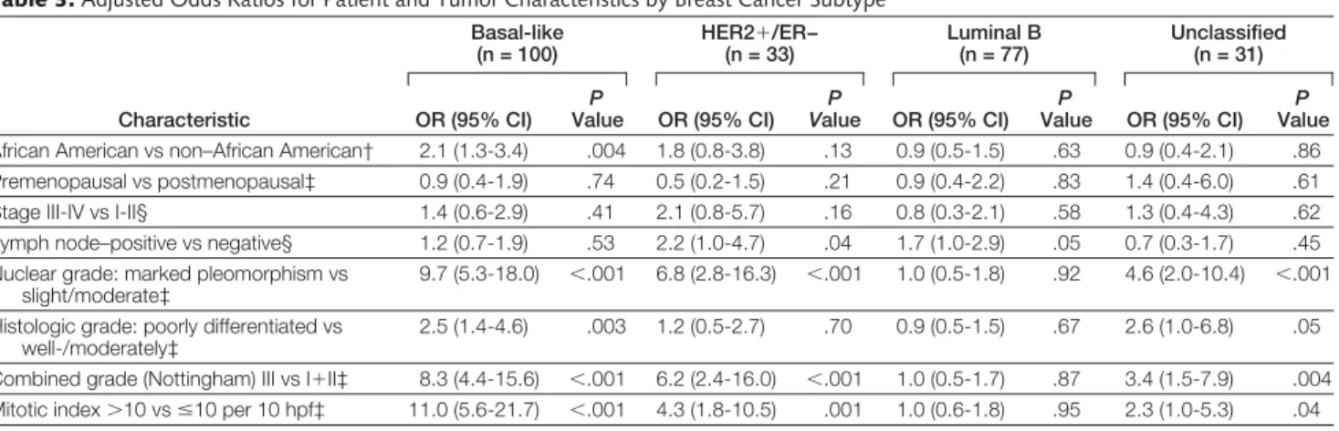
Odds ratios for the association of breast cancer subtypes with lymph node status, histologic grade, and mitotic in-dex are presented inTABLE3, with the luminal A subtype (the most common IHC subtype representing 51% of the cases) serving as the referent group. Odds ratios were adjusted for age, stage, and race. Compared with the luminal A
subtype, patients with basal-like tu-mors were 2.1 times more likely to be African American (P= .004). Likeli-hood ratio tests showed a significant interaction between race and meno-pausal status for developing the basal-like subtype (P= .02), but not
HER2⫹/ER− (P= .49), luminal B
(P=.62), or unclassified tumors (P=.58) compared with luminal A. In compari-son with luminal A tumors and after ad-justment for age, race, and stage, the basal-like subtype was 11 times more likely to have high mitotic index (P⬍.001), 9.7 times more likely to have high nuclear grade (P⬍.001), and 2.5 times more likely to have high histo-logic grade (P=.003). The basal-like sub-type was not associated with the pres-ence of positive axillary lymph nodes at the time of diagnosis (P=.53), whereas
both HER2⫹ subtypes (HER2⫹/ER−
and luminal B) were significantly more likely to have positive lymph nodes at presentation (P=.04). Notably, a strong association with high histologic, nuclear, and mitotic grade was seen for both sub-types of ER− tumors, namely the
basal-like and HER2⫹/ER− tumors.
How-ever, the HER2⫹/ER− subtype was not significantly associated with race or menopausal status.
TheTP53sequence-based mutation
analysis was performed on 330 of the 496 IHC classified breast cancer cases,
of which 84 (25%) had TP53
muta-tions. The presence of TP53
muta-tions differed significantly with IHC subtype: 44% (28 of 63) of basal-like tumors and 43% (10 of 23) of HER2⫹/
ER− subtype tumors contained TP53
mutations, whereas only 23% (12 of 52) of luminal B and 15% (25 of 175) of lu-minal A were mutation-positive (P⬍.001). These findings were in agree-ment with previous comparisons of the breast tumor intrinsic subtypes and TP53mutation status14as well as
pre-vious demonstration of a high
propor-tion of p53-mutant tumors inBRCA1
and cytokeratin 5/6–positive tumors.39,40
A subset of CBCS patients were
screened for BRCA1 germline
muta-tions.31Of the 496 cases assayed, 211
were screened for mutations inBRCA1, with 4 carriers and 1 variant of un-Table 2.Prevalence of Breast Cancer Subtypes According to Race and Menopausal Status
Tumor Status
All Cases
No. (%)
African American* Non–African American†
Premenopausal (n = 97)
Postmenopausal (n = 99)
Premenopausal (n = 164)
Postmenopausal (n = 136)
Basal-like 100 38 (39) 14 (14) 26 (16) 22 (16)
HER2⫹/ER− 33 9 (9) 7 (7) 9 (6) 8 (6)
Luminal A 255 35 (36) 58 (59) 83 (51) 79 (58)
Luminal B 77 9 (9) 16 (16) 30 (18) 22 (16)
Unclassified 31 6 (6) 4 (4) 16 (10) 5 (4)
*P⬍.001,2test for basal-like vs other tumor types in premenopausal vs postmenopausal African American women.
†P= .94,2test for basal-like vs other tumor types in premenopausal vs postmenopausal non–African American women.
Table 3.Adjusted Odds Ratios for Patient and Tumor Characteristics by Breast Cancer Subtype*
Characteristic
Basal-like (n = 100)
HER2⫹/ER−
(n = 33)
Luminal B (n = 77)
Unclassified (n = 31)
OR (95% CI)
P
Value OR (95% CI)
P
Value OR (95% CI)
P
Value OR (95% CI)
P
Value
African American vs non–African American† 2.1 (1.3-3.4) .004 1.8 (0.8-3.8) .13 0.9 (0.5-1.5) .63 0.9 (0.4-2.1) .86
Premenopausal vs postmenopausal‡ 0.9 (0.4-1.9) .74 0.5 (0.2-1.5) .21 0.9 (0.4-2.2) .83 1.4 (0.4-6.0) .61
Stage III-IV vs I-II§ 1.4 (0.6-2.9) .41 2.1 (0.8-5.7) .16 0.8 (0.3-2.1) .58 1.3 (0.4-4.3) .62
Lymph node–positive vs negative§ 1.2 (0.7-1.9) .53 2.2 (1.0-4.7) .04 1.7 (1.0-2.9) .05 0.7 (0.3-1.7) .45 Nuclear grade: marked pleomorphism vs
slight/moderate‡
9.7 (5.3-18.0) ⬍.001 6.8 (2.8-16.3) ⬍.001 1.0 (0.5-1.8) .92 4.6 (2.0-10.4) ⬍.001
Histologic grade: poorly differentiated vs well-/moderately‡
2.5 (1.4-4.6) .003 1.2 (0.5-2.7) .70 0.9 (0.5-1.5) .67 2.6 (1.0-6.8) .05
Combined grade (Nottingham) III vs I⫹II‡ 8.3 (4.4-15.6) ⬍.001 6.2 (2.4-16.0) ⬍.001 1.0 (0.5-1.7) .87 3.4 (1.5-7.9) .004
Mitotic index⬎10 vsⱕ10 per 10 hpf‡ 11.0 (5.6-21.7) ⬍.001 4.3 (1.8-10.5) .001 1.0 (0.6-1.8) .95 2.3 (1.0-5.3) .04
Abbreviations: CI, confidence interval; hpf, high-power field; OR, odds ratio.
*The luminal A (ER⫹and/or progesterone receptor positive, HER2−) subtype acts as the referent group. †Adjusted for age (11-level ordinal variable) and stage (I, II, III⫹IV).
known effects being identified. The BRCA1mutation carriers comprised 1 luminal A tumor, 1 unclassified tumor, and 2 basal-like tumors. Al-though these numbers were very small, the data were consistent with
earlier findings that most BRCA1
mutant tumors show the basal-like phenotype15,41,42and that mostBRCA1
mutant tumors do not show HER2 posi-tivity.43
Survival by IHC Subtype
The maximum duration of follow-up for the CBCS phase 1 cases was 11.2 years (minimum of 8.1 years). During this pe-riod of observation, the study patients had 73% overall survival (232 deaths among 861 cases). Of the 232 deaths, 170 were considered breast cancer-specific, giving an overall disease-specific survival of 80% (691 of 861). Af-rican AmeAf-rican cases had worse breast cancer-specific survival (74%) com-pared with non–African American cases
(84%) (P⬍.001). Age, race,
meno-pausal status, stage, ER status, PR sta-tus,TP53mutation status, mitotic in-dex, nuclear grade, and histologic grade were also significant predictors of breast cancer-specific survival (P⬍.001 for each).
The breast cancer subtypes also dif-fered significantly in breast cancer-specific survival (P⬍.001): basal-like
subtype (75%), HER2⫹/ER− subtype
(52%), luminal A (84%), luminal B (87%), and unclassified (77%). Kaplan-Meier survival curves for breast cancer-specific survival are presented in FIGURE2. A steep fall in breast cancer– specific survival was observed in the first 4 to 5 years for the basal-like and
HER2⫹/ER− tumors, with
particu-larly poor survival for the HER2⫹/ ER− subtype. A similar early relapse
pat-tern has been described forBRCA1
tumors.44,45Over the entire
observa-tion period, breast cancer–specific sur-vival was significantly worse among basal-like (hazard ratio, 1.8; 95% CI, 1.1-2.9;P=.03) and HER2⫹/ER− breast cancer patients (hazard ratio, 3.5; 95% CI, 1.9-6.2;P⬍.001) compared with lu-minal A as the referent group.
The difference in survival by breast cancer subtype was seen both among lymph node–positive patients (P= .01) and lymph node–negative patients (P= .03). Data were sparse after strati-fying on lymph node status and should be interpreted with caution. Breast can-cer–specific survival within lymph no-de–positive patients by subtype was the following: basal-like (51%), HER2⫹/ ER− (39%), luminal A (65%), luminal B (83%), and unclassified (44%). Within the lymph node−negative pa-tients, breast cancer-specific survival was the following: basal-like (93%),
HER2⫹/ER− (71%), luminal A (94%),
luminal B (92%), and unclassified (91%).
The outcomes in premenopausal Af-rican AmeAf-rican cases did not become more similar to the other groups when basal-like cases were removed. The breast cancer−specific survival by ra-cial and menopausal subsets without basal-like breast cancers still differed significantly: premenopausal African American 64%, postmenopausal Afri-can AmeriAfri-can 81%, premenopausal non–African American 81%, and post-menopausal non–African American
91% (P⬍.001). These data suggest that factors other than subtype, such as ac-cess to treatment, could also be influ-encing survival in younger African American women.
COMMENT
Gene expression profiling has identi-fied breast cancer intrinsic subtypes that predict distinct clinical outcomes14,15
and which have been shown to be pres-ent in women of multiple ethnici-ties.46The basal-like subtype has been
associated with poor clinical out-comes,15,16which likely reflect this
sub-type’s high proliferative capacity14-16as
well as the lack of directed therapies since basal-like tumors do not typi-cally express ER− or overexpress HER2.17To facilitate investigation of the
population-based frequencies of the basal-like breast cancer subtype, we re-fined an IHC-based assay to identify the main breast tumor intrinsic subtypes. We used the IHC method for catego-rization and determined for the first time the population-based prevalence of these subtypes. Although IHC-based assays do not provide as much biological insight into tumor biology as Figure 2.Survival Analysis of the Carolina Breast Cancer Study Cases Grouped Using the Refined Breast Tumor Immunohistochemical Intrinsic Subtypes
P <.001
Survival
Basal-like Luminal B Luminal A Unclassified
HER2+/ER–
No. at Risk
89 78 72 71 25
Basal-like
76 67 65 63 17
Luminal B
245 226 213 197 81
Luminal A
30 28 25 23 9
Unclassified
30 27 21 20 7
HER2+/ER– 0 1.0
0.8
0.6
0.4
0.2
2 4 6 8 10
Disease-Specific Survival Time, y
mRNA-based assays containing thou-sands of genes, this IHC assay allowed classification of tumors into catego-ries that have demonstrated associa-tions between intrinsic subtypes and proliferation rates, overall survival, TP53status, andBRCA1mutation
sta-tus.14,15,17,29,41,42The reproducible
cor-relations across different studies and when using different assays (IHC and DNA microarray expression profiles) shows that we are tracking common tu-mor subtypes with similar biologic characteristics and clinical behaviors across distinct patient sets. The IHC-based classification system also allows analyses of subtypes to be conducted in patient populations where fresh tis-sue is not available.
In the population-based CBCS, the prevalence of the basal-like and lumi-nal A breast cancer subtypes was strongly influenced by race and meno-pausal status; the highest prevalence of basal-like and lowest prevalence of lu-minal A tumors were observed among premenopausal African American breast cancer patients. Because the CBCS is a population-based sample, within de-fined race and age groups estimates of prevalence are likely to be representa-tive of the underlying North Carolina population.2Differences between the
CBCS and breast cancer patients re-ported to the North Carolina Central Cancer Registry include a lower pro-portion of African American women be-tween the ages of 40 and 59 years with higher-stage tumors and lower partici-pation among women from lower so-cioeconomic and educational strata.2,23
Each of these factors could actually pro-duce an underestimate of the preva-lence of more aggressive breast cancer
subtypes (basal-like and HER2⫹/
ER−) among younger African Ameri-can cases enrolled in the CBCS. How-ever, this potential bias may have been partially offset by the fact that the analy-sis of IHC markers in the CBCS was based on patients with larger tumors. A high frequency of basal-like tu-mors was observed in a study of breast cancer in Nigerian women, among whom ER-negative and
HER2-nega-tive tumors comprised 87 of 148 women, or 59% of total cases.47According to gene
expression studies, ER-negative breast tumors fall into 1 of 2 categories,14,15
namely basal-like tumors (ER−, PR−, and
HER−) and the HER2⫹/ER− subtype
(HER2⫹/ER−) (Figure 1). The HER2⫹/ ER– group, which is also a high-grade and ER-negative tumor group, did not vary significantly with age or race. These findings suggest that associations be-tween premenopausal breast cancer, race, and hormone status in the CBCS was driven by an excess of the basal-like subtype. Breast cancers that
de-velop amongBRCA1mutation carriers
are generally basal-like.15,41,42However,
very fewBRCA1mutation carriers were present in the CBCS, with 2 out of the 4 known carriers falling into the basal-like category. NoBRCA1carriers were identified among the African American cases tested in the CBCS and only a single variant of unknown biological sig-nificance was identified.31ThusBRCA1
variants are unlikely to explain the high prevalence of basal-like breast cancer in younger African American patients in this study.
Basal-like breast cancers in the CBCS exhibited aggressive features, includ-ing high proliferative capacity (mea-sured by mitotic index), high histo-logic grade, high nuclear grade, and frequentTP53mutations. Even after ad-justment for age, race, and stage, the as-sociation of basal-like and HER2⫹/ ER− subtypes with aggressive features remained significant. These findings were expected given the high expres-sion of the proliferation cluster of genes in microarray analyses of basal-like and HER2⫹/ER− subtype tumors.13-15,48The
association of race with high-grade breast tumors and ER negativity has been previously reported.2,3However,
our study suggests that this associa-tion is driven by the increased preva-lence of basal-like tumors and not by an increase in HER2⫹/ER− subtype.
The observation that the intrinsic breast cancer subtypes carry different prognoses was confirmed in the CBCS. Disease-specific survival was signifi-cantly lower among breast cancer cases
with basal-like and HER2⫹/ER− tumors, and more favorable among cases with luminal A tumors. The HER2⫹/ER− sub-type appears particularly prone to early and frequent relapse, befitting the clini-cal experience with HER2 overexpress-ing tumors49; the CBCS cases in this study
were diagnosed between 1993 and 1996 and were not treated with the anti– HER2 monoclonal antibody trastu-zumab. Basal-like tumors were more fre-quent in younger African American women in the CBCS, and could contrib-ute to their poor prognosis compared with other breast cancer patients. How-ever, when cases of basal-like tumors were removed, the breast cancer– specific survival remained significantly worse among premenopausal African American cases. As noted previously, this may reflect the impact on prognosis of access to care, treatment, or other dif-ferences. In other words, while the high incidence of the poor-prognosis basal-like subtype may contribute to their rela-tively worse outcome, it does not entirely explain the poor outcomes seen in younger African Americans. We lacked treatment data in the CBCS, so we could not examine interactions between IHC subtypes and efficacy of cancer therapy. Examination of tumor microarray data using patients treated with surgery alone also suggests that these subtypes are prognostic and reflect the natural his-tory of these tumors.15Interestingly,
unlike HER2⫹/ER− and luminal B
tumors, the basal-like subtype was not associated with involvement of posi-tive axillary lymph nodes, a finding that was previously noted in a study of cyto-keratin 5/6–positive tumors that over-sampledBRCA1tumors.40Since
basal-like breast cancers still carried a poor prognosis, it is possible, as suggested by others,40that this finding reflects a
pre-dominantly hematogenous, rather than lymphatic, pattern of dissemination. Fur-ther studies are needed to address this issue.
race and breast cancer, it is important that race be evaluated in the context of other variables such as stage at diag-nosis and tumor histology. Informa-tion on breast cancer risk factors will help to determine whether basal-like tu-mors have a different underlying etiol-ogy compared with other types of breast cancer. SinceBRCA1carriers tend to de-velop basal-like tumors, there may be other inherited genetic variants that pre-dispose to developing specific sub-types of breast cancer.15,21 The
ab-sence ofBRCA1carriers among African American breast cancer patients in the CBCS suggests that genes other than BRCA1could predispose women to basal-like breast cancers; however, en-vironmental and socioeconomic fac-tors could also play a role in the ob-served distribution of breast cancer subtypes. Notably, in the CBCS, the
prevalence ofBRCA1mutations was 0
in African Americans and low (3.3%) in non–African Americans.31Most
im-portantly, our data suggest that epide-miological studies of breast cancer in African American women should con-sider the joint distribution of ER, PR, and HER2 status (ie, subtypes), rather than rely on ER and PR status alone. Previous analyses typically group to-gether HER2⫹/ER− tumors with basal-like tumors under the ER-negative des-ignation; however, in the CBCS,
HER2⫹/ER− tumors were not
associ-ated with race or menopausal status. The high prevalence of basal-like tu-mors in younger African American women could contribute to their higher breast cancer mortality. Additional studies of long-term survival among pa-tients with specific breast cancer sub-types are needed. Clinical trials aimed at identifying therapeutic approaches to the management of basal-like breast cancer are also needed, especially for young African American women.
Author Contributions:Dr Millikan had full access to all of the data in the study and takes responsibility for the integrity of the data and the accuracy of the data analysis.
Study concept and design:Carey, Perou, Moorman, Millikan.
Acquisition of data:Livasy, Dressler, Conway, Edmiston, Deming, Geradts, Cheang, Nielsen, Moorman, Millikan.
Analysis and interpretation of data:Carey, Perou, Livasy, Dressler, Cowan, Conway, Karaca, Troester, Tse, Deming, Cheang, Nielsen, Earp, Millikan.
Drafting of the manuscript:Carey, Perou, Tse, Nielsen, Millikan.
Critical revision of the manuscript for important in-tellectual content:Carey, Perou, Livasy, Dressler, Cowan, Conway, Karaca, Troester, Edmiston, Deming, Geradts, Cheang, Nielsen, Moorman, Earp, Millikan.
Statistical analysis:Tse, Deming, Cheang, Millikan.
Obtained funding:Perou, Dressler, Conway, Earp, Millikan.
Administrative, technical, or material support:Carey, Livasy, Dressler, Cowan, Conway, Karaca, Troester, Edmiston, Nielsen, Moorman, Millikan.
Study supervision:Carey, Perou, Dressler, Conway, Edmiston, Nielsen, Millikan.
Financial Disclosures:None reported.
Funding/Support:This work was supported by an award to the University of North Carolina for a Breast Cancer Specialized Program of Research Excellence (SPORE) from the National Cancer Institute (NIH/ NCI P50-CA58223), a grant from the General Clini-cal Research Centers Program of the Division of Re-search Resources/National Institutes of Health (M01RR00046 awarded to Dr Carey), and by the NCI (RO1-CA-101227-01 awarded to Dr Perou). Role of the Sponsor:All study funding was from pub-lic grants for scientific research. The funding organi-zations had no role in the design and conduct of the study; the collection, analysis, and interpretation of the data; or the preparation, review, or approval of the manuscript.
Previous Presentation:This work was presented in part at the 40th Annual Meeting of the American Society of Clinical Oncology; New Orleans, La; June 2004. Acknowledgment:For their critical review, we thank Barbara Rimer, PhD, School of Public Health; and Paul Godley, MD, PhD, and Matthew G. Ewend, MD, School of Medicine, University of North Carolina. They were not compensated for their time.
REFERENCES
1.Surveillance, Epidemiology, and End Results Pro-gram (SEER) SEER Stat Database: Mortality—All COD, Public-Use With State, Total U.S. for Expanded Races/ Hispanics (1990-2001). National Cancer Institute, DC-CPS, Surveillance Research Program, Cancer Statis-tics Branch, released April 2004. http://www.seer .cancer.gov.
2. Furberg H, Millikan R, Dressler L, Newman B, Geradts J. Tumor characteristics in African American and white women.Breast Cancer Res Treat. 2001;68: 33-43.
3.Porter PL, Lund MJ, Lin MG, et al. Racial differ-ences in the expression of cell cycle-regulatory proteins in breast carcinoma.Cancer. 2004;100:2533-2542.
4.Eley JW, Hill HA, Chen VW, et al. Racial differ-ences in survival from breast cancer: results of the Na-tional Cancer Institute Black/White Cancer Survival Study.JAMA. 1994;272:947-954.
5.Clegg LX, Li FP, Hankey BF, Chu K, Edwards BK. Cancer survival among US whites and minorities: a SEER (Surveillance, Epidemiology, and End Results) Pro-gram population-based study.Arch Intern Med. 2002; 162:1985-1993.
6.Shavers VL, Brown ML. Racial and ethnic dispari-ties in the receipt of cancer treatment.J Natl Cancer Inst. 2002;94:334-357.
7.del Carmen MG, Hughes KS, Halpern E, et al. Ra-cial differences in mammographic breast density.
Cancer. 2003;98:590-596.
8.Ademuyiwa FO, Olopade OI. Racial differences in genetic factors associated with breast cancer.Cancer Metastasis Rev. 2003;22:47-53.
9.Joslyn SA. Racial differences in treatment and
sur-vival from early-stage breast carcinoma.Cancer. 2002; 95:1759-1766.
10. Mountain JL, Risch N. Assessing genetic contributions to phenotypic differences among “racial” and “ethnic” groups.Nat Genet. 2004; 36(11 suppl):S48-S53.
11.Schwartz RS. Racial profiling in medical research.
N Engl J Med. 2001;344:1392-1393.
12. Tate SK, Goldstein DB. Will tomorrow’s medi-cines work for everyone? Nat Genet. 2004; 36(11 suppl):S34-S42.
13. Perou CM, Sorlie T, Eisen MB, et al. Molecular por-traits of human breast tumours.Nature. 2000;406:747-752.
14.Sorlie T, Perou CM, Tibshirani R, et al. Gene ex-pression patterns of breast carcinomas distinguish tu-mor subclasses with clinical implications.Proc Natl Acad Sci U S A. 2001;98:10869-10874.
15. Sorlie T, Tibshirani R, Parker J, et al. Repeated ob-servation of breast tumor subtypes in independent gene expression data sets.Proc Natl Acad Sci U S A. 2003; 100:8418-8423.
16. Sotiriou C, Neo SY, McShane LM, et al. Breast can-cer classification and prognosis based on gene expres-sion profiles from a population-based study.Proc Natl Acad Sci U S A. 2003;100:10393-10398. 17.Nielsen TO, Hsu FD, Jensen K, et al. Immunohis-tochemical and clinical characterization of the basal-like subtype of invasive breast carcinoma.Clin Can-cer Res. 2004;10:5367-5374.
18.Finlin BS, Gau CL, Murphy GA, et al. RERG is a novel RAS-related, estrogen-regulated and growth-inhibitory gene in breast cancer.J Biol Chem. 2001;276: 42259-42267.
19. Usary J, Llaca V, Karaca G, et al. Mutation of GATA3 in human breast tumors.Oncogene. 2004;23: 7669-7678.
20. Hedenfalk I, Duggan D, Chen Y, et al. Gene-expression profiles in hereditary breast cancer.N Engl J Med. 2001;344:539-548.
21. van ’t Veer LJ, Dai H, van de Vijver MJ, et al. Gene expression profiling predicts clinical outcome of breast cancer.Nature. 2002;415:530-536.
22.Newman B, Moorman PG, Millikan R, et al. The Carolina Breast Cancer Study: integrating population-based epidemiology and molecular biology.Breast Can-cer Res Treat. 1995;35:51-60.
23.Moorman PG, Newman B, Millikan RC, Tse CK, Sandler DP. Participation rates in a case-control study: the impact of age, race, and race of interviewer.Ann Epidemiol. 1999;9:188-195.
24.Millikan RC, Pittman GS, Newman B, et al. Ciga-rette smoking, N-acetyltransferases 1 and 2, and breast cancer risk.Cancer Epidemiol Biomarkers Prev. 1998; 7:371-378.
25.Genestie C, Zafrani B, Asselain B, et al. Compari-son of the prognostic value of Scarff-Bloom-Richardson and Nottingham histological grades in a series of 825 cases of breast cancer: major impor-tance of the mitotic count as a component of both grading systems.Anticancer Res. 1998;18(1B): 571-576.
26.Huang WY, Newman B, Millikan RC, Schell MJ, Hulka BS, Moorman PG. Hormone-related factors and risk of breast cancer in relation to estrogen receptor and progesterone receptor status.Am J Epidemiol. 2000;151:703-714.
27. Landis JR, Koch GG. The measurement of ob-server agreement for categorical data.Biometrics. 1977;33:159-174.
28.Millikan R, Eaton A, Worley K, et al. HER2 codon 655 polymorphism and risk of breast cancer in Afri-can AmeriAfri-cans and whites.Breast Cancer Res Treat. 2003;79:355-364.
29.van de Rijn M, Perou CM, Tibshirani R, et al. Ex-pression of cytokeratins 17 and 5 identifies a group of breast carcinomas with poor clinical outcome.
30.Conway K, Edmiston SN, Cui L, et al. Prevalence and spectrum of p53 mutations associated with smoking in breast cancer.Cancer Res. 2002;62:1987-1995.
31.Newman B, Mu H, Butler LM, Millikan RC, Moor-man PG, King MC. Frequency of breast cancer attributable to BRCA1 in a population-based series of American women.JAMA. 1998;279:915-921.
32.Rich-Edwards JW, Corsano KA, Stampfer MJ. Test of the National Death Index and Equifax Nationwide Death Search.Am J Epidemiol. 1994;140:1016-1019.
33.SAS Support technical FAQ (354). What exact and Monte Carlo methods are available and in which pro-cedures and releases do they appear? http://support .sas.com/faq/003/FAQ00354.html. Accessed Febru-ary 8, 2006.
34. Hosmer DW, Lemeshow S.Applied Logistic Regression.New York, NY: Wiley & Sons; 1989. 35. Bland JM, Altman DG. Survival probabilities (the Kaplan-Meier method).BMJ. 1998;317: 1572.
36.Bland JM, Altman DG. The logrank test.BMJ. 2004;328:1073.
37.Cox DR, Oakes D.Analysis of Survival Data. Lon-don, England: Chapman & Hall; 1984.
38.Dupont WD, Plummer WD. Power and sample size calculations: a review and computer program. Con-trol Clin Trials. 1990;11:116-128.
39.Crook T, Brooks LA, Crossland S, et al. p53 Mu-tation with frequent novel condons but not a mutator phenotype in BRCA1- and BRCA2-associated breast tumours.Oncogene. 1998;17:1681-1689.
40.Foulkes WD, Brunet JS, Stefansson IM, et al. The prognostic implication of the basal-like (cyclin E h i g h / p 2 7 l o w / p 5 3⫹/ g l o m e r u l o i d m i c r o -vascular-proliferation⫹) phenotype of BRCA1-related breast cancer.Cancer Res. 2004;64:830-835.
41.Foulkes WD, Stefansson IM, Chappuis PO, et al. Germline BRCA1 mutations and a basal epithelial phe-notype in breast cancer.J Natl Cancer Inst. 2003;95: 1482-1485.
42.Olopade OI, Grushko T. Gene-expression pro-files in hereditary breast cancer.N Engl J Med. 2001; 344:2028-2029.
43.Grushko TA, Blackwood MA, Schumm PL, et al. Molecular-cytogenetic analysis of HER-2/neu gene in BRCA1-associated breast cancers.Cancer Res. 2002; 62:1481-1488.
44.Foulkes WD, Wong N, Brunet JS, et al. Germ-line BRCA1 mutation is an adverse prognostic factor
in Ashkenazi Jewish women with breast cancer.Clin Cancer Res. 1997;3:2465-2469.
45.Stoppa-Lyonnet D, Ansquer Y, Dreyfus H, et al. Familial invasive breast cancers: worse outcome related to BRCA1 mutations.J Clin Oncol. 2000;18: 4053-4059.
46.Yu K, Lee CH, Tan PH, Tan P. Conservation of breast cancer molecular subtypes and transcriptional patterns of tumor progression across distinct ethnic populations.Clin Cancer Res. 2004;10:5508-5517.
47.Olopade OI, Ikpatt FO, Dignam JJ, et al. “Intrin-sic gene expression” subtypes correlated with grade and morphometric parameters reveal a high propor-tion of aggressive basal-like tumors among black women of African ancestry. Paper presented at: Ameri-can Society of Clinical Oncology Annual Meeting; June 2004; New Orleans, La.
48. Whitfield ML, Sherlock G, Saldanha AJ, et al. Iden-tification of genes periodically expressed in the hu-man cell cycle and their expression in tumors.Mol Biol Cell. 2002;13:1977-2000.