Journal of Global Pharma Technology
Available Online at:
www.jgpt.co.in
RESEARCH ARTICLE
Walking Analysis Using the Angles around the Midpoint between
Print Length and Toe Spread by Four Different Color Footprints
and SFI in Sciatic Nerve-Injured Rat Model Treated With
Curcumin
Ria Margiana
1,3*
,Silvia Werdhy Lestari
2,3,Sasanthy Kusumaningtyas
1,3,Khoirul Ima
3,Kamila Alawiyah
11. Department of Anatomy, Faculty of Medicine, Universitas Indonesia, Jakarta, Indonesia. 2. Department of Medical Biology, Faculty of Medicine, Universitas Indonesia, Jakarta, Indonesia.
3. Master’s Programme in Biomedical Sciences, Faculty of Medicine, Universitas Indonesia, Jakarta,
Indonesia.
*Corresponding Author: Ria Margiana
Abstract
SFI, the sciatic function index, has been applied as an evaluation method in walking analysis over a long period. It is a method that addresses the functionality of nerves using footprints during walking analysis. Other techniques developed previously make use of one-color ink. The method has difficulties in interpretation due to footprints mingled among the mice specimen's four legs. There exists a possibility of the rats not walking straight, and this leads to overlapping, and the result is complicated measurements. SFI, as a method, entails the application of a mathematical formula that is special. Researchers have questioned this method since it is affected by muscles' contraction, automatic mutilation, and rat motion speed. During this study, the idea is to use four different colors, allowing easier and faster interpretation of the footprints results. Moreover, the study will also modify tools based on the already existing available tools in pre-existing literature. The modified tool gives a measurement of print leg denoted as PL, toe spread TS, and inter- toe spread IT’S, all belonging to the SFI component. The tool's validity and reliability are essential when applied in the measurement of components used in the functional analysis of nerves. Confirmation from the study is that results from SFI and TOA (toe out of angle) all describe footprint. Toe out of angle is an essential measurement in the walking analysis as it has an impact on the alteration of abduction-adduction as well as the internal-external navigation of the hind legs. The study will help develop a method that assesses nerves' functional analysis using the sciatic functional analysis and total out of angle spread. The angle at the intersection point of the print length and the toe spread.
Keywords: SFI, TOA and ITS.
Introduction
One of the most re-known and peripheral injuries of the nerves is suffered by 3-10 % of patients [1]. Periphery injury of the nerves is the leading cause of disability in young male adults in society. Injury of the nerves takes a long for patient care and is propelled by several underlying factors. The sciatica epidemiology definition has seen itself lack of clarification in the epidemic’s literature. For example, in the United Kingdom, sciatica's prevalence is 3.1 % in males and 1.3 % in the opposite gender [2]. Like in the most common
research, the study did not apply more strict criteria in diagnosing sciatica [35].
make it less responsive to any treatment. The importance of this study is mainly to assess the clinical progress related to nerve injury.
Animal specimens are used to determine the functional indexes of nerves, and the instruments used are footprints obtained from rats in motion [4]. In general, rats' footprints are hard to perform an analysis, and hence the rats are allowed to walk under restriction. Basic motion in rats’ hind limbs includes abduction, extension, rotation, flexion, and adduction.
These motions are under the body posture influence when walking either in a straight or zigzag motion [5]. Toe out of angle TOA is an explanation of the biomechanical effects of the stance phase, usually the external revolving motion in the rat's walking mode [6]. The changes in the toe out of angle usually occur since the lateral and the medial parts of the rat's paw innervated by tibial and fibular types of nerves are divided from the sciatic nerve, and they can be easily influenced in the case of an injury [7].
The study incorporates a fundamental assumption that is a developed parameter that is consistent and bares a positive relationship with the other parameters. Based on this, the study considers an extra parameter, which is the angle formed when two lines form an intersection. These lines exist between the heel plus the third toe and between the thumb plus the little toe. Angles formed can provide the impacts of abduction-adduction motions, the external-internal rotation of the hind limb’s rats, and the possibility of a direct proportionality to other functional parameters [8].
Materials and Methods
Rat Specimen
It is established that 36 and 12 weeks old male rats weighing 250-300 grams were randomly identified and assigned into two groups [9]. These two groups comprised a control group n=6 and an experimental group that was subject to surgery [12]. Out of the experimental group, six rats were collected randomly during the first to the sixth week, excluding the fifth after the surgery. These further created subgroups, namely 1w, 2w, 3w, 4w, and 6w. These were further used for the footprint analysis and evaluation of SFI [10].
Examination using Growth Associated Protein GAP-43
Frozen tissues are harvested while still fresh from the rats, thoroughly cleaned and later immersed in cold PBS [11]. A suture is tied around the distal end to facilitate orientation and fast embedded in the appropriate medium, frozen and ready for sectioning. Paraffin embedded tissues in holding cassettes will be immersed in paraffin during the processing final stage. Application of the paraffin embedder with a cold plate ensures that the embedder is heated first almost 3 degrees the melting point of wax [12].
All the pellets of paraffin are in liquefied form while the cold plate is being chilled first. A test to the embedding block is done to ensure that the machine is fully operational seeing that the paraffin hardens following a short time on the cold plate. The cassette is then carefully removed from the processor and placed on the heating block so as to keep the wax melted. An already labelled embedding block is placed in a stainless-steel base mold and placed on the heating block [13].
Using one hand; hold the embedding block as well as the base of the mold under the paraffin dispenser. Using forceps, the vessel is carefully picked so as to allow embedding in the right orientation inside the embedding block. Care should be taken when releasing the vessel from the forceps since the wax can cause them to stick together and dislocate the vessel out of its original vertical position.
After a short time. It is expected that the block will start to harden [6]. Upon complete solidification of paraffin, the block is moved from the cold plate and the steel base mold is released. The block is then stored at room temperature waiting sectioning.
Specimen Surgery
the surrounding tissues, a crush injury of 2 mm long was formed at the point directly under the gluteal tuberosity. The latter was done with the use of a standard hemostat used for surgical. The injury site was then marked using a stereomicroscope having a single 9-0 nylon stitch epineural at the injury end for identification later [16]. Muscles and skin were then wrapped and closed using 4-0 sutures made of nylon, and the rats returned to their cages.
Specimen Sciatic Function Index
Analysis
It was done in a wooden walking alley with bilateral walls of plastic plates prepared in advance [17]. The wooden way was prepared prior and constituted of a special alley box at the end. For the floor specification, the alley was wrapped using paper tape and a brush used for ink smearing entirely over the rat's plantar pedis.
The rat was then put at the alley's entry point and would always walk following a straight path into the special alley [14, 18, 19].While this took place, the rat left footprints on the paper. About three footprints were selected, which appeared legible from the various prints that had been left by each rat. The prints' identification was
followed by measurement of three
parameters that included the print length defined as the distance from the heel and the third toe PL and the distance from the second to the fourth toe defined as the intermediate toe spread [13]. SFI was thus obtained and calculated from the formula
SFI = -38.3 ((EPL –NPL ) / NPL) + 109.5 ((ETS – NTS)/ NITS) –8.8
Where E stands for the experimental side and N, the normal side, the value of 0
indicates a normal function while a value of -100 is an indication of total impairment [20].
Statistical Analysis
Data collected were presented as average. The variations were assessed for significance with the application of a single way ANOVA [21]. Dunnet test was applied to analyze the importance of the variations between the control experiment specimen and the experimental specimen. Tukeys HSD test was performed to comparisons within the groups following surgery [12]. The primary focus was on performing comparisons of the coinciding groups for the study as alterations in these data portrayed improvements in most of the circumstances. The change in SFI did not show any trend towards progress from 1w heading to 2w [21].
Results
The value of SFI (Sciatic function index) generally improved upwards. However, there was no trend indicated in the improvement from 1w to 2w [11]. The value of SFI recorded by the specimen in the control experiment was -7.88 ± 3.10. As time proceeded, there were changes in the value of SFI as -84.89 ± 2.38 in week one, 87.76 ± 2.52 in week two, -34.67 ± 2.54 in week three, -13.66 ± 1.93 in week four, and -10.92 ± 1.84 in week six. 1w, 2w, and 3w groups had significant variations as compared to the control experiment group (p < 0.01) [31]. Comparisons of the adjacent groups showed indications of variations in 2w and 3w (p < 0.01) while that of 3w and 4w (p < 0.01).
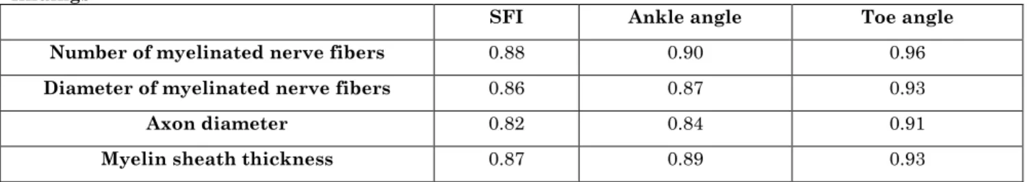
Relationship between the sciatic function index, the ankle angle, and the toe angle with the number of myelinated nerve fibers, myelinated nerve fibers diameters, axon diameter as well as the thickness of the myelin sheath is as represented in the table.
Table 1: Relationship between sciatic function indexes (SFI), ankle angle, and toe angle summary
findings
SFI Ankle angle Toe angle
Number of myelinated nerve fibers 0.88 0.90 0.96
Diameter of myelinated nerve fibers 0.86 0.87 0.93
Axon diameter 0.82 0.84 0.91
Myelin sheath thickness 0.87 0.89 0.93
From the table above, P < 0.05 while p < 0.01
SFI analysis was performed by identifying
as mention at the Margiana R et al research report [22]. An evaluation on the relationship between the number of myelinated nerve fibers, diameter of the myelinated nerve fibers as well as the axon diameter showed
that the values for the toe angle were considerably higher compared to the ankle angle and SFI.
‘
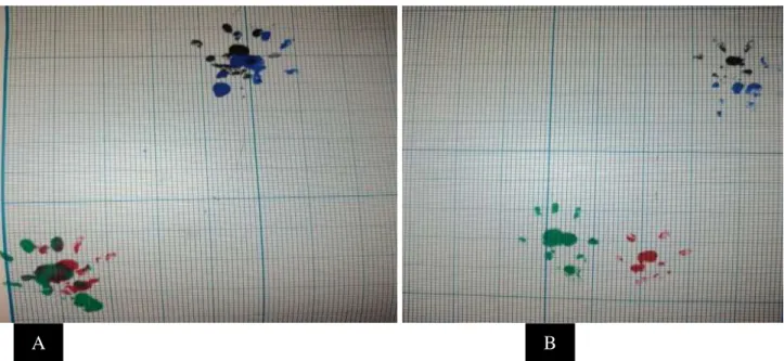
Fig. 1: Representative image of rat footprints after ink smearing over the rat's planta pedis. The surgical region and sciatic nerve-injured represented by the green footprint. Experimental group (A), Control group (B)
Discussion
From the results obtained from the experimental analysis, they demonstrate an increase for both the ankle and the toe angles [23]. These are very consistent with several changes, including the numbers of the myelinated fibers of the nerves, the diameters of the myelinated nerve fibers, the axon's diameter, and the sheath's thickness after surgery [24]. However, the ankle did not show any improvements after two weeks of operation.
Of important is that the ankle angle made improvements between 2w and 6w, and the toe angle also made improvements from 1w to 6w. In comparison to the control experiment group, there were significant variations until 4w for the toe angle value. For the ankle angle, the change only occurred until the 3w and sciatic function index.
On the centrally, SFI did not have any trend that suggested improvement for the first two weeks after operation [25]. There only occurred minimal growth improvement from 4w as well as 6w [26]. To summarize, the ankle angle plus the toe angle were extensive indicators compared to the SFI for this study. SFI is very easy and obvious to perform and is applicable optimally in the rat sciatic nerve
crush model for injury despite being prone to underperformance in the rat sciatic conduit model, which results from its low sensitivity in addition to the intrinsic limitations [25].
GAP43 is examined in this research as its
important role in peripheral nerve
regeneration. GAP43 is involved in
peripheral nerve regeneration is supported by the findings that GAP43 is one of the factor that is having important role in many signalling pathway in peripheral ner regeneration such as cyclic adenosine monophosphate signaling pathway (cAMP) etc., as the candidate of neuroregeration marker and also from bioinformatics study. [26, 27, 28].2Curcunin can be thought as a potential agent to add to the stand art management therapy of sciatic nerve injury. As already reported in other research, many additional agents can give advantage to the therapy result of sciatic ner injury. [29].
Conclusion
In conclusion, SFI, ankle angle, and toe angle have a strong correlation. However, the ankle angle and the toe angle measurements are more accurate than the SFI in the model. In the injury model to the rat sciatic nerve, the
toe angle gives a better prediction compared to the ankle angle. The consideration of the toe angle as a parameter, which is very informative is very innovative, and the results are a picture of the improvements relating to the functional evaluation as well as increased sensitivity in comparison to the ankle angle.
More research and study will be essential in the future to determine the basic mechanism that toe angle is a more sensitive
measurement in contrast to the ankle angle in the toe-off phase of nerve regeneration during the early stages.
Acknowledgment
We would like to thank for the support of funding by Penelitian Dasar Unggulan Perguruan Tinggi from Indonesia Ministry of Research, Technology and Higher Education
with contract number
NKB-35/UN2.RST/HKp.05.00/2020.
References
1. Chamanara M, Rashidian A, Mehr SE,
Dehpour A-R, Shirkohi R, Akbarian R, et al (2019) Melatonin ameliorates TNBS-induced colitis in rats through the melatonin receptors: involvement of TLR4/MyD88/NF-κB signalling pathway. Inflammopharmacology, 27: 361- 71. 2. Chauderlier A, Delattre L, Buée L, Galas
M-C (2017) In vivo hyperthermic stress model: an easy tool to study the effects of
oxidative stress on neuronal tau
functionality in mouse brain. Methods Mol. Biol., 1523: 369-73.
3. Chang HM, Liu CH, Hsu WM, Chen LY,
Wang HP, Wu TH, et al (2014) Proliferative effects of melatonin on S chwann cells: implication for nerve regeneration following peripheral nerve injury. J. Pineal Res., 56: 322-32.
4. Coletti, D, Teodori L, Lin Z, Beranudin J F, AdamoS (2013) Restoration versus reconstruction: cellular mechanisms of skin, nerve and muscle regeneration
compared. Regenerative Medicine
Research, 1: 1:4.
5. Dent EW, Gupton SL, Gertler FB (2011) The Growth Cone Cytoskeleton in Axon Outgrowth and Guidance. Cold Spring Harbor Perspectives in Biology, 3(3): a001800.
6. Neumüller RA, Knoblich JA (2009)
Dividing cellular asymmetry: asymmetric cell division and its implications for stem cells and cancer. Genes & Development, 2675-2699.
7. Uittenbogaard M, Martinka DL,
ChiaramelloA (2003) The basic helix-loop-helix differentiation factor Nex1/MATH-2 functions as a key activator of the GAP-43gene. Journal of Neurochemistry, 84(4):678-688. 8.
8. Godenschwege AT, Simpson JH, Xiaoliang
S, Bashaw GJ, Goodman CS, Murphey RK (2002) Ectopic Expression in the Giant Fiber System of Drosophila Reveals Distinct Roles for Roundabout (Robo), Robo2, and Robo3 in Dendritic Guidance and Synaptic Connectivity. The Journal of Neuroscience, 22(8):3117-3129.
9. Dumont JR, Amin E, Poirier GL, Albasser
MM, Aggleton JP (2012) Anterior thalamic nuclei lesions in rats disrupt markers of neural plasticity in distal limbic brain regions. Neuroscience, 224 (2): 81-101. 10.Ghosh S, Banerjee S, Sil PC (2015) The
beneficial role of curcumin on
inflammation, diabetes and
neurodegenerative disease: A recent update. Food Chem. Toxicol., 83: 111-24.
11.Jones S, Eisenberg H, Jia X (2016)
Advances and future applications of augmented peripheral nerve regeneration. Int. J. Mol. Sci., 17: E1494.
12.Jeong W, Kung H, Cheng CC, Lim C, Jung
MJ, Lee J, et al (2017) Dexmedetomidine to help nerve regeneration in a rat sciatic nerve injury model. Pain Res Manag., 2017: 9045608.
13.Ivell R, Teerds K, Hoffman GE (2014) Proper Application of Antibodies for Immunohistochemical Detection: Antibody Crimes and How to Prevent Them. Endocrinology, 155(3):676-687.
14.Holahan MR, Honegger KS, Tabatadze N,
Routtenberg A (2007) GAP-43 gene expression regulates information storage. Learning & Memory, 14(6): 407-415.
15.Houschyar K, Momeni A, Pyles M, Cha J,
16.Williams RR, Venkatesh I, Pearse DD,
UdvadiaAJ, Bunge MB (2015)
MASH1/Ascl1a Leads to GAP43
Expression and Axon Regeneration in the Adult CNS. PLoS ONE 10(3):0118918. 17.Saijilafu HEM, Jiao LCM, Xu WL, Zhou
FQ (2013) PI3K-GSK3 signalling regulates mammalian axon regeneration by inducing
the expression of Smad1. Nature
Communications, 4: 2690.
18.Margiana R, Jusuf AA, Aman RA, Kurnia
I (2015) A New Method in Walking Analysis Using the Angles around the Midpoint between Print Length and Toe Spread by Four Different Color Footprints. Int. J. Sci. Basic Appl. Res (IJSBAR). 21(1):117-28.
19.Anna K Methods and techniques to
evaluate sciatic nerve recovery in rats.
20.Tedeschi A, Bradke F (2013) The DLK
signalling pathway a double-edged sword in neural development and regeneration. EMBO Reports, 14(7): 605-614.
21.PuttaguntaR, Giovanni S (2011) Retinoic acid signalling in axonal regeneration. Frontiers in Molecular Neuroscience, 4: 59. 22.Moore DL, Apara A, Goldberg JL (2011)
Kruppel-Like Transcription Factors in the Nervous System: Novel players in neurite
outgrowth and axon regeneration.
Molecular and Cellular Neurosciences, 47(4): 233-243.
23.Moore DL, Blackmore MG, Hu Y, Kaestner
KH, Bixby JL, Lemmon VP, Goldberg JL (2009) KLF Family Members Regulate Intrinsic Axon Regeneration Ability. Science (New York, N.Y.) 326(5950): 298-301.
24.NeumannS, Woolf CJ (1999) Regeneration
of Dorsal Column Fibers into and beyond the Lesion Site following Adult Spinal Cord Injury. Neuron., 23(1): 83-91.
25.Neumüller RA, Knoblich JA (2009)
Dividing cellular asymmetry: asymmetric cell division and its implications for stem cells and cancer. Genes & Development, 2675-2699.
26.Margiana R, Kusumaningtyas S, Lestari
SW, Mukdisari Y, Ima K (2019) Review on cyclic adenosine monophosphate signaling pathway (cAMP), DLK signaling pathway,
RAS/RAF signaling, retinoic acid
signaling, phosphatidylinositol 3-kinase (PI3K) as the signaling pathways involved in peripheral neuronal generation. Journal of Global Pharma Technology, 1: 11(1):181-198.
27.Margiana R, Kusumaningtyas S, Lestari
SW, Mukdisari Y, Ima K (2019) Analysis on advantages and disadvantages Gap 43 as neuroregenaration marker examination in rat sciatic nerve injury. Journal of Global Pharma Technology, 1: 11(1):147-153.
28.Margiana R (2016) Markers of peripheral neuronal regeneration: A bioinformatic study. Research journal of pharmaceutical, biological and chemical sciences, 7: 2643-2654.
29.Margiana R, Pasaribu SR, Haikal HH