Designing T-cells
with desired T-cell
receptor make-up
for immunotherapy
Designing T-cells with desired T-cell receptor make-up for immunotherapy
Proefschrift ter verkrijging van
de graad van Doctor aan de Universiteit Leiden, op gezag van Rector Magnificus prof.mr. P.F. van der Heijden,
volgens besluit van het College voor Promoties te verdedigen op woensdag 20 april 2011
klokke 15:00 uur
PROMOTIECOMMISSIE
Promotor: Prof.dr. J.H.F. Falkenburg
Copromotor: Dr. M.H.M. Heemskerk
Overige leden: Prof.dr. E.A.J.M. Goulmy Prof.dr. T.N. Schumacher
Prof.dr. H.J Stauss, University College London
FINANCIAL SUPPORT
The work described in this thesis was financially supported by the Dutch Cancer Society (KWF). Printing of this thesis was financially supported by the Dutch Cancer Society, J.E. Jurriaanse Stichting, BD Biosciences, Beckman Coulter, and Lonza.
COLOFON
Published material in chapters 2, 3, 4 and 5 was reprinted with per-mission from their respective publishers.
© M.M. van Loenen, 2011
Cover design and layout by J. Folmer
Contents
1. INTRODUCTION 7
Stem cell transplantation 7
Donor lymphocyte infusion / T-cell based
im-munotherapy 8
T-cells 9
T-cell differentiation 11
Gene therapy 14
2. RAPID RE-EXPRESSION OF RETROVIRALLY INTRODUCED VERSUS ENDOGENOUS TCRS IN ENGINEERED T-CELLS AFTER
ANTIGEN-SPECIFIC STIMULATION 23
Abstract 23
Introduction 24
Results 25
Discussion 30
Materials and Methods 33
Acknowledgements 35
3. KINETIC PRESERVATION OF DUAL-SPECI-FICITY OF COPROGRAMMED MINOR HIS-TOCOMPATIBILITY ANTIGEN-REACTIVE
VIRUS-SPECIFIC T-CELLS 37
Abstract 37
Introduction 38
Materials and Methods 39
Results 41
Discussion 46
4. MIXED TCR DIMERS HARBOR
POTENTIAL-LY HARMFUL NEOREACTIVITY 49
Abstract 49
Introduction 50
Results 51
Discussion 56
Materials and Methods 59
Acknowledgements 61
Supporting information 62
5. OPTIMIZATION OF THE HA-1-SPECIFIC T-CELL RECEPTOR FOR GENE THERAPY OF
HEMATOLOGICAL MALIGNANCIES 67
Abstract 67
Introduction 68
Design and methods 69
Results 71
Discussion 79
Authorship and disclosures 81
6. SUMMARY AND DISCUSSION 83
Summary 83
General discussion 86
Characteristics of the introduced TCR 86
Specificity of the endogenous TCR 90
Clinical study 94
7. NEDERLANDSE SAMENVATTING 97
Achtergrond 97
Dit proefschrift 99
Klinische studie 103
8. NAWOORD 105
9. CURRICULUM VITAE 107
List of publications 109
Allogeneic stem cell transplantation (allo-SCT) is a treatment option with curative potential for patients with various malig-nant and non-maligmalig-nant hematological diseases(1). Conventional
myeloablative transplantation includes pre-transplantation con-ditioning with high dose chemo- and radiotherapy to eradicate residual disease and recipient (host) immunity in preparation for healthy donor-derived hematopoietic stem cells (graft). Allo-SCT is performed to replace the lethally damaged hematopoietic stem cells from the patient by donor hematopoietic stem cells that have the ability to proliferate and differentiate into mature blood cells and reconstitute the patient’s hematopoietic system with donor-derived healthy blood cells.
Unfortunately, T-cells present in the stem cell graft from the donor can lead to severe damage to various tissues, named graft versus host disease (GvHD). GvHD is characterized by lesions of the skin, gut and the liver and is clinically subdivided in four degrees of severity. GvHD is one of the main causes of morbidity and mortality after allo-SCT. GvHD after allo-SCT can be inhibited by administering immunosuppressive agents that affect T-cell activation and proliferation. To prevent GvHD, T-cell
depleted allo-SCT can be applied, resulting in a decreased inci-dence and severity of GvHD(2-4).
However, T-cell removal resulted in increased incidence of relapse of leukemia after allo-SCT and did not result in sig-nificantly improved overall survival(5,6). In line with this finding
was the association of the occurrence of GvHD with a decreased likelihood of relapse of the leukemia after allo-SCT(7,8). These
observations indicated that donor derived T-cells present in the stem cell graft not only mediate GvHD, but can also mediate a Graft versus leukemia (GvL) effect. Indirectly, the role of T-cells in GvL effect was demonstrated by the induction of remissions in patients after withdrawal of immunosuppression(9,10). The
obser-vation that allo-SCT was associated with a lower risk of relapse and better disease-free survival than autologous SCT indicated that the T-cells mediating the GvL effect had to be from donor origin(11,12). In addition, the finding of higher relapse rates in
recipi-ents of syngeneic compared to allogeneic transplants indicated that genetic disparities between patient and donor are neces-sary for the GvL effect(13,14). The demonstration that infusions of
lymphocytes from the original marrow donor without additional
Introduction
1
chemotherapy could eradicate the recurrence of leukemia after allo-SCT provided the first direct evidence for a GvL effect(15,16).
The recognition that donor derived T-cells could mediate GvL activity laid the foundation for the subsequent development of non-myeloablative allo-SCT. The high intensity of myeloabla-tive treatment aims at efficient killing of malignant stem cells. Regimen-related toxicity of the myeloablative treatment, however, limits this procedure to younger patients. The perception that donor T-cells were capable of efficiently eradicating leukemic cells resulted in development of reduced intensity conditioning regimens (RIC) in order to be able to perform nonmyeloabla-tive allo-SCT in patients of older age, or with comorbidities(17-21).
These regimens do not eradicate all residual disease but result in sufficient immunoablation to permit engraftment of donor hematopoietic stem cells and induce a state of host-versus-graft tolerance that gives donor derived T-cells the opportunity to recognize and eliminate residual malignant stem cells. Although RIC regimens have been shown to permit engraftment with lower toxicity, GvHD is still an important complication, with consider-able morbidity and mortality(22).
DONOR LYMPHOC YTE INFUSION / T-CELL BASED IMMUNOTHER APY
The acknowledgement that donor derived T-cells have the capac-ity to specifically recognize and eradicate malignant cells initiated the development of T-cell based immunotherapy. After allo-SCT
relapse of the hematological malignancy can occur that can be treated with donor lymphocyte infusion (DLI) from the original stem cell donor(23-25). Treatment with DLI after allo-SCT can induce
sustained complete remissions(23,24). The best responses to DLI
occur in patients with chronic myeloid leukemia (CML). Close to 80% of patients with relapsed chronic-phase CML after transplant will achieve a complete remission in response to unmanipulated
DLI(15,23,24,26-28). Patients with other malignancies respond less
frequently to DLI(23,27,29-32). Response rates of 25-50% have been
reported in hematological malignancies like multiple myeloma (MM), chronic lymphocytic leukemia (CLL) and myelodysplasia (MDS). In acute lymhoblastic (ALL) and acute myeloid leukemia (AML), remissions have been documented even less frequently. Possibly, the time of donor T-cells to respond is too long in rapidly growing acute leukemia. Alternatively, the difference in responses to DLI may be due to intrinsic differences in susceptiblity of the diverse tumor types to adoptive immunotherapy.
Next to the beneficial GvL effect, induction of detrimen-tal GvHD can be a severe complication of the application of DLI, especially in HLA-mismatched allo-SCT(33). It remains challenging
are thought to be the prime mediators of both GvL and GvHD after HLA-identical allo-SCT(34). To selectively induce GvL, more
defined T-cell populations with restricted anti-leukemic specific-ity should be used.
The possibility to isolate antigen-specific T-cells and reinfuse them to patients to reconstitute antigen-specific immu-nity has been demonstrated in immunodeficient bone marrow transplant recipients at risk for developing Cytomegalovirus (CMV) disease(35-39), Epstein-Barr virus (EBV) reactivation or
devel-opment of EBV positive B cell lymphomas(40-43). These
opportun-istic infectious diseases form a major clinical problem during the post-transplant period of immunodeficiency. It has been demon-strated that these viral diseases can be both prevented or cured by adoptive transfer of CMV-(35-39) and EBV-specific T-cells(40-43)
isolated from the donor. This adoptive transfer was demonstrat-ed not only to be effective but also to be safe without the induc-tion of GvHD. Long-term persistence of the virus-specific donor T-cells could be demonstrated(44).
The isolation of therapeutic T-cells resulting in GvL without induction of GvHD has proven more difficult. Some pa-tients with leukemia that were treated with MiHA-specific T-cells selected on bases of recognition of patient’s normal hemat-opoietic and malignant cells but no recognition of non-hemato-poietic cells like fibroblasts experienced exclusive GvL effect, whereas other patients suffered from GvHD without apparent GvL effect(45). Previously, we reported the successful treatment of
a patient with accelerated phase CML refractory to DLI infusion who received in vitro generated leukemia-reactive donor T-cells
resulting in a molecular complete remission(46). Based on this
evi-dence that GvL can be separated from GvHD by using defined leukemia-reactive donor T-cells we have recently completed a phase I/II feasibility study analyzing the possibility of large scale in vitro generation of leukemia-reactive T-cells to treat patients with relapsed leukemia after allo-SCT(47). Despite some evidence
of clinical benefits, this technique is complex and very time-con-suming and not feasible for every patient. In addition, it is now recognized that long in vitro culture periods negatively influence the in vivo functional activity of the T-cells(48-50).
In conclusion, adoptive transfer of donor derived T-cells with defined specificity directed against patient’s malignant cells may be a potential strategy to separate the GvL effect from the GvHD. However, current approaches to obtain leukemia-specific donor T-cells are complex and time-consuming, and need to be customized for every patient.
T-CELLS
TCR rearrangement and selection
In the thymus selection takes place of T-cells expressing useful T-cell receptors (TCRs) able to engage self-HLA molecules(51), and
lineage commitment to either CD4+ helper or CD8+ cytotoxic T-cells(52). First, the rearrangement of TCR genes leading to
TCR-cell surface expression is essential for progression during T-TCR-cell development. The TCR consists of a transmembrane heterodimer of TCRα and TCRβ chains linked with a disulfide bond. Each TCR locus consists of variable (V), joining (J), and constant (C) region genes, and the β chain locus also contains diversity (D) gene segments. Both TCR chains are the result of a complex process of random combination of different gene segments (V-D-J-C). The rearrangement of first the TCRβ and subsequently the TCRα genes result in the formation of TCRs with unique extracellular variable regions and a constant intracellular region(53). During
positive selection immature T-cells expressing TCRs with no or too low an affinity for self-HLA die by neglect, whereas immature T-cells with a TCR with intermediate affinity receive a survival signal. These immature T-cells subsequently commit to the CD4 or CD8 T-cell lineage with their precise lineage fate being deter-mined by the HLA-restriction of their TCR. Immature T-cells that receive signals through HLA class II-restricted TCRs differentiate into CD4+ T-cells, whereas immature T-cells that receive signals through HLA class I-restricted TCRs differentiate into CD8+ T-cells. CD4+ and CD8+ T-cells then undergo negative selection resulting in elimination of T-cells with too high affinity to self-peptides in the context of self-HLA molecules.
Generally, the T-cells that end up in the periphery rec-ognize via their unique TCR a particular conformation of an HLA
molecule and antigenic peptide. The antigen-binding surface of a TCR is formed by three complementarity-determining regions (CDR) contributed by the TCRα and three contributed by the TCRβ chain. Whereas TCR CDR1 and CDR2 are well conserved throughout different TCRα and TCRβ subfamilies, the CDR3 region in contrast shows high diversity and plays a central role in peptide recognition(54). The antigenic peptides that are
rec-ognized by T-cells, called epitopes, are derived from degraded proteins and can be presented in HLA class I or class II mol-ecules(55). The strength by which a T-cell binds to a target cell is
called avidity. The T-cell avidity is determined by the affinity of the TCR for the antigen in the context of an HLA molecule, and addi-tional interactions between T-cell and target cell via adhesion and costimulatory molecules that interact with different molecules on the target cells.
CD4 and CD8 coreceptors
Two molecules that play a role in enhancing the interaction between T-cell and target cell are the CD4 and CD8 co-receptors. CD4 and CD8 co-receptors are transmembrane proteins with extracellular domains that promote TCR engagement of HLA-ligands and, in addition, intracellular domains that enhance TCR signal transduction. The CD4 molecule is a coreceptor that enhances the overall avidity of the interaction between the T-cell and the target cell by binding to the β2 domain of the HLA class II molecule(56-58). Whereas both the α and β chain of CD8 can
contact with the HLA-molecule. The CD8 co-receptor enhances binding between the T-cell and the target cell by binding to the alpha 3 domain of the HLA class I molecules(59,60). While CD8
on peripheral T-cells mostly consists of disulfide-linked CD8αβ heterodimers, intestinal T-cells, γδ T-cells, and NK-cells express CD8αα homodimers(61-66). CD8β protein requires association with
CD8α for its stable expression at the cell surface. This is not due to the inability of CD8β molecules to form homodimers, which can be formed intracellularly, but these are unstable and rapidly degrade(67-69). When expressed as cell-surface molecules, however,
the coordinated binding of CD8αβ with TCR-engaged HLA class I is much stronger as compared to membrane-bound CD8αα(70-72).
Both CD4 and CD8 are molecules that promote signaling by HLA-restricted TCRs. The intracellular domains of CD4 and CD8 associate with the protein tyrosine kinase LCK which initiates TCR signal transduction when it is enzymatically activated(56,73-77).
By binding to the same peptide-HLA complexes that have engaged the TCR, CD4 and CD8 bring intracellular LCK, which is present in lipid rafts, into physical proximity with the cytosolic domains of the engaged TCR to initate signaling(59,78-80).
CD4+ and CD8+ T-cell functions
In response to antigen-recognition, several biological responses take place. The major effector functions of CD4+ helper T-cells are the secretion of cytokines acting on other T-cells and promot-ing CD8+ T-cell effector functions(81,82), as well as upregulation
of CD40L promoting B-cell activation(83). The major effector
functions of CD8+ T-cells are the secretion of lytic granules that
kill antigen positive target cells, as well as the production of cytokines(84). In addition, after antigen-recognition, T-cells
down-regulate their TCR resulting in a so called refractory period(85,86).
This refractory period enables T-cells to transcribe DNA, result-ing in proliferation generatresult-ing high numbers of antigen-specific T-cells and execution of different effector functions.
As mentioned before, CD4+ T-cells recognize peptides bound to HLA class II molecules. The major source of peptides that bind in HLA class II molecules are extracellular proteins. The major source of peptides that bind in HLA class I molecules and can be recognized by CD8+ T-cells are intracellular proteins found in the cytosol of antigen-presenting cells (APCs) or target cells. Although most CD8+ T-cells are cytotoxic T-cells recogniz-ing antigens in the context of HLA class I and most CD4+ T-cells are helper T-cells recognizing antigens in the context of HLA class II, the existence of CD4+ T-cells with cytolytic capacity has been demonstrated previously(87). In addition, CD8+ T-cells have
been described recognizing an antigen in the context of HLA class I as well as an antigen in the context of HLA class II(88).
T-CELL DIFFERENTIATION
immune responses upon subsequent exposure to the antigen. The different naïve, effector and memory subsets can be distin-guished both by their differential expression of several cell sur-face antigens and by their distinct functional properties(89-91).
In CMV- and EBV-specific immune responses different memory subsets are raised(92,93). Even within one virus-specific
memory response distinct memory subsets can be found. For example, the CD8+ memory T-cells specific for EBV lytic anti-gens predominantly have a more differentiated effector memory phenotype, consisting of mainly CD45RO+, CD27-, CD28-, CCR7- and CD62L- T-cells. CD8+ memory T-cells specific for EBV latent antigens predominantly have a central memory phenotype, con-sisting mainly of CD45RO+, CD28+, CCR7+ and CD62L+ T-cells(94).
Based on phenotypic characteristics CD8+ memory T-cells specific for CMV are mainly effector-type or late memory T-cells(95).
These experimental findings suggest that the memory phenotype reflects the frequency in which T-cells encounter their antigen and thus reflects the activation state in vivo of these T-cells .
MiHAs / HA-1 and HA-2
After HLA-identical allo-SCT donor derived T-cells recogniz-ing MiHAs may induce both GvL effects, as well as GvHD(34).
Individuals are genetically disparate due to SNPs in the hu-man genome that can result in small differences in amino acid sequence of several proteins. If these polymorphic peptides presented in the context of HLA on patient’s cells elicit a strong immune response of donor derived T-cells these anti-gens are called MiHAs. Based on our current understanding of
antigen-processing, different mechanisms can explain the great immunogenicity of MiHAs. First, if single or multiple amino acid substitutions are present within the peptide that is processed and bound into the groove of the HLA molecules and presented on patient-derived cells, these polymorphisms can be recognized as “foreign” by donor T-cells(96-100). Second, polymorphisms in amino
acids within the peptide that do not have direct contact with the TCR but are essential for binding of the peptide to the HLA class I molecules on the target cells may lead to differential expression of the peptide-HLA complex on the cell membrane between patient- and donor-derived cells(101-104). If the peptide is not
ap-propriately processed in donor cells and therefore cannot be presented in HLA molecules by cells from the donor but only by cells from the patient, donor T-cells will not have been educated to recognize this antigen as self, and a T-cell response against the epitope presented on patient’s cells may occur. In addition, (partial) deletion of the gene coding for the protein involved has been described as a mechanism by which MiHAs can arise(105).
Polymorphic peptides of various lengths can also be presented in the context of HLA class II molecules. Although the mechanism by which peptides are processed and presented in HLA class II molecules is different from that of HLA class I and less clearly understood, several mechanisms for the generation of MiHAs are similar(106-109).
T-cells recognizing MiHAs exclusively expressed on cells of the hematopoeitic system will selectively induce GvL by eliminating all patient hematopoietic cells, including the malignant cells, but not donor hematopoietic cells. MiHAs that are expressed only on cells of the hematopoietic system, and are also expressed on leukemic precursor cells are optimal target antigens for a cura-tive strategy without induction of GvHD. HA-2 and HA-1 were the first MiHAs described to be expressed solely on cells of the hematopoietic system and to be present on clonogenic leukemic precursor cells(103,110,111).
HA-2 is a HLA-A*0201-restricted epitope with a popula-tion frequency of 70%- 95%(112-114). HA-2 is derived from a diallelic
gene encoding a novel human class I myosin with selective high-level expression on hematopoietic cells(102,103,110). Target cells
recog-nized by HA-2-specific T-cells contained a single A-to-G transition at nucleotide 49 of the gene sequence that alters the sequence of the HA-2 epitope from YIGEVLVSM (HA-2M) to the immuno-genic YIGEVLVSV (HA-2V). Although HA-2-specific T-cells are able to recognize both HA-2V and HA-2M variants when the synthetic peptides are exogenously pulsed onto HLA-A*0201+ target cells, experiments indicated that endogenously processed HA-2M peptide is not expressed on the cell surface(103). It is not
complete clear whether this failure of HA-2M to be presented is due to differently proteosomal cleavage or inefficient translation.
HA-1 is an epitope presented in the context of HLA-A*0201(97). HA-1 is derived from a diallelic gene with a yet
unknown function with selective hematopoietic expression, and, in addition, shows expression on epithelial cancer cells(110,111,115).
HA-1 has a population frequency of 35-69%(97,113,114). A two
nu-cleotide difference alters the sequence of the HA-1 epitope from VLRDDLLEA 1R) into the immunogenic VLHDDLLEA (HA-1H)(97). HA-1-specific T-cells required 10,000 times the
concentra-tion of exogenously pulsed HA-1R peptide compared to HA-1H peptide for HA-1-specific TNF-α production, and in concordance with this finding, HA-1H but not HA-1R transfected HeLa cells were recognized by HA-1-specific T-cells(97). It was demonstrated
that the HA-1R peptide is extremely rapidly dissociated from HLA-A*0201 when compared with the HA-1H peptide, and most likely, the HLA-A*0201/HA-1R complexes never reach the cell surface but already dissociate intracellularly(116).
Both MiHA HA-1 and HA-2 induce high-affinity T-cell responses and are shown to be induced frequently in vivo in HLA-A*0201+ patients that received allo-SCT(114,117-119). Previously
in MiHA HA-1 and/or HA-2 incompatible donor–recipient pairs a direct association between the emergence of MiHA HA-1 or HA-2 tetramer+ cytotoxic T-cells and the complete disappear-ance of malignant recipient cells was shown(120). The observation
that T-cell responses against HA-1 are capable of eliminating leukemic precursor cells capable of engrafting into immunode-ficient NOD/SCID mice confirmed the ability of these T-cells to prevent outgrowth of leukemia(121).
exclusively expressed on cells of the hematopoietic system, these MiHAs are attractive target antigens for adoptive immunotherapy.
GENE THER APY
TCR gene transfer
Adoptive transfer of T-cells is a strategy used to target both solid tumors and leukemia. Patients with relapsed hematological ma-lignancies after allo-SCT can be successfully treated with donor lymphocyte infusion (DLI)(23,24), and patients with solid tumors can
be effectively treated with tumor infiltrating lymphocytes (TILs) cultured from tumor tissue(122). The beneficial GvL effect of DLI
mediated by the recognition of MiHAs is, however, often accom-panied by GvHD. Furthermore, isolation and expansion of TILs is feasible only for a fraction of patients with solid tumors. Since the antigen-specificity of a T-cell is purely defined by the TCRα and β chains, consequently, the transfer of TCR-chains into recipient T-cells can be used as a strategy to transfer T-cell immunity, as was demonstrated for the first time by the group of Steinmetz(123). By
introducing a well-characterized TCR, large numbers of T-cells with defined antigen-specificity can be obtained. Furthermore, TCR gene transfer allows the introduction of TCRs that have spe-cificities that are not present in the endogenous T-cell repertoire of the recipient.
Different studies have shown the effectiveness of TCR transfer, both in vitro(123-128) and in vivo(129-131). Recently, patients
with advanced melanoma have been treated by adoptive transfer
of lymphocytes genetically modified with a TCR specific for MART1(Melan-A)(130,132) and a TCR specific for gp100(132). In the first
clinical study, persistence of TCR gene-modified T-cells in indi-vidual patients was variable, and expression of the introduced MART-I-specific TCR was markedly lower than endogenous TCR expression (130). Perhaps because of this, with a response rate of
4/31, clinical effectiveness of TCR gene transfer was clearly less than that of prior trials by the Rosenberg group that involved in-fusion of ex vivo expanded tumor-infiltrating lymphocytes (TILs)
(122,133,134). Also in the second clinical trial, although high affinity
TCRs were used in this study, clinical response rate was still lower compared to the use of TILs. Although the clinical response rate was lower than anticipated, the results support the therapeutic potential of TCR gene-modified lymphocytes as an anti-tumor treatment.
Vector systems
could be used to deliver genes into nondividing cells. However, cytokine stimulation of quiescent human T-cells is still needed to transduce the T-cells using lentiviral vectors(135).
The most successful gene therapy trial using a Mo-MuLV-based retroviral vector was by Alain Fischer on children suffering from a fatal form of SCID, SCID-X1. This disease is an X-linked hereditary disorder characterized by an early block in the development of T- and NK-cells because of mutations in the γc cytokine receptor subunit. Hematopoietic stem cells (HSCs) from patients were stimulated and transduced ex vivo with a retroviral vector expressing the γc cytokine receptor subunit, and were then reinfused into the patients(136). During a 10-month
follow up, γc-expressing T- and NK-cells could be detected, and cell counts and function were comparable to age-matched controls. The selective proliferation advantage of the transduced lymphocyte progenitors due to expression of the γc cytokine receptor subunit contributed considerably to the success of this study. While 9 of 10 patients were successfully treated, 4 of the 9 children developed T-cell leukemia 31-68 months after gene therapy which were found to be due to insertional
mutagen-esis(137). In 2 of these cases, blast cells contained activating vector
insertions near the LIM domain-only 2 (LMO2) proto-oncogene. In two other patients, integrations near the proto-oncogene BMI1 and CCND2 were found. Chemotherapy led to sustained remission in 3 of the 4 cases of T-cell leukemia, but failed in the fourth. Successful chemotherapy was associated with restora-tion of polyclonal transduced T-cell popularestora-tions. As a result, the treated patients continued to benefit from therapeutic gene
transfer. Untill now, 20 SCID-X1 patients have been treated, with 5 children developing T-cell leukemia and the immunodeficiency corrected in 17 of the 20 patients(138).
Similarly, patients with adenosine deaminase (ADA)-deficiency SCID were treated with genetically corrected HSCs. ADA-SCID is a complex metabolic and immunological disorder, characterized by a severe immunodeficiency. Due to the absence of enzymatic activity of ADA, purine metabolites accumulate in plasma and cells, leading to lymphopenia, absent cellular and humoral immunity, failure to thrive, and recurrent infection. In 19 of the 27 patients treated with transduced HSCs the immunode-ficiency was corrected(139). In contrast to the SCID-X1 trial, none
of the 27 patiens with ADA-deficiency treated with genetically modified HSCs showed any adverse effects up to 8 years after treatment(140).
Although the risk of insertional mutagenesis in retro-viral integration has been subject to debate, in contrast to hem atopoietic stem cells, retroviral vector integration into mature T-cells has been found to be safe. In the first clinical trial in the early nineties that attempted to treat patients suffer-ing from ADA-deficiency with retrovirally transduced mature T-lymphocytes long-term reconstitution from transduced pro-genitor cells was observed at low levels, without in vivo clonal expansion or malignant transformation up to 4 years after treatment(141). However, multiple infusions of corrected T-cells
long-term engraftment of donor lymphocytes genetically engi-neered with the suicide gene thymidine kinase of herpes simplex virus (HSV-tk) after allo- SCT(142,143). In addition, gene transfer to
T-cells using retroviral constructs con taining the marker gene truncated nerve growth factor receptor and subsequent infu-sion of more than 1011 transduced cells into 31 patients did not result in undesirable side effects(144). Recently, the susceptibility
of mature T-cells and hematopoietic stem cells to transformation after retroviral gene transfer with potent T-cell oncogenes was di-rectly compared in an animal model(145). All animals that received
transplants of hematopoietic stem cells transduced with a T-cell oncogene developed leukemia/lymphoma. In contrast, none of the animals that received transplants of mature T-cells transduced with a T-cell oncogene developed a hematological malignancy.
These studies indicate that introduction of therapeutic genes using retroviral integration into mature T-cells is a safe strategy.
High affinity TCRs
TCRs introduced via gene transfer have to compete for cell surface expression with not only the endogenous TCR, but also with mixed TCR dimers that can be formed by pairing of the endogenous TCR chains with the introduced TCR chains (Figure 1). Therefore, gene transferred TCRs need to exhibit high affinity for their specific peptide-HLA complex. One strategy is to obtain TCRs that recognize foreign antigens in self-HLA. MiHA-TCRs like the HA-1- and HA-2-TCR derived from an immune response after allo-SCT of a HLA-A*0201 and HA-1/HA-2 positive patient
with stem cells from a HLA-A*0201 positive but HA-1/HA-2 negative donor are examples of high-affinity TCRs recogniz-ing foreign antigens in self-HLA. In contrast, TCRs recognizrecogniz-ing tumor associated antigens (TAA) are mostly derived from T-cell responses against solid tumors and are directed against self-HLA molecules presenting peptides derived from self-proteins over-expressed in tumor tissue. Therefore, most of these TAA-specific TCRs are of low affinity. Several strategies have been explored to increase the affinity of TAA-specific TCRs, inducing variations in TCRα and β sequences and screening for TCRαβ complexes that exhibit improved binding affinity for the specific MHC-peptide combination(146-151).
Alternatively, chimeric antigen receptors (CARs) can be engineered that combine antigen-specificity with the high affinity of an antibody and T-cell activating properties in a single fusion molecule(152). Generally, first generation CARs consisted
of a single-chain antibody-derived antigen-binding motif that is coupled to signalling modules that are normally present in the TCR complex, such as the CD3ζ-chain. First generation CARs effectively redirected T-cell cytotoxicity, but failed to enable T-cell proliferation and survival upon repeated antigen exposure. Since then different second generation CARs have been engineered containing costimulatory signalling domains of CD28 or 4-1BB to reduce activation induced cell death (AICD) and improve persis-tence(153-158). The value of second generation CARs has still to be
Suicide genes
TCR gene transfer poses different safety issues, that might war-rant the inclusion of a suicide gene. First, different strategies to improve affinity of TCRs might pose the risk of unwanted on-tar-get toxicity. Recently, it has been described that administration of high affinity TAA-specific T-cells directed against the renal cell carcinoma antigen carboxy anhydrase IX (CAIX) resulted in se-vere cholestasis based on the overlooked CAIX expression by the bile duct epithelial cells(159). Likewise, in the second clinical study
of Rosenberg and colleagues that used a high-affinity MART-1-specific TCR on-target autoimmune destruction of melanocytes in ear, skin and hair that required treatment was observed in several patients(132). Furthermore, in the case of unexpected
off-target reactivity, inclusion of a suicide gene as a safety switch can abrogate unwanted toxicity directed against healthy tissue.
Several suicide genes or safety switches have been reported. HSV-tk is a well-established suicide gene that has been successfully used to control GvHD following DLI after
allo-SCT(142,160,161). Transfer of HSV-tk to DLI preserved the beneficial
anti-tumor effect and allowed in vivo elimination of donor T-cells using ganciclovir if severe GvHD occured. In immunocompetent patients receiving HSV-tk gene modified DLI late after transplan-tion, however, gene modified lymphocytes rapidly disappeared due to induction of HSV-tk-specific immunity(162,163). Another
disadvantage of the HSV-tk suicide gene is that ganciclovir used to eliminate HSV-tk modified T-cells is first line therapeutic agent used in transplanted patients with CMV reactivations, a common
complication after allo-SCT. Administration of ganciclovir to control CMV replication to patients after allo-SCT who received anti-leukemic TCR and HSV-tk modified T-cells will result in de-pletion of the TCR modified T-cells and terminate the beneficial anti-leukemic immune response.
Another well-studied suicide gene is the CD20 cell surface molecule(164,165). CD20 is a transmembrane calcium
chan-nel that is believed to play a role in B-cell activation, prolifera-tion and differentiaprolifera-tion. It is first expressed on pre-B-cells and persists until later in differentiation, but is absent on terminally differentiated plasma cells. Since CD20 is already expressed on the cell surface of B-cells, it is unlikely that CD20 expressed on T-cells to function as a suicide gene will be immunogenic. We have demonstrated that human CD20 may be used as a safety switch in adoptive immunotherapy without affecting normal antigen-specific T-cell functions(166). Rituximab is a therapeutic
anti-CD20 antibody, which is widely used in the clinic, and upon ligation of CD20 triggers various effector mechanisms, includ-ing complement-dependent cytotoxicity (CDC). At present, only Rituximab and ganciclovir are available as clinical-grade thera-peutic reagents.
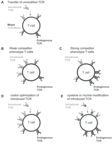
TCR make up of host cells
Figure 1. Simplified representation of TCR cell surface make up after TCR gene transfer in different T-cells using different strategies to improve cell surface expression of the introduced TCR.
Figure 1: (A) Transfer of unmodified TCR will result in
cell surface expression of the endogenous TCR, the
in-troduced TCR and mixed TCR dimers composed of the
introduced TCRα chain pairing with the endogenous
TCRß chain and the endogenous TCRα chain pairing
with the introduced TCRß chain. (B) T-cells with a
weak competitor phenotype predominantly express the
introduced TCR after TCR gene transfer, and to a lesser
extent the endogenous TCR on their cell surface. (C)
T-cells with a strong competitor phenotype
predomi-nantly express their endogenous TCR after TCR gene
transfer, and to a lesser extent the introduced TCR on
their cell surface. (D) Codon optimization is a strategy
that improves cell surface expression of the introduced
TCR by changing the nucleotide sequence to obtain
op-timal codon usage. This opop-timal codon usage results in
identical amino acid sequence of the TCR chains,
how-ever, improves mRNA stability and translation efficacy
of the introduced TCR chains, resulting in improved
introduced TCR cell surface expression. (E) Inclusion
of cysteine residues or murinization of the constant
domains of the introduced TCR chains induces
prefer-ential pairing of the introduced TCR chains. Cell surface
expression of the introduced TCR chains is improved
since reduced numbers of mixed dimers are formed,
resulting in less competition for cell surface expression.
Additionally, forced preferential pairing might offer
advantages for the introduced TCR of capturing more
CD3 complexes.
high number of different mixed TCR dimers with un-known specificity can be formed due to pairing of the retrovirally introduced TCR chains with the endoge-nously expressed TCR chains, increasing the probabil-ity of the formation of autoreactive mixed TCR dimers. Theoretically, the introduction of a TCR into a T-cell will result in formation of two mixed TCR dimers, con-sisting of the endogenous TCRα chain pairing with the introduced TCRβ chain and vice versa (Figure 1A). Therefore, usage of unselected PBMCs with a broad TCR repertoire as host cells for TCR transfer will in-crease the risk of formation of mixed TCR dimers with a harmful off-target reactivity. An alternative strategy to prevent formation of mixed TCR dimers would be to transduce γδ-T-cells, since the γδ-TCR chains are not able to pair with αβ-TCR chains(167). Human
γδ-T-cells redirected with αβ-TCRs were fully functional in vitro and were capable of recognizing chronic my-eloid leukemic cells. In addition, in murine studies we were able to show functional activity in vivo and per-sistence of the cells(168). However, further analyses will
TCR dimers harboring harmful specificities will be limited. Another possible advantage of the use of virus-specific T-cells is the exclusion of regulatory T-cells from the pool of TCR modi-fied lymphocytes that can possibly disturb the immune reaction. Furthermore, adoptive immunotherapy with EBV-specific T-cells in patients with post-transplant proliferative disease and CMV-specific T-cells as prophylaxis for CMV reactivation(61-63) in patients
after SCT has proven to be a therapeutic strategy without toxic-ity or GvHD, and long-term persistence of these T-cells has been demonstrated(44). Since EBV and CMV are examples of latent
viruses, we hypothesize that due to frequent encounter with viral antigens and subsequent triggering of the endogenous TCR, TCR transferred virus-specific T-cells will survive for a prolonged period of time in vivo. Moreover, it was recently shown in a mouse model that tolerization of one TCR could be overcome by signaling via the other TCR. In this model the function of the tol-erized self-tumor-reactive TCR of dual-T-cell receptor transgenic T-cells was rescued by proliferation induced via the virus-specific TCR, underlining the potency of TCR transfer into virus-specific T-cells(173). In addition, expression of the transgene under
regula-tion of a viral promotor is enhanced upon T-cell activaregula-tion(174-177).
Using T-cells specific for latently present viruses may result in repetitive stimulation via the endogenous TCR and increased expression of the introduced TCR due to T-cell activation.
We have previously reported differences between TCRs in the capacity to compete for cell surface expression(178), and we
described weak competitor phenotype TCRs exhibiting low cell surface expression (Figure 1B) and strong competitor phenotype
TCRs (Figure 1C) exhibiting high cell surface expression after gene transfer. Probably interchain pairing of the introduced TCR and competition for CD3-complex formation may both play a role. Because the TCR is expressed only at the cell surface when noncovalently bound to the CD3 complex composed of CD3γ, CD3ε, CD3δ, and CD3ζ, correct assembly of all these subunits with TCRα- and β-chains is required to assure optimal membrane expression of the TCR-CD3 complex in T-cells(179-181).
Single subunits and partial receptor complexes redundant for the assembly process retain in the ER where these products are highly susceptible to proteolysis(182,183). We speculate that weak
and strong competitor phenotype can be explained by two mechanisms. Possibly, strong competitor phenotype TCRs have a higher interchain affinity, which results in rapid formation of TCRαβ complexes and hinders degradation of the single TCRα and β chains. Alternatively, strong competitor phenotype reflects the ability of particular TCR-chains to more efficiently capture CD3 and thus be preferentially transported to and expressed at the cell surface. Ideally, TCRs selected for the purpose of gene transfer should exhibit both high interchain affinity and a high TCR-CD3 intrinsic affinity to generate T-cells that preferentially express the transferred-TCR, resulting in a strong competitor phenotype. Alternatively, weak competitor phenotype T-cells could be selectively used as host cells. Recently, we have de-scribed that weak competitor phenotype of virus-specific T-cells is, to some extent, correlated with specificity(166). However,
Furthermore, to ensure persistence of TCR modified T-cells, we would like to preserve the endogenous virus-specific TCR cell surface expression. Introduction of a strong competitor pheno-type TCR into weak competitor phenopheno-type virus-specific T-cells might result in loss of cell surface expression of the endogenous virus-specific TCR. Several strategies to improve expression of the introduced TCR have been described. mRNA and protein stability and translation efficacy of the introduced TCR chains can be enhanced by codon optimization(184) (Figure 1D). Furthermore,
matched pairing of the introduced TCR chains can be facilitated by murinization(185-187) or introduction of cysteine residues in the
constant regions of the introduced TCR chains, resulting in for-mation of an extra disulfide bond(188,189) (Figure 1E).
In conclusion, TCR gene transfer is a promising strategy to rapidly engineer therapeutically relevant amounts of anti-tu-mor specific T-cells. However, future application of TCR modified T-cells in clinical trials might benefit from increased knowledge how to improve cell surface expression of the introduced TCR and persistence of TCR modified T-cells.
Aim of the study
TCR gene transfer is a strategy that enables the rapid engineer-ing of anti-leukemic T-cells with defined specificity, resultengineer-ing in a so called ‘off the shelf’ therapy. An elegant strategy to promote persistence of TCR modified T-cells may be TCR gene transfer into CMV- and EBV-specific T-cells, which exhibit proper memory and effector phenotypes. Furthermore, these virus-specific T-cells do not induce GvHD after HLA identical allo-SCT, and can thus
be safely administered. For efficient anti-leukemic reactivity of the introduced TCR coinciding with enhanced in vivo survival, a bal-ance between cell surface expression of the introduced and en-dogenous TCR is required. The aim of this thesis was to optimize the efficacy of TCR gene transfer, study possibilities and restric-tions of virus-specific T-cells as host cells for TCR gene transfer and characterize the occurrence of potentially harmful mixed TCR dimers and strategies to prevent their formation.
Since the introduced TCR chains have to compete for cell surface expression with the endogenous TCR, the introduced TCR chains are under control of a strong viral promotor, which, in contrast to the endogenous promotor, is constitutively active. In Chapter 2, we analyzed whether physiological TCR downregula-tion resulting in a protective refractory period was preserved in TCR modified T-cells. For this purpose, CMV- and EBV-specific T-cells were retrovirally transduced with the hematopoietic minor histocompatibility antigen HA-2-specific TCR (HA-2-TCR). TCR transduced T-cells were antigen-specifically triggered via either the introduced TCR or the endogenous virus-specific TCR. At various time points after stimulation TCR cell surface expression as well as TCR-responsiveness and activation induced cell death (AICD) was measured to analyze preservation of the protective refractory period.
the introduced anti-leukemic MiHA-TCR, persistence in vivo of TCR modified virus-specific T-cells capable of controlling the he-matological malignancy may fall short. In Chapter 3, we analyzed whether the dual-specificity of the TCR transferred T-cells after repetitive stimulation via either the introduced anti-leukemic TCR or the endogenous virus-specific TCR was preserved. Purified CMV-specific T-cells were transduced with the HA-2-TCR and either repetitively stimulated via the endogenous CMV-TCR to mimick a period of minimal residual disease (MRD) or via the introduced HA-2-TCR to mimic relapse, and preservation of dual-specificity was analyzed.
It has been described that introduction of TCR chains into T-cells results in mixed TCR dimer formation, consisting of the introduced TCR chains pairing with the endogenous TCR chains. Since the specificity of mixed TCR dimers is unpredict-able, hazardous specificities may be formed. In Chapter 4, we investigated whether TCR transfer can lead to the generation of mixed TCR dimers exhibiting new detrimental reactivities. To ad-dress this issue we created T-cells expressing mixed TCR dimers. To be able to discriminate between the functionality of the en-dogenous TCR, the introduced TCR as well as mixed TCR dimers, we transduced different defined virus-specific T-cells with seven different well characterized antigen-specific TCRs and tested these for newly acquired reactivities against an HLA-typed EBV-LCL panel covering all prevalent HLA class I and II molecules, and against different normal cell subsets. Furthermore, we explored the introduction of cysteine residues in the constant domains of the introduced TCR resulting in formation of an extra
disulfide bond as a strategy to avoid expression of neoreactive mixed TCR dimers.
The MiHA HA-1 is an attractive candidate antigen for clinical study, as it is exclusively expressed on hematopoietic cells. However, previously it has been demonstrated that HA-1-TCRs are poorly expressed after gene transfer. In Chapter 5 we therefore sought to improve HA-1-TCR expression after gene transfer. TCR-deficient jurkat-cells were used to study pairing capacities of the HA-1-TCR chains. The role of the CDR1 region of the always identical HA-1-TCR BV6S4 chain in low HA-1-TCRβ expression was analyzed by exchanging this region. Furthermore, two well described strategies, namely the inclusion of cysteine residues in the TCR constant domains and codon optimization were explored for improvement of HA-1-TCR cell surface expres-sion after gene transfer in virus-specific T-cells known to pos-sess endogenous TCRs which strongly compete for cell surface expression.
To broaden the applicability of cellular immunotherapy, adop-tive transfer of T-cell receptor (TCR) transferred T-cells may be an attractive strategy. Using this approach, high numbers of defined antigen-specific T-cells can be engineered. Since the introduced TCR has to compete for cell surface expression with the en-dogenous TCR, the introduced TCR chains are under control of a strong viral promotor, which, in contrast to the endogenous promotor, is constitutively active. We examined whether this difference in regulation would result in differences in TCR inter-nalization and re-expression of the introduced and endogenous TCR on dual TCR engineered T-cells as well as the antigen-responsiveness of both TCRs. We demonstrated comparable
TCR downregulation of TCRs expressed under regulation of a retroviral promotor or the endogenous promotor. However, the introduced TCRs were rapidly re-expressed on the cell surface after TCR stimulation. Despite rapid re-expression of the intro-duced TCR, T-cells exerted similar antigen-sensitivity compared to control T-cells, illustrating that cell mechanisms other than TCR cell surface expression are involved in antigen-sensitivity directly after antigen-specific stimulation. These results demon-strate that TCR transduced T-cells are functionally not different from non-transduced T-cells and can potentially be used as an effective treatment strategy.
Rapid re-expression of retrovirally introduced
versus endogenous TCRs in engineered
T-cells after antigen-specific stimulation
J Immunother. 2011 Mar;34(2):165-74. Reprinted with permission.
Marleen M. van Loenen, Renate S. Hagedoorn, Renate de Boer, Esther H.M. van Egmond, J.H. Frederik Falkenburg, Mirjam H.M. Heemskerk
2
INTRODUC TION
Adoptive transfer of TCR transduced (td) T-cells may be an attrac-tive strategy to obtain high numbers of defined antigen-specific T-cells for cellular immunotherapy without complicated isolation strategies and labour intensive culturing procedures(1). Different
studies have shown the effectiveness of TCR transfer, both in vitro(2-7) and in vivo(8-11), and recently the feasibility of this approach
was demonstrated in clinical trials(8,10).
In TCR td T-cells, the introduced TCR has to compete for cell surface expression with the endogenous TCR. For optimal efficacy of TCR modified T-cells in vivo, the cell surface expression of the introduced TCR has to be high, allowing the TCR td T-cells to recognize clinically relevant target cells expressing endog-enously processed antigen. One of the strategies to acquire high TCR cell surface expression on TCR gene modified T-cells, is to use a strong retroviral promotor to regulate the introduced TCR. However, retroviral promotor regions are constitutively active, and in addition, it has been described that viral promotor activity will increase by T-cell activation(12-14). In contrast, the endogenous
promotor regions regulating the endogenous TCR expression have been demonstrated to be transiently inactivated after TCR triggering. TCRαβ mRNA expression decreases within 4-7h after TCR triggering, followed by normalization of mRNA levels 24h after activation(15,16). In addition, T-cell activation induced by TCR
triggering has been demonstrated to induce internalization of the TCR-CD3-complexes. It has been suggested that internaliza-tion of TCR-CD3-complexes and transient inactivainternaliza-tion of the
promotor regions regulating the endogenous TCR result in a refractory period of activation in which all effector-target in-teractions are terminated(17-20). This latter effect is supported by
the observation that TCR-CD3 downregulation results in a loss of cellular sensitivity to subsequent stimulation for 72 hours or longer(18,20), and vice versa, the inhibition of receptor
downregula-tion leads to enhanced signaling(17,21,22). Thus, the control of TCR
expression by internalization of TCR-CD3 complexes and degra-dation of all its subunits(23-25) is speculated to result in a refractory
period important to prevent harmful hyperstimulation resulting in activation induced cell death (AICD).
RESULTS
Rapid re-expression of the introduced TCR-CD3 complex on TCR td T-cells
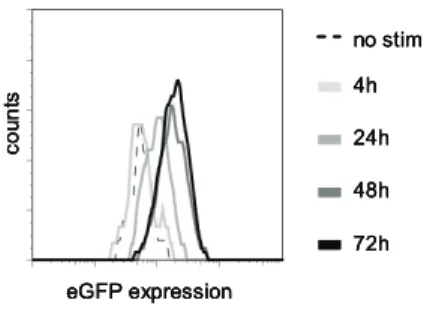
To ensure high and stable expression of the introduced TCR, most TCR gene transfer studies to date use retroviral vectors for transgene delivery. Expression of the introduced TCRs in these studies will be regulated by the retroviral long terminal repeats (LTRs), whereas endogenous promotor regions will regulate the endogenous TCR expression. We assessed whether TCR-triggering of TCR td virus-specific T-cells resulted in increased protein levels under regulation of the retroviral LTR by analyzing eGFP expression as a marker using flow cytometric analyses. As shown in Figure 1, eGFP expression was increased at 24h after antigen-specific TCR triggering, and showed further increase up till 48h after TCR triggering, confirming previous observations that protein levels under regulation of a viral promotor increase upon TCR stimulation(12-14). To determine whether
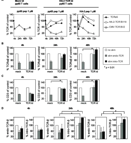
antigen-specif-ic stimulation would result in changed TCR modulation between the introduced TCR under regulation of a viral promotor and the endogenous TCR, we sorted TCR td virus-specific T-cells based on double positivity for eGFP and truncated nerve growth factor receptor (NGF-R). These TCR engineered T-cells with dual-specificity were stimulated antigspecifically via their en-dogenous or introduced TCR, and analyzed for TCR cell surface expression. We analyzed three different TCR td T-cells; HA-2-TCR td or CMV-TCR td EBNA3A-specific T-cells, and HA-2-TCR td pp65-specific T-cells. T-cells were stained at different time points
after antigen-specific stimulation with TCRαβ-, CD3- or TCRβ-specific mAbs to determine the TCR-CD3 cell surface expression and to dissect between the endogenous and introduced TCRβ chains. Unfortunately no mAbs are available to stain for the endogenous or introduced TCRα chains. TCR downregulation of the different TCR-CD3 complexes in TCR td virus-specific T-cells was compared to TCR downregulation of mock td virus-specific T-cells. In Figure 2A a representative example of the kinetics of TCR cell surface expression after antigen-specific stimulation is depicted. The HA-2-TCR td pp65 T-cells demonstrate down-regulation of the cell surface expressed TCRαβ complexes after 4h of stimulation similar to mock td pp65 T-cells. With mAbs specific for the endogenous and introduced TCRβ chain we observed after antigen-specific stimulation via the endogenous TCR (pp65 pep 1 µM) downregulation of both the endogenous as well as the introduced TCRβ chains. Likewise, we observed after antigen-specific stimulation via the introduced TCR (HA-2 pep 1 µM) downregulation of both the introduced as well as the
!iigguurree 11 MM..MM.. vvaann LLooeenneenn
nnoo ssttiimm
44hh
2244hh
4488hh
7722hh
eeGG!PP eexxpprreessssiioonn
ccoo
uunn
ttss
!iigguurree 11 MM..MM.. vvaann LLooeenneenn
nnoo ssttiimm
44hh
2244hh
4488hh
7722hh
eeGG!PP eexxpprreessssiioonn
ccoo
uunn
ttss
nnoo ssttiimm
44hh
2244hh
4488hh
7722hh
eeGG!PP eexxpprreessssiioonn
ccoo
uunn
ttss
Figure 1. Protein levels under regulation of a viral promotor increase after stimulation.
Figure 1: Sorted virus-specific T-cells
trans-duced with vectors containing TCRα chains
in combination with the marker gene eGFP
and TCRβ chains in combination with the
marker gene NGF-R were stimulated via their
endogenous TCR using peptide pulsed target
cells and eGFP expression was measured
us-ing FACS as an indication of viral promotor
activity. eGFP expression of T-cells without
stimulation (black dotted line), 4h after
stim-ulation (light grey line), 24h after stimstim-ulation
(grey line), 48h after stimulation (dark grey
line) and 72h after stimulation (black line) is
shown. Data is representative for several TCR
td as well as mock td T-cells in six
!iigguurree 22 AABBCCDD;;; MM..MM.. vvaann LLooeenneenn
!** **
nnoo ssttiimm
ssttiimm eennddoo--TTCCRR
ssttiimm iinnttrroo--TTCCRR
**pp << 00..0011 TTCCRRĮßß HHAA--22--TTCCRR BBVV1188
CCMMVV--TTCCRR BBVV22 AA
pppp6655 ppeepp 11 µµMM HHAA--22 ppeepp 11 µµMM HHAA--22 TT!RR ttdd
pppp6655 TT cceellllss
114400 114400
pppp6655 ppeepp 11 µµMM MMoocckk ttdd pppp6655 TT cceellllss
114400 %% TT CC RR oo
ff
ccoo nn ttrr oo ll %% TT CC RR oo
ff
ccoo nn ttrr oo ll %% TT CC RR oo
ff
ccoo nn ttrr oo ll 2200 6600 110000 114400 2200 6600 110000 114400 2200 6600 110000 114400 **** 2200 6600 110000 114400 %% iinn ttrr oo --TT CC RR --ȕ 2200 6600 110000 114400 %% ee nndd oo --TT CC RR --ȕ 2200 6600 110000 114400 %% iinn ttrr oo --TT CC RR --ȕ 2200 6600 110000 114400 %% ee nndd oo --TT CC RR --ȕ 2200 6600 110000 114400 %% iinn ttrr oo --TT CC RR --ȕ 2200 6600 110000 114400 %% ee nndd oo --TT CC RR --ȕ
44hh 2244hh 4488hh
DD 44hh 2200 6600 110000 114400
mmoocckk TTCCRR ttdd
2244hh
2200 6600 110000 114400
mmoocckk TTCCRR ttdd
4488hh
2200 6600 110000 114400
mmoocckk TTCCRR ttdd BB %% TT CC RR Į ȕ oo
ff
ccoo nn ttrr oo ll %% TT CC RR Į ȕ oo
ff
ccoo nn ttrr oo ll %% TT CC RR Į ȕ oo
ff
ccoo nn ttrr oo ll **
44hh 2244hh 4488hh
%%
CC
DD
33
oo
ff
ccoo nn ttrr oo ll %% CC DD 33 oo
ff
ccoo nn ttrr oo ll %% CC DD 33 oo
ff
ccoo
nn
ttrr
oo
ll
mmoocckk TTCCRR ttdd mmoocckk TTCCRR ttdd mmoocckk TTCCRR ttdd
2244hh 4488hh 7722hh 110000 6600 2200 110000 6600 2200
2244hh 4488hh 7722hh 110000
6600
2200
2244hh 4488hh 7722hh 44hh 44hh
44hh
** **
!
**** **
nnoo ssttiimm
ssttiimm eennddoo--TTCCRR
ssttiimm iinnttrroo--TTCCRR nnoo ssttiimm
ssttiimm eennddoo--TTCCRR
ssttiimm iinnttrroo--TTCCRR
**pp << 00..0011 TTCCRRĮßß HHAA--22--TTCCRR BBVV1188
CCMMVV--TTCCRR BBVV22 TTCCRRĮßß HHAA--22--TTCCRR BBVV1188
CCMMVV--TTCCRR BBVV22 AA
pppp6655 ppeepp 11 µµMM HHAA--22 ppeepp 11 µµMM HHAA--22 TT!RR ttdd
pppp6655 TT cceellllss
114400 114400
pppp6655 ppeepp 11 µµMM MMoocckk ttdd pppp6655 TT cceellllss
114400 %% TT CC RR oo
ff
ccoo nn ttrr oo ll %% TT CC RR oo
ff
ccoo nn ttrr oo ll %% TT CC RR oo
ff
ccoo nn ttrr oo ll 2200 6600 110000 114400 2200 6600 110000 114400 2200 6600 110000 114400 **** 2200 6600 110000 114400 %% iinn ttrr oo --TT CC RR --ȕ 2200 6600 110000 114400 %% ee nndd oo --TT CC RR --ȕ 2200 6600 110000 114400 %% iinn ttrr oo --TT CC RR --ȕ 2200 6600 110000 114400 %% ee nndd oo --TT CC RR --ȕ 2200 6600 110000 114400 %% iinn ttrr oo --TT CC RR --ȕ 2200 6600 110000 114400 %% ee nndd oo --TT CC RR --ȕ
44hh 2244hh 4488hh
DD ******** 2200 6600 110000 114400 %% iinn ttrr oo --TT CC RR --ȕ 2200 6600 110000 114400 %% ee nndd oo --TT CC RR --ȕ 2200 6600 110000 114400 %% iinn ttrr oo --TT CC RR --ȕ 2200 6600 110000 114400 %% ee nndd oo --TT CC RR --ȕ 2200 6600 110000 114400 %% iinn ttrr oo --TT CC RR --ȕ 2200 6600 110000 114400 %% ee nndd oo --TT CC RR --ȕ
44hh 2244hh 4488hh
DD 44hh 2200 6600 110000 114400
mmoocckk TTCCRR ttdd
2244hh
2200 6600 110000 114400
mmoocckk TTCCRR ttdd
4488hh
2200 6600 110000 114400
mmoocckk TTCCRR ttdd BB %% TT CC RR Į ȕ oo
ff
ccoo nn ttrr oo ll %% TT CC RR Į ȕ oo
ff
ccoo nn ttrr oo ll %% TT CC RR Į ȕ oo
ff
ccoo nn ttrr oo ll ** 44hh 2200 6600 110000 114400
mmoocckk TTCCRR ttdd
2244hh
2200 6600 110000 114400
mmoocckk TTCCRR ttdd
4488hh
2200 6600 110000 114400
mmoocckk TTCCRR ttdd BB %% TT CC RR Į ȕ oo
ff
ccoo nn ttrr oo ll %% TT CC RR Į ȕ oo
ff
ccoo nn ttrr oo ll %% TT CC RR Į ȕ oo
ff
ccoo nn ttrr oo ll ****
44hh 2244hh 4488hh
%%
CC
DD
33
oo
ff
ccoo nn ttrr oo ll %% CC DD 33 oo
ff
ccoo nn ttrr oo ll %% CC DD 33 oo
ff
ccoo
nn
ttrr
oo
ll
mmoocckk TTCCRR ttdd mmoocckk TTCCRR ttdd mmoocckk TTCCRR ttdd
2244hh 4488hh 7722hh 110000 6600 2200 110000 6600 2200
2244hh 4488hh 7722hh 110000
6600
2200
2244hh 4488hh 7722hh 44hh 44hh
44hh 2244hh 4488hh 7722hh
110000 6600 2200 110000 6600 2200
2244hh 4488hh 7722hh 110000
6600
2200
2244hh 4488hh 7722hh 44hh 44hh
44hh
**** ****
Figure 2. TCR td T-cells demonstrate fast TCR re-expression in comparison with mock td T-cells due to fast introduced TCR re-expression. Figure 2: (A) As an example, the kinetics of total TCR re-expression
(TCRαβ; black diamonds) or re-expression of the endogenous
TCR-β (CMV-TCR BV2; grey triangles) or introduced TCR-β
(HA-2-TCR BV18; white circles) of mock and HA-2 TCR td pp65 T-cells
after stimulation with LCLs pulsed with either 1 µM of pp65 or HA-2
peptide is depicted. The cell surface expression of T-cells stimulated
with unpulsed LCL (control) was set at 100%. Per timepoint the
percentage cell surface expression was calculated as follows: [MFI
of T-cells with peptide pulsed LCL / MFI of control T-cells] * 100. The
average MFI of control stimulated mock td T-cells stained with
anti-TCRαβ = 673; anti-TCR-BV2 = 736. The average MFI of control
stimulated HA-2-TCR td T-cells stained with anti-TCRαβ = 744;
anti-TCR-BV2 = 296; anti-TCR-BV18 = 29.
(B)/(C)/(D) Mock td pp65 and mock td EBNA3A T-cells (mock), HA-2-TCR td pp65 and HA-HA-2-TCR td EBNA3A T-cells and CMV-TCR
td EBNA3A T-cells (TCR td) were stimulated with LCL-Z (control
stim; white bars) or with either LCL-Z pulsed with pp65 peptide or
HLA-B7 td LCL-Z pulsed with EBNA3A peptide, respectively (stim
endogenous TCR; grey bars) or with LCL-Z pulsed with either HA-2
or pp65 peptide (stim introduced TCR; black bars) and analyzed at
the indicated time points for B) TCRαβ expression, C) CD3
expres-sion, and D) endogenous and introduced TCRβ expression. The cell
surface expression of T-cells stimulated with unpulsed LCL (control)
was set at 100%. Per timepoint the percentage cell surface
expres-sion was calculated as follows: [MFI of T-cells with peptide pulsed
LCL / MFI of control T-cells] * 100. Reported P values were
consid-ered statistically different if <0.01 and values statistically different
are indicated with an asterisk. Data from six independent
endogenous TCRβ chains. HA-2-TCR td pp65 T-cells, however, re-expressed TCRαβ complexes faster at their cell surface com-pared to mock td T-cells, both when stimulated via their endog-enous or introduced TCR. Already 24h after stimulation TCRαβ complexes were re-expressed on TCR td pp65 T-cells, while TCRαβ expression of mock td pp65 T-cells was still decreased 72h after TCR stimulation. Also TCRαβ expression of the parental HA-2-specific T-cell clone was still decreased 72h after TCR stimu-lation (data not shown). Using mAbs specific for the endogenous and introduced TCRβ chain we demonstrated that re-expression of TCRαβ complexes 24h and 48h after TCR triggering correlated with recovery of the introduced TCRβ chain, whereas the endog-enous TCRβ chain was still downregulated 72h after stimulation. Moreover, recovery of the endogenous TCRβ chain appeared to be even slower compared to mock transduced T-cells, indicating that the abundance of introduced TCR chains may compete for cell surface expression with the endogenous TCR chains. Since the introduced TCRα and TCRβ chain are both regulated via a similar retroviral LTR, it seems plausible that the recovered TCR complexes that stained with the mAb specific for the introduced TCRβ early after stimulation are primarily composed of the intro-duced TCRα and TCRβ chains. To demonstrate that the kinetics of TCR expression in dual TCR engineered T-cells is not influ-enced by the specificity of the virus-specific T-cells or the trans-ferred TCR, we performed similar experiments with HA-2-TCR or CMV-TCR td EBNA3A T-cells. Figure 2B, 2C and 2D demonstrate that the TCR td pp65 and TCR td EBNA3A T-cells showed similar TCR expression kinetics using mAbs against TCRαβ, CD3 and
against the endogenous and introduced TCRβ chains, respec-tively. Eventually, TCR make up as expressed by both mock and TCR td T-cells before stimulation was re-established 7 days after stimulation.
Based on these results, we conclude that although the retrovirally introduced and endogenous TCRs demonstrate a similar rapid downregulation after antigen-specific stimulation, the introduced TCR is re-expressed significantly faster at the T-cell surface compared to the endogenous TCR.
Fast re-expression of introduced TCR is not reflected in restored tetramer binding