Elucidating G protein signaling and ubiquitin conjugation in Entamoeba histolytica
Dustin Eli Bosch
A dissertation submitted to the faculty of the University of North Carolina at Chapel Hill in partial fulfillment of the requirements for the degree of Doctor of Philosophy in the School of Medicine (Pharmacology)
Chapel Hill 2013
Approved by:
ABSTRACT
DUSTIN ELI BOSCH: Elucidating G protein signaling and ubiquitin conjugation in Entamoeba histolytica
(Under the direction of Dr. David Peter Siderovski)
The intestinal parasite Entamoeba histolytica is responsible for an estimated 50 million infections and 100,000 deaths per year worldwide. The causative agent of amoebic colitis and systemic amoebiasis is spread primarily through contaminated food and drinking water sources. Although reasonably effective treatments have long been available for invasive amoebiasis, imperfect patient response rates, drug side effects, and concern for emerging drug resistance all warrant exploration of new pharmacological targets in E. histolytica. This work describes structural, biochemical, and cell biological explorations of heterotrimeric G protein and Rho family GTPase signaling and ubiquitination in E. histolytica.
of its catalytic domain in the inactive state. However, co-expression with constitutively active EhGα1 and EhRacC lead to EhRGS-RhoGEF activation in cells.
The Rho family GTPase EhRho1 lacks a signature Rho insert helix and sensitivity to C3 exoenzyme. Crystal structures of EhRho1 indicate unique nucleotide-contacting residues that confer fast intrinsic nucleotide exchange activity. However, EhRho1 functions like its homologs in engaging a diaphanous-related formin effector to regulate actin polymerization. EhFormin1 is autoinhibited and is activated by Rho GTPase binding. A crystal structure of the EhRho1/EhFormin1 complex indicates similarity to human RhoC/mDia1, despite an absent secondary binding site at the Rho insert helix.
Multiple ubiquitin-proteasome pathway genes were differentially transcribed upon perturbed EhGα1 expression. EhUbiquitin was activated by the E1 enzyme EhUba1 and conjugated by the E2 enzyme EhUbc5, indicating a conserved ubiquitination cascade in E. histolytica. Crystal structures of EhUbiquitin and EhUbc5 suggested potentially unique polyubiquitin linkages, but a likely conserved mode of ubiquitin chain elongation.
I dedicate my work:
To my wife Samantha for unconditional support, encouragement, and motivation,
as well as preservation of my sanity.
ACKNOWLEDGEMENTS
I am grateful for and indebted to my dissertation advisor and mentor, Dr. David
Siderovski, who has most impacted my development as a scientist. His approachability and
generosity with both time and resources has been a foundation for my successful graduate
training experience, as well as those of other former trainees. I have come to expect from him
a cogent word of advice at times when I need guidance, and unfettered freedom to pursue and
struggle with my own scientific ideas. Despite his career transitions, David has maintained
my training at UNC as a priority, giving freely of his after-hours time to exchange emails and
revise manuscripts. From him I have learned the rarity and value of great mentorship in both
scientific training and career development, an example that I will strive to emulate in the
future.
I also thank other members of the Siderovski lab who have shared their skills and
experience and made the lab a fantastic place to work: Brian Buckley, Stephanie Hutsell, Dr.
Staci Cohen, Dr. Emily Oestreich, Dr. Patrick Giguere, Dr. Genevieve Laroche, and Dr.
Vince Setola. I particularly thank former graduate student Dr. Adam Kimple for his
investment in my initial training, his example of success, and for many fruitful
collaborations.
I thank fellow members of the UNC MD/PhD training program, in particular, Will
Jeck, Erin Steinbach, Jeff Federspiel, Tricia Lenhart, Tom Jarrett, Isaac Chan, and Chris
MD/PhD program administrators have also been instrumental, including Dr. Eugene
Orringer, Dr. Kim Rathmell, Alison Regan, and Carol Herion.
I thank the Pharmacology Department staff that has assisted with grants, finances, and
navigation of administrative tasks, including Kathy Justice, Tangi Covington, Eddie Gill,
Chris Turner, and Alfred Dolge.
Finally, I thank the members of my dissertation committee: Dr. Ken Harden, Dr.
Brian Kuhlman, Dr. Scott Plevy, and Dr. Henrik Dohlman for giving of their time and effort
to guide my scientific development. Extra thanks goes to Drs. Kuhlman and Plevy for their
commitments as co-mentors on my training grant, for research collaborations, and for a
refreshing clinical experience during my graduate school training. I also particularly thank
Dr. Ken Harden, whose freely given mentorship and guidance convinced me to pursue a
PhD, in addition to an MD, following an undergraduate research experience in his lab, and
who was instrumental in helping me find an excellent MD/PhD training opportunity at UNC.
TABLE OF CONTENTS
LIST OF TABLES………..….……xv
LIST OF FIGURES……….…...xvi
LIST OF ABBREVIATIONS………...……...xxi
CHAPTER 1 GENERAL INTRODUCTION ...1
1.1 OVERVIEW ...1
1.2 Entamoeba histolytica CAUSES AMOEBIC COLITIS AND SYSTEMIC AMOEBIASIS ...2
1.2.1 Epidemiology, disease sequelae, and current treatment options ...2
1.2.2 Parasite factors in pathogenesis ...4
1.3 HETEROTRIMERIC G PROTEINS AND RAS SUPERFAMILY GTPases...5
1.4 REGULATION OF THE GUANINE NUCLEOTIDE CYCLE...6
1.4.1 Heterotrimeric G proteins ...6
1.4.2 Ras superfamily GTPases ...7
1.5 HETEROTRIMERIC G PROTEIN SIGNALING IN E. histolytica ...8
1.6 RAS SUPERFAMILY GTPases IN E. histolytica...12
1.6.1 Ras family GTPases...13
1.6.2 Rho family GTPases ...14
1.6.3 Rab family GTPases ...21
1.7 THE UBIQUITIN-PROTEASOME SYSTEM IN E. histolytica...25
CHAPTER 2 HETEROTRIMERIC G-PROTEIN SIGNALING IS CRITICAL TO PATHOGENIC PROCESSES IN
Entamoeba histolytica ...42
2.1 OVERVIEW ...42
2.2 INTRODUCTION...43
2.3 EXPERIMENTAL PROCEDURES...45
2.3.1 Cloning of E. histolytica G-protein subunits ...45
2.3.2 Protein purification ...46
2.3.3 Crystallization and structure determination...47
2.3.4 qRT-PCR of E. histolytica gene transcription ...49
2.3.5 Western blotting...51
2.3.6 Fluorescence complementation and co-immunoprecipitation ...52
2.3.7 Nucleotide binding, hydolysis and EhGα1 activation...52
2.3.8 Evolutionary analysis...53
2.3.9 Surface plasmon resonance...53
2.3.10 Trophozoite stable transfection...53
2.3.11 Trophozoite migration and Matrigel transmigration ...54
2.3.12 Host cell attachment...54
2.3.13 Cell killing ...55
2.3.14 Whole transcriptome shotgun sequencing ...55
2.3.15 Cysteine protease activity ...57
2.4 RESULTS...58
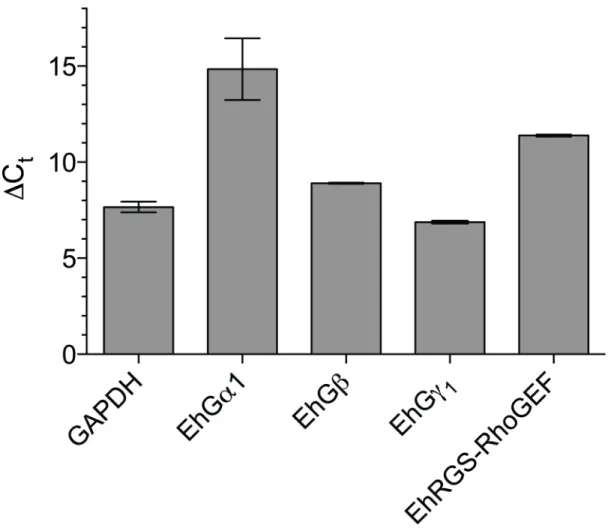
2.4.1 Identification of E. histolytica heterotrimeric G-protein subunits...58
2.4.2 Functional assessments of E. histolytica G-protein subunits...58
2.4.3 EhGα1 functional mutants...60
2.4.5 A crystal structure of EhGα1...62
2.4.6 G-protein signaling perturbation modulates trophozoite migration, Matrigel transmigration, and host cell attachment and killing ...64
2.4.7 Regulation of transcription by perturbed heterotrimeric G-protein signaling...66
2.5 DISCUSSION ...69
2.6 REFERENCES ...98
CHAPTER 3 STRUCTURAL DETERMINANTS OF RGS-RhoGEF SIGNALING CRITICAL TO Entamoeba histolytica PATHOGENESIS...105
3.1 OVERVIEW ...105
3.2 INTRODUCTION...106
3.3 EXPERIMENTAL PROCEDURES...108
3.3.1 Cloning and protein purification...108
3.3.2 Crystallization and structure determination...110
3.3.3 Single turnover nucleotide hydrolysis ...111
3.3.4 Surface plasmon resonance...112
3.3.5 NTA affinity co-precipitation ...112
3.3.6 Trophozoite stable transfection...113
3.3.7 Chemotactic migration...113
3.3.8 Host cell attachment...113
3.3.9 Cell killing ...114
3.3.10 Cysteine protease activity ...114
3.3.11 S2 cell culture and spreading assay ...115
3.3.12 Immunofluorescence microscopy ...116
3.4 RESULTS...117
3.4.2 EhGα1 and EhRacC activate EhRGS-RhoGEF to promote
Rho-dependent cell spreading...118
3.4.3 EhRGS-RhoGEF modulates pathogenic processes of E. histolytica trophozoites...120
3.4.4 A crystal structure of EhRGS-RhoGEF...121
3.4.5 The inhibitory helix coordinates occlusion of the Rho GTPase binding site...123
3.4.6 Convergent evolution of the EhGα1/EhRGS-RhoGEF interface...124
3.5 DISCUSSION ...125
3.6 REFERENCES ...146
CHAPTER 4 UNIQUE STRUCTURAL AND NUCLEOTIDE EXCHANGE FEATURES OF THE Rho1 GTPase OF Entamoeba histolytica ...150
4.1 OVERVIEW ...150
4.2 INTRODUCTION...151
4.3 EXPERIMENTAL PROCEDURES...154
4.3.1 Bioinformatic analysis of the E. histolytica Rho GTPase family ...154
4.3.2 Protein expression and purification ...154
4.3.3 Crystallization of EhRho1·GDP and EhRho1·GTPgS and structure determination ...157
4.3.4 Surface plasmon resonance (SPR) binding assays ...159
4.3.5 GST-EhRhoGDI affinity co-precipitation ...160
4.3.6 Fluorescent guanine nucleotide exchange assays ...160
4.3.7 Actin stress fiber quantification...161
4.4 RESULTS...162
4.4.1 Comparison of human and E. histolytica Ras superfamily GTPases ...162
4.4.2 Structures of EhRho1 in the active and inactive states...163
4.4.3 EhRho1 interacts with an mDia homolog, EhFormin1...164
4.4.5 EhRho1 stimulates stress fiber formation in mammalian cells...166
4.4.6 Unique nucleotide interactions in EhRho1 ...166
4.4.7 Non-conserved residues in EhRho1 contribute to a restrained nucleotide exchange rate...168
4.5 DISCUSSION ...169
4.6 REFERENCES ...185
CHAPTER 5 Entamoeba histolytica Rho1 REGULATES ACTIN POLYMERIZATION THROUGH A DIVERGENT, DIAPHANOUS-RELATED FORMIN ...189
5.1 OVERVIEW ...189
5.2 INTRODUCTION...190
5.3 EXPERIMENTAL PROCEDURES...193
5.3.1 Protein purification ...193
5.3.2 Actin co-sedimentation ...194
5.3.3 Actin polymerization in vitro...195
5.3.4 Surface plasmon resonance...196
5.3.5 Crystallization and structure determination...196
5.4 RESULTS...198
5.4.1 E. histolytica Formin1 modulates actin filament formation ...198
5.4.2 EhFormin1 is autoinhibited by N- and C-terminal interactions ...199
5.4.3 Interaction of the EhFormin1 GBD-FH3 domain tandem with EhRho1 reverses autoinhibition of the FH2 domain ...200
5.4.4 Structural features of the EhRho1/EhFormin1 complex...201
5.5 DISCUSSION ...205
5.6 REFERENCES ...226
CHAPTER 6 STRUCTURAL DETERMINANTS OF UBIQUITIN CONJUGATION IN Entamoeba histolytica ... 230
6.2 INTRODUCTION...231
6.3 EXPERIMENTAL PROCEDURES...234
6.3.1 Cloning and protein purification...234
6.3.2 Crystallization and structure determinations of EhUbiquitin and EhUbc5...236
6.3.3 In vitro ubiquitin transfer assay ...237
6.3.4 In vitro polyubiquitin chain formation assay...238
6.3.5 PPi:ATP radioisotope exchange assay ...238
6.3.6 Surface plasmon resonance (SPR) assays...239
6.4 RESULTS...240
6.4.1 Structural features of a divergent ubiquitin from Entamoeba histolytica...240
6.4.2 EhUbiquitin is activated by the E1 enzyme EhUba1...242
6.4.3 EhUba1 engages the E2 enzyme EhUbc5 and transfers activated EhUbiquitin...244
6.4.4 Structural features of the E2 ubiquitin conjugating enzyme EhUbc5 and its non-covalent interaction with EhUbiquitin...245
6.4.5 EhUbc5 engages a RING family E3 ubiquitin ligase ...246
6.5 DISCUSSION ...248
6.6 REFERENCES ...263
CHAPTER 7 CLINICAL IMPLICATIONS AND FUTURE DIRECTIONS ...267
7.1 TARGETING HETEROTRIMERIC G PROTEIN SIGNALING ...267
7.1.1 Identification of a GPCR and ligand in E. histolytica...268
7.1.2 Investigating other heterotrimeric G protein signaling components ...271
7.2 RHO FAMILY GTPase SIGNALING SPECIFICITY ...273
7.2.1 Structure and function of the EhRacC/EhPAK4 complex...273
7.3 DE-UBIQUITINATING ENZYMES IN E. histolytica
LIST OF TABLES
Table:
2.1 Data collection and refinement statistics for lysine-methylated
selenomethionine EhGα1………. 95
2.2 Genes differentially transcribed in E. histolytica trophozoites expressing EhGα1 or EhGα1S37C with known roles in
pathogenesis or putative vesicular trafficking functions……….. 96
3.1 Rho family GTPase signaling and actin-associated genes differentially transcribed in E. histolytica trophozoites
expressing EhGα1 or the dominant negative EhGα1S37C………. 144
3.2 Data collection and refinement statistics for selenomethionine
EhRGS-RhoGEF……….. 145 4.1 Data collection and refinement statistics for EhRho1………….. 184
5.1 Data collection and refinement statistics for the EhRho1·GTPγS/EhFormin1 complex……….. 225
6.1 Ubiquitin and proteasome system genes differentially transcribed in E. histolytica trophozoites expressing EhGα1 or
the dominant negative EhGα1S37C……… 261
LIST OF FIGURES
Figure:
1.1 Nucleotide cycle regulation of heterotrimeric and Ras superfamily G
proteins……… 28
1.2 Model of heterotrimeric G protein signaling in E. histolytica………. 29
1.3 EhRho1 and EhRacA signaling modulate pathogenic behaviors in E.
histolytica………. 30
2.1 The genome of Entamoeba histolytica encodes heterotrimeric
G-protein subunits……… 72
2.2 Heterotrimeric G-protein signaling components are expressed in E.
histolytica………. 74
2.3 E. histolytica G-protein subunits form a heterotrimer in a
nucleotide-dependent manner……… 75
2.4 EhGα1 cycles between an active, GTP-bound state and an inactive,
GDP-bound state……….. 76
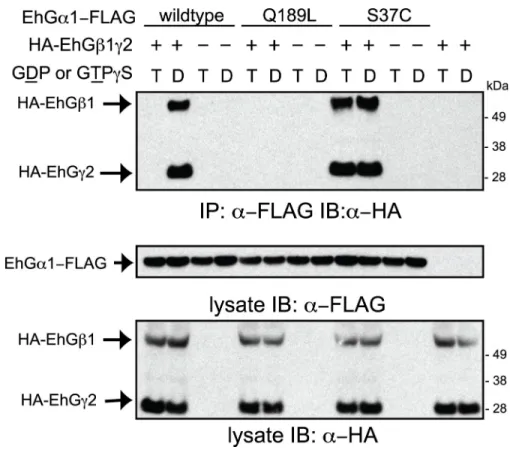
2.5 The inactive EhGα1(S37C) constitutively binds to EhGβ1γ2, while the constitutively active EhGα1(Q189L) mutant does not…………... 78
2.6 Evolutionary relationship of Gα subunits and identification of
EhRGS-RhoGEF as a putative effector for activated EhGα1……….. 79 2.7 Mammalian Gα subfamily homology analyses……… 81
2.8 Structural comparison of EhGα1 with Hs transducin and switch 2
crystal contacts………. 82
2.9 Structure of EhGα1 reveals a conserved fold with unique features…. 83
2.10 Electron density map of the guanine nucleotide binding pocket of EhGα1………... 85
2.11 Heterotrimeric G-protein signaling increases trophozoite migration across porous membranes and Matrigel layers………. 86
2.12 Expression of EhGα1wt or EhGα1S37C does not significantly alter
2.13 E. histolytica transfected with empty vector is not affected by tetracycline treatment………... 88
2.14 Heterotrimeric G-protein signaling positively regulates E. histolytica attachment to host cells as well as host cell killing……….. 89
2.15 Microscopic analysis of perturbed E. histolytica attachment to host cells upon overexpression of EhGα1wt or EhGα1S37C……….. 90
2.16 Heterotrimeric G-protein signaling alters E. histolytica transcription to modulate cysteine protease secretion………... 92
2.17 RT-PCR analysis of differentially transcribed genes and altered expression of amoebapore A protein……… 94
3.1 Multiple sequence alignment of the EhRGS-RhoGEF RGS and DH-PH domains……….. 129
3.2 Circular dichroism (CD) of EhRGS-RhoGEF(E39K) and nucleotide-dependent interaction of EhGα1 with the isolated RGS domain……. 131
3.3 EhRGS-RhoGEF is an EhGα1 effector that accelerates its GTP hydrolysis………. 132
3.4 EhRGS-RhoGEF activation by constitutively active EhGα1 and EhRacC leads to Rac-dependent S2 cell spreading……….. 133
3.5 EhRGS-RhoGEF expression inhibits host cell attachment and killing, cysteine protease secretion, and chemotactic migration by E. histolytica trophozoites……… 134
3.6 The structure of EhRGS-RhoGEF reveals inter-relationship between RGS and DH/PH domains……… 135
3.7 Crystal contacts of the EhRGS-RhoGEF RGS domain and inhibitory helix……….. 137
3.8 The EhRGS-RhoGEF RGS domain adopts a canonical fold and interacts with the DH domain………... 138
3.9 The EhRGS-RhoGEF PH domain contacts the DH domain………… 139
3.10 The EhRGS-RhoGEF inhibitory helix engages both the DH and PH domains………. 140
3.11 Electron density maps of the inhibitory helix and RGS/DH domain interface regions………... 141
3.13 EhGα1 diverges from the Gα12/13 subfamily despite sharing a function in binding an RGS-RhoGEF protein……….. 143
4.1 Identification of 19 expressed Rho family GTPases in E. histolytica.. 173
4.2 Electron density map derived from EhRho1·GTPγS diffraction data.. 174
4.3 Sequence similarity among Rho and Ras family GTPases from Entamoeba histolytica and humans……….. 175
4.4 The structural models of EhRho1 in two nucleotide states reveal a conserved mechanism of nucleotide-dependent activation with Rho- and Ras-like characteristics……….. 176
4.5 EhRho1 interacts with EhFormin1, a homolog of the human Rho effector mDia……… 178
4.6 EhRho1 interaction with EhRhoGDI is favored by the inactive conformation and prenylation at the CaaX motif………. 179
4.7 EhRho1 induces stress fiber formation in mammalian fibroblasts…... 180
4.8 Unique guanine nucleotide binding pocket residues of EhRho1…….. 181
4.9 Non-conserved nucleotide binding pocket residues moderate an otherwise fast rate of nucleotide exchange on EhRho1……… 182
5.1 EhFormin1 domain structure and constructs used in this study……... 209
5.2 The FH2 domain of EhFormin1 modulates actin filament formation.. 210
5.3 Multiple sequence alignment of FH2 domains and DAD motifs……. 212
5.4 EhFormin1 is autoinhibited by N- and C-terminal domain interactions………... 213
5.5. EhRho1 activates EhFormin1 through interaction with the GBD-FH3 domain tandem………. 214
5.6 EhRho1·GTPγS/EhFormin1 fusion binding parameters……….. 215
5.7 Sequence comparisons of Rho family GTPases and Diaphanous-related formins……….. 216
5.8 The crystal structure of EhRho1·GTPγS bound to the GBD-FH3 tandem of EhFormin1………... 218
5.10 The GBD of EhFormin1 does not impinge on the putative DAD motif-binding region of the DID, in contrast to the known structures
of mDia1………... 220
5.11 The conformation of EhRho1 in the EhRho1·GTPγS/EhFormin1 complex resembles that of free EhRho1·GTPγS and of human RhoC………. 221
5.12 Crystal contacts between EhRho1 molecules………... 222
5.13 Structural determinants of EhRho1/EhFormin1 binding specificity… 223 5.14 A representative electron density map of the region surrounding EhRho1 Arg83……….. 224
6.1 Sequence variations in EhUbiquitin cluster on a single surface……... 251
6.2 The divergent EhUbiquitin surface is not frequently utilized by structurally elucidated ubiquitin binding proteins……… 253
6.3 The ubiquitin activating enzyme EhUba1 catalyzes EhUbiquitin thioester formation and transfer to the E2 enzyme EhUbc5…………. 254
6.4 EhUba1 interacts directly with EhUbc5 through its ubiquitin-fold domain, and affinity is enhanced by the presence of activated EhUbiquitin………... 256
6.5 The crystal structure of EhUbc5 is highly similar to human UbcH5B despite sequence divergence………. 258
6.6 EhUbc5 engages EhUbiquitin through covalent thioester and non-covalent “backside” interactions………... 259
6.7 EhUbc5 exhibits a conserved mode of interaction with a RING-family E3 ligase……… 260
7.1 EhGα2 engages EhRGS-RhoGEF in a nucleotide state-independent fashion………... 281
7.2 The mammalian PAK inhibitor IPA-3 impairs E. histolytica chemotactic migration……….. 282
7.3 The EhPAK4 and EhPAK5 PBDs selectively bind activated EhRacC………. 284
7.4 The crystal structure of EhRacC·GTP/EhPAK4 PBD reveals similarity to mammalian Rho/PBD complexes……… 285
7.6 De-ubiquitinating enzyme inhibitors impair trophozoite proliferation……….. 288
ABBREVIATIONS
Å Angstrom
A (Ala) alanine
AlF4- Aluminum tetrafluoride (or tetrafluoroaluminate) ion AMP adenosine monophosphate
APS Advanced Photon Source A. thaliana Arabidopsis thaliana ATP adenosine triphosphate
BiFC bimolecular fluorescence complementation
C (Cys) cysteine
Ca2+ calcium ion
cAMP cyclic adenosine monophosphate C. botulinum Clostridium botulinum
cDNA complementary DNA
C. elegans Caenorhabditis elegans CHO Chinese Hamster Ovary cells
ConA concanavalin A
Ct threshold cycle number C-terminus carboxyl terminus
CTX cholera toxin
D (Asp) aspartic acid
DAD Diaphanous autoinhibitory domain D. discoideum Dictyostelium discoideum
DH Dbl homology domain
DMEM Dulbecco’s modified eagles medium DNA Deoxyribonucleic acid
DRF Diaphanous-related formin
DTT dithiothreitol
DUB deubiquitinating enzyme E (Glu) glutamic acid
E1 (Uba) ubiquitin activating enzyme E2 (Ubc) ubiquitin conjugating enzyme E3 ubiquitin ligating enzyme EDTA ethylenediaminetetraacetic acid E. dispar Entamoeba dispar
E. histolytica Entamoeba histolytica E. invadens Entamoeba invadens E. moshkovskii Entamoeba moshkovskii E. terrapinae Entmaoeba terrapinae
ELISA Enzyme-linked immunosorbent assay
ER endoplasmic reticulum
F (Phe) phenylalanine F-actin filamentous actin
FBS fetal bovine serum
FH1 formin homology 1 domain FH2 formin homology 2 domain FH3 formin homology 3 domain
FPKM fragments per kilobase of exon per million fragments mapped Fmoc fluoromethoxy-carbonyl
FP Fluorescence polarization
g acceleration by gravity
G (Gly) glycine
G-protein Guanine nucleotide binding protein GAP GTPase accelerating protein
GAPDH glyceraldehyde-3-phosphate dehydrogenase
GBD GTPase binding domain
GDI guanine nucleotide dissociation inhibitor GDP guanosine-5’-diphosphate
GEF guanine nucleotide exchange factor GFP green fluorescence protein
GGL G-gamma like
GoLoco Gi/o-Loco interaction motif GPCR G-protein coupled receptor GRK G-protein receptor kinase GST glutathione S transferrase GTP guanosine-5'-triphosphate GTPase GTP hydrolase
Gα alpha subunit of heterotrimeric G protein Gαi “inhibitory” G protein of adenylyl cyclase Gαo “other”; most abundant G protein in bovine brain
Gαq “queer”; G protein that is not a substrate for ADP-ribosylation Gαs “stimulatory” G protein of adenylyl cyclase
Gαt “transducin”; abundant G protein in retinal photoreceptor cells Gβ beta subunit of heterotrimeric G protein
HA hemagluttinin epitope tag
HECT homology to E6AP C-terminal domain HEK Human Embryonic Kidney cells
HEPES 4-(2-hydroxyethyl)-1-piperazineethanesulfonic acid His6 hexahistidine epitope tag
HRP horse radish peroxidase I (Ile) isoleucine
IP3 inositol 1,4,5-trisphosphate
IPTG isopropyl-beta-D-thiogalactopyranoside IQF internally quenched fluorescence
K (Lys) lysine
KD dissociation constant kD (kDa) kilodalton
Km Michaelis-Menton constant kobs observed rate constant koff dissociation rate constant kon association rate contant
L (Leu) leucine
LARG leukemia associated RhoGEF LPA lysophosphatidic acid
M (Met) methionine
M molar
MAPK mitogen activated protein kinase
Mg2+ magnesium ion
min minutes
MLY N-dimethyl lysine
N (Asn) asparagine
NMR nuclear magnetic resonance N-terminus amino terminus
Na+ sodium ion
NaCl sodium chloride
NHS N-Hydroxysuccinimide
Ni2+ nickel ion
nm nanometers
nM nanomolar
NTA nickel-nitrilotriacetic acid
ORF open reading frame
P (Pro) proline
PAK p21-activated kinase
PBD p21 binding domain
PBS phosphate buffered saline PCR Polymerase chain reaction
PDB Protein Data Bank
PDZ PSD-95/Dlg/ZO-1 homology domain PEG polyethylene glycol
PH Pleckstrin homology domain PHMB p-hydroxy-mercuribenzoic acid
PKA protein kinase A
PLC phospholipase-C
P-loop phosphate binding loop
PPi pyrophosphate
PPV pre-phagosomal vacuole
Q (Gln) glutamine R (Arg) arginine
RBD Ras binding domain rgRGS RhoGEF RGS-like domain RGS regulator of G-protein signaling
RhoGEF Rho family GTPase guanine nucleotide exchange factor RING really interesting new gene related domain
RMSD (r.m.s.d.) root mean square deviation
RNA ribonucleic acid
ROCK Rho activated kinase RT-PCR reverse transcriptase PCR
RU resonance units
S (Ser) serine
S200 sephadex 200 gel exclusion column SAD single-wavelength anomalous dispersion SAXS small angle X-Ray scattering
S. cerevisiae Saccharomyces cerevisiae SDS sodium dodecyl sulfate
SDS/PAGE SDS polyacrylamide gel electrophoresis S.E.M. standard error of the mean
SeMet selenomethioine protein derivative SPR surface plasmon resonance
T (Thr) threonine
TBS tris-buffered saline
TBS-T TBS with 0.001% tween-20 TCA tricholoroacetic acid
TEV tobacco etch virus
TLS translation/libration/screw parameters TRIS tris(hydroxymethyl)aminomethane Ubi (Ub) ubiquitin
UFD ubiquitin fold domain ULM ubiquitin-like modifier
UNC University of North Carolina at Chapel Hill V (Val) valine
Vmax maximum reaction velocity W (Trp) tryptophan
w/v weight-to-volume
w/w weight-to-weight Y (Tyr) tyrosine
CHAPTER 1
GENERAL INTRODUCTION1
1.1 OVERVIEW
The parasite Entamoeba histolytica causes amoebic colitis and systemic amoebiasis. Among
the known amoebic factors contributing to pathogenesis are signaling pathways involving
heterotrimeric and Ras superfamily G proteins. Here, we review the current knowledge of the
roles of heterotrimeric G protein subunits, Ras, Rho, and Rab GTPase families in E.
histolytica pathogenesis, as well as of their downstream signaling effectors and nucleotide
cycle regulators. Heterotrimeric G protein signaling likely modulates amoebic motility and
attachment to and killing of host cells, in part through activation of an RGS-RhoGEF
effector. Rho family GTPases, as well as RhoGEFs and Rho effectors (formins and PAKs)
regulate the dynamic actin cytoskeleton of E. histolytica and associated pathogenesis-related
cellular processes, such as migration, invasion, phagocytosis, and evasion of the host immune
response by surface receptor capping. A remarkably large family of 91 Rab GTPases plays
multiple roles in a complex amoebic vesicular trafficking system required for phagocytosis
and pinocytosis and secretion of known virulence factors, such as amoebapores and cysteine
proteases. Although much remains to be discovered, recent studies of G protein signaling in
E. histolytica have enhanced our understanding of parasitic pathogenesis and suggested
possible targets for pharmacological manipulation.
1.2 Entamoeba histolytica CAUSES AMOEBIC COLITIS AND SYSTEMIC
AMOEBIASIS
1.2.1 Epidemiology, disease sequelae, and current treatment options
The parasite Entamoeba histolytica is the causative agent of infectious amoebic colitis and
systemic amoebiasis [1]. The worldwide prevalence of E. histolytica infection is not precisely
known, with the most recent published estimates (World Health Organization, 1997 [2])
being approximately 50 million infections and 100,000 deaths, annually. Epidemiological
estimates have been historically complicated by limitations of diagnostic tests, as well as
difficulty in differentiating E. histolytica from the morphologically similar, but typically
nonpathogenic related Entamoeba species, E. dispar and E. moshkovskii [3]. However, more
recently developed antigen detection and PCR-based modalities with improved sensitivity
and specificity have allowed more accurate regional estimations of E. histolytica infections
[4, 5]. The prevalence of E. histolytica infection is particularly high among susceptible
populations with limited access to clean water. For instance, a study of preschool-aged
children in Bangladesh revealed annual infections in 40-50% of subjects [6], a profile of
Orang Asli ethnic groups in Malaysia found an overall prevalence of E. histolytica positive
stool samples to be 15-20% [7], and E. histolytica was detected by PCR in 10-15% of a rural
Mexican population [8]. The prevalence of antibodies specific for E. histolytica in sera of a
Chinese population varied from 0.5 to 14%, depending on geographical location [9]. An
interrelationship between host nutritional status and susceptibility to E. histolytica infection
relatively rare in developed countries, such as the United States, it does occur among
travelers, immigrants, and select susceptible subpopulations [11, 12]. Furthermore, outbreaks
of E. histolytica have occurred due to contaminated municipal water supplies, for example
[13].
The life cycle of E. histolytica consists of an interchange between an encysted form
and the motile, pathogenic trophozoite form. Entamoeba histolytica cysts, shed in the feces
of infected human hosts, are transmitted primarily by ingestion of contaminated water or
food [1]. Excystation occurs in the small intestine, and the resultant E. histolytica
trophozoites may then colonize the large intestine while evading the host immune response
[3]. Although the majority of E. histolytica infections are asymptomatic, trophozoites can
penetrate the intestinal mucous barrier, resulting in colitis [1]. Amoebic colitis is
characterized by trophzoite-mediated killing of intestinal epithelial cells and responding
immune cells, as well as local tissue destruction [14]. In rare cases, E. histolytica
trophozoites can enter the blood stream and spread systemically, giving rise to abscesses,
primarily in the liver and less frequently in the lungs and brain [3]. Although systemic
amoebiasis requires prior intestinal infection, amoebic liver abscesses can develop in the
absence of symptomatic colitis [14, 15] and are known to appear months or years following
exposure [16]. Thus, treatment is recommended for patients with E. histolytica infection,
even in the absence of symptomatic disease [3].
Nitroimidazoles, such as metronidazole, are the current best drugs for the treatment of
invasive amoebiasis [3]. Approximately 90% of patients with mild or moderate amoebic
colitis respond to nitroimidazole therapy, although persistent intestinal infection often
eradication [3]. However, a significant fraction of patients with E. histolytica infection do not
respond to nitroimidazoles, and relatively rare side effects such as allergic reactions,
neuropathies, and additional gastrointestinal symptoms can also affect treatment tolerance
[17]. Resistance of E. histolytica infection to nitroimidazoles and paromomycin has not yet
emerged as a major limitation to treatment; however, numerous examples of antibiotic
resistance in other microorganisms warrants further exploration of alternative
pharmacological therapeutics [18]. A recent study identified auranofin, an FDA-approved
rheumatoid arthritis drug, as a potent inhibitor of E. histolytica thioredoxin reductase and
demonstrated its protective effects in a mouse model of amoebic colitis [19]. Other classes of
compounds have also recently been pursued as nanomolar-potency inhibitors of E. histolytica
growth in culture [20, 21]. Despite existing effective therapies, E. histolytica infection and
associated disease remains endemic in many parts of the world, particularly in areas with
contaminated drinking water and food sources [6, 8]. Problems with sanitation
implementation and access to appropriate therapeutics could potentially be circumvented by
the development of an E. histolytica vaccine, and efforts toward this goal are ongoing (e.g.
[22]).
1.2.2 Parasite factors in pathogenesis
A number of E. histolytica molecular components have been thoroughly established as
contributors to pathogenesis. During initial intestinal colonization, E. histolytica adheres to
the colonic mucin layer primarily through a galactose-inhibitable lectin, known as the
Gal/GalNAc lectin (reviewed in [23]). The trimeric surface protein is also a dominant factor
interdependently with the dynamic actin cytoskeleton of E. histolytica [23]. Trophozoites
also secrete pore-forming peptides known as ‘amoebapores’ that assemble within host cell
membranes to trigger cell death (reviewed in [24]). A relatively large family of E. histolytica
-encoded cysteine proteases also contributes to host cell killing, as well as degradation of the
extracellular matrix during invasive amoebic infection and evasion of the host immune
response through proteolysis of immunoglobulins and complement (reviewed in [25]). Many
regulators of the actin-rich cytoskeleton within E. histolytica are also emerging as
contributors to pathogenesis-related processes, such as phagocytosis of host cells, trophozoite
motility and tissue invasion, and shedding of host antibodies by surface receptor capping
(reviewed in [26-29]).
1.3 HETEROTRIMERIC G PROTEINS AND RAS SUPERFAMILY GTPases
Sequencing of the complete E. histolytica genome [30] and genome-wide expression studies
(e.g. [31]) have revealed large numbers of putative cell signaling molecules expressed in the
single-celled parasite, including a substantial family of >300 kinases [32]. Also prominent
within the E. histolytica are genes encoding heterotrimeric G protein subunits (Gα, Gβ, and
Gγ) and a large number of small, ~21 kDa G proteins belonging to the Ras superfamily [30].
Gα subunits and Ras GTPases are molecular switches and cellular signaling nodes that bind
guanine nucleotides (GTP or GDP) through highly conserved, nucleotide-interacting
sequencing motifs [33, 34]. As mammalian G proteins are known to be master regulators of
cellular functions spanning cell division and proliferation, cytoskeletal dynamics, vesicular
trafficking, and specific responses to extracellular cues [33, 35], it is likely that E. histolytica
signaling pathways are also notable for amenability to pharmacological manipulation;
particularly, heterotrimeric G protein signaling via G protein-coupled receptors (GPCRs) is
the target of approximately one third of all currently FDA-approved drugs [36, 37].
1.4 REGULATION OF THE GUANINE NUCLEOTIDE CYCLE
1.4.1 Heterotrimeric G proteins
A Gα subunit in the inactive, GDP-bound state forms a heterotrimer with the obligate Gβγ
dimer (Figure 1.1A). A seven-transmembrane G protein-coupled receptor, when activated by
an extracellular ligand, engages the heterotrimer and catalyzes the release of GDP from the
Gα subunit [38]. Thus the GPCR is a guanine nucleotide exchange factor (GEF) for the Gα
subunit, promoting GDP release and subsequent binding of GTP, which is present in a higher
concentration than GDP in the cytoplasm [34]. Nucleotide exchange is accompanied by
structural rearrangement of three switch regions in the Ras-like domain of the Gα subunit,
resulting primarily from nucleotide-binding pocket interactions with the γ-phosphoryl group of GTP [39]. The activated Gα·GTP separates from the Gβγ dimer, and both components are free to signal through downstream effectors [34]. Mammalian Gα subtypes engage different effectors: Gαs activates, while Gαi/o inhibits, cyclic AMP generation by adenylyl cyclase;
Gαq stimulates phospholipase Cβ activity and subsequent release of intracellular calcium
stores; and Gα12/13 signaling leads to Rho GTPase activation through RhoGEFs [34, 40].
Signaling is terminated by the intrinsic GTPase activity of the Gα subunit, leading to release
of free phosphate and repeated formation of the Gαβγ heterotrimer (Figure 1.1A). Gα
subunit-mediated GTP hydrolysis, and thus signal termination, is accelerated a by family of
proteins) [41]. RGS proteins do not directly contribute to the GTP hydrolysis reaction, but
instead stabilize the Gα switch regions to allow for efficient hydrolysis [42]. Some Gα subunit effectors also enhance GTPase activity; particularly, phospholipase Cβ serves as a GAP for Gαq, and the Gα12/13 subfamily RGS-RhoGEF effectors possess a
GTPase-accelerating domain (the rgRGS domain) with distant homology to RGS proteins [40, 43].
An additional class of Gα regulators is the GoLoco motif protein family, members of which serve as guanine nucleotide dissociation inhibitors (GDIs) by binding directly to Gα·GDP
and preventing nucleotide release [44].
1.4.2 Ras superfamily GTPases
The nucleotide cycle of Ras superfamily G proteins and its regulators closely parallel that of
heterotrimeric G proteins. Inactive, GDP-bound Ras GTPases are activated by guanine
nucleotide exchange factors (GEFs) in a process that involves structural rearrangement of
two switch regions within the G protein to promote release of GDP and the Mg2+ cofactor
(Figure 1.1B) [33, 45]. Following binding of GTP, activated Ras superfamily GTPases
engage a host of downstream effectors. In contrast to heterotrimeric G proteins, the intrinsic
GTPase activity of Ras superfamily members is typically very slow. Thus, Ras
superfamily-specific GAPs truly ‘activate’ GTP hydrolysis (rather than merely accelerate hydrolysis as is
the case with Gα GAPs) by contributing directly to the reaction, as typified by the “arginine finger” of p120GAP [46, 47]. In another distinct difference with Gα subunits, Ras superfamily GTPases typically possess a C-terminal cysteine residue that is isoprenylated in
cells by specific lipid moiety transferases, a posttranslational modification that promotes
exchange and utilize an isoprenyl group binding site to extract GTPases from, and shuttle
them between, cellular membranes [48, 49].
1.5 HETEROTRIMERIC G PROTEIN SIGNALING IN E. histolytica
Prior to completion of the E. histolytica genome-sequencing project [30], indirect evidence
for heterotrimeric G protein signaling components existing within E. histolytica accumulated
in the literature, but specific genes and associated protein products had not been identified.
Studies on the effects of histamine and serotonin, typical G protein coupled receptor agonists,
on E. histolytica trophozoites revealed alterations in pathogenicity and phagocytic activity, as
well as enhancement of virulence in a mouse model [50-53], suggesting the possible presence
of a hormone-sensing G protein signaling pathway within E. histolytica. Exposure of E.
histolytica to fibronectin fragments resulted in actin cytoskeleton rearrangements, as well as
changes in intracellular calcium and cAMP levels [54-56], raising the possibility of
fibronectin-responsive Gαq, Gαs, and/or Gαi/o signaling in trophozoites. Additional indirect
evidence arose from studies utilizing cholera toxin (CTX) and pertussis toxin (PTX), factors
know to ADP-ribosylate and activate Gαs or inhibit Gαi/o signaling, respectively. Both CTX
and PTX were seen to ADP-ribosylate multiple proteins of diverse molecular weights in
trophozoite lysates, and cholera toxin treatment lead to increased cAMP formation in both
cytoplasmic and cell membrane preparations, as well as increased adhesion to a
fibronectin-coated surface [57]. Studies in the related species Entamoeba invadens further suggested the
possibility of heterotrimeric G protein signaling in Entamoeba. The catecholamines
epinephrine and norepinephrine, classic GPCR agonists in mammals, were found to promote
traditional concentration-response pattern was not observed [58]. The authors hypothesized
the presence of a β1 adrenergic receptor-like entity on trophozoite cell surfaces, as further supported by radioligand binding with a specific antagonist. Furthermore, chromatography
techniques identified catecholamines within E. histolytica extracts, suggesting a potential
autocrine G protein signaling loop [58]. Additional studies implied that CTX or PTX
treatment, as well as the adenylyl cyclase-stimulating compound forskolin could also
promote cAMP accumulation in and encystation of E. invadens, while application of an
adenylyl cyclase inhibitor was reported to have opposite effects [59]. Together with
epinephrine-induced binding of GTPγS on trophozoite membranes, these findings were
suggestive of an adrenergic signal transduction cascade involving Gαs- and/or Gαi/o-like
proteins with opposing regulatory effects on an adenylyl cyclase.
However, the sequenced E. histolytica genome [30], as well as those of E. dispar and
E. invadens, have revealed the presence of two putative Gα subunits, a single Gβ subunit,
and at least two Gγ subunits [60, 61]. Absent from the genome are clear homologs to
mammalian phospholipase Cβ, as well as G protein-regulated adenylyl cyclases or cyclic
nucleotide phosphodiesterase [30]. Thus, although exposure of E. histolytica to stimuli such
as fibronectin and catecholamines may lead to cAMP accumulation or increased intracellular
calcium levels, it is unlikely that these effects are mediated by traditional Gαs/adenylyl
cyclase, Gαi/o/adenylyl cyclase, or Gαq/phospholipase Cβ signaling pathways. Also, we have
been unable to identify within the E. histolytica genome clear homologs of adrenergic,
histamine, and serotonin receptors (unpublished data and [60]), suggesting that the functional
effects of these biogenic amines on trophozoites may not be mediated by traditional
Analysis of both the sequence and structure of the Gα subunit EhGα1 revealed a lack
of homology to mammalian Gα subfamilies, including Gαs and Gαi/o [60]. This finding,
together with a lack of the C-terminal cysteine required for ADP ribosylation by PTX,
suggests that EhGα1 is unlikely to be specifically be modified by bacterial toxins [60]. The
observed effects of CTX and PTX treatment on Entamoeba trophozoites might instead result
from off-target effects, a hypothesis supported by CTX- and PTX-mediated ADP ribosylation
of multiple proteins of diverse molecular weights in E. histolytica trophozoite lysates [57].
Despite its lack of phylogenetic relationship to any particular mammalian Gα subfamily [60],
EhGα1 shares functional similarity with Gα12/13 subunits in engaging and contributing to the
activation of an RGS-RhoGEF effector (Figure 1.2) [62]. An evolutionary origin of E.
histolytica heterotrimeric G protein signaling independent from but functionally convergent
with that of mammalian Gα12/13/RGS-RhoGEF pathways is suggested by multiple factors,
including sequence divergence of EhGα1, the canonical nature of its interaction with the
EhRGS-RhoGEF RGS domain (i.e. as opposed to the rgRGS domain found in mammalian
RhoGEFs), and the structural features of the autoinhibited EhRGS-RhoGEF [60, 62].
Expression of constitutively active EhGα1 and EhRacC mutants, together with the effector
EhRGS-RhoGEF, leads to Rho family GTPase activation in Drosophila S2 cells [62],
suggesting that heterotrimeric G protein and Rho family GTPase signaling pathways
communicate in E. histolytica (Figure 1.2). However, no specific Rho family GTPase has yet
been identified as an EhRGS-RhoGEF substrate. Overexpression of either wild type EhGα1
or a dominant negative, constitutively EhGβγ-bound mutant has opposing effects on
trophozoite migration, invasion, and host cell attachment and killing, suggesting that
Perturbation of EhGα1 expression also leads to significant changes in the E. histolytica
transcriptome and altered the secretion of cytotoxic cysteine proteases [60], suggesting a
possible functional overlap with Rab family GTPases (see below). Overexpression of
EhRGS-RhoGEF has similar effects on trophozoite function when compared to the dominant
negative EhGα1, consistent with its function as an EhGα1 GAP (also demonstrated in vitro)
and, thus, a negative regulator of heterotrimeric G protein signaling in the context of its
overexpression [60, 62]. Nucleotide exchange is rate-limiting in the EhGα1 nucleotide cycle
[60], as seen in mammalian Gα subunits, suggesting that GEF activity is needed for signal
activation. Yet the E. histolytica genome lacks homologs of non-receptor GEFs for
heterotrimeric G proteins such as Ric-8 and GIV [30, 63], leading to the hypothesis that E.
histolytica may express one or more GPCRs (i.e. a putative cell surface-spanning,
EhGα1-directed GEF; Figure 1.2). Although a bona fide heterotrimeric G protein couple receptor has
not yet been identified in this organism, one or more receptor/ligand pairs would provide
valuable tools for manipulating G protein signaling in E. histolytica and also potentially serve
as a candidate drug discovery target [36, 60].
A second, putative Gα subunit (AmoebaDB acc. no. EHI_186910) exhibits a unique
domain structure, with an N-terminal Gα-like fold easily identifiable despite substantial
sequence divergence from mammalian Gα subunits, and a C-terminal PP2C-related
phosphatase domain [61]. The Gα-like region lacks determinants for CTX- or PTX-mediated
ribosylation (as does EhGα1); furthermore, this putative Gα subunit lacks the otherwise very
well-conserved nucleotide binding motifs shared among all G proteins, suggesting a lack of
functional assessment of its Gα-like domain and its unique relationship to the adjacent
phosphatase domain.
EhGβ1 dimerized with one of two E. histolytica Gγ subunits when expressed in
mammalian cells, and the EhGβγ dimer in turn bound EhGα1 in a nucleotide state-selective
fashion [60]. Gβγ subunits also frequently engage downstream effectors, even when the
associated Gα subunits lack a major known effector, as seen in the case of Arabidopsis
thaliana sugar sensing and yeast phermone signaling [65, 66]. Signaling downstream of
EhGβγ is a distinct possibility for E. histolytica and may contribute to the phenotypic effects
of perturbed EhGα1 expression [60]; however, no EhGβγ effectors have yet been identified.
1.6 RAS SUPERFAMILY GTPases IN E. histolytica
The E. histolytica genome encodes a remarkably large number of small GTPases for a
single-celled parasite (>170 annotated in AmoebaDB, [67]), suggesting a prominent role for Ras
superfamily G protein signaling. The Ras superfamily can be divided into the Ras subfamily,
typically regulating cell proliferation and survival; the Rho family that regulates actin
organization, the cell cycle, and gene expression; the Ran family, implicated primarily in
nucleocytoplasmic transport; and the Rab and Arf families, known as regulators of vesicular
transport and trafficking (reviewed in [33]). Ten Ras proteins and two related Rap homologs
have been described in E. histolytica [68, 69], although the complete set of Ras homolog
genes has not been described since completion of the E. histolytica genome sequencing
project. At least 20 Rho family GTPases, including Rho, Rac, and Cdc42 homologs are
transcribed by E. histolytica trophozoites [70-72]. The Rab family is the most numerous
yet described in the literature, putative Ran and Arf family GTPases also exist in the E.
histolytica genome [67]. While a small fraction of E. histolytica Ras superfamily GTPases
has been investigated, the extent of functional redundancy, signaling specificity, and
nucleotide cycle regulation among small G proteins remain largely unknown. Given the poor
genetic tractability of E. histolytica trophozoites, investigations of G protein signaling in this
organism have largely been limited to overexpression studies. While overexpression is
certainly an informative genetic perturbation, it should be noted that overexpressed G
proteins, or nucleotide cycle-impaired mutants thereof, are subject to potential
mislocalization and non-physiological functions.
1.6.1 Ras family GTPases
An initial study in E. histolytica trophozoites identified two Ras genes and two related Rap
genes, as well as a single protein that apparently cross-reacted with a mammalian anti-Ras
antibody [68]. Ras family GTPases in mammals and yeast are isoprenylated with either a
geranylgeranyl or a farnesyl group at the characteristic C-terminal CaaX motif, where “a” is
an aliphatic amino acid and the final residue is predictive of either geranylgeranylation or
farnesylation [33]. Expression of EhRap2, EhRas1, and CaaX motif mutants thereof, in
mammalian reticulocytes revealed that E. histolytica Ras GTPases can be isoprenylated, but
that their CaaX motif sequences are less predictive of the specific isoprenyl group added than
mammalian counterparts [74]. An E. histolytica farnesyltransferase (EhFT), consisting of two
subunits, was later cloned and shown to farnesylate human H-Ras and EhRas4, to the
exclusion of three other E. histolytica Ras isoforms, indicating a distinct CaaX motif
transferase inhibitors, precluding their use as tools in studying Ras GTPase function in E.
histolytica trophozoites. Ras GTPases and related signaling machinery have been the targets
of much pharmaceutical development effort, given the centrality of oncogenic Ras signaling
to cellular proliferation and survival in many human malignancies [75]. However, no studies
of perturbed Ras signaling in E. histolytica have yet emerged. Similarly, putative regulators
of Ras nucleotide cycling (e.g. GEFs and GAPs) and candidate Ras effectors are currently
understudied in E. histolytica.
1.6.2 Rho family GTPases
Entamoeba histolytica possesses a highly dynamic, actin-rich cytoskeleton that participates
in many pathogenesis-related processes (reviewed in [28]) as well as two major
actin-associated myosins (reviewed in [29, 76]). Remarkably rapid actin remodeling is apparent in
trophozoite motility [77], a process regulated by extracellular matrix interactions [78] as well
as self-generated chemokines [79]. Cytoskeletal remodeling is also intimately associated with
E. histolytica phagocytosis [26] and surface receptor capping [27]. As master regulators of
the actin cytoskeleton, as well as cell division and transcription in mammals, Rho GTPases
and their associated proteins have been a focus of intense investigation in E. histolytica.
The first identified Rho family GTPase in E. histolytica was EhRho1, also later
referred to as EhRhoA1 (Figure 1.3) [70]. As a homolog of human RhoA, EhRho1 was a
natural candidate substrate for the Rho-inhibiting C3 exoenzyme from Clostridum botulinum,
a protein whose ectopic expression in E. histolytica trophozoites leads to ribosylation of an
~25 kDa protein and reduces both proliferation and host cell killing [80]. However,
instead glucosylated in vitro by both C. difficile toxin B and C. novyi α-toxin [82]. However,
use of these two Clostridium toxins to study EhRho1 function in vivo is impaired by a lack of
trophozoite membrane permeability [82]. A structural study of EhRho1 has more recently
highlighted its conserved conformational difference between the GDP- and GTP-bound
states, as well as its distinct lack of a “Rho insert helix” -- a structural feature that
differentiates all other Rho family GTPases from the greater Ras superfamily [72]. EhRho1
also differs from its homologs at a key nucleotide-binding residue, a feature found to confer
rapid intrinsic nucleotide exchange, but not constitutive activity [72, 81]. However, EhRho1
does exhibit a signature activity of other Rho family GTPases; expression of a constitutively
active mutant in human cells promotes actin stress fiber formation [72]. Activated
EhRho1·GTP binds a Diaphanous-related formin effector protein, EhFormin1, in contrast to
multiple other E. histolytica Rho family GTPases (Figure 1.3) [72, 83]. EhFormin1 is known
to modulate actin polymerization, to be autoinhibited by an N- to C-terminal intramolecular
interaction like its well-studied mammalian homologs [84], and to be specifically activated
by EhRho1·GTP [83]. A recent crystal structure of the EhRho1·GTPγS/EhFormin1 complex
revealed a similar mode of interaction compared to mammalian RhoC/mDia1. However, the
E. histolytica complex lacks a secondary binding site involving the Rho insert helix.
Structure-based mutagenesis also yielded insights into specificity requirements for Rho
GTPase/effector pairings [83]. EhFormin1 (also called EhDia) belongs to a family of eight E.
histolytica formin proteins, three of which were Diaphanous-related (i.e. containing tandem
Rho GTPase binding domains (GBDs) and formin homology three domains (FH3s)) [85, 86].
Overexpressed EhFormin1 in trophozoites localizes to pseudopodia, the microtubular
Furthermore, EhFormin1- and EhFormin2-overexpressing amoebae exhibit cell division
defects, with an increased number of nuclei per cell and increased average DNA content per
nucleus [86], suggesting that EhRho1/EhFormin1 signaling may be involved in actin
polymerization in pseudopodia and/or trophozoite cell division (Figure 1.3).
EhRho1 has also been implicated in signaling downstream of lysophosphatidic acid
(LPA), an agent that promotes actin polymerization and associated F-actin structures, alters
concanavalin A (ConA)-induced surface receptor capping, increases migration and invasion,
and modulates erythrophagocytosis in E. histolytica trophozoites [54, 87]. LPA treatment (on
the order of 10 µM concentration) has been reported to promote EhRho1 activation in E.
histolytica, as measured by a GST-Rhotekin Rho binding domain (RBD) pull-down assay
[54, 87]. However, we and others have been unable to observe nucleotide-specific interaction
between GST-Rhotekin RBD and either epitope-tagged EhRho1 expressed in cells or
purified recombinant EhRho1 (unpublished data), suggesting that EhRho1 binding observed
in other studies [54, 87] may be the result of non-specific interactions, or that the employed
anti-EhRho1 antibody may cross-react with one or more other E. histolytica Rho family
GTPases. LPA-induced EhRho1 activation has also been assessed by
co-immunoprecipitation with a human antigen-derived anti-Rho kinase 2 (ROCK-2) antibody
[54]; however no ROCK homologs in E. histolytica have yet been described or are apparent
in the genome [67].
A number of Rac homologs are also expressed in E. histolytica [71], including
EhRacA. Overexpression of a constitutively active EhRacA(G12V) in trophozoites leads to
delayed cell division, as well as defects in phagocytosis of bacteria, human erythrocytes, and
EhRacA was seen to specifically engage the p21-binding domain (PBD) of the p21-activated
kinase EhPAK2, both in amoebic lysates and in the context of purified recombinant proteins
(Figure 1.3) [89]. PAKs are effectors for canonical Rho family GTPases, and their
serine/threonine kinase activities and/or localizations are modulated by binding of activated
G proteins (reviewed in [90]). Trophozoites engineered to overexpress the kinase domain of
EhPAK2, but not the full-length protein or the N-terminal regulatory region, exhibit defects
in collagen matrix invasion, surface receptor capping, and cytokinesis [89]. Phenotypic
overlap between EhRacA(G12V) and EhPAK2 kinase domain strains suggests a role for
EhRacA/EhPAK2 signaling in surface receptor capping and regulation of cell division.
EhRacG has also been identified as a contributor to pathogenesis-related functions in
E. histolytica. Overexpression of constitutively active EhRacG(G12V) in trophozoites leads
to formation of a minor population of giant multinucleated cells, indicating a likely
cytokinesis defect [91]. Filamentous actin arrangements and surface receptor capping are also
altered with EhRacG(G12V) expression, and electron microscopy observations suggest
increased budding of membrane vesicles [91]. Endogenously expressed EhRacG is enriched
in ConA-induced uroids, together with filamentous actin and myosin II, consistent with its
regulatory role in surface receptor capping via modulation of the actin cytoskeleton [91].
Activated EhRacC was recently shown to directly engage the heterotrimeric G protein
effector EhRGS-RhoGEF [62]. Expression of constitutively active EhRacC, together with
constitutively active EhGα1 is required to achieve EhRGS-RhoGEF activation in Drosophila
S2 cells [62], suggesting a convergence with heterotrimeric G protein signaling. However,
the contributions of EhRacC to cellular processes in E. histolytica remain to be directly
A number of other putative Rho family GTPase effectors have been described in E.
histolytica, although without unequivocally associated G proteins. For instance, two other
diaphanous-related formins with GBD-FH3 domain tandems are encoded by the E.
histolytica genome, in addition to the EhRho1 effector EhFormin1 [86]. Overexpressed
EhFormin2 in trophozoites, like EhFormin1, is localized in pseudopodia and pinocytic and
phagocytic vesicles, and results in mitosis and cytokinesis defects [86], suggesting some
functional redundancy among diaphanous-related formins despite differences in their
Rho-GTPase binding sites and, thus, likely differences in Rho activator specificity ([83] and
unpublished data). A fourth GBD-FH3 tandem protein, the actin-binding EhNCABP166, has
also been implicated as a modulator of phagocytosis, chemotactic migration, and possibly
proliferation in trophozoites [92]. The small G protein specificity of the EhNACAP166
GBD-FH3 domain tandem has been investigated; however, these binding experiments were
conducted with denatured Rho GTPases [92], and intact Rho tertiary structure is required for
the typical Rho/GBD-FH3 association (e.g. [83]). Some of the seven identified PAK family
members, in addition to the EhRacA effector EhPAK2, have also been studied in E.
histolytica. EhPAK (also called EhPAK1) localizes to pseudopods during amoebic migration
and to the uroid upon ConA-induced capping [93]. The N-terminus of EhPAK1 was found to
bind human Rac1 with typical nucleotide specificity (i.e. dependent on the GTP-bound state)
despite the lack of an identifiable PBD; trophozoites overexpressing the EhPAK1 kinase
domain exhibit reduced migration, an increased number of membrane extensions, and an
increased rate of erythrocyte phagocytosis [94]. EhPAK3 is also expressed in trophozoites,
and both recombinant protein purified under denaturing conditions and protein
Putative regulators of Rho family GTPase nucleotide cycling are also prominent in
the E. histolytica genome [30], including ~70 Dbl homology (DH) domain-containing
candidate RhoGEFs, ~70 encoded RhoGAP domain-containing proteins, and a single
RhoGDI. Although no studies of RhoGAP proteins have yet emerged, they are likely to
regulate pathogenesis-related functions like their associated GTPases. Recombinant purified
EhRhoGDI binds EhRho1 in a nucleotide state- and isoprenylation-dependent fashion [72].
As the only apparent RhoGDI, it is likely that this protein also engages other inactive Rho
GTPases in E. histolytica to impair nucleotide exchange and regulate their subcellular
localization.
Better studied are a number of Dbl family RhoGEFs. For example, overexpression of
EhGEF1 in trophozoites decreases total cellular filamentous actin, reduces amoebic
migration, and alters killing of mammalian cells [96, 97]. In vitro nucleotide exchange assays
indicate that EhGEF1 likely catalyzes exchange on EhRacG and EhRho1 (the latter
illustrated in Figure 1.3A), although concentrations of GEF protein employed in these assays
as well as a concentration-response analysis were not included in this report [97]. Later
studies utilized structural homology models to predict EhGEF1 Dbl homology domain (DH)
point mutations that impaired GEF activity toward EhRho1 and EhRacG, as indicated by
maximal nucleotide analog fluorescence at a single time point [98]. However, kinetic
analysis is a preferable measure of GEF activity, as maximal fluorescence readings are
subject to artifacts due to differing specific activities of recombinant proteins, non-specific
binding, fluctuations in instrumentation settings, and/or “buffer shifts” in fluorescence that
vary among protein preparations. EhGEF1 small-molecule inhibitors have also been pursued,
structures (~50% or less sequence similarity) [99]. Five compounds were assessed for
EhGEF1 inhibition by in vitro nucleotide exchange assays and found to be active at ~50-100
µM concentrations [99]. However, exchange kinetics were not assessed, a typical
concentration-response pattern was not obtained, and direct binding of compounds to
EhGEF1 (or potentially Rho GTPases) has not yet been demonstrated in these studies.
Furthermore, the specificity of these potential pharmacological tools, for instance across
other E. histolytica RhoGEFs, remains to be determined.
The armadillo-repeat containing EhGEF2 has been implicated in erythrocyte
phagocytosis, trophozoite proliferation, and chemotaxis, based upon an E. histolytica strain
engineered to overexpress a dominant negative point mutant [100]. Both the N-terminal and
DH domain regions were seen to contribute to EhGEF2 membrane localization. EhGEF2 was
also suggested to activate EhRacA-D, EhRacG-H, and EhCdc42 in vitro [100], although no
kinetic analysis was provided in this report and the fluorescence time courses shown appear
to be caused by buffer shifts upon GEF addition rather than a single exponential binding
event per se. Which Rho substrates and dominant negative mutant-impaired signals are
relevant for the observed in vivo effects are currently unknown.
The DH-PH domain tandem of a third Dbl family RhoGEF, EhGEF3, stimulates
nucleotide exchange on EhRacA and EhRho1 in vitro [101]. Simultaneous EhGEF3 and
EhRacA overexpression in E. histolytica leads to increased migration toward fibronectin,
while a dominant negative EhGEF3 mutant has the opposite effect. Overexpressed EhGEF3,
but not the dominant negative point mutant, also promotes EhRacA activation in
trophozoites, as assessed by a GST-EhPAK2 PBD pulldown assay, suggesting a role for
EhRacA co-localize in caps induced by ConA treatment, suggesting a possible additional role
in surface receptor capping [101].
Members of a family of eleven RhoGEFs in E. histolytica each contain a FYVE
domain, known to associate with inositol phospholipids and to decorate early phagosomes in
trophozoites [102]. A GFP-tagged mammalian FYVE domain overexpressed in trophozoites
was observed to translocate to phagocytic cups and phagosomes during host cell
phagocytosis [103]. One overexpressed FYVE domain-containing RhoGEF, EhFP4, is also
recruited to phagocytosis-related structures [103], and overexpression of the isolated FYVE
domain from EhP4 impairs trophozoite phagocytosis. Interaction of EhFP4 or its DH-PH
domains with recombinant E. histolytica Rho family GTPases has been assessed with
pull-down assays. EhFP4 was seen to interact with EhRacC, EhRacD, and two unnamed G
proteins, although nucleotide state selectivity was not assessed, and the authors reported
inability to detect nucleotide exchange activity [103]. Thus, it remains to be established
whether these FYVE domain-containing RhoGEFs exhibit GEF activity and whether there is
any functional interplay between their FYVE and DH-PH domains.
1.6.3 Rab family GTPases
The E. histolytica genome encodes a remarkable 91 Rab family G proteins, many of which
are not clear homologs of mammalian Rabs, suggesting an unusually high degree of
complexity underlying vesicular trafficking regulation in trophozoites [73, 104, 105].
Endosomes isolated from E. histolytica by magnetic fractionation are associated with
virulence-associated cysteine protease activity, as well as enrichment of Rab GTPases, such

![Figure 1.2. Model of heterotrimeric G protein signaling in E. histolytica. Activated EhGα1, together with GTP-bound EhRacC, engages the autoinhibited EhRGS-RhoGEF, leading to Rac GTPase signaling in Drosophila S2 cells [62],](https://thumb-us.123doks.com/thumbv2/123dok_us/8222556.2179968/56.918.134.815.103.502/figure-heterotrimeric-signaling-histolytica-activated-autoinhibited-signaling-drosophila.webp)