Functional protein networks
unifying Limb Girdle Muscular
Dystrophy
Reprinted with kind permission of the Rijksmuseum.
Layout by E Savelkoul, CV de Morrée and A de Morrée
Printing by Off Page, www.offpage.nl
ISBN 978-94-90371-60-9
Copyright 2010 by Antoine de Morrée. All rights reserved. Copyright of the individual chapters rests with the authors, with the following exceptions: Chapers 2 and 4: Public Library of Science
Chapter 3: Oxford University Press
No part of this book may be reproduced, stored in a retrieval system, or
Limb Girdle Muscular Dystrophy
Proefschrift
ter verkrijging van
de graad van Doctor aan de Universiteit Leiden,
op gezag van Rector Magnificus prof.mr. P.F. van der Heijden,
volgens besluit van het College voor Promoties
te verdedigen op woensdag 12 januari 2011
klokke 16.15 uur
door
Antoine de Morrée
Promotores:
Prof. Dr. Ir. Silvère M van der Maarel
1Prof. Dr. Rune R Frants
1Overige leden:
Prof. Dr. Isabel Illa
2Prof. Dr. Peter ten Dijke
3Prof. Dr. Rene Toes
41 Afdeling Humane Genetica, Leids Universitair Medisch Centrum, Leiden, Nederland 2 Laboratori de Neurologia Experimental, Universitat Autònoma de Barcelona, Spanje. 3 Afdeling Moleculaire Cel Biologie, Leids Universitair Medisch Centrum, Leiden, Nederland 4 Afdeling Reumatologie, Leids Universitair Medisch Centrum, Leiden, Nederland
The studies described in this thesis have been performed at the Leiden University Medical Center, department of human genetics. This work was financially supported by the Dutch Prinses Beatrix Fonds (MAR05-0112) and the Jain Foundation.
Abbreviations 11
Foreword 15
1. Protein networks unifying Limb Girdle Muscular Dystrophy 19
2. Proteomic analysis of the Dysferlin protein complex unveils its importance for sarcolemmal maintenance and
integrity 33
-Chapter 2 is currently in press, PLoS
ONE-3. Calpain 3 is a modulator of the Dysferlin protein complex in skeletal
muscle 63
-Chapter 3 has been published, HMG
2008-4. Calpain 3 is a rapid-action, unidirectional proteolytic switch central to muscle remodeling 85 -Chapter 4 has been published, PLoS ONE
2010-5. Self-regulated alternative splicing at
the AHNAK locus 105
-Chapter 5 has been submitted for
publication-6. AHNAK redistributes upon Integrin inhibition in cultured myoblasts 131
7. Crosstalk between Dysferlin and Integrin ß3 regulates cell contacts in
human monocytes 147
8. Maintenance and remodeling are central to skeletal muscle physiology and disturbed in Limb Girdle Muscular
Dystrophy 171
Supplement 189
References 213
Summary 237
Nederlandse samenvatting zonder
vakjargon 241
Curriculum Vitae 249
Publication list 251
ACTN Actinin
ADP Adenosine diphosphate ANXA1 Annexin A1
ANXA2 Annexin A2 AP2 Adaptin 2
ATP Adenosine triphosphate ATP5b ATP Synthase ß Subunit AuC Area under the ROC Curve BCA Bicinchoninic acid protein
assay
BG Beta-Galatosidae-GFP fusion protein
CAPN3 CAPN3
CHAPS 3-[(3-cholamidopropyl)- dimethylammonio]-1-propanesulfonate CLTA Clathrin alpha
CMT Charcot-Marie-Tooth disease, demyelinating
DACM Distal Anterior Compartment Myopathy
DAPI 4’,6-diamidino-2-phenylindole
DAVID Database for Annotation, Visualization and Integrated Discovery
DFNB9 Neurosensory nonsundromic recessive deafness 9
DGC Dystophin Glycoprotein Complex
DHPR Dihydropyridene Receptor DMD Dystrophin
DYSF Dysferlin ECL Enzymatic
Chemiluminescence ELISA Enzyme-Linked
ImmunoSorbent Assay ER Endopplasmic Reticulum FCS Fetal Calf Serum
Fer1 Ferlin Fer1L Ferlin-like FLNC Filamin C
FSHD Facioscapulohumeral muscular dystrophy GFP Green Fluorescent Protein GO Gene Ontology
GST Glutathione S-transferase HCAb Heavy Chain Antibody IGF2R Mannose-6 phosphate
receptor
IP Immunoprecipitation IPTG Isopropyl
and Genomes
L-AHNAK Large AHNAK isoform LGMD Limb Girdle Muscular
Dystrophy
L-PRX Large Periaxin isoform LVGCC L-Type Voltage-Gated
Calcium Channel MD Muscular Dystrophy
MG53 Matsagumin 53, also known as Trim72
MH Malignant hyperthermia MM Miyoshi Myopathy MS Mass Spectrometry MTJ Myotendinous Junctions NCBI National Center for
Biotechnology Information NES Nuclear Export Signal NLS Nuclear Localization Signal NP40 nonyl
phenoxypolyethoxylethanol OPMD Oculopharyngeal muscular
dystrophy
PABPN1 Poly(A) Binding Protein N1 PAGE Poly-Acrylamide Gel
Electrophoresis
PBS Phosphate Buffered Saline PCR Polymerase Chain Reaction PIAS Protein Inhibitor of Activated
STATs
PKA(B)(C) Protein Kinase A(B)(C) PRX Periaxin
PVDF Polyvinylidene difluoride ROC Receiver operating
characteristic RT Room Temperature S-AHNAK Small AHNAK isoform SC35 Spliceosome Component 35 SDS Sodium-Dodecyl-Sulphate S-PRX Small Periaxin isoform SUMO Small Unbiquitin-like Modifier TEA TriEthanolAmine
TLN Talin
TPM TropoMyosin TUBA Alpha-Tubulin
UMLS Unified Medical Language System
VHH Variable domain of a Heavy Chain Antibody
One of the most versatile tissues of the human body is the muscle. There are three different types of muscle: cardiac muscle, smooth muscle and skeletal muscle. Cardiac muscle cells make up the heart, which drives blood circulation. The smooth muscle cells are part of the blood vessel walls and aid blood flow by maintaining blood pressure. Finally, the skeletal muscle connects skeletal structures to allow movement of body parts in relation to one another.
Muscle tissue is highly adaptive, which is apparent in growth, exercise and degeneration and regeneration. During normal contractions it needs effective and efficient mechanisms to repair the continuous damage to the cytoskeletal anchors, the contractile apparatus, and the cellular membrane. When repair is not effective, the damaged fibers die and are rapidly cleared by immune cells. A new fiber will replace the former.
Each of these maintenance and repair processes can be affected by genetic mutations, resulting in a muscular dystrophy phenotype. Much can be learned about normal muscle function from such mutations. Such knowledge can be used in the development of treatments for these diseases. Limb Girdle Muscular Dystrophy (LGMD) is a rare heterogeneous disorder that can be caused by mutations in at least 21 different genes. These genes encode proteins with highly differing functions and yet mutations in all of them give rise to a similar clinical presentation.
In this thesis I will explore a potential molecular mechanism that unifies the different genetic defects that individually can cause a limb girdle muscular dystrophy. I will start with a thorough survey of recent literature, venturing the hypothesis that all forms of LGMD suffer from impaired muscle maintenance capacity (Chapter 1). I then describe several lines of experimental research that investigate this hypothesis; work that started with the membrane repair (and maintenance) protein Dysferlin.
Introduction
Limb Girdle Muscular Dystrophy (LGMD) is a rare progressive disorder that mainly affects the skeletal muscles of the pelvic and shoulder girdle. LGMD is characterized by a period of good health until disease onset, which depending on the gene involved can start ranging from early teens, to midlife. After disease manifestation, LGMD progresses and within several years the muscle tissue shows severe wasting and increasing weakness, and patients often end wheelchair bound [32]. While in most cases the proximal muscles are first affected, with time other more distal muscles follow. LGMD is not deadly per se, but in many cases there is an increased risk for cardiac and respiratory failure [32].
Because of these risk factors it is important that a correct diagnosis is reached rapidly [32]. LGMD is genetically heterogeneous and can be transmitted in an autosomal dominant (LGMD1) and autosomal recessive (LGMD2) manner. For many LGMD loci the disease gene has been identified. Strikingly, there are at least 21 different genes that when mutated give rise to an LGMD phenotype (Table 1) [160]. Being this heteregoneous, diagnosis of LGMD is not straightforward. It has been estimated however, that it should be possible to reach a precise diagnosis in roughly 75% of the LGMD patients [32]. Diagnosis is based on clinical presentation of affected muscles, creatine kinase levels, and often western blotting and DNA analysis [32]. An additional complication with LGMD is that the genes involved are often causally linked to other muscle diseases as well. Phenotypes may be partially overlapping, resulting in a risk of misdiagnosis (for examples see below) [32].
With so many genes involved in a single phenotype LGMD is often considered a group of progressive muscle disorders [32,160]. However, while it is certainly possible to regard the distinct subtypes as separate disease entities, the possibility of a common mechanism underlying all forms of LGMD is worthwhile to explore; certainly in the light of a potential intervention strategy. Most LGMD genes are not solely expressed in muscle, yet their phenotype seems to be restricted predominantly to skeletal muscle. For most of the encoded proteins a function has been ascertained (Table 1) [160].
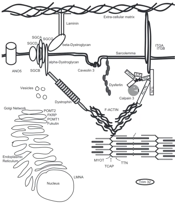
VINC
TLN
PARVB ILK
ACTN
ITGA ITGB
Caveolin 3
Sarcolemma
F-ACTIN Dysferlin
Calpain 3
Trim 32 FKRP
LMNA
MYOT SGCA
SGCB SGCG SGCD
TCAP TTN
ANO5
Fukutin POMT2
POMT1 Dystrophin
Laminin
beta-Dystroglycan
alpha-Dystroglycan
Extra-cellular matrix
Endoplasmic Reticulum
Nucleus Golgi Network
Vesicles
LGMD and membrane stability
The skeletal muscle fibers provide contractile strength to the body. Such force is generated by the internal sarcomere. At the Z-disk the sarcomere connects to the internal Actin cytoskeleton. The filamentous Actin network is bound by the Dystrophin-glycoprotein complex (DGC) which links to the extracellular matrix and thereby allows for transduction of the mechanical force (Figure 1) [16]. Mutations in partners of the DGC have been identified in various muscle disorders, the most severe and prevalent disorder being Duchenne Muscular Dystrophy, which is caused by mutations in the DMD gene encoding for Dystrophin [115]. Dystrophin localizes to the intercellular side of the sarcolemma. It is a modular rod-like protein with an Actin binding domain at its N-terminus and a large C-terminus, connected by a large number of repeat domains. Dystrophin directly interacts with filamentous Actin and to a large protein complex at the sarcolemma. It thereby provides a molecular link between the internal Actin cytoskeleton and the extracellular matrix.
The central part of the anchoring complex is built by the transmembrane glycoprotein Dystroglycan. Dystroglycan is post-translationally cleaved. The resulting α-Dystroglycan is fully extracellular and interacts with extracellular matrix proteins such as Laminin. These interactions are mediated via glycosyl groups that are enzymatically added to Dystroglycan as it travels through the ER and Golgi. Interestingly, several mutations in the genes encoding of at least four different glycosylation enzymes have been causally linked to LGMD (Table 1). The extracellular α-Dystroglycan stays in complex with the transmembrane β-Dystroglycan. This protein interacts on the cytosolic side of the sarcolemma with Dystrophin.
The anchoring complex is stabilized in the sarcolemma by a group of proteins called Sarcoglycans, together with Sarcospan [221]. These proteins prevent extreme movement of the anchor when much force is generated by the muscle. The four different Sarcoglycans form a tight complex, and loss of one often results in secondary loss of the others, indicating that they depend on each other’s presence for correct localization and function [221]. Mutations in all four Sarcoglycan genes have been identified in LGMD [221]. Intriguingly, α-Sarcoglycan contains a putative ATP binding site [221], suggesting additional functionalities for this protein.
LGMD and membrane repair
(www.dmd.nl/dysf). Moreover, mutations in the Dysferlin gene can also cause other disease entities, such as Miyoshi Myopathy (MM [165]) and Distal Anterior Compartment Myopathy (DACM [126]. All of them are collectively referred to as “Dysferlinopathies” [160]. Intriguingly, it has been reported that a single Dysferlin mutation resulted in LGMD in one patient and MM in another [255]. The Dysferlinopathies are characterized by a late onset at age 17-25. Proteomic analysis indicated a decrease in glycolytic type I fiber marker proteins and a concomitant increase in oxidative type II marker proteins, suggesting differences in muscle fiber composition [70]. In addition, Dysferlinopathy patients have a higher number of immature fibers than healthy controls [45]. The muscle tissue is characterized by a strong inflammation of mainly monocytes and macrophages [193], and the disease is often misdiagnosed as polymyositis [193], an autoimmune disease of the muscle.
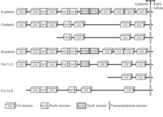
Transmembrane domain O toferlin
Fer1L6 Dysferlin
M yoferlin
C2 C2 Fer1Fer1 C2 Fer1Fer1 Fer1Fer1 DYS F DYS F C2 C2 C2
C2 Fer1Fer1 C2 Fer1Fer1 C2 C2 C2
C2 C2 Fer1Fer1 C2 Fer1Fer1 Fer1Fer1 DYS F DYS F C2 C2
C2 C2 Fer1Fer1 C2 Fer1Fer1 Fer1Fer1 DYS F C2 C2 C2
C2 C2 C2
Fer1 Fer1
C2 Fer1Fer1 C2 Fer1Fer1 C2 C2
C2 C2
C2
C2 C2
Fer1L5
Cytosolic Extra cellular
DysF domain
DYS F
Ferlin domain
Fer1 Fer1
C2 domain
C2 C2
The Dysferlin gene encodes a 230 kDa protein that contains a single pass transmembrane domain at its C-terminus, and has seven C2 domains, three ferlin domains and two DysF domains (Figure 2). The larger part of Dysferlin is intracellular. The C2 domains are calcium-sensitive phospholipid-binding domains and are believed to be essential for Dysferlin function [243,244]. For the first C2 domain, C2A, calcium-dependent phospholipid-binding activity has been shown [243]. Through the C2 domains Dysferlin can also interact with other proteins, in certain cases also in a calcium-sensitive manner (AHNAK2 [119]). The function of the Ferlin and DysF domains are not clear. Based on structural experiments it is thought that mutations in this domain cause the protein to misfold [198], and such misfolding might affect the entire protein.
Cellular studies showed that Dysferlin is a vesicular protein that travels to and from the sarcolemma, and responds to changes in intracellular free calcium [13]. When the plasma membrane of cultured myotubes is ruptured, for instance by laser-mediated wounding, Dysferlin is rapidly recruited to the plasma membrane in a calcium-dependent manner, and accumulates at the site of the lesion [13]. This recruitment is essential for patch-fusion repair of the damaged membrane [12,13], and it has been hypothesized that a disturbed membrane repair capacity underlies the different Dysferlinopathies [214].
Dysferlin is part of the Ferlin family of proteins named after the Caenorhabditis elegans gene Ferlin (fer1) [15]. Ferlin is involved in vesicular function in C. elegans spermatids [253]. Mutations in Ferlin result in infertility [253]. Recently it was shown that these worms also have a muscle phenotype [150]. The human genome encodes six Ferlin-like (Fer1L) genes.
Otoferlin (Fer1L2) is expressed in hair cells of the inner ear, and participates in cellular transmission of sound [217]. Mutations in Otoferlin cause an autosomal recessive form of congenital deafness (DFNB9 MIM#601071) [187]. Otoferlin is expressed in a long and a short isoform (Figure 1) [262]. In humans, the long isoform is found in brain, while the short isoform is expressed in cochlea, vestibule and brain [27,262]. In mice, only the long isoform is observed, yet a mutant mouse model recapitulates the hearing impairment [166]. Otoferlin binds to Syntaxin 1a and Snap25 (both through C2F) and is involved in calcium-regulated vesicle trafficking [209]. In addition Otoferlin interacts with the L-type voltage gated calcium channel (via C2D domain) and aids in regulating neurotransmission in hair cells [209]. Interestingly, the C2 domains that have thus far been described in Otoferlin protein-protein interactions, are present in both Otoferlin isoforms.
with differentiation [75]. While Dysferlin is mostly found at the sarcolemma of mature skeletal muscle fibers, Myoferlin is predominantly found at peri-nuclear membranes [75]. No pathogenic mutations in Myoferilin have yet been described [63]. Knockdown of Myoferlin in endothelial cells attenuated membrane repair [21], suggesting that Myoferlin has a membrane repair function as well and that it might be able to substitute Dysferlin as a potential strategy for therapy. However, while the introduction of human Myoferlin in Dysferlin deficient mice yielded improved the membrane resealing capacity of isolated myoblasts, the general dystrophic phenotype was not rescued [2]. This suggests that Dysferlin has functions beyond membrane repair which cannot be readily substituted by Myoferlin.
The Fer1L4, Fer1L5 and Fer1L6 proteins have not yet been characterized.
Several mouse models have been described that have altered or absent Dysferlin expression [13,22]. These models recapitulate the human phenotype and show a mild progressive muscular dystrophy, and a disturbed membrane repair capability, both in vivo and in cell culture [13,22]. A high level (10-fold) of Dysferlin overexpression also causes a muscle pathology [93], different from membrane repair defects, with increased Endoplasmic Reticulum (ER) stress levels. Like Dysferlinopathy patient muscle tissue, Dysferlin deficient mouse models show strong immune infiltrate after massive muscle damage, either through repeated eccentric contractions [215] or through repeated injections with the snake venom notoxin [45,168].
It is believed that infiltration of immune cells in skeletal muscle tissue is required for normal physiology [9,231]. Throughout life and development there is ongoing communication between muscle tissue and immune cells. This communication is not only important for the clearing of pathogens and dead cells, but also seems to be important for maintenance and differentiation of skeletal muscle tissue [231]. When muscle fibers are damaged, immune cells (first neutrophils, and then monocytes/macrophages) are recruited to clean up the damaged cells that are beyond repair [9,231]. This is mediated by M1 pro-inflammatory macrophages. In a second phase of the immune response M2 contra-inflammatory macrophages are needed to stimulate regeneration [9]. It has been shown that soluble factors from such macrophages can activate satellite cells [252].
between monocyte and muscle.
Recent studies showed Dysferlin expression in cells of non-muscle lineage [6]. These cells include neuronal cells and monocytes [6]. While it is interesting to speculate that Dysferlin is a membrane repair protein in these cell types as well, this is less likely in the case of monocytes/macrophages. These cells destroy other cells, clear up debris and matrix proteins, and participate in cytokine signaling, and should one be damaged others will be recruited and take its place.
It was shown that monocytes from Dysferlin knockout mice, and Dysferlinopathy patients are more aggressive in phagocytosis assays, indicating altered function [193]. This might contribute to the phenotype, but is unlikely to explain all of the pathology. In recent experiments Dysferlin was introduced in Dysferlin knockout mouse muscle tissue, by transgenesis [185] and by AAV treatment [168]. Both studies reported a complete rescue of the contraction induced phenotype [168,185]. The presence of Dysferlin in monocytes however is indicative of Dysferlin functions other than membrane repair (see Chapter 7 for further reading). And indeed, at later ages the rescued mouse models still developed a mild phenotype [168], indicative of a role of non-muscle Dysferlin in the pathogenicity.
LGMD and structural stability
Skeletal muscle is very adaptive and continuously undergoes cycles of degeneration and regeneration. To do this it needs to remodel its cytoskeleton and contractile apparatus. The contractile apparatus is formed around the giant protein Titin (3500 kDa). Titin functions as a molecular ruler along which the contractile proteins are aligned [147]. Upon contraction-induced damage this giant protein complex needs to be disassembled, to remove toxic protein fragments, and to allow for rapid reassembly [17]. It is believed that the cysteine protease CAPN3 is centrally involved in this process.
CAPN3 is a member of the Calpain family of cysteine proteases which participate in limited proteolysis in response to free calcium levels [139]. It is the only disease causing member of the family, and its expression is limited to muscle [139]. Contrary to its ubiquitous family members it is non-responsive to the endogenous inhibitor Calpastatin (which in fact is a substrate [195]), and it is extremely sensitive to fluctuations in intracellular free calcium levels (within nanomolar range [191,195]). CAPN3 is an unstable protease that autolyses upon activation. Its estimated in vitro half-life is less than ten minutes [138] and consequently not much is known of its substrates, or function. Within skeletal muscle most of the CAPN3 is found in its full-length non-autolysed form of 94 kDa [139]. This form is believed to be proteolytically inactive [72]. It localizes for 90% to the sarcomere [17], and directly interacts with Titin [107,195]. It is hypothesized that the Titin interaction is used to store inactive CAPN3, to allow for local proteolysis upon activation [107,138]. Interestingly, mutations in Titin at the CAPN3 binding site also cause LGMD [104]. Elegant proof for this model comes from experiments with transgenic mice. Mice that overexpress human CAPN3 were crossed with mice that had mutations in the CAPN3 binding domain in Titin [122,195]. While the CAPN3 transgenic mouse itself had no phenotype, the Titin mutant mouse showed a mild muscular dystrophy. Crossing both mouse models aggravated the muscle phenotype, strongly indicating the importance of Titin in buffering CAPN3 activity [122,195].
was found in hind-limb suspension experiments [153]. Where wild-type muscle showed strong muscle atrophy, degeneration and sarcomere remodeling during the experiment, and clear regeneration thereafter, this process was impaired in CAPN3 deficient muscle [153]. Therefore, CAPN3 is considered to be essential for muscle atrophy and possibly degeneration and regeneration. In agreement with this, the very few substrates reported are all structural proteins [98,242], and ectopic overexpression of CAPN3 causes cell rounding and detachment, indicative of cytoskeleton remodeling [242] (see Chapter 3 and 4 for further reading).
Over 400 pathogenic CAPN3 mutations have been described and roughly one third of these are miss sense (www.dmd.nl/capn3). There is no clear mutational hotspot [151]. It has been estimated that roughly one third of all mutations does not impair the proteolytic activity [184]. Modeling experiments on the experimentally determined structure of Calpain 2 indicated that several mutations affect the autolytic activity of CAPN3, showing that a change in the activity time span might already be deleterious [87,135]. This resulted in a model where locally stored inactive CAPN3 (on Titin molecules [107]) allows for local proteolysis upon activation [17]. However, the activation signal remains elusive and biochemical support for this hypothesis is wanting [17] (see Chapter 4 for further reading).
Due to the identification of CAPN3 in the Dysferlin protein complex, the hypothesis was put forward that CAPN3 functions in membrane repair [118]. It has recently been shown that ubiquitous Calpains, Calpain 1 and 2, are essential components of the non-muscle membrane repair system [183]. However, membrane repair assays on CAPN3 deficient primary myoblasts showed no clear difference in membrane repair capacity [182], indicating that the link between LGMD2A and 2B is not that straightforward.
Linking LGMD proteins
In all forms of LGMD muscle tissue is correctly formed and functional. Concomitantly, in all three above described pathogenic mechanisms deregulation of muscle maintenance seems to be central, rather than muscle development. In all forms of LGMD an increased level of regeneration has been observed. This suggests that when muscle maintenance processes fail, damaged muscle fibers need to be replaced. Muscle has a high capacity for regeneration, but it is not unlimited [56,57].
possible. This stress on the satellite cell population results in a quicker loss of regenerative capacity [31]. When regeneration can no longer match the loss of existing fibers, and the rate of myofiber loss exceeds the rate of replenishment, LGMD disease onset would eventually follow.
In line with this model the earliest onset of disease is described for CAPN3 (LGMD2A) patients [28]. Extrapolating from the mouse models[152,153], these patients likely have an impaired muscle degeneration and regeneration capacity, making them especially prone to defects in muscle maintenance. Dysferlin patients (LGMD2B) can still regenerate their myofibers. Onset of LGMD2B is thus later than for CAPN3 patients.
To maintain correct muscle function specific molecular signaling pathways exist. Pathways that mechanically or chemically sense contraction and resulting damage, and that need to respond appropriately. The molecular interactions between Dysferlin and Caveolin 3 [172] and CAPN3 [120] provide a first indication for a connecting molecular network that underlies muscle maintenance and LGMD pathogenicity. Maintenance processes require rapid and clear-cut communication at a molecular level. An intriguing protein in such network might be AHNAK, which has been implicated in many different processes that are important to muscle, ranging from membrane repair, vesicle trafficking, excitation-contraction coupling, and (neuronal) remodeling [3,19,52,53,90,100,106,118,119,146,175,176,20 6,220,222] (for further reading on AHNAK see Chapters 3, 5 and 6). AHNAK consists of a large body of repeat units that fold into a B-propellor [146], and a N- and C-terminus that are intrinsically disordered [113,146,227]. Given the large number of hitherto described ANAK binding partners (>10) [5,99,177], and its partially disordered folding, we propose that AHNAK functions as a Hub protein, which are important central components of protein-protein networks [109].
Table 1
Disease MIM Gene Protein Expression Function
LGMD1A #159000 MYOT Myotillin Skeletal muscle, heart * Sarcomere component
LGMD1B #159001 LMNA Lamin A/C Ubiquitous Nuclear lamina component
LGMD1C #607801 CAV3 Caveolin 3 Skeletal muscle, heart, brain, adrenal gland
Essential component of caveolae
LGMD1D %603511 unknown Unknown
LGMD1E %602067 Unknown Unknown
LGMD1F %608423 Unknown Unknown
LGMD1G %609115 Unknown Unknown
LGMD2A #253600 CAPN3 CAPN3 Skeletal muscle, heart * Proteolytic regulation of the cytoskeleton
LGMD2B #253601 DYSF Dysferlin
Skeletal muscle, heart, monocytes, kidney, trophoblast
Membrane repair
LGMD2C #253700 SGCG γ-sarcoglycan
Skeletal muscle, heart, monocytes, mesenchym
Dystrophin glycoprotein complex stability
LGMD2D #608099 SGCA α-sarcoglycan Skeletal muscle, heart, monocytes Dystrophin glycoprotein complex stability
LGMD2E #604286 SGCB β-sarcoglycan Skeletal muscle, heart Dystrophin glycoprotein complex stability
LGMD2F #601287 SGCD δ-sarcoglycan Skeletal muscle, heart, intestine Dystrophin glycoprotein complex stability
LGMD2G #601954 TCAP Theletonin Skeletal muscle and heart Sarcomere component
LGMD2I #607155 FKRP Fukutin-Related Protein
Skeletal muscle, heart, thymus, kidney, placenta *
Glycosylation enzyme
LGMD2J #608807 TTN Titin Skeletal muscle, heart,
trophoblast
Sarcomere component
LGMD2K #609308 POMT1 Protein O-Mannosyl transferase 1
Skeletal muscle, heart, cerebellum, lymph node, placenta *
Glycosylation enzyme
LGMD2L #611307 ANO5 Tmem16 Skeletal muscle, heart, brain* Calcium-dependent Chloride channel
LGMD2M #611588 FKTN Fukutin Heart, intestine, trophoblast Glycosylation enzyme
LGMD2N *607439 POMT2 Protein O-Mannosyl transferase 2
Adrenal gland, thyroid gland, monocytes, kidney
Glycosylation enzyme
Proteomic analysis of the Dysferlin protein
complex unveils its importance for
sarcolemmal maintenance and integrity
Antoine de Morrée 1, Paul J. Hensbergen2, Herman H.H.B.M. van Haagen1, Irina Dragan2, André M Deelder2, Peter A.C. ’t Hoen1, Rune R. Frants1, Silvère M. van der Maarel1
1 Department of human genetics, Leiden University Medical Center, Leiden, The Netherlands 2 Biomolecular Mass Spectrometry Unit, Department of parasitology, Leiden University
Medical Center, Leiden, The Netherlands
5;5(11):e13854.-Abstract
Author summary
Introduction
Dysferlin (DYSF, MIM*603009) is a 230 kDa large transmembrane protein highly expressed in striated muscle and to a lesser extent in other tissues, including monocytes, syncytiotrophoblast, endothelium, brain, pancreas, and kidney [6]. Dysferlin is found intracellularly on vesicles and at the plasma membrane. Upon laser-inflicted membrane damage Dysferlin rapidly accumulates at the site of the lesion in a calcium-dependent manner, and participates in patch-fusion repair. In the absence of Dysferlin the membrane tear is not adequately repaired and the myofiber will undergo necrosis [13].
Mutations in the Dysferlin gene cause a spectrum of adult-onset progressive muscular dystrophies including Limb Girdle Muscular Dystrophy type 2B (LGMD2B, MIM#253601), Myoshi Myopathy (MM, MIM#254130), and Distal Anterior Compartment Myopathy (DACM, MIM#606768), commonly referred to as Dysferlinopathies [15,126,165]. There is no clear genotype-phenotype correlation and the ~150 described mutations cover the complete open reading frame (www.dmd.nl/dysf). It is therefore unclear how defects in the DYSF gene cause muscular dystrophy. It has been suggested that the skeletal muscle membrane is continuously subject to mechanical wear and tear, and that the Dysferlin deficiency phenotype results from inefficient membrane repair in response to continued membrane damage [214]. Dysferlin knockout mice develop a phenotype similar to the Dysferlinopathies [13].
Dysferlin contains seven C2 domains, two DysF domains and a C-terminal transmembrane domain. C2 domains are calcium-sensitive phospholipid-binding domains, as was also shown for the first C2 domain (C2A) of Dysferlin [243], and are thought to be important for regulating Dysferlin trafficking. These domains have also been shown to interact with proteins [119]. The function of the DysF domain remains unclear [198].
have not yet been characterized.
While the role of Dysferlin in membrane repair is well established, it is less clear how the protein is regulated. Likely it requires binding partners that aid in vesicle nucleation, localization, targeting and recycling. To date only few of such cofactors have been identified, yet they yielded important insight into Dysferlin function.
MG53 (TRIM72, MIM *613288) is a redox sensor that participates in vesicle nucleation [34]. It can interact with Dysferlin [36]. Together with the Dysferlin interacting protein caveolin 3 it is involved in the trafficking of Dysferlin to and from the sarcolemma [36,110,111]. In addition, annexins A1 and A2 can bind Dysferlin in a calcium-dependent manner [162]. These proteins are membrane fusogens that participate in lysosome exocytosis [179]. They are thought to aid in Dysferlin vesicle targeting. The cysteine protease Calpain 3 (CAPN3, MIM *114240) co-immunoprecipitates with Dysferlin in skeletal muscle [118,120]. It is hypothesized to play a role in cytoskeleton remodeling [17,118], and is predicted to remodel cytoskeletal structures to allow for patch fusion repair [118,182,183]. Finally, AHNAK (MIM*103390) is found on enlargosomes which have been implicated in membrane enlargement and repair [26,52,167]. AHNAK interacts with Dysferlin in skeletal muscle, an interaction that is regulated by CAPN3 activity [118]. These protein interactions have thus yielded some information on Dysferlin function in membrane repair.
Recent data however, suggest that Dysferlin is more than a membrane repair protein. It has been shown to be involved in cytokine [45] and chemokine [68] secretion and associates with developing t-tubules [143]. In fertilized sea urchin embryo’s Dysferlin participates in extracellular ATP signaling [60]. Moreover, a defect in membrane repair cannot fully explain the patient’s phenotype, which has been reported to include renal [130] and cardiac failure [169]. In addition, Dysferlin is considered to be a very dynamic protein, which is found in the cytosol in myoblasts and regenerating myofibers, but shows a more prominent membranous localization in mature skeletal muscle tissue [45]. It was shown that Dysferlin trafficking and endocytosis depend on its direct interaction with caveolin 3 [110,111,172].
Dysferlin from human skeletal muscle followed by mass-spectrometry and western blotting we could previously identify CAPN3 and AHNAK to interact with Dysferlin [119,120].
In this study we compared Dysferlin protein complexes from proliferating myoblasts, differentiated myotubes and mature skeletal muscle tissue. We show that 1) we can efficiently and reproducibly immunoprecipitate Dysferlin protein complexes from these sources, 2) bioinformatics analyses of mass spectrometry identified proteins confirm a role for Dysferlin as a vesicle protein, and 3) such analyses reveal new layers of Dysferlin function which we substantiated by further exploring interactions with Myoferlin, and focal adhesion components.
Results
Robust and reproducible isolation of Dysferlin protein complexes
As Dysferlin expression is reported to increase with myogenic differentiation we hypothesized that the composition of its protein complex might also change during this process. Therefore we aimed to isolate Dysferlin protein complexes from different stages of myogenic differentiation. We used the IM2 cell model [189] as a source for Dysferlin protein complexes to establish the optimal immunoprecipitation conditions. This cell line can be grown indefinitely under
250 kDa
120 kDa
100 kDa
72 kDa
55 kDa
Loading control
CAPN3
DYSF
Day 0 Day 1 Day 3 Day 5
permissive conditions, and switched to myogenic differentiation by serum deprivation. The IM2 cell model performs more consistently than the C2C12 cell line in terms of myogenic capacity and differentiates within a shorter time-span. We first established by western blotting that IM2 cells express Dysferlin (Figure 1). Dysferlin expression increases with differentiation, in line with previous data from C2C12 myoblast cells [81]. CAPN3, an established partner in the Dysferlin protein complex is also expressed in these cells (Figure 1).
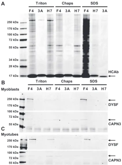
We previously reported two Dysferlin specific llama VHH heavy chain antibody fragments (HCAb) that can specifically immunoprecipitate Dysferlin from human skeletal muscle [23]. We adapted the immunoprecipitation method for cultured cells by testing different buffer conditions (Figure 2). We used the two reported Dysferlin HCAb fragments, F4 and H7 [23], and compared their performance to a non-specific HCAb (3A), selected against amyloid-β [40] and which does not recognize Dysferlin. We selected three different lysis buffers for our experiments. The first buffer is of low ionic strength and contains 0.2% Triton. Triton is a comparatively mild non-denaturing non-ionic detergent. The second buffer is of high ionic strength containing SDS and TEA and has a strong solubilizing capacity. At the used concentration (0.1%) SDS acts as a partially denaturing detergent that is efficient at breaking low-affinity protein-protein interactions. The third buffer contains CHAPS (0.15%) and is of intermediate strength. CHAPS is a nondenaturing zwitterionic detergent that is useful for solubilizing membrane proteins. The CHAPS buffer has been used previously to precipitate Dysferlin protein complexes from muscle tissue with HCAb [23] and with conventional antibodies [37].
therefore preferred as lysis buffer. Because it is more difficult to homogenize tissue than cultured cells, we decided to use CHAPS buffer for skeletal muscle tissue as we have done previously [120].
Triton Chaps SDS
F4 3A H7 F4 3A H7 F4 H7 3A
HCAb
250 kDa 170 kDa 100 kDa 72 kDa 55 kDa
34 kDa 28 kDa
17 kDa
250 kDa 170 kDa 100 kDa 72 kDa 55 kDa
DYSF
CAPN3
250 kDa 170 kDa 100 kDa 72 kDa 55 kDa
DYSF
CAPN3
Triton Chaps SDS
F4 3A H7 F4 3A H7 F4 3A H7
Myoblasts
Myotubes
A
B
C
Figure 2:Reproducible Dysferlin immunoprecipitation under different conditions.
Myoblast Myotube Muscle
F4 3A H7 F4 3A H7 F4 3A H7
Myoblast Myotube Muscle
F4 3A H7 F4 3A H7 F4 3A H7
DYSF 250 kDa
170 kDa
100 kDa
72 kDa
55 kDa
34 kDa
28 kDa
250 kDa
170 kDa
M yoblasts: M yotubes: Muscle:
D ysferlin D ysferlin D ysferlin
AH N AK AH N AK AH N AK
C alpain 3 C alpain 3 C alpain 3 Annexin A2 Annexin A2
Tubulin Tubulin Tubulin
A
B
C
Described interaction partners validated by western blotPARVB PARVB PARVB
DHPR DHPR
Dysferlin interactions in myoblasts, myotubes, and skeletal muscle tissue
We continued with preparing lysates for IM2 myoblasts, IM2 myotubes at 5 days post-differentiation (when spontaneous contraction was observed), and human skeletal muscle tissue. After cell lysis in Triton buffer or tissue homogenization in CHAPS buffer, Dysferlin protein complexes were immunoprecipitated from these lysates using HCAb H7, separated on SDS-PAGE gels and stained with Coomassie-blue for further mass spectrometry analysis (Figure 3A). We monitored the IP procedure with western blot (Figure S1). The IP was confirmed by western blot with a conventional antibody specific for Dysferlin (Figures S1 and 3B). With western blotting we could confirm six of the described interactions (TUBA [41] (Figure S1) CAPN3 [23], AHNAK [8] (both in Figure S2), PARVB [42], DHPR [43], ANXA2 [21] (data not shown), summarized in Figure 3C. Mass spectrometry analysis confirmed the presence of one additional reported Dysferlin interaction partner (MG53 [18] Table S1), but also resulted in the identification of many new putative binding partners (complete lists in table S1). Skeletal muscle cells contain a large amount of sarcomeric proteins, and components of the dystrophin glycoprotein complex (DGC). We did not observe the core DGC components in the IP fractions, and the amount of sarcomeric proteins is low, consistent with the fact that the HCAb specifically recognize and immunoprecipitate Dysferlin. We conclude that the IP method efficiently and reproducibly enriches for Dysferlin protein complexes.
In our IP dataset for myoblasts we identified 521 proteins annotated in online databases, 344 proteins in myotubes and 229 proteins in skeletal muscle tissue (full lists in supplemental tables S1 and S2). 115 proteins are shared by all datasets (Figure 4). These proteins we therefore consider a core-set of most likely
115 126
84 0
103 196
30
Tissue 229
M yotube 344 M yoblast 521
Dysferlin interacting proteins, or representative of functionalities of Dysferlin that do not change during differentiation. As mentioned before, the tissue IP was performed under more stringent conditions. Interestingly, 85% of the proteins identified in the tissue IP were also identified in the cultured cells. This underlines the reproducibility of the IP experiment, and indicates a relatively low number of proteins that nonspecifically co-immunoprecipitate with Dysferlin. An unexpected finding was that there are no proteins shared by IP from tissue and myotube alone. This might be explained by the more stringent conditions used for tissue homogenization.
Potential interaction between Dysferlin and Myoferlin
and Dysferlin can be part of the same protein complex, and opens the possibility of their co-existence in a single protein complex in muscle.
250 kDa
130 kDa
100 kDa
250 kDa
130 kDa
DYS F
MYOF
F4 H7
U2OS
Non-bound
Immunoprecipitation
F4 H7 F4 H7
Wild type
Dysferlin
Mock
Wt
Dysf
Mock
F4 F4 F4
250 kDa
F4
Myoblast
3A
H7
F4
Myotube
3A
H7
F4
Muscle
3A
H7
MYOF
A
B
*
130 kDa
Bioinformatics analysis of all identified complex partners
We continued with the three datasets of putative Dysferlin interacting proteins and performed bioinformatics analyses to characterize the Dysferlin protein complex. We first analyzed the datasets with the STRING interaction database (http://string-db.org/). STRING contains information of known annotated protein-protein interactions. 14 Dysferlin protein-protein-protein-protein interactions are recorded in STRING for human and mouse, of which 6 are direct interactions described in PubMed references (CAV3, CAPN3, AHNAK, PARVB, ANXA1, ANXA2). The others are interactions inferred from co-occurrence in MedLine abstracts and protein databases. Protein-protein interactions described in recently published studies, such as TUBA [10], are not yet annotated, and therefore not included in STRING. The remaining 8 predicted Dysferlin interactions in STRING are not supported by experimental evidence and include DGC partners. These interactions are not identified in our datasets, and we consider them unlikely given the existing literature on Dysferlin. Thus, STRING does not contain any information on the Dysferlin protein complex partners, and therefore we consider the majority of interactions identified with our IP experiments as novel Dysferlin interactions.
Literature analysis of the Dysferlin protein complex
of concepts associate with the Dysferlin protein complex we clustered the identified proteins and subsequently annotated with concepts. Representative concepts for the observed clusters are shown in Table 1B (full table in S3), and include vesicle related concepts such as Kinesin Activity and Membrane Protein Traffic, consistent with the vesicular localization of Dysferlin. However, also many novel associations are uncovered, such as Translation Initiation, Actin Cortical Patch, and Chaperonin Activity. These concepts may refer to novel Dysferlin functionalities.
Given that Dysferlin expression is not restricted to skeletal muscle, we next evaluated co-expression of the Dysferlin protein complex partners over multiple tissues, using the database Gene Atlas. We obtained an AuC of ~0.7 (Figure S3), where an AuC of 0.5 would mean a random level co-expression and an AuC of 1.0 would mean complete co-expression. This result suggests that we do not mainly identify muscle specific proteins.
Gene Ontology analysis of the Dysferlin protein complex
Gene Ontology (GO) category analysis is commonly used for bioinformatics analysis of large datasets. We used the DAVID web tool to analyze GO category enrichment by performing a cluster analysis (Table 2, full table in S4). Protein lists were uploaded in DAVID and compared to a random background dataset (shown) or a random set of muscle genes (not shown) with comparable results. The output is a ranked set of GO term clusters that are significantly overrepresented in the IP datasets (Table 2). Many of the highest ranked clusters relate to vesicle trafficking (in bold). Examples of such proteins are Clathrin and Rab GTPases. This corroborates the reported function of Dysferlin as a vesicular protein. In addition to vesicle trafficking, several other clusters are significantly represented, which may point to potential novel roles for Dysferlin function.
Many identified proteins relate to ATP binding and purine synthesis. This finding is interesting as a recent study showed that Dysferlin is involved in extracellular ATP and ADP signaling. One of the indentified target proteins, found in all datasets, is ATP synthase (ATP5b).
A large number of mitochondrial proteins is indentified in the Dysferlin protein complex. 18 proteins are observed in the core-set of 115 proteins. This includes proteins that localize to the outer and inner mitochondrial membrane, indicating that large parts of mitochondrial membranes co-purify with Dysferlin. Intriguingly, the alternative first exon of Dysferlin [202] encodes a putative mitochondrial targeting signal (not shown), raising the possibility that Dysferlin is targeted to mitochondria in myoblasts.
possible explanation for this is that ER-residing Dysferlin is immunoprecipitated from the cells.
KEGG pathway analysis of the Dysferlin protein complex
We proceeded with a KEGG pathway analysis, which gives a better visualization how the identified Dysferlin protein complex partners relate to each other (table S5). Interestingly, the strongest vesicle-related KEGG pathway in all three datasets is Endocytosis, rather than Exocytosis. This is consistent with previous studies which suggested that Dysferlin undergoes rapid endocytosis in the absence of caveolin 3 on the cell membrane [111]. Additionally, Dysferlin negative cells show an upregulation of endocytotic proteins [193], again suggesting a role for Dysferlin in endocytosis. Most proteins that are found upregulated by Nagaraju
et al [193] are coimmunoprecipitated with Dysferlin, and include Mannose-6 phosphate receptor (CIMPR, or IGF2R), adaptin (AP2), clathrin-α (CLTA) and the GTPase RAC2 (Table S1). Interestingly, this KEGG pathway analysis also revealed several immunological pathways, such as Fc gamma R-mediated phagocytosis. As Dysferlin is also expressed in monocytes [66,114,256], this indicates that common pathways may exist between muscle and monocytes.
Multiple pathways are ranked higher than Endocytosis, but do not relate directly to vesicles. The top-ranked pathway is Metabolic Enzymes, which includes most of the mitochondrial proteins identified in the IPs. This indicates a role for Dysferlin in energy metabolism. However, due to the high content of metabolic enzymes in muscle, some aspecific interactions cannot be excluded. Of interest is the presence of creatine kinase M and B. Serum levels of creatine kinase are exceptionally high in Dysferlinopathy patients, reaching 100 fold above normal [29].
Finally, the pathways Focal Adhesion and Tight Junction are ranked high in all 3 datasets. Focal adhesions and tight junctions are two structures involved in cell-cell and cell-matrix contacts, in which the intracellular Actin cytoskeleton is linked by transmembrane proteins to extracellular proteins. These adhesion sites exist independently of the DGC, and previous cellular fractionation experiments have shown that Integrins, which are part of focal adhesions, are restricted to different membrane sections than the DGC [190]. While we do not identify DGC components in the Dysferlin protein complex, several focal adhesion proteins were identified in all three sources, including Vinculin (VINC), Actinin (ACTN), and Talin (TLN). This hints at a role for Dysferlin in cell adhesion.
Dysferlin can interact with the focal adhesion component Vinculin
To validate our bioinformatics analysis we further investigated one of the newly identified pathways: Focal Adhesion (Figure 6). Vinculin has strong conceptual overlap with Dysferlin (VCL in Table 1A). Interestingly, the previously reported Dysferlin interactor PARVB localizes to and regulates focal adhesions [174]. PARVB can directly interact with ILK and Dysferlin, and regulates focal adhesion turnover [173,174,261]. In the absence of Dysferlin, PARVB is no longer recruited to the sarcolemma [174]. To confirm the existence of focal adhesion components in the Dysferlin protein complex we verified that both Dysferlin HCAbs can co-immunoprecipitate full-length Vinculin (VINC) (Figure 7A). A Vinculin doublet, corresponding to two Vinculin isoforms [194], is detected consistently in the Dysferlin HCAb IP fractions, but not in controls. Interestingly, the relative amount of immunoprecipitated Vinculin decreases with differentiation, suggesting that the interaction is dynamic. To further support the validity of this interaction we performed the reverse experiment and used conventional antibodies against Vinculin to co-immunoprecipitate Dysferlin (Figure 7B). These antibodies can efficiently co-immunoprecipitate Dysferlin from cultured myotubes, unlike isotype control antibodies, indicating the validity of their interaction. We performed additional immunofluorescent analysis of human skeletal muscle cryosections. As shown in Figure 7C, Vinculin is found predominantly at the sarcolemma in cross-sections, and colocalizes with Dysferlin.
VINC
Myoblast
Myotube
Muscle
F4
3A
H7
F4
3A
H7
F4
3A
H7
120 kDa
A
B
Vinculin
Dysferlin
DAPI
Merge
C
Myoblast
Dysferlin
250 kDa
Input
F4
NB
3A
B
NB
B
B
B
VINC
Control
Vinculin
120 kDa
Discussion
We have identified a large set of potential Dysferlin protein complex partners in a myogenic context. We showed that many new complex partners participate in vesicle trafficking, which corroborates previous data that Dysferlin is involved in this process and suggest that its interaction partners, such as Clathrin, assist in this function. In addition we identified several potential novel roles for Dysferlin, including cell adhesion, metabolism, mitochondrial association, and immune cell function.
F4 and H7 show a different performance in the IP as shown in Figure 2. Such differences in performance are not unique to HCAb, and it is difficult to predict and establish why this occurs. It might be that the antibodies compete with interaction partners at the site of antigen binding, or that the epitope availability is not uniform. The latter has been described for Dysferlin as C2 domains are calcium-sensitive [7], and VHH are known to recognize three-dimensional folds [55].
It must be noted that the IP procedure does not discriminate between direct and indirect interactions. We aimed to limit the potential problem of non-specific binders by analyzing two Dysferlin specific HCAb and using different myogenic sources and buffers with different solubilizing properties. Although it cannot be excluded that some co-immunoprecipitated proteins are non-specific binders, the large overlap of immunoprecipiated proteins in all three sources under different stringency conditions, and their absence in the control experiments, indicates that most IP proteins are valid complex partners of Dysferlin, whether direct or indirect. In addition, we tested and confirmed 7 of 9 established protein-protein interactions of Dysferlin, and provided biochemical evidence for the novel interactions with Myoferlin and Vinculin. Finally, an additional indication for the validity of the method is that no components of the DGC were identified.
Myoferlin was consistently identified in the Dysferlin protein complex. Given the specificity of all antibodies used in this study we do consider this interaction to be genuine. It is interesting to speculate that Dysferlin could form oligomers with its homologous family member Myoferlin, and how this contributes to protein function, possibly patch-fusion repair.
We often observed in our western blots that the relative amounts of immunoprecipitated proteins often changes with differentiation. Examples of such dynamic protein-protein interactions of Dysferlin are CAPN3 (Figure 2B, C), Myoferlin (Figure 5A), and Vinculin (Figure 7A). This suggests that the Dysferlin protein complex has a dynamic nature as a function of myogenic differentiation. When we further compared the specific proteins for each dataset, we noticed that a high number of mitochondrial proteins was co-immunoprecipitated from myoblasts (20%, not shown). This amount decreases with differentiation and is lowest in tissue (12%). In contrast, the relative amount of metabolic enzymes from the glycolysis pathway is enriched with differentiation (going from 2% to 4%). This may indicate that the function of Dysferlin changes with differentiation. Unfortunately our qualitative analysis does not allow for solid quantitative conclusions due to its dependence on experimental conditions. Future studies using quantitative proteomic approaches may give a clearer impression of the relative abundance of the protein identified in the dysferlin protein complex during certain processes.
Among the identified mitochondrial proteins is ATP synthase (ATP5b). ATP synthase is a protein complex enriched in mitochondria and part of the respiratory chain [56]. In addition ATP5b is also found at the cell membrane, where it maintains extracellular ATP and ADP levels, and functions as a membrane receptor for HDL [56]. It was recently shown that Dysferlin is involved in extracellular ATP and ADP signaling [34]. It is tempting to speculate that the putative interaction between Dysferlin and ATP synthase allows for the formation of vesicle with ATP content to participate in the repair process.
Many of the identified novel Dysferlin protein interactions relate to protein translation and folding. It was recently suggested that pathogenic mutations in Dysferlin result in improper folding of the protein and therefore in dysfunction and degradation [258]. Moreover, it has been shown that the ER can function as an intracellular storage compartment of recombinant Dysferlin [83], which is in line with elongated translation/folding time. Therefore we feel that the identified interactions between Dysferlin and several ER proteins are not an artifact but reflect the amount of effort that the muscle cells invest into proper folding of the 230 kDa protein.
Dysferlin in vivo. In addition, it was shown that recombinant Dysferlin interacts with the chaperones VCP and SEC61 [83] as part of the ERAD pathway. Both VCP and SEC61 co-immunoprecipitate with endogenous Dysferlin.
Based on the high ranking of the Focal Adhesion KEGG pathway in all datasets we focused our attention on this pathway. Mutations in the focal adhesion components ITGA7 [108], and VINC [194] result in a muscle pathology. Interestingly, Integrins and FAK have been shown to be essential for myogenic differentiation and localize at the site of fusion [74,204,205]. Dysferlin shows a similar localization in differentiated myotubes [143]. This indicates that Dysferlin might be involved in regulation of cell-cell contacts. Interestingly, a recent screen identified Integrins (the transmembrane components of focal adhesions) to be essential for endocytosis in HeLa cells [55]. This is in line with our finding that Dysferlin co-immunoprecipitates endocytic proteins.
Dysferlin was previously described to be involved in membrane repair. For membrane repair to occur, the cell needs to patch the damaged membrane, and remodel the local cytoskeleton [183]. It has been shown that at the site of damage, cytoskeletal proteins Talin and Vimentin are proteolytically processed by Calpains [183]. Moreover, at this site Integrins and Actin rapidly accumulate [183], suggesting cytoskeletal remodeling of the cortical cytoskeleton, and the adhesion sites. It is tempting to speculate that Dysferlin is involved in both processes. Through its membrane association it can target vesicular membrane stores, and through its interaction with focal adhesion components it can coordinate cytoskeletal remodeling. Previous work showed that Dysferlin recruits PARVB to the sarcolemma [174]. PARVB can directly interact with Integrin linked kinase, and is important for stabilizing focal adhesions [173,261]. In the absence of Dysferlin PARVB does not localize to the sarcolemma, but the functional consequence of this is unknown [174]. CAPN3 is also in complex with Dysferlin. Through its proteolytic activity it can modulate the direct interaction between Dysferlin and the giant protein AHNAK [118]. Thus, Dysferlin might act as a sensor to coordinate the remodeling of structural proteins in addition to aiding patch-fusion of membranes.
Methods
Cell cultureMouse IM2 myoblast were maintained under permissive conditions at 33°C and 10% CO2 in DMEM (61965, GIBCO) supplemented with L-Glutamine, 1% Pen/ Strep (100IU/100UG/ML, Gibco-BRL), 20% FCS, IFN-γ, and Chick embryo extract. For differentiation cells were grown to 70% confluency, washed with PBS and grown in DMEM (61965) supplemented with 10% HS, L-Glutamine, and Pen/Strep.
Antibodies
The following antibodies were used in this study: MaCAPN3 (NCL-12A2 Novocastra) at 1;500 for western blot. MaVSV (P5D4 gift from Dr. J. Fransen, Nijmegen, The Netherlands) at 1;5,000 for IF. MaDYSF (Hamlet, Novocastra) at 1;300 for western blot and IHC. MaMYOF (gift from Dr. R. Bashir), GaMousealexa488 (Molecular Probes, Eugene, OR, USA), GaRabbitalexa594 (Molecular Probes) at 1;1,000 and 1;2,000, respectively. GaRabbitIRDye800 and GaMouseIRDye680 (Westburg) were used at 1;5,000 for western blotting.
Immunoprecipitation
The following lysis buffers were used: Triton buffer (50mM TrisHCl, pH 7.5, 150mM NaCl, 0.2% Triton X100, 1x protease inhibitor cocktail (Roche)), CHAPS buffer (50 mM Tris-HCl pH 7.5, 150 mM NaCl, 0.15% CHAPS and 1x protease inhibitor cocktail), or SDS buffer (20mM triethanolamine 0,14M NaCl, 0,1% DOC, 0,1% SDS, 0,1% triton X100, protease inhibitor cocktail). Cultured cells were prepared freshly by washing in PBS and lysed by scraping on ice in cold lysis buffer. Proteins Snap-frozen human skeletal muscle tissue was homogenized in CHAPS buffer. All homogenates were spun down at maximum speed, 4°C, 20 min. The pellet was discarded as debris. The supernatant containing the solubilized proteins was cleared with protein A Sepahrose CL-4B (Amersham). The Sepharose was first pre-equilibrated by a 3x wash in lysis buffer and added to the homogenates for 1h, at 4°C tumbling. Sepharose was removed and antibody added (50µg HCAb, or 10µg conventional antibody) for O/N incubation at 4°C tumbling. Thereafter pre-equilibrated Sepharose was added and incubated for 2h, 4°C, tumbling). Homogenates were spun down at 500g and supernatant stored as non-bound fraction. The Sepharose was washed 5x (>5min tumbling at 4°C). Finally, all fluid was removed and protein eluted by boiling in sample buffer. This was analyzed as the IP or bound fraction.
Western blot
gels. Proteins were separated and blotted onto PVDF membranes. Blots were washed, blocked in 4% mPBS for 30 min, and incubated with primary antibody diluted in blocking buffer for 2h at RT. Subsequent washes in PBS-tween were followed with 1h incubation with secondary antibody in the dark. Blots were washed and scanned with an odyssey scanner (Licor, Lincoln, Nebraska, USA).
Immunostaining
For immunohistochemical examinations, muscle cryosections of 6 µm thickness were fixed in 3.7% formaldehyde in PBS containing 0.1% Triton X-100 for 30 min, following by pre-incubation with 4% skimmed milk (Marvel) in PBS at room temperature for 2h. The sections were incubated with primary antibody o/n at 4˚C, and subsequently by incubation of fluorescein-labeled secondary antibody for 1h min at RT. The sections were washed with PBS, dehydrated with (subsequently) 70, 90, 100% ethanol and mounted in a DAPI (50 ng/µl)/ Vectashield mounting medium (Burlingame). Final preparations were analysed with a Leica Aristoplan fluorescence microscope and images were obtained using a Cytovision (Applied imaging) digital system.
In-gel tryptic digestion
IP fractions were separated on SDS-PAGE gels, and proteins were visualized with Coomassie (SimplyBlue, Invitrogen). Gel lanes were sliced into 25-30 bands, cut into small pieces and washed with 25 mM NH4HCO3 followed by two rounds of dehydration with 100% acetonitrile for 10 min. For reduction and alkylation, gel particles were first incubated with 10 mM dithiothreitol for 30 minutes at 56 °C. Following dehydration with acetonitrile, gel plugs were subsequently incubated in 55 mM iodoacetamide for 20 minutes at room temperature. After two rounds of washing with 25 mM NH4HCO3 and dehydration with 100% acetonitrile, the gel particles were completely dried in a centrifugal vacuum concentrator (Eppendorf, Hamburg, Germany). Dried gel particles were re-swollen for 15 min. on ice by addition of 15 µl of a trypsin solution (12.5 ng/µl in 25 mM NH4HCO3, Sequencing grade modified trypsin, Promega, Madison, WI). Following this, 20 µl of 25 mM NH4HCO3 was added and samples were kept on ice for an additional 30 min. Tryptic digestion was subsequently performed overnight at 37˚C. Following tryptic digestion, the overlaying digestion-solution was collected. Two additional rounds of extraction with 20 µl 0.1 % TFA were used to extract peptides from the gel plugs and all extracts were pooled.
Nano LC ESI MS/MS
A volume of 10 µL of sample was injected onto a C18 PepMapTM 0.3mm×5mm trapping column (Dionex) and washed with 100% A (2% acetonitrile in 0.1% formic acid in MQ water, v/v) at 20µL/min for 15 min. Following valve switching, peptides were separated on a C18 PepMap 75µm×150mm column (Dionex) at a constant flow of 200nL/min. The peptide elution gradient was from 10 to 60% B (95% acetonitrile in 0.1% formic acid in MQ water v/v) over 50min. The nanoflow LC system was coupled to an HCTultra IonTrap (Bruker Daltonics, Bremen, Germany) using a nano-electrospray ionisation source. The spray voltage was set at 1.2 kV and the temperature of the heated capillary was set to 165 °C. Eluting peptides were analyzed using the data dependent MS/MS mode over a 300-1500 m/z range. The five most abundant ions in an MS spectrum were selected for MS/ MS analysis by collision-induced dissociation using helium as the collision gas.
Mass spectrometry data analysis
Peak lists were generated using DataAnalysis 4.0 software (Bruker Daltonics) and exported as Mascot Generic (MGF) files. These files were searched against the mouse (myoblast and myotube samples) or the human (muscle sample) IPI database using the Mascot (version 2.2.1) search algorithm (Matrix Science, London, UK) and data from 1 lane were merged using Mascot Deamon. An MS tolerance of 0.6 Da (with # 13C=1) and a MS/MS tolerance of 0.5 Da was used. Trypsin was designated as the enzyme and up to one missed cleavage site was allowed. Carbamidomethylcysteine was selected as a fixed modification and oxidation of methionine as a variable modification. Only significant protein hits with at least one unique peptide with a score above 30 were selected. All identified proteins were assigned to concepts. Proteins for which insufficient data was available to create a concept were discarded. To make sure that the remaining “single peptide” hits did not strongly influence the data analysis we performed the bioinformatics analyses of the myoblast data with a more stringent dataset requiring a minimum of 2 unique peptides, and obtained comparable results (not shown).
Text-mining analysis
score between two concept profiles is taken as the inner product of the concept profile vectors.
Co-expression analysis
Microarray co-expression data (human GNF1H chip) were downloaded from Gene Atlas (http://biogps.gnf.org/downloads/). First the log was taken from the MAS5.0 normalized expression values for each tissue (78 in total). Then a Pearson correlation was calculated over these values.
Bioinformatics
IPI numbers retrieved from Mascot were mapped to GENE identifiers using the NCBI mapping. GENE lists were analyzed using the webtool DAVID (http://david.abcc. ncifcrf.gov/), using functional annotation clustering. The following settings were used: GOTERM_BP_FAT, GOTERM_CC_FAT, GOTERM_MF_FAT, COG_ONTOLOGY, SP_PIR_KEYWORDS, UP_SEQ_FEATURE.
KEGG Pathways were analyzed separately. The analyses were done with a random gene set as background, and subsequently with a muscle specific (obtained from C2C12 micro-array experiments) gene set, for confirmation.
A Functional Annotation Clustering was performed, and rank determined by the software, based on an enrichment score calculated from the separate p-values for each GO-term associated with a cluster. Whole output tables are included in the supplement. Representative GO terms for each cluster are included in summarizing Table 1.
Separately, the gene lists were analyzed with the web tool KEGG (http://www. genome.jp/kegg/), using object search in KEGG pathways. Random gene lists were used as reference.
Acknowledgements
Table 1:Concept analysis of the identified Dysferlin protein complex partners. A) For each dataset the proteins were assigned to concept profiles (summary of all concepts such as proteins, diseases and GO terms to associate with a given protein identifier), and conceptual overlap with Dysferlin was calculated. The 9 proteins with strongest conceptual overlap are listed and include described interaction partners (in bold). B) Identified proteins were first clustered and subsequently annotated with concepts. Representative concepts for the uncovered clusters are shown for each dataset (full table in S3).
Table 1
A Myoblasts Myotubes Tissue
1 MYOF MYOF MYOF
2 VCL TRIM72 VCL
3 EIF3B LDB3 EIF3B
4 CAPZB VCL DCTN1
5 DCTN1 EIF3B LMNA
6 ANXA2 CAPZB HSP90B1
7 UTRN ACTN2 NEDD4L
8 EIF2S1 TTN HSPA5