Ministry of Health, NSW 73 Miller Street North Sydney NSW 2060 Locked Mail Bag 961 North Sydney NSW 2059 Telephone (02) 9391 9000 Fax (02) 9391 9101 http://www.health.nsw.gov.au/policies/ space space
Central Venous Access Device Insertion and Post Insertion Care
space
Document Number PD2011_060 Publication date 22-Sep-2011
Functional Sub group Clinical/ Patient Services - Surgical
Clinical/ Patient Services - Medical Treatment Clinical/ Patient Services - Nursing and Midwifery
Clinical/ Patient Services - Governance and Service Delivery Population Health - Infection Control
Summary To minimise complications from the insertion, management and access of
central venous access devices (CVADs) and to reduce central line associated bacteraemia blood stream infections in NSW Health facilities.
Author Branch Clinical Excellence Commission
Branch contact Clinical Excellence Commission 02 9269 5500
Applies to Local Health Districts, Board Governed Statutory Health Corporations,
Chief Executive Governed Statutory Health Corporations, Specialty Network Governed Statutory Health Corporations, Government Medical Officers, Public Health Units, Public Hospitals
Audience Hospital administration, nursing, medical, surgical, clinical governance,
clinical staff
Distributed to Public Health System, Divisions of General Practice, Government
Medical Officers, Ministry of Health, Private Hospitals and Day Procedure Centres, Tertiary Education Institutes
Review date 22-Sep-2016 Policy Manual Patient Matters
File No. 07/9113 Status Active
Director-General
space
This Policy Directive may be varied, withdrawn or replaced at any time. Compliance with this directive is mandatory for NSW Health and is a condition of subsidy for public health organisations.
PD2011_060 Issue date: September 2011 Page 1 of 2
CENTRAL VENOUS ACCESS DEVICE INSERTION AND POST
INSERTION CARE
PURPOSE
The purpose of this Policy is to minimise complications from the insertion, management and access of central venous access devices (CVADs) and to reduce central line associated bacteraemia blood stream infections1
MANDATORY REQUIREMENTS
in NSW Health facilities.
This Policy applies to all percutaneously peripherally and centrally inserted CVADs using the Seldinger technique.
All clinical staff who insert CVADs or care for a patient with a CVAD must comply with this Policy. For each insertion, the CVAD Insertion Record must be completed.
This Policy does not cover the indications for CVAD insertion; the Seldinger technique; the choice of CVAD sites in neonates; CVADs inserted by direct surgical access of veins; or placement, in operating theatres, of closed system non-Seldinger peripheral CVADs for peri-operative pressure monitoring.
IMPLEMENTATION
Department of Health:• Provides the mandatory requirements, standards and tools for implementation of this Policy.
• Monitors compliance with this Policy. Clinical Excellence Commission:
• Work with clinical staff and Executive Sponsors to support rapid implementation across NSW.
• Review effectiveness and clinical outcomes of the program and recommend changes as required through the relevant CEC Committee structures.
• To advise the Department on issues affecting implementation. Chief Executives:
• Assign responsibility and personnel to implement this Policy.
• Provide line managers with support for implementation of this Policy in clinical areas. Directors of Clinical Governance:
• Promote safe practices for insertion and post insertion care of CVADs.
• Ensure successful implementation of this Policy within the Local Health District/Network. • Ensure clinical audit includes review of compliance with this Policy.
1
Centers for Disease Control and Prevention. Guidelines for the Prevention of Intravascular Catheter-Related
PD2011_060 Issue date: September 2011 Page 2 of 2
Hospital, facility, clinical stream and unit managers:
• Ensure that systems and practices prescribed in this Policy are successfully implemented.
• Ensure that clinicians complete a CVAD Insertion Record for every CVAD insertion and that this is part of the Health Care Record.
• Ensure that only experienced clinicians who have been trained and assessed as competent supervise CVAD insertion (refer to definitions).
• Ensure staff caring for patients with CVADs post insertion are trained and assessed as competent to care for patients with CVADs.
Attending Medical Officers:
• Demonstrate leadership in improving clinical practice in relation to CVAD insertion. • Ensure a CVAD Insertion Record is documented for every CVAD insertion in the health
care record policy.
• Ensure compliance with this Policy for every insertion of a CVAD. • Ensure compliance with this Policy for post insertion care of a CVAD. Clinical Staff Inserting Devices:
• Comply with this Policy for every insertion of a CVAD.
• Are trained and competent in accordance with this Policy in inserting devices. Clinical Staff Caring for Devices:
• Comply with this Policy for post insertion care of a CVAD.
• Are trained and competent in accordance with this Policy in the care of CVADs.
All staff undertaking or supervising CVAD insertion, or caring for a CVAD post insertion must comply with this Policy
REVISION HISTORY
Version Approved by Amendment notes
September 2011 (PD2011_060)
Deputy Director-General Health System Quality Performance Improvement
New policy directive
ATTACHMENTS
•
Central Venous Access Device Insertion and Post Insertion Care: Policy Standard
ASSOCIATED DOCUMENTS/RESOURCES
NSW Health NSW Health Po NSW Health (PD2010_026) NSW HealthCentral Venous Access Device Insertion and Post
Insertion Care
POLICY STANDARD
Issue date: September 2011 PD2011_060
PD2011_060 Issue date: September 2011 Contents page
CONTENTS
1 BACKGROUND ... 1 1.1 Introduction ... 1 1.2 Purpose ... 1 1.3 Key definitions ... 12 REQUIREMENTS FOR THE INSERTION OF CENTRAL VENOUS ACCESS DEVICES ... 4
3 ENVIRONMENTAL, MONITORING AND EMERGENCY REQUIREMENTS ... 5
4 NECESSARY EQUIPMENT ... 5
5 PATIENT FACTORS ... 5
6 CATHETER SELECTION ... 6
7 PERSONAL PROTECTIVE EQUIPMENT (PPE) ... 6
8 ASEPTIC TECHNIQUE ... 7
9 LOCAL ANAESTHETIC ... 7
10 ULTRASOUND ... 7
11 CONFIRMATION OF VENOUS ACCESS... 7
12 DILATOR INSERTION ... 8
13 GUIDE WIRE ... 8
14 STIFFENING WIRE (PICCs) ... 8
15 CONFIRMATION OF CENTRAL VENOUS ACCESS DEVICE PLACEMENT ... 8
16 DEPTH OF INSERTION ... 9
17 SWABABLE CAPLESS VALVES ... 9
18 ESCALATION PROCEDURE ... 9
19 SECURING AND DRESSING THE CVAD AFTER INSERTION ... 9
20 ROUTINE OBSERVATIONS ... 10
21 DOCUMENTATION ... 10
22 POST INSERTION CARE OF CVADS ... 10
23 REMOVAL OF CVAD ... 12
24 REPORTING NON-INFECTIVE COMPLICATIONS ... 13
APPENDIX 1 – CVAD INSERTION STEPS FOR BEST PRACTICE ... 14
APPENDIX 2 – SUGGESTED EQUIPMENT FOR CVAD INSERTION... 17
APPENDIX 3 – CVAD SITE SELECTION ... 18
APPENDIX 4 – ANTISEPTIC SOLUTIONS ... 20
APPENDIX 5 – COMPLICATIONS AND SUGGESTED MANAGEMENT ... 21
APPENDIX 6 - IMPLEMENTATION PLANNER ... 23
PD2011_060 Issue date: September 2011 Page 1 of 24
CVAD INSERTION AND POST INSERTION CARE
POLICY
STANDARD
1
BACKGROUND
1.1 Introduction
Central venous access device (CVAD) insertion is a complex procedure that has the potential for immediate as well as delayed complications.
CVAD insertion is performed in a variety of clinical settings, for example in emergency departments (ED), intensive care units (ICU), operating theatres (OT), cardiac and
interventional radiology suites and inpatient wards by a variety of clinicians including emergency department specialists, anaesthetists, intensivists, radiologists, cardiologists, surgeons, nurses and midwives.
This document is relevant to all clinicians required to insert or manage a CVAD in the scope of their practice and those clinicians caring for patients post insertion.
1.2 Purpose
This Policy sets out the requirements for the safe insertion and post insertion care of CVADs in NSW Health facilities. Except where specified all instances of CVAD insertion must comply with this Policy regardless of the clinical setting. The Policy also sets out to minimise complications from the insertion, management and access of CVADs.
It is recognised that in an emergency, it may not be possible to fully comply with this Policy. CVADs placed when the requirements set out in this Policy are not met should be replaced as soon as practical.
1.3 Key definitions
Acute Care Facility An acute care facility provides immediate care for trauma and injuries, severe or sudden illness, or recovery from surgery. Generally, stays are brief in acute care and patients are sent home, are managed through ambulatory care or are transferred to non-acute facilities.
Antimicrobial A substance that is capable of destroying or inhibiting the growth of
micro-organisms.1
Antiseptics Antimicrobial substances that are applied to the skin to reduce the
number of microflora. Examples include topical alcohols, chlorhexidine and iodine.2
Assistant A trained or experienced clinician who supports or aids a clinician
inserting a CVAD. Attending Medical
Officer
The medical officer primarily responsible for the clinical care of the patient/client for the episode of care. The AMO is responsible for ensuring that adequate standards of medical documentation are maintained for each patient/client under their care.
PD2011_060 Issue date: September 2011 Page 2 of 24
Central Venous Access Device (CVAD)
A catheter introduced via a large vein into the superior vena cava or right atrium for the administration of parenteral fluids, medications or for the measurement of central venous pressure. For the purposes of this Policy this includes femoral venous catheters.
Centrally inserted central venous catheters have a skin entry point in the neck or trunk. Peripherally inserted central catheters (PICC) have a skin entry point on a limb or the scalp.
Non-Tunnelled CVAD - the catheter insertion and exit points are the same
Tunnelled CVAD - the catheter is inserted through one point and then “tunnelled” under the skin to a remote exit point.
CVAD is also called a central venous line or central venous catheter.
CVADABSI CVAD associated bacteraemia bloodstream infection
Clinician For the purpose of this Policy, a clinician is defined as a medical
practitioner (including Locum Medical Officers), nurse or midwife Experienced
clinician
A clinician:
• with a high level of competence in CVAD insertion and a comprehensive understanding of the management of potential complications.
Escalation An untrained clinician who fails to cannulate a vein after three passes,
or causes an arterial or lung puncture, should make no further attempts at cannulation at that site and seek assistance from a more experienced proceduralist before attempting another site.
The number of passes by an experienced clinician should be governed by clinical judgement (taking into account the experience of the
clinician). If insertion failure occurs despite multiple passes, the clinician should consider using an alternate site, the use of ultrasound or radiological guidance or a change of proceduralist (including seeking insertion by a Radiologist or Surgeon).
Insertion failure Unsuccessful insertion of a CVAD at a particular insertion site. Locum Medical
Officer
A suitably qualified, registered and authorised medical practitioner introduced to a Public Health Organisation by a Medical Locum Agency that is listed on the NSW Health Register of Medical Locum
Agencies at
employed in a casual or temporary capacity to provide cover for an absent member of the permanent non-specialist medical staff or when shifts are unable to be filled by overtime or casual medical employees. This document applies to all junior medical staff employed as Locums, including cover for Interns, Residents, Registrars, and Career Medical
Officers (NSW Health Policy
Maximum sterile protection
Surgical mask, hat (head and facial hair cover), eye protection, sterile gown and sterile gloves.
PD2011_060 Issue date: September 2011 Page 3 of 24
Multiple pass More than one pass at the same insertion site.
Must Indicates a mandatory action.
Pass Each complete insertion of the needle that is intended to cannulate the
central vein. This excludes passes with a small gauge seeking needle (e.g., 21g or smaller).
Public health organisation
For the purpose of this Policy a Public Health Organisation is: • Local Health District
• Statutory health corporation that provides inpatient services, or • Affiliated health organisation in respect of its recognised
establishments that provide inpatient services.
Seldinger Technique Procedure to obtainaccess to blood vessels. The desired vessel is punctured with a needle (using ultrasound guidance where
appropriate). A guidewire is advanced through the lumen of the
needle which is then withdrawn. A dilator is passed over the guidewire into the vessel. The dilator is withdrawn and a catheter passed over the guidewire into the vessel. The guidewire is removed.
Should Indicates an action that ought to be followed unless there are
justifiable reasons for taking a different course of action.
Supervisor An experienced clinician (also refer to definition of experienced
clinician).
Trained clinician Clinician who has completed a training program consistent with best practice for the insertion of CVADs.
Untrained clinician Clinician who has commenced, but not completed, a training program consistent with best practice for the insertion of CVADs.
PD2011_060 Issue date: September 2011 Page 4 of 24
2
REQUIREMENTS FOR THE INSERTION OF CENTRAL VENOUS
ACCESS DEVICES
2.1 CVAD insertion should only be conducted:
• with adequate physical conditions, equipment, monitoring and trained assistance (refer to Appendix 1 – Central Venous Access Device Insertion Steps for Best Practice);
• by trained or supervised clinicians. Untrained clinicians must not insert a CVAD without supervision of an experienced or trained clinician and must complete a training program consistent with best practice. The level of supervision required by a clinician for a particular CVAD insertion should be appropriate for the experience of the operator and the clinical condition of the patient. An escalation procedure should be in place to minimise patient harm when difficulties arise (e.g. multiple passes, complications, patient’s condition deteriorates);
• where there is a clear indication for its use and when the benefits obtained from CVAD access outweigh the risks of insertion. CVADs should be removed as soon as practical;
• using aseptic technique which must be applied during all CVAD insertions to reduce the risk of local or systemic infection;
• where clinicians take steps to minimise the risks of guidewire/stiffening wire embolisation, shearing and tip damage and
• if using a dilator after it has been determined that the vessel is a vein and not an artery.
2.2 Other factors to note:
• Where the CVAD insertion and type of catheter is inserted should be determined on the basis of patient/situational factors and the risks inherent in the sites considered. The number of lumens and the catheter diameter should be minimised;
PD2011_060 Issue date: September 2011 Page 5 of 24
• All connections/ports attached to the CVAD should be able to be decontaminated prior to injection of fluids/drugs or aspiration of blood (refer to 22.3 Care of
administration sets).
• Antibiotic prophylaxis and/or routine replacement of CVADs (ie. weekly changes) should not be used as a means to reduce CVAD associated bacteraemia
bloodstream infections (CVADABSI).3
• Tunnelled CVADs have a lower rate of infection and may be more suitable when long-term (greater than 30 days) access is required.3
• Appropriate post insertion care is vital to minimise complications. Inexperienced clinicians should be supervised when providing post insertion care and the level of supervision required by a clinician for post insertion care of a CVAD should be appropriate for that clinician.
3
ENVIRONMENTAL, MONITORING AND EMERGENCY
REQUIREMENTS
CVAD insertion should only be performed in areas that have: 1. Adequate lighting
2. Adequate space around the patient for ease of movement 3. Easy maintenance of aseptic technique
4. Electrical safety support
5. Immediate access to cardiac resuscitation equipment and drugs 6. Skilled staff able to assist.
7. All patients must have electrocardiograph (ECG) monitoring during the procedure. If there is a reduced level of consciousness (e.g. sedation), blood pressure (BP) and pulse oximetry (SpO2) monitoring must be used. End tidal CO2 monitoring may also be of value. Supplemental oxygen must be available and administered when there is a reduced level of consciousness.
4 NECESSARY EQUIPMENT
All equipment required to insert the CVAD must be sterile and immediately available, including a variety of catheters, guidewires, capless valves, sterile, drapes, syringes, needles and preparation solutions (refer to Appendix 2 – Suggested equipment for central venous access device insertion).
5
PATIENT FACTORS
The following patient factors should be considered before inserting a CVAD:
5.1 Patient risk factors:
a) Obesity
b) Coagulopathy (platelets < 50,000/mm3, International Normalised Ratio (INR) > 1.5, activated partial thromboplastin time (APTT) > 50 seconds)
c) Patients on anti-platelet medications especially clopidogrel/ticlopidine d) Hypotension
PD2011_060 Issue date: September 2011 Page 6 of 24
e) Previous surgery at or near the same central vein location f) Previous CVAD insertion at the same site
g) Infection at the insertion site
h) Presence of left bundle branch block (LBBB) on the ECG (pulmonary catheters) i) Lymph node dissection/removal
j) Previous DVT in limb
5.2
Complications and risks inherent to each insertion site and their particular relevance in a given clinical setting e.g., pneumothorax occurring on mechanical ventilation during surgery (refer to Appendix 3 – Central Venous Access Device site selection).5.3
Risks related to the transmission of blood borne pathogens, for example, humanimmunodeficiency virus (HIV), hepatitis B or C.
5.4
Likely duration of CVAD placement.5.5
Whether or not the CVAD is intended for use by the patient outside an acute care facility.6
CATHETER SELECTION
The following should be considered when selecting a catheter:
6.1
Lumens: The number of lumens, connectors and ports and the diameter of the catheter should be minimised.6.2
Risk of infection: The use of antimicrobial catheters should not be standard practice for patients requiring a short-term CVAD. Coated catheters should be considered for immunosuppressed patients (e.g., burns, transplants, haematology, and mechanical cardiac or circulatory support).6.3
Multiple Infusions and / or total parenteral nutrition (TPN): Where multiple infusions or where TPN is being administered, a single lumen mustbe reserved exclusively for that purpose. Prior to commencing TPN, the lumen shouldnot have been used for any other purpose.6.4
Likely duration of CVAD placement.6.5
Whether or not the CVAD is intended for use outside an acute care setting facility.7
PERSONAL PROTECTIVE EQUIPMENT (PPE)
The clinician inserting the CVAD and assistant/supervisor (if they are entering the sterile field) must use the following personal protective equipment – surgical mask, hat (head and facial hair cover), eye protection, sterile gown and sterile gloves.3
The following order for donning PPE must be followed: • routine hand hygiene
• don hat
PD2011_060 Issue date: September 2011 Page 7 of 24
• procedural hand hygiene • don sterile gown
• don gloves.
Other staff involved in insertion of the CVAD must use the following personal protective equipment – surgical mask, hat and eye protection. Additional personal protective equipment may be required (e.g. if transmission based precautions are required for the patient).
All staff must perform hand hygiene consistent with current policy and immediately prior to putting on personal protective equipment.
8
ASEPTIC TECHNIQUE
The clinician performing the insertion, the supervisor or the assistant, must stop the insertion procedure if aseptic technique is not maintained except in an emergency.
Aseptic technique must include: • Two minute hand hygiene 4
• Full sterile gown and sterile gloves, and surgical mask/hat/eye protection
• Meticulous skin preparation with an antiseptic solution, preferably alcoholic chlorhexidine (unless contraindicated e.g., flammability issues, children under 2 months of age, allergy) and allowing it to dry before insertion. See Appendix 4 – Antiseptic solutions for a list of approved solutions3
• Sterile drape/s fully covering the patient and their bed (unless this is impractical). • Hair at the insertion site, which would interfere with the sterile field or adherence of the
dressing, should be removed using clippers. Shaving is not recommended.
• Equipment to be used in placing a CVAD should be opened as close to the time of line insertion as practical.
9
LOCAL ANAESTHETIC
Use local anaesthetic if appropriate for the insertion site and adjacent subcutaneous tissues in a conscious patient.
10 ULTRASOUND
Ultrasound should always be used if available and the operator trained in the use of the device for CVAD insertions particularly for internal jugular, femoral and PICCs when the peripheral veins are not visible or palpable.5
11 CONFIRMATION OF VENOUS ACCESS
To reduce the risk of potentially damaging a major artery with the dilator, the clinician inserting the CVAD should confirm that the guidewire is in a vein by:
• running a cannula over the wire removing the wire; attaching a short extension tube to this and holding it vertically to act as a simple manometer; or
PD2011_060 Issue date: September 2011 Page 8 of 24
• visualising intravenous placement using ultrasound; or • blood gas analysis.
12 DILATOR INSERTION
The dilator should only be inserted to a sufficient depth to dilate the subcutaneous tissue track at the puncture site. Insertion beyond this distance does not provide any further advantage and increases risk of vessel intimal damage, guidewire damage and/or vessel perforation.
13 GUIDEWIRE
To prevent embolisation of the guidewire into the patient, part of it must remain visible and the clinician must hold or otherwise control it at all times during the insertion and positioning process. The clinician must have control over the distal (external) end of the guidewire before advancing the catheter through the skin.
After the guidewire has been removed, the proceduralist should confirm that the guidewire is complete and the tip has not been damaged and document this on the CVAD Insertion Record. The assistant should confirm this if available.
14 STIFFENING WIRE (PICCs)
PICCs sometimes have a stiffening wire to aid advancement. The wire must never be trimmed or the PICC cut while the stiffening wire is in the catheter. Where shortening is unavoidable the manufacturer’s instructions must be followed.6
The wire must be removed after PICC insertion. The proceduralist should confirm that the stiffening wire is complete and the tip has not been damaged and document this on the CVAD Insertion Record. The assistant should confirm this.
15 CONFIRMATION OF CENTRAL VENOUS ACCESS DEVICE
PLACEMENT
After insertion, a chest x-ray is required to confirm the tip is in the correct position (except for short femoral catheters in adults). The tip of a CVAD (excluding femoral lines in adults) should lie in the superior vena cava, outside the right atrium in order to prevent arrhythmias or atrial perforation. Pneumothorax should also be excluded for neck or subclavian line insertions. Prior to this, other methods may be used to confirm venous placement, (e.g., manometry, ultrasound, transduction, image intensifier).
For children:
1. If the CVAD insertion was done under a general anaesthetic then the tip position should be confirmed by x-ray or image intensifier prior to waking the child up.
PD2011_060 Issue date: September 2011 Page 9 of 24
16 DEPTH OF INSERTION
It is reasonable to expect that a CVC tip should be: 1. in the superior vena cava
2. above the cephalic limit of the pericardial reflection
3. at a level corresponding to the carina on a chest radiograph
17 SWABABLE CAPLESS VALVES
Swabable capless valves should be used on all connections to CVADs as they have a lower rate of infection than capped valves/three way taps and are less prone to accidentally being opened to air.7
18 ESCALATION PROCEDURE
Where there is not a continuous flow of fluid through the lumen of the catheter then a technique to prevent catheter blockage should be used (e.g. positive displacement swabable capless valve, “heparin locking”).
An untrained clinician who fails to cannulate a vein after three passes, advances catheter or causes an arterial or lung puncture, shouldmake no further attempts at cannulation at that site and seek assistance from a more experienced clinician before attempting another site.
The number of passes by an experienced clinician should be governed by clinical judgement (taking into account the experience of the clinician). If insertion failure continues to occur despite multiple passes, the clinician should consider using an alternate site, the use of ultrasound or radiological guidance or seek assistance of another clinician (including seeking insertion by a radiologist or surgeon).
Any complication should be managed appropriately and reported as required in the Incident Information Management System (IIMS) (refer to Appendix 5 – Complications and suggested
management).
19 SECURING AND DRESSING THE CVAD AFTER INSERTION
Following insertion of a CVAD, the site should be cleaned using an acceptable skin antiseptic (refer to Appendix 4 – Antiseptic solutions). The CVAD must be secured at the skin insertion point by catheter clamp or direct suturing and at the anchor point (if present) by suture or sutureless fixation device (to prevent catheter migration).A catheter that requires repositioning by way of pulling back if in too far should have the existing stabilisation removed, the catheter repositioned and then the stabilisation reapplied in a manner that will prevent the mal-positioning recurring. Aseptic technique must be applied during this procedure.
Adhesive tape alone should not be used to secure the CVAD. Refer to 22.6 Dressing the CVAD and insertion site.
PD2011_060 Issue date: September 2011 Page 10 of 24
20 ROUTINE OBSERVATIONS
Refer to section 3 which outlines the monitoring requirements for CVAD insertion. Where a complication has been identified refer to Appendix 5 for suggested management of
complications
21 DOCUMENTATION
A CVAD Insertion Record must be completed by the clinician inserting the CVAD or their assistant for all CVAD insertions, except those inserted by direct surgical means and non-Seldinger peripheral CVAD insertions for peri-operative pressure monitoring.
The Record must be placed in the patient’s health care record.
22 POST INSERTION CARE OF CVADS
822.1 CVAD Change: Daily Review
Hand hygiene must be performed consistent with current policy prior to and after any manipulation of the CVAD or intravenous (IV) administration sets. This includes, but is not limited to:
• Assessment of the line
• Safety checks
• Accessing any ports such as administration of drugs or blood sampling.
Inpatient CVAD insertion sites must be examined daily for:
• erythema • drainage • tenderness • pain • redness • swelling • integrity of suture • dressing integrity • catheter position • ongoing need for line.
CVADs should not be changed 'routinely' and if there are no signs of the above, lines should not be changed.
The findings must be documented in the patient’s health care record. The daily review should decide whether the CVAD should stay in or be removed. This decision should be based on a risk/benefit analysis. Factors to consider include the clinical benefit from retaining the catheter; the presence and/or severity of signs of local infection, thrombosis or bacteraemia; how difficult or hazardous it might be to re-insert the catheter; and how much longer the CVAD is likely to be needed.
PD2011_060 Issue date: September 2011 Page 11 of 24
For PICCs, if limb swelling is suspected, compare the mid-upper limb circumference with the initial value recorded on the CVAD Insertion Record to quantify this. If a significant increase in circumference is confirmed, venous thrombosis should be considered and investigated
appropriately.
Patients and/or carers should be trained to examine and report signs or symptoms as above if the CVAD is to be used outside the acute care facility and investigated appropriately.
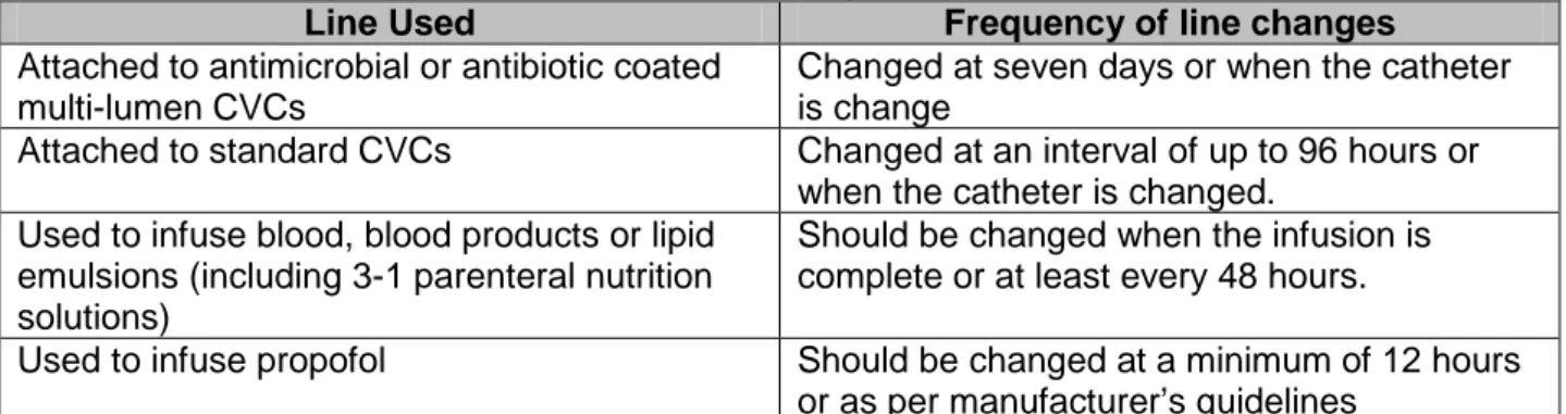
22.2 Administration set changes
IV administration sets include both the IV lines and any additional attachments such as bungs, three-way stopcocks, multi-flow adaptors and extension tubing that may be added.
The frequency for changing IV administration sets will be influenced by: • Type of central venous access device
• Type of infusion fluid • Patient characteristics.
For patients in intensive care, high dependency and general medical-surgical wards the frequency of change of administration sets is recommended in Table 1:
Table 1: Recommended Frequency of Line Change
Line Used Frequency of line changes
Attached to antimicrobial or antibiotic coated multi-lumen CVCs
Changed at seven days or when the catheter is change
Attached to standard CVCs Changed at an interval of up to 96 hours or
when the catheter is changed. Used to infuse blood, blood products or lipid
emulsions (including 3-1 parenteral nutrition solutions)
Should be changed when the infusion is complete or at least every 48 hours.
Used to infuse propofol Should be changed at a minimum of 12 hours
or as per manufacturer’s guidelines
22.3 Care of administration sets
Care of IV administration sets includes, but is not limited to:
1. Decontaminating all bungs, needleless injection sites and/or sampling ports with an alcohol/chlorhexidine swab before and after use to remove any organic substance or drug (particulate matter).
2. Clamping unused CVAD lumens and multi-flow adaptors to prevent air emboli and backflow of blood, protein or lipid solutions.
22.4 Intravenous fluid bag changes
1. Change crystalloid solutions (such as normal saline or 5% dextrose) without drug additives only when the administration set or the CVAD is changed OR the infusion is
PD2011_060 Issue date: September 2011 Page 12 of 24
complete.
2. All flasks or bottles of blood or blood products should be infused within four hours or as per infusion service guidelines except for Factor VIII or IX prepared for continuous infusion9
22.5 Antiseptic solutions and cleaning of the skin and CVAD
.Refer to Appendix 4 – Antiseptic solutions.
22.6 Dressing the CVAD and the insertion site
A sterile, transparent semi-permeable dressing should be used to protect the site from extrinsic contamination, allow continuous observation of the insertion site, and to stabilise and secure the catheter except in the following circumstances:
• where a patient is diaphoretic or has excessive bleeding or oozing from the site, or other contraindications: use sterile gauze secured with a sterile, transparent, semi-permeable dressing over it.
• when a patient has multiple catheters (note: except where the puncture sites are close together, each catheter must be dressed separately).
• in some instances when a commercially available chlorhexidine impregnated sponge dressing may be used.
Regardless of the dressing type it must:
• be positioned so that the CVAD insertion site is in the centre of the dressing • cover the CVAD from the insertion site to the hub
• create a complete seal from the CVAD hub through to the insertion site. Ongoing care of the dressing involves:
• Changing the transparent dressing every seven days (except in those paediatric patients in whom the risk of dislodging the catheter outweighs the benefit) or sooner if :
- The dressing is not intact, i.e., there is no longer a seal;
- There is evidence of inflammation and/or discharge from exit site, or
- There is excessive accumulation of blood or moisture.
• Sterile gauze and tape dressing should be changed daily, and whenever loose, soiled, or moist.
23 REMOVAL OF CVAD
A CVAD may only be removed by trained or supervised clinicians.
• Removal of a CVAD will be undertaken using an aseptic technique (refer to Section 8 Aseptic Technique
)
that will minimise the risk of infection.PD2011_060 Issue date: September 2011 Page 13 of 24
• The patient is to be positioned supine with head slightly down (if tolerated) during CVAD removal. This is to increase the pressure in the large veins to above that of atmospheric pressure, which reduces the risk of aspirating air into the venous circulation.
• Following CVAD removal, the site must be sealed with an airtight dressing which remains insitu for at least 24 hours to reduce the risk of late air embolism.
• The patient must remain in supine position (or Semi-Fowlers if supine not tolerated) for between 30 and 60 minutes following CVAD removal. At least one set of observations should be done during this period, as well as immediately prior to retrieving the patient to the upright position.
• The removal of a CVAD and the presence of the intact tip must be noted in the patient’s health record.
• If a blood stream infection is suspected as the result of a CVAD, blood cultures must be attended and the CVAD tip sent for culture following removal (note: tips should not be sent for culture routinely).
• Following removal, the CVAD site will need daily review and dressing until healed. • Routine observations are to be conducted after the removal of the CVAD.
24 REPORTING NON-INFECTIVE COMPLICATIONS
Non-infective complications must be reported in the Incident Information Management System (IIMS) under the incident type “clinical management” and classified according to current policy. Non-infective complications associated with the insertion of a CVAD include arterial puncture, bleeding, pneumothorax, haemopneumothorax, air embolus, thrombus, vascular foreign body, vascular damage and malposition.
Malposition leading to adverse events and requiring catheter removal must be reported in the IIMS under the incident type “clinical management”. Minor adjustments to reposition the catheter prior to use do not need to be reported.
PD2011_060 Issue date: September 2011 Page 14 of 24
APPENDIX 1 – CVAD INSERTION STEPS FOR BEST PRACTICE
Steps for CVAD insertion Relevant section in policy
1. Consider whether or not a CVAD is indicated and the most appropriate site
2 Requirements For The Insertion Of CVADs
5 Patient Factors 2. Choose the type, size, length and number of
lumens of the catheter and check that it is suitable for intended therapy and duration
6 Catheter Selection
3. Seek procedural support from an assistant and/or supervisor
2 Requirements For The Insertion Of CVADs
4. Move the patient to an area suitable for CVAD insertion
3 Environmental, monitoring and emergency
5. Perform hand hygiene before patient contact 7 Personal protective equipment
6. Clip hair at the insertion site, if required 8 Aseptic Technique
7. Perform hand hygiene again if a peripheral venous access device needs to be inserted
8 Aseptic Technique
8. Establish ECG, pulse oximetry, end-tidal carbon dioxide (ETCO2) and blood pressure (BP) monitoring (as appropriate) if this has not been done already
3 Environmental, monitoring and emergency
9. Consider the use of sedation in the conscious patient (making sure of compliance with sedation standards)
3 Environmental, monitoring and emergency
10. Give supplemental oxygen if sedation is used or there is a decreased level of consciousness
3 Environmental, monitoring and emergency
11. Position the patient head down to distend the veins (internal jugular/subclavian line insertion). The degree to which this can be done will depend on the patient’s condition (e.g. head injuries,
congestive cardiac failure) and whether or not they are conscious. The assistant may do this after skin antiseptic preparation, if appropriate. Use a venous tourniquet for PICCs.
5 Patient Factors
12. Put on surgical mask, hat (head and facial hair) and eye protection.
7 Personal protective equipment
PD2011_060 Issue date: September 2011 Page 15 of 24
Steps for CVAD insertion Relevant section in policy
antiseptic handwash
14. Put on sterile gown and sterile gloves 7 Personal protective equipment
15. Inspect, open and check the CVAD kit and ensure that all necessary sterile items are present
4 Necessary Equipment
16. Perform meticulous skin preparation with an antiseptic solution preferably alcoholic chlorhexidine (unless contraindicated e.g., flammability issues, children under 2 months of age, allergy) and allow it to dry before insertion. See Appendix 4 – Antiseptic solutions for a list of approved solutions3
8 Aseptic Technique
17. Fully cover the patient and their bed with sterile drape/s (unless this is impractical)
8 Aseptic Technique
18. Maintain asepsis throughout the procedure. Insertion must stop if asepsis is breached
8 Aseptic Technique
19. Always use local anaesthetic for the insertion site and adjacent subcutaneous tissues in a conscious patient
9 Local Anaesthetic
20. Ultrasound should always be used if available when inserting a CVAD
10 Ultrasound
21. Cannulate the vein with an introducing
cannula/needle. If a wire through needle technique is used, insert the wire and remove the needle, maintaining control of the guidewire at all times
11 Confirmation of Venous Access 13 Guide Wire
22. Confirm venous (not arterial) placement (e.g., manometry, ultrasound, transduce catheter, contrast injection).
11 Confirmation Of Venous Access
23. Maintain control of the guidewire at all times 13 Guide Wire
24. Insert dilator only to a depth sufficient to dilate the vein puncture site
12 Dilator Insertion
25. Remove the guidewire 13 Guide Wire
PD2011_060 Issue date: September 2011 Page 16 of 24
Steps for CVAD insertion Relevant section in policy
wire)
27. Remove stiffening wire if used (PICCs) 14 Stiffening Wire (PICCs) 28. After the guidewire/stiffening wire has been
removed, confirm that it is complete and the tip has not been damaged
13 Guide Wire
14 Stiffening Wire (PICCs)
29. Secure and dress the CVAD 19 Securing the CVAD
22.6 Dressing the CVAD and the insertion site
30. Confirm CVAD position before use 15 Confirmation of CVAD
Placement
31. Dispose of all sharps in sharps bin and perform hand hygiene
8 Aseptic Technique
32. Complete a CVAD Insertion Record 21 Documentation
33. Perform and check chest x-ray to confirm the tip is in the correct position (excluding femoral lines in adults)
PD2011_060 Issue date: September 2011 Page 17 of 24
APPENDIX 2 – SUGGESTED EQUIPMENT FOR CVAD INSERTION
• Necessary PPE including sterile gown and gloves• Selection of catheters
• Antiseptic handwash and running water
• Antiseptic solution for skin preparation (refer to Appendix 4 – Antiseptic solutions) • 0.9% sodium chloride solution
• Large trolley drapes • Sterile procedure trays • Gauze squares
• Sterile sheet/s to ensure adequate draping of the procedure site. • Local anaesthetic
• Needles and syringes • Suture equipment
• Small sterile container to isolate used sharps on the sterile field
• Swabable capless valves or similar for each lumen. Positive displacement swabable capless valve for when a lumen will be clamped.
• Heparin for “heparin locking” catheter lumen • Transducers, blood gas syringes
• Electrocardiograph (ECG) monitor • Blood pressure monitor
• Pulse oximeter (where necessary) • Capnography (where necessary) • Ultrasound machine
• Appropriate dressings • Sharps bin/trolley.
If the equipment needed to place a CVAD is not usually available at the location where the insertion will take place, a trolley that includes all necessary equipment for inserting or re-wiring a CVAD should be dedicated for this purpose. This may include a sterile procedure pack with necessary equipment included.
Ensure items have not expired.
If used, the trolley should be set up for the insertion in a manner consistent with operating theatre sterile technique. The trolley should be set up as close to the time of the CVAD placement as practical.
PD2011_060 Issue date: September 2011 Page 18 of 24
APPENDIX 3 – CVAD SITE SELECTION
Site
Selection CriteriaInfraclavicular Subclavian
• Suitable long-term site
• When considering infection risk only, the subclavian vein has lowest rates of infection compared with other CVAD sites
• Should be avoided for dialysis catheter insertion due to risk of subclavian vein stenosis
• If arterial puncture should occur, the site has the least ability to control bleeding
• Least suitable insertion site for patients with potentially severe lung pathology due to the risk of pneumothorax
• Least suitable insertion site for patients with uncorrected coagulopathy, as it is associated with the greatest risk of uncontrollable haemorrhage
• Right infraclavicular subclavian is more likely to result in satisfactory catheter location than the left
Internal Jugular • Convenient site for short-term CVAD access
• Preferred site for intraoperative access
• Route with the least acute complications (after PICC) especially if ultrasound guidance is used
• Preferred site for pulmonary artery catheter insertion
• Suitable site for dialysis catheter insertion
• More suitable for patients at special risk of pneumothorax or haemorrhage
• Right internal jugular approach is more likely to result in satisfactory catheter location
External Jugular • Insertion can be technically difficult due to the presence of valves, however if the guidewire can be passed then placement of the line is usually possible. It is not suitable for dialysis catheter insertion due to the catheter size and rigidity.
• May be a useful alternative particularly in coagulopathic patients
• May be a site for surgically inserted, tunnelled CVADs (e.g., Portacaths).
Femoral • Increased risk of infection at this site compared with other sites.
• Another useful site when internal jugular or subclavian sites are not appropriate or in coagulopathic patients
• Suitable site for short-term dialysis catheter insertion
• Arterial puncture, if it occurs is easily compressed
• Patient does not need to be tilted head down
PD2011_060 Issue date: September 2011 Page 19 of 24
Arm Veins (PICC)
• Low risk of serious complications
• Recommended for long-term, low flow rate access
• Not recommended when more than two lumens or high flow rate infusions required
• Not recommended for dialysis insertion
• Can be used for pulmonary artery catheter insertion if other sites not available Arm Veins Closed system, non-Seldinger peripheral CVADs
• Primarily used for perioperative pressure monitoring
• If the clinical need for a CVAD persists past this period, a CVAD should be inserted at another site.
PD2011_060 Issue date: September 2011 Page 20 of 24
APPENDIX 4 – ANTISEPTIC SOLUTIONS
Based on available evidence alcohol-containing antiseptics, particularly with chlorhexidine, are recommended for preparation of the for CVAD insertion. Their use is associated with a
significantly lower risk of intravenous line-associated (and surgical site) infections compared with the use of aqueous solutions.
The drying time of alcohol-based antiseptic increases with the increase in alcohol content as does the risk of fire in theatre. However, any risk of fire from alcoholic solutions is negligible if the solutions are used with care and as recommended (i.e. not in the presence of free oxygen, diathermy or laser instruments) and allowed to dry.
The risk of fire is minimized by following the FDA approved use of applicators: Do not use 26-ml applicator for head and neck surgery, do not use on an area smaller than 21.3cm × 21.3cm, use a smaller applicator instead. Do not drape or use ignition source until solution is completely dry (minimum of 3 minutes on hairless skin, up to 1 hour in hair). Do not allow solution to pool; remove wet materials from prep area10.
The following solutions are available:
1. Topical chlorhexidine 2% in 70-80% alcohol (lower concentrations of chlorhexidine (0.5-1%) may also be used (e.g. for infants).
2. Topical povidone Iodine 10% in 70% alcohol
Other antiseptic alternatives for use on keratinized skin are inferior to the above products but include:
3. Topical chlorhexidine 1-2% in water. 4. Topical povidone Iodine 10% in water Notes:
1. All solutions must be allowed area to dry before beginning CVAD insertion. Alcoholic solutions will dry more rapidly.
2. Alcoholic chlorhexidine solutions now contain colour to allow easier identification.
3. Sterile saline or water solutions alone are not acceptable antiseptic solutions and should only be used to clean the skin of gross contaminants prior to applying antiseptic solution.
PD2011_060 Issue date: September 2011 Page 21 of 24
APPENDIX 5 – COMPLICATIONS AND SUGGESTED MANAGEMENT
Complication Suggested Management
Vein entered but unable to pass guidewire
Remove needle and guidewire together to avoid mediastinal injury or shearing off the guidewire inside the patient.7
Arterial puncture with needle or transducing cannula (16 gauge or smaller)
Generally this can be managed by removal of the needle/cannula with local pressure until haemostasis is secured (at least 5 minutes). If a significant haematoma has occurred another site should be
considered. Ultrasound may assist further attempts at the same site. With subclavian arterial puncture, keep in mind the possibility of occult bleeding leading to delayed cardiovascular compromise. Haemothorax or mediastinal widening (aortic dissection) must be looked for in the post-procedure chest X-ray.
These patients should have hourly observations for at least four hours after the procedure. Immediate vascular consultation should be sought if there is any suspicion that either of these complications has
occurred.
With carotid puncture the patient should be observed for any neurological changes after pressure is applied.
If local pressure cannot establish haemostasis or a carotid arterial haematoma leads to the airway being compromised, then immediate vascular consultation should be sought.
Arterial puncture with large bore catheter or dilator
Complications are much more likely the bigger the catheter inserted into the artery. Unless you are able to repair the artery yourself do NOT remove the catheter or dilator. Consult the vascular surgical team for further management.
If the patient is going to be heparinised intraoperatively, consider leaving the catheter or dilator in until the heparin has been reversed. Acute airway obstruction may occur after removal of a dilator or large bore catheter from the carotid artery. If the line was placed prior to the induction of an anaesthetic, the airway should be secured before the dilator or catheter is removed.
Guidewire lost inside patient
Consult an interventional radiologist who can attempt removal.
Pneumothorax A surgical (ideally cardiothoracic) consultation must be sought.
Usually a small (10-20% pneumothorax) is managed conservatively with follow up serial chest x-rays. A larger pneumothorax may require a chest tube and underwater drain. If the patient is mechanically ventilated, consider inserting a drain regardless of the size of the
PD2011_060 Issue date: September 2011 Page 22 of 24
pneumothorax to prevent a tension pneumothorax occurring. This is particularly important if the surgical positioning would make inserting an urgent chest drain difficult (e.g., prone neurosurgical operations). If a drain is not inserted, this complication must be strongly considered if subsequent hypotension occurs.
PD2011_060 Issue date: September 2011 Page 23 of 24
APPENDIX 6 - IMPLEMENTATION PLANNER
Note: This action planner is NOT mandatory – it is a tool for public health organisations to use to monitor implementation of this
Policy.
ACTION PLANNER FOR:
Chief Executives, Health Service Executives, Managers, Directors of Clinical Governance
DATE:
No education and training of clinicians undertaking CVAD insertion.
Comments/Actions:
Clinicians do not follow any established guidelines for CVAD insertion.
Comments/Actions:
Clinicians do not record CVAD insertions. Comments/Actions:
IMPLEMENTATION STANDARD
Current compliance status () Actions Required Assigned to Target Completion Date N o t S ta rte d P ar ti al ly C o m p let ed C o m p let edAdequate stocks of “necessary equipment” , “skin preparation”, personal protective equipment and appropriate catheters available
CVAD Insertion Record used for every CVAD insertion
Clinicians, including LMOs (undertaking or supervising CVAD insertion) have completed a training program consistent with best practice for the insertion of CVADs.
Monitoring of CVAD associated bloodstream infections (CVADABSI)
COMPLIANCE STANDARD
Local monitoring of clinician CVAD insertionpractices.
Only trained/experienced clinicians undertake or supervise CVAD insertion
All reportable CVADABSI events are reviewed at the facility on a case by case basis to identify potential opportunity for clinical practice improvement
LEADERSHIP STANDARD
Facility wide monitoring of clinician CVADinsertion practices as per NSW Department of Health compliance program
Diagnosis and treatment of CVADABSI is contemporaneous and multidisciplinary
100% compliance with this Policy Statement and Policy Standard achieved facility-wide
PD2011_060 Issue date: September 2011 Page 24 of 24
REFERENCES
1 The Free Dictionary
2 Centers for Disease Control and Prevention. Guideline for Hand Hygiene in Health-Care Settings: Recommendations of the Healthcare Infection Control Practices Advisory Committee and the HICPAC/SHEA/APIC/IDSA Hand Hygiene Task Force. MMWR
2002;51(No. RR-16):[inclusive page numbers]
(accessed 11 April 2011)
3 Centers for Disease Control and Prevention. Guidelines for the Prevention of Intravascular
Catheter-Related Infections. 2011
4 NSW Health Hand Hygiene Policy
5 National Institute for Clinical Excellence Technology Appraisal No. 49. Guidance on the use of ultrasound locating devices for placing central venous catheters, September 2002.
2011)
6 Therapeutic Goods Administration (TGA). Peripherally inserted central catheters (PICC lines)
- safety alerts and advisori
27 July 2011)
7 Bouza, E, Munoz, P, Lopez-Rodriguez, J et al. A needless closed system device (CLAVE) protects from intravascular catheter tip and hub colonization: a prospective randomized study. Journal of Hospital Infection 2003; 54(4):279-287.
8 Intensive Care Coordination and Monitoring Unit (ICCMU) (2007). Nursing Care of Central
Venous Catheters in Adults Intensive Care: NSW Health Statewide Guidelines for Intensive Care.
9
NSW Health Blood – Fresh Components – Management
10
Darouiche RO, Mosier MC. Chlorhexidine-Alcohol versus Povidone – Iodine for Surgical Site Antisepsis NEJM 2010; 362 (1): 18-26.