Eastern Kentucky University
Encompass
Online Theses and Dissertations
Student Scholarship
2013
Relationship Between a Proxy of Prenatal
Testosterone (2D:4D) and Determinants of
Endurance Running Performance
Simon D. Holzapfel
Eastern Kentucky University
Follow this and additional works at:
http://encompass.eku.edu/etd
This Open Access Thesis is brought to you for free and open access by the Student Scholarship at Encompass. It has been accepted for inclusion in Online Theses and Dissertations by an authorized administrator of Encompass. For more information, please [email protected].
Recommended Citation
Holzapfel, Simon D., "Relationship Between a Proxy of Prenatal Testosterone (2D:4D) and Determinants of Endurance Running Performance" (2013).Online Theses and Dissertations.Paper 110.
RELATIONSHIP BETWEEN A PROXY OF PRENATAL TESTOSTERONE (2D:4D) AND DETERMINANTS OF ENDURANCE RUNNING PERFORMANCE
By
SIMON D. HOLZAPFEL Bachelor of Arts Eastern Kentucky University
Richmond, Kentucky 2011
Submitted to the Faculty of the Graduate School of Eastern Kentucky University
in partial fulfillment of the requirements for the degree of
MASTER OF SCIENCE May, 2013
ii
Copyright © Simon D. Holzapfel, 2013
All rights reserved
iii
ACKNOWLEDGMENTS
I would like to thank my adviser and Chair of my thesis committee, Dr. Matthew J. Sabin, for his guidance and patience. I would also like to thank the other committee members, Dr. Peter J. Chomentowski for his comments and investment of time and energy in the data collection process and Dr. Louisa A. M. Summers for her comments and assistance in the recruitment of participants. I would like to express my thanks to Christopher Perry, Matthew Creech, Kaitlyn Krizman, and Ariana Mason for their help in the data collection process. My appreciation goes to the track & field and cross country teams of Eastern Kentucky University and Berea College for their cooperation and participation in this project. Lastly, I would like to thank Rob Sica, who was instrumental in the identification of the research topic for my thesis.
iv
ABSTRACT
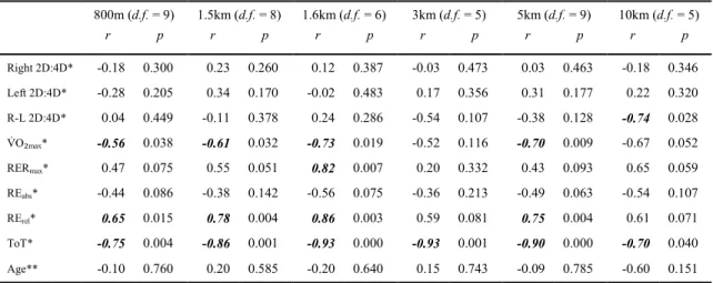
Low ratios of the length of the second finger to the length of the fourth finger in the right (right 2D:4D) and the left hand (left 2D:4D) have been linked to high prenatal testosterone concentrations. Low R-L 2D:4D (subtracting left 2D:4D from right 2D:4D) has been associated with high androgen sensitivity as indicated by low numbers of cytosine-adenine-guanine (CAG) triplet repeats on exon-1 of the androgen receptor gene. Endurance training has led to higher increases in maximal oxygen uptake capacity V O₂max) in men with relatively low numbers of CAG triplet repeats, suggesting a relationship between R-L 2D:4D and V O₂max. Moreover, Low right and left 2D:4D are associated with superior performances in sports such as fencing, rugby, soccer, basketball, and sumo wrestling. The strongest associations, however, have been found between right 2D:4D and endurance running performance (r² = 0.25). An inverse relationship between R-L 2D:4D and V O₂max, running velocity at V O₂max, and peak lactate concentration in pubertal boys has been reported.
The purpose of the present investigation was to examine the relationships between measures of digit ratios and performance variables (V O₂max, maximal respiratory exchange ratio (RERmax), absolute
running economy (REabs), relative running economy (RErel), and total time on the treadmill (ToT)) in
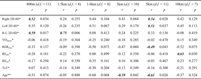
post-pubertal sedentary populations and in trained endurance runners. The relationship between digit ratios and endurance running performance in the form of personal records (PRs) over different race distances in the trained runners was also examined in order to explore which performance variable moderates the relationship between prenatal testosterone and/or testosterone sensitivity and endurance running
performance. A significant negative relationship between left 2D:4D and V O₂max (r = 0.49) was found only in the female sedentary group after removing the effects of weight using first order partial correlation analyses. In both the male and female trained runners, measures of digit ratio and V O₂max showed a trend to be positively related. REabs related negatively to digit ratios in the male and female sedentary groups, while
RErel was negatively related to digit ratios in only the male sedentary group. All other relationships between
digit ratios and performance variables were highly inconsistent across groups and often within groups. We found fairly consistent and moderately strong positive relationships between digit ratios and PRs which do not seem to be moderated by V O₂max. However, The associations between 2D:4D and endurance running performance seemed be mediated by RERmax in male endurance runners, indicating that the capacity to
buffer and/or clear lactic acid moderates the relationship between prenatal testosterone stimulation and endurance running capabilities. We recommend the investigation of the relationships among lactate threshold, endurance running performance and 2D:4D.
v
TABLE OF CONTENTS
CHAPTER
PAGE
I.
INTRODUCTION ...1
Prenatal Testosterone and Sexual Dimorphism ...1
2D:4D and Sex Steroid Stimulation ...1
Athletic Performance and 2D:4D ...2
Adult Testosterone Levels and Cardiovascular Health
Components ...3
Right and Left 2D:4D and Maximal Oxygen Uptake ...4
R–L 2D:4D and Maximal Oxygen Uptake ...5
Purpose of this Investigation ...6
Population ...7
Hypotheses ...7
Limitations ...8
Delimitations ...9
II.
LITERATURE REVIEW ...10
2D:4D – A Sexually Dimorphic Trait ...10
Links between Prenatal Sex Steroid Concentrations and
Digit Ratios ...12
Association of Digit Ratios with Postnatal Testosterone
Concentrations ...17
Androgen Sensitivity and Digit Ratios ...17
Athletic Performance and 2D:4D ...20
2D:4D as a Biomarker of Cardiovascular Fitness...24
Influence of Exercise on Serum Testosterone Levels ...28
Postnatal Testosterone‟s Influence on Components of
Cardiovascular Fitness ...32
Testosterone Sensitivity and Cardiovascular Fitness ...35
Conclusion ...37
III.
METHODS ...39
Introduction ...39
Participants ...39
Instruments and Apparatuses ...40
vi
Procedures ...42
Statistical Analyses ...43
Pilot Study ...43
IV.
MANUSCRIPT ...45
Introduction ...45
Methods...48
Participants
...48
Finger Length Measurements
...48
Measurement of Performance Variables and PRs
...48
Statistical Analyses
...49
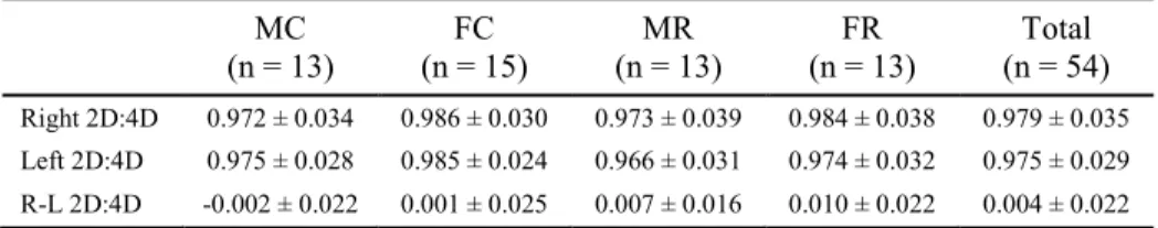
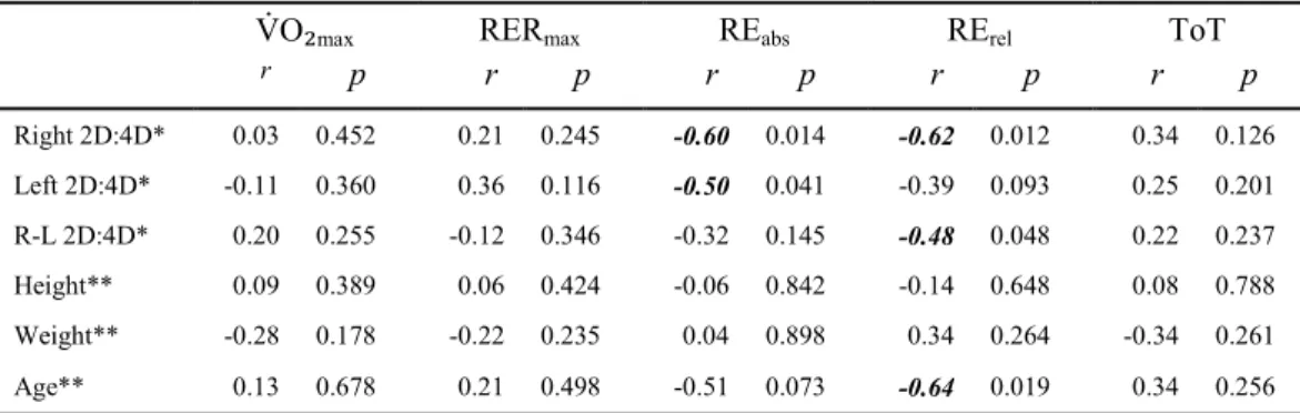
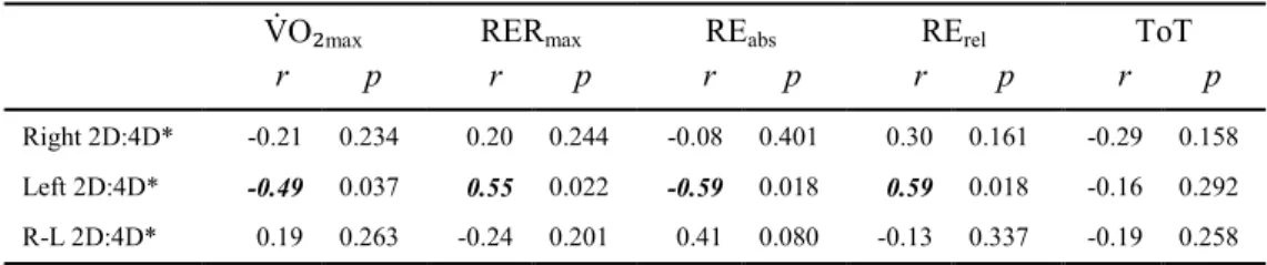
Results ...49
Intraobserver Reliability of Finger Length
Measurements and 2D:4D Descriptive Statistics
...49
Other Descriptive Statistics and Group Differences
...50
Correlations for MC
...51
Correlations for FC
...51
Correlations for MR
...52
Correlations for FR
...53
Discussion ...54
Conclusion ...58
LIST OF REFERENCES ...60
APPENDICES ...71
A. Tables ...71
B. Figures...77
C. Pre-Test Instructions ...80
D. Informed Consent Form ...84
E. PAR-Q & YOU ...94
F. Pre-Test Questionnaires ...96
G. Demographics Questionnaires ...101
H. Treadmill Test Instructions ...104
I. OMNI RPE Scale ...106
J. Bruce Protocol for the Treadmill ...108
vii
LIST OF TABLES
TABLE
PAGE
A-1.
2D:4D in Individuals With CAIS and Controls (Mean ± SD) ...72
A-2.
Descriptive Statistics of Digit Ratios by Group and Total
Sample (Mean ± SD) ...72
A-3.
Descriptive Statistics of Age, Height, Weight, Body Mass
Index (BMI), Percentage of Body Fat (%BF), and
Performance Variables by Group and Total Sample
(Mean ± SD) ...72
A-4.
Descriptive Statistics of PRs by Race Distance and Group
(Mean [min:sec] ± SD [sec]) ...73
A-5.
Relationships (
r, d.f.
= 11) Between Right 2D:4D, Left 2D:4D,
R-L 2D:4D, Height, Weight, and Age, and Performance
Variables in Male Sedentary College Students (MC) ...73
A-6.
Relationships (
r, d.f.
= 13) Between Right 2D:4D, Left 2D:4D,
R-L 2D:4D, Height, Weight, and Age, and Performance
Variables in Female Sedentary College Students (FC)...73
A-7. First Order Partial Correlations Between Right 2D:4D,
Left 2D:4D, and R-L 2D:4D, and Performance Variables
Controlled for Weight in Female Sedentary College
Students (FC) ...74
A-8.
Relationships (
r, d.f.
= 11) Between Right 2D:4D, Left 2D:4D,
R-L 2D:4D, Height, Weight, and Age, and Performance
Variables in Male Intercollegiate Long Distance
Runners (MR) ...74
A-9.
Relationships Between Right 2D:4D, Left 2D:4D, R-L 2D:4D,
Performance Variables, and Age, and PRs in Male
viii
A-10. Relationships (
r, d.f.
= 11) Between Right 2D:4D, Left 2D:4D,
R-L 2D:4D, Height, Weight, and Age, and Performance
Variables in Female Intercollegiate Long Distance
Runners (FR) ...75
A-11. Relationships Between Right 2D:4D, Left 2D:4D, R-L 2D:4D,
Performance Variables, and Age, and PRs in Female
Intercollegiate Long Distance Runners (FR) ...76
A-12. First Order Partial Correlations Between Right 2D:4D,
Left 2D:4D, and R-L 2D:4D, and PRs Controlled for
₂
maxix
LIST OF FIGURES
FIGURE
PAGE
B-1.
Sex steroid receptors as link between 2D:4D and postnatal traits ...78
B-2.
Standardized correlations of right-hand (A) and left-hand (B)
2D:4D with running performance by distance ...78
B-3.
A schematic representation of the determination of digit ratios ...79
1
CHAPTER 1
INTRODUCTION
Prenatal Testosterone and Sexual Dimorphism
A mounting body of evidence suggests that prenatal testosterone exerts a permanent
organizational effect on sexually dimorphic traits and characteristics. It determines to what degree a person possesses male or female characteristics. Pre-natal testosterone has organizational effects on a person‟s anatomy and physiology, masculinizing behavior, appearance, and physical ability (Cohen-Bendahan, Buitelaar, Van Goozen, Orlebeke, & Cohen-Kettenis, 2005; Hönekopp, Manning, & Müller, 2006). Sex differences upon birth seem to be caused by differences in testosterone concentrations and not estrogen concentrations as estrogen levels between male and female fetuses were found to be equal (McIntyre, 2006). Evidence exists that the degree of masculinization is a correlate of prenatal androgen levels as the absence of fetal testosterone leads to female phenotypical development (Zitzmann & Nieschlag, 2003). Research conducted with non-human animals has shown that the artificial elevation of prenatal testosterone levels leads to the masculinization of a variety of characteristics. Ethical concerns prohibit such
experimentation with human fetuses; therefore investigators are limited to correlational studies to explore the relationships between early androgen stimulation and proxies thereof and adult characteristics in humans (Breedlove, 2010).
2D:4D and Sex Steroid Stimulation
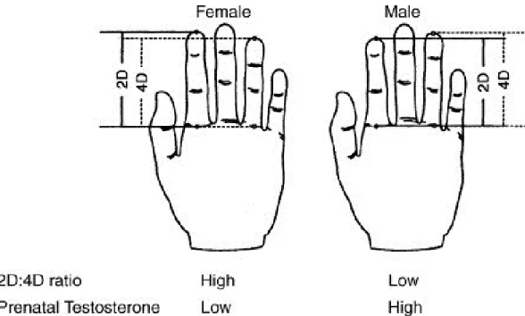
The ratio of the length of the second digit to the length of the fourth digit (2D:4D) on the human hand is a sexually dimorphic trait, whereas men have lower 2D:4D than women (Hönekopp & Watson, 2010). The amount of testosterone the fetus produces between the 12th and 24th week, as measured by analysis of amniotic fluid (amniocentesis), negatively correlates with 2D:4D (Lutchmaya, Baron-Cohen, Raggatt, Knickmeyer, & Manning, 2004). This means that greater testosterone production by the gonads of the fetus leads to a shorter index finger relative to the ring finger of the same hand (Lutchmaya et al., 2004). Besides amniocentesis other methods of establishing the link between in utero testosterone and digit ratios have been employed. These include the comparison of 2D:4D between females with congenital adrenal hyperplasia (CAH) and healthy controls (e.g. Buck, Williams, Hughes, & Acerini, 2003), the comparison of 2D:4D in dizygotic twins of the same and opposite sex (e.g. Anders, Vernon, & Wilbur, 2005), the comparison of 2D:4D between women with polycystic ovary syndrome and healthy controls
2
(e.g. Cattrall, Vollenhoven, & Weston, 2005), and the comparison of 2D:4D between men with Klinefelter‟s syndrome KS) and healthy controls (Manning, Kilduff, & Trivers, 2013).
Typically, the index finger is slightly shorter than the ring finger, which causes the value of 2D:4D to be below 1.0 in most individuals. The sex differences in 2D:4D are more pronounced in the right hand (Hönekopp & Watson, 2010) and it seems that the fourth or ring finger is equipped with more androgen and estrogen receptors than the second or index finger especially in the right hand (Zheng & Cohn, 2011). Estrogen stimulates metaphyseal tissue to calcify while testosterone promotes bone growth (Weise et al., 2001). Evidence also suggests that testosterone sensitivity governed by the androgen receptor gene influences the development of sexually dimorphic traits (Breedlove, 2010). The ability of androgen receptor genes to transcribe the testosterone stimulus depends on their number of cytosine-adenine-guanine (CAG) triplet repeats (Chamberlain, Driver, & Miesfeld, 1994; Kazemi-Esfarjani, Trifiro, & Pinski, 1995). The higher the number of CAG triplet repeats the more inhibited the transcription process is (La Spada, Wilson, Lubahn, Harding, & Fishbeck, 1991). CAG triplet repeat length has been shown to positively correlate with 2D:4D and with difference between right and left 2D:4D (R-L 2D:4D) (Manning, Bundred, Newton, & Flanagan, 2003). Breedlove (2010) thus concludes that 2D:4D reflects total androgen
stimulation which is proportionate to the concentration of and sensitivity to androgens. 2D:4D has been commonly used as a putative measure of in utero testosterone concentration and as an indicator of testosterone sensitivity.
Athletic Performance and 2D:4D
It has been shown that athletic performances, which are influenced by the sexually dimorphic traits such as muscular strength, muscular endurance, speed, and cardiovascular endurance, have a negative correlation with 2D:4D and R-L 2D:4D and thus a positive correlation with prenatal testosterone levels (Bennett, Manning, Cook, & Kilduff, 2010; Manning, 2002a; Manning, Morris, & Caswell, 2007; Tamiya, Lee, & Ohtake, 2011). Researchers found that a longer ring finger relative to the index finger in either hand predicts better performances in fencing (Voracek, Reimer, Ertl, & Dressler, 2006), skiing (Manning, 2002b), soccer (Manning & Taylor, 2001), field-based fitness tests (Hönekopp et al., 2006), sumo wrestling (Tamiya, Lee, & Ohtake, 2011), basketball (Tester & Campell, 2007), 50m dash (Manning & Hill, 2009), a hand-grip strength test (Fink, Thanzami, Seydel, & Manning, 2006), and 2,000m ergometer rowing (Longman, Stock, & Wells, 2011). The effect size of the relationship between 2D:4D of the right hand and athletic prowess (r = -0.26) is highly significant (Hönekopp & Schuster, 2010). The relationship between left 2D:4D and athletic prowess shows a similar and also significant effect size (r = -0.24). It seems that neither hand is a better predictor of athletic performance. The correlation between 2D:4D and endurance running performance measured in finishing position or time ranged from r = 0.30 to r = 0.51. The average
3
variance in athletic performance accounted for by 2D:4D ranges from 1% to 16% (Hönekopp & Schuster, 2010). The variance in endurance running performance explained by 2D:4D is 25% (Manning et al., 2007). Therefore, it seems that prenatal testosterone exerts greater effects on the function of the cardiovascular system than on other physiological variables, as endurance running is an activity that requires greater aerobic efficiency than many of the aforementioned activities (Manning et al., 2007).
Adult Testosterone Levels and Cardiovascular Health Components
While prenatal testosterone exposure seems to have beneficial effects on cardiovascular fitness, i.e. it makes one a better endurance runner, the effects of postnatal testosterone stimulation on
cardiovascular health should also be discussed as digit ratio relates to adult testosterone sensitivity
(Manning et al., 2003). Therefore, digit ratio may reflect not only prenatal testosterone stimulation but also, to some degree, adult testosterone stimulation. The effects of exercise on serum testosterone concentrations and the influence of serum testosterone levels on physiological variables in adults have been studied. Strength training and endurance training led to significant increases in serum testosterone, peaking approximately 20 minutes after commencement of training (Jensen et al., 1991; Vogel, Books, Ketchum, Zauner, & Murray, 1985). Elevations in serum testosterone levels have been shown to stimulate
erythropoiesis, elevations in serum hemoglobin concentrations, Type I muscle fiber growth, growth of the myocardium, reductions in serum low-density lipoprotein levels, and elevations in serum high-density lipoprotein levels (Hartgens & Kuipers, 2004; Rebuffe-Scrive et al., 1991; Vermeulen et al., 1999; Zitzmann & Nieschlag, 2007). Testosterone supplementation has also caused decreases in diastolic and systolic blood pressure as well as resting heart rates (Zitzmann & Nieschlag, 2007). However, it has been shown that very high endurance training volumes led to chronic depressions in serum testosterone levels (Häkkinen, Pakarinen, Alén, Kauhanen, & Komi, 1988; MacConnie et al., 1986). The physiology of endurance athletes is characterized by most if not all of the benefits provided by elevated serum
testosterone levels. It seems counterintuitive that endurance athletes would have depressed resting serum testosterone levels. Endurance athletes may thus be a very useful population when studying the effects of prenatal testosterone levels on human physiology as their depressed resting serum testosterone levels should cause less of the inter-individual variation in physiological parameters and thus making variation due to prenatal testosterone stimulation more easily detectable. Moreover, the elevation in serum testosterone associated with the repeated testosterone stimulus provided by endurance training could be reflected in 2D:4D as Kilduff, Cook, Bennett, Crewther, Bracken, and Manning (2012) found a significant negative correlation between R-L 2D:4D and free salivary testosterone in rugby players following a physical challenge. Therefore, testosterone sensitivity, with R-L 2D:4D as a postnatal proxy, and elevations
4
in serum testosterone associated with exercise could be the mediating variable between testosterone stimuli and physiological performance variables.
Evidence also suggests that cardiovascular risk factors and certain diseases are less prevalent in individuals with low 2D:4D (Abbott, Dumesic, & Franks, 2002; Fink, Manning, & Neave, 2006; Manning & Bundred, 2001; Manning, Taylor, & Bundred, 2003; Rebuffe-Scrive, Marin, & Bjorntrop, 1991; Singh, 1994; Vermeulen, Goemaere, & Kaufman, 1999). These findings seem to point towards beneficial effects of prenatal testosterone on cardiovascular health and fitness components (English, Mandour, Steeds, Diver, Jones, & Channer, 2000). The validation of 2D:4D as a biomarker of in utero androgen concentrations as well as sensitivity is important to provide for opportunities of quick and inexpensive studies and
examinations of the genetic predisposition for health risk factors because direct measures of in utero
androgen concentrations are not practical when studying their relationship to cardiovascular risk factors in adulthood
.
Right and Left 2D:4D and Maximal Oxygen Uptake
The physiological measure that is widely accepted as the best measure of cardiovascular fitness is the maximal oxygen uptake or V O₂max (ACSM, 2010. Bassett & Howley, 1999. Mitchel & Blomqvist, 1971). V O₂max is the maximum amount of oxygen an individual can consume for energy production during vigorous exercise (Mitchel & Blomqvist, 1971). V O₂max can reflect poor cardiovascular fitness and inactivity, established as primary risk factors for cardiovascular heart disease (Blair & Kohl, 1989). Hill, Simpson, Manning, and Kilduff (2012) explored the relationship between digit ratios and V O₂max, running velocity at V O₂max (v- V O₂max), and peak lactate concentration (LAmax) in young athletic teenage boys (age:
13.9 ± 1.3 years) during an incremental treadmill test. All of these variables have been shown to be sexually dimorphic, whereas men display consistently higher values than women (Bouchard et al., 1998; Daniels & Daniels, 1991; Esbjörnsson-Liljedahl, Sundberg, Norman, & Jansson, 1999). Therefore, a significant negative relationship between 2D:4D and these variables was expected. However, Hill and colleagues (2012) did not find a significant relationship between right or left hand 2D:4D and V O₂max, v- V O₂max, or LAmax. These findings are somewhat surprising as Manning and colleagues (2007) found that
right and left hand 2D:4D correlated significantly with endurance running performance. As lower right and left hand 2D:4D is associated with higher prenatal testosterone concentrations, these findings do not support strong favorable organizational effects conducive to cardiovascular health and fitness of prenatal testosterone on the human physiology. The lack of a significant correlation between 2D:4D and V O₂max in Hill‟s and colleagues 2012) study could be explained by the inclusion of a variety of sports, including soccer, squash, table tennis, and athletics (track and field), which the participants played. These sports require different amounts of running and movement which elicit varying acute cardiovascular responses
5
and therefore varying chronic adaptations of the cardiovascular and neuromuscular systems. For example, table tennis might not elicit heart rates as high as other athletic activities (e.g. track & field, soccer, etc.) and it certainly does not involve a comparable amount of locomotion in the sagittal plane (running). Therefore, table tennis players with low 2D:4D might have a low V O₂max compared to track & field runners with greater 2D:4D. The pre-pubertal and pubertal age of Hill‟s at al. 2012) sample presents further possible confounders because of the heterogeneity in age at the onset of puberty, the continuous elevation of testosterone levels, and the acceleration of growth and maturation during puberty (Mantzoros, Flier, & Rogol, 1997).
Attempts of synthesizing Manning‟s et al. 2007) and Hill‟s et al. 2012) reports to explain the influence of in-utero testosterone concentrations on determinants of endurance running performance bears only limited value because of significant methodological differences between those two studies. The participants in the study of Hill and colleagues (2012) were Middle-Eastern boys with a mean age of 13.9 ± 1.3 years who played a variety of sports and whose digit length was measured from photocopies of their hands. Manning and colleagues‟ 2007) sample consisted of trained Caucasian male and female distance runners with mean ages ranging from 24.04 ± 8.82 to 33.58 ± 9.25 years whose digit lengths were measured directly with steel vernier calipers. Further research is needed controlling for age to explore a possible influence of fetal testosterone stimulation on V O₂max related training effects.
R–L 2D:4D and Maximal Oxygen Uptake
Interestingly, in the same study by Hill and colleagues (2012), significant negative correlations were found between R-L 2D:4D and V O₂max (b = -0.33), v- V O₂max (b = -0.47), and LAmax (b = -0.50). As
mentioned, low R-L 2D:4D, more strongly than right or left 2D:4D, has been associated with low numbers of cytosine-adenine-guanine triplet repeats, which in turn are associated with high testosterone sensitivity (Manning et al., 2007). Men with relatively few cytosine-adenine-guanine triplet repeats in exon 1 of the androgen receptor gene have reacted with larger increases in V O₂max than those with relatively large numbers of cytosine-adenine-guanine triplet repeats in response to 30-day hypoxic training (Wang et al., 2010). An increase in hematocrit has also been reported in males with relatively few cytosine-adenine-guanine triplet repeats in response to an elevation in testosterone levels caused by exercise or the administration of testosterone (Wang et al., 2010; Zitzmann & Nieschlag, 2007). These findings suggest that low R-L 2D:4D indicates a heightened sensitivity to elevations in postnatal testosterone levels which causes favorable changes in the human physiology training effects). Hill‟s et al. 2012) findings could be explained by this sensitivity theory, despite the variety of sports which the boys played. High sensitivity to testosterone and pubertal elevations in serum testosterone might outweigh the effects of different types of training. For instance, a table tennis player with high testosterone sensitivity might have a higher V O₂max
6
than a soccer player with low testosterone sensitivity. It is also possible that the boys with low -L 2 :4 gravitated towards the sports that elicit large changes in and require high values of V O₂max for success. However, a similar study examining the influence of the relationship between 2 :4 and V O₂max on the relationship between 2 :4 and endurance running while controlling for training effects and fluctuations in serum testosterone levels is warranted. The gender differences in the relationship of digit ratios to V O₂max
are also unknown.
Purpose of this Investigation
In summary, a commonly used putative measure of prenatal testosterone stimulation (2D:4D) did explain 25% of the variance in endurance running performances in trained runners (Manning et al., 2007). However, right or left 2D:4D did not explain differences in V O₂max whereas -L 2 :4 did Hill et al., 2012). The samples in previous studies were diverse, the evidence about the organizational effects of prenatal testosterone concentrations on the human physiology, in particular V O₂max, are inconclusive, and the effects of long-term high volumes of endurance training on the relationship of maximal oxygen uptake to digit ratio are largely unknown. Therefore, the purpose of this investigation was to examine the influence of in-utero testosterone stimulation via 2D:4D on V O₂max and the effects of long-term endurance running training on this relationship by controlling for age and training. Due to the association of right and left 2D:4D with endurance running performance but the lack of association of right and left 2D:4D with V O₂max, we also included an analysis of the relationship of 2 :4 with running economy ). is the volume of oxygen V O₂) consumed at a certain constant running speed. For instance, runner one consuming 30 ml*kg-1*min-1 of O₂ is more economical in absolute terms (RE
abs) than runner two
consuming 34 ml*kg-1*min-1 of O₂ if both runners are running at the same speed and incline on two
identical treadmills aniels aniels, 1992). However, if runner one has a V O₂max of 40 ml*kg-1*min-1 and runner two has a V O₂max of 68 ml*kg-1*min-1, it means that runner two displays better relative RE
(RErel) running at 50 of his her V O₂max than runner one running at 75 of his her V O₂max (Daniels &
Daniels, 1992). RE has been demonstrated to be a reliable predictor of endurance running performance among trained runners and it is also a sexually dimorphic trait, whereas men generally exhibit better RErel
than women (Daniels & Daniels, 1992). Moreover, RE is dependent on a number of sexually dimorphic traits including body composition, height, weight, leg mass, and flexibility (Pate, Macera, Bailey, Bartoli, & Powell, 1992; Saunders, Pyne, Telford, & Hawley, 2004). This makes RE a potential correlate of digit ratio. Maximal respiratory exchange ratio (RERmax) will also be included as a dependent variable because
of the association of -L 2 :4 and maximal lactate concentrations in Hill‟s et al. 2012) investigation. While digit ratio is the independent variable, V O₂max, RERmax, REabs, RErel, total time on the treadmill
7
quasi-experimental (or categorical) variable of endurance running training. It is assumed based on past research that right and left 2D:4D reflect variations in prenatal testosterone levels while R-L 2D:4D is a stronger biomarker of testosterone sensitivity. Hence, we assumed that a negative correlation between measures of 2D:4D and V O₂max is moderated by prenatal testosterone and testosterone sensitivity. The investigation of the relationships between measures of 2 :4 , V O₂max, RE, RERmax, and running
performance within the same population allowed for a more direct comparison between the association of 2 :4 to V O₂max, RE, and RERmax and the association of 2 :4 to endurance running performance. or
this purpose, partial correlation analysis was used to explore which performance variable V O₂max, REabs,
RErel, or RERmax) moderates the relationship between 2D:4D and endurance running performance.
Population
For this purpose, two different college aged post-pubertal populations were recruited: Highly endurance trained and sedentary. The highly trained population consisted of 26 (13 female) individuals presently involved in competitive collegiate athletics (cross country and track & field) while the sedentary population (n = 28; 15 female), serving as the control group in terms of long term endurance training, was recruited from the general student population. Both groups included males and females. All participants were between 18 and 25 years of age to control for pubertal changes in testosterone levels as testosterone levels tend to level off after age 14 (Crabbe, Christiansen, Rødbro, & Transbøl, 1979).
Hypotheses
We expected to find a negative correlation between right, left, and -L 2 :4 and V O₂max in all samples. requent training stimuli may cause changes in V O₂max independent of training induced testosterone or prenatal testosterone stimulation due to other exercise induced hormonal changes and enzymatic activities. However, there should be a detectable negative relationship between 2 :4 and V O₂max in the highly trained runners as the physiological effects of serum testosterone spikes seem to be moderated by testosterone sensitivity (Wang et al., 2010) which is reflected in 2D:4D. Variations in V O₂max
in the sedentary population, however, should be largely attributable to genetic variation such as prenatal testosterone exposure and testosterone sensitivity. In accordance with the previous report of a significant correlation between 2D:4D and endurance running performance (Manning et al., 2007), we expected to find a positive relationship of similar strength between measures of digit ratio and personal records in terms of time per race distance (PRs). In comparison, the relationship between V O₂max and 2D:4D was expected to be weaker due to other determinants of endurance running performance such as the lactate threshold, RE, as well as neuromuscular and biomechanical factors which could also be affected by prenatal testosterone
8
and/or testosterone sensitivity assett Howley, 2000; Kyr l inen, elli, Komi, 2001; Nummela et al., 2006). ue to these variables and because Hill et al. 2012) found a stronger association between 2 :4 and peak running velocity than between 2 :4 and V O₂max, we expected to find a stronger relationship between 2 :4 and ToT compared to 2 :4 and V O₂max or . In accordance with Hill‟s et al. 2012) report of a negative relationship between R-L 2D:4D and LAmax, we also expected digit ratios to correlate
negatively to RERmax. At equal running velocities, men generally exhibit greater oxygen consumption per
kilogram of body weight V O₂) than women (REabs). Thus, we anticipated negative correlations between
measures of 2D:4D and REabs and positive correlations between measures of 2D:4D and RErel, as RErel and
V O₂max are inversely related. In summary, we expected negative correlations between measures of digit ratio right 2 :4 , left 2 :4 , -L 2 :4 ) and V O₂max, REabs, RERmax, and ToT, and positive correlations
between digit ratios, RErel, and PRs.
Limitations
The intercollegiate runners have been subjected to the selective process of recruitment which is likely to eliminate those with poor genetic predispositions. This might cause the polymorphism of 2D:4D in the runners to be relatively small. However, the 2D:4D polymorphism in the sedentary population could be comparably small as well, as they might have poor genetic predispositions in common which might be a reason for their relative inactivity. This potential lack of relative variation may make it hard to detect associations between digit ratios and dependent variables. Another limitation is that highly trained endurance runners tend to have fairly homogenous V O₂max values (Daniels & Daniels, 1992). This decreased diversity in V O₂max values makes it less likely to detect differences and to correlate them with potentially small differences in 2D:4D. The sampling of female collegiate distance runners from one mid-sized southeastern university and one small southeastern college presents a limitation as the training stimuli between the runners from these two institutions are not equal. The male runners were all recruited from one mid-sized southeastern university. Furthermore, the years of exposure to college level training was diverse among the runners as their status varied from first year college students to fourth year college students. This difference in training status could cause difference in V O₂max which would not be explainable by 2D:4D. Moreover, we will not measure peak lactate concentrations or any other lactate variable. In depth analyses of the relationship between 2D:4D and blood lactate concentrations during and after exercise are warranted as Hill et al. (2012) found the strongest correlation to be between R-L 2D:4D and LAmax.
9
Delimitations
Delimitations in this study include homogenous groups of participants in regard to age (18-25 years), the limitation of training modalities to running, and the limitation of runners to intercollegiate runners. This allows age and training status to be fairly controlled because age and years of training have been demonstrated to influence V O₂max vans, avy, Stevenson, Seals, 1995; ranch, Madsen, jurhuus, Pedersen, 1998; Jones, 1998; Ogawa et al., 1992). espite the large differences in cardiovascular fitness between groups, the V O₂max of all participants will be measured with the ruce treadmill protocol to allow comparison of V O₂max, REmax, REabs, RErel, and ToT between groups. Digit
length will be measured directly with a digital offset steel vernier caliper to reduce the distortion of soft tissue in the fingers which is often associated with measures obtained from photocopies of the ventral surface of the hand due to the pressure of the hand exerted against the glass of the photocopy machine. In order to control possible extraneous variables in the form of biochemical substances and physical fatigue, participants will be asked not to change their training routines, not to exercise the day before or the day of testing, not take any new medications or discontinue the use of routine medication unless the doctor told them to do so, and not to consume any alcohol, nicotine, or caffeine during the 24 hours prior to testing. Those who use performance enhancing drugs will also be excluded.
10
CHAPTER 2
LITERATURE REVIEW
2D:4D – A Sexually Dimorphic Trait
Dr. John T. Manning is known as the pioneer of research employing digit ratios as a putative measure of prenatal sex steriods. The likelihood of a predictive relationship between digit ratio and prenatal sex steroids seemed promising due to the chronological overlap in digit and urino-genital system
development and the control of the development of both types of tissue by the same group of Hox genes (Kondo et al., 1997; McIntyre, 2006; Peichel, Prabhakaran, & Voght, 1997). In 1998, Manning, Scutt, Wilson, and Lewis-Jones were the first to explore the relationship between the second to fourth digit length ratio (2D:4D) and prenatal testosterone as well as estrogen among other measures. For the first part of this 1998 study, the length of the second (2D) and fourth digit (4D) from the basal crease proximal to the palm to the tip of the finger on both hands was measured in 340 male and 340 female participants whose numbers were equally distributed between the ages of two years to 18 years. In addition, 60 male and 60 female participants between the ages of 19 and 25 were included. These researchers reported a 2D:4D of 0.98 ± 0.002 (Mean ± SD) for men and a 2D:4D of 1.00 ± 0.002 for women in the right hand. Similar results were found for the left hand. An unpaired t-test revealed that the difference in 2D:4D between men and women is highly significant. These results have been supported by numerous studies since (Hönekopp & Watson, 2010). Hence, this ratio is well established as a sexually dimorphic trait with men tending to have a shorter index finger relative to the ring finger of the same hand compared to women. This data indicates that digit ratios, specifically 2D:4D, are stable after two years of age. However, a long term study was needed to provide evidence for the stability of digit ratio after birth. Data from the Fels Longitudinal Study supports the relative stability of digit ratios from age one through 18 in 52 female and 59 male participants (McIntyre, Ellison, Lieberman, Demerath, & Towne, 2005). Sex differences in digit ratio in children correlate to sex differences after puberty (r² = 0.20) indicating that digit ratios remain stable throughout puberty (McIntyre et al., 2005). Further evidence for the postnatal stability of digit ratios is provided by Trivers, Manning, and Jacobson (2006). The digit ratios of 54 Afro-Caribbean girls and 54 Afro-Caribbean boys aged 9.68 ± 1.39 years were compared to their digit ratios four years later. Their 2D:4D was sexually dimorphic and average 2D:4D decreased a little over the four years. Right 2D:4D (r = 0.78) and left 2D:4D (r = 0.79) remained stable from the first to the second measurement (Trivers et al., 2006). Therefore, digit ratio seems to be determined in utero and/or within the first year after birth and remains stable throughout puberty. In fact, digit ratios have been shown to be sexually dimorphic in utero
11
(Malas, Dogan, Evcil, & Desdicioglu, 2006). Malas et al. (2006) demonstrated the stability of 2D:4D from week 9 through week 40 of gestation in 161 human fetuses (78 female) and they found significantly greater 2D:4D in the female fetuses than the male fetuses.
The digit lengths of 69 men and 62 women were analyzed for the second part of Manning‟s and his colleagues‟ 1998) study. lood samples of 58 men and 40 women were used to assay testosterone concentration in men and luteinizing hormone (LH), follicle stimulating hormone (FSH), estrogen, and prolactin in both genders. The results indicate that testosterone concentration in men significantly and negatively correlates with 2D:4D. Thus, low 2D:4D seems to predict high serum testosterone
concentrations in adult men. LH, estrogen, and prolactin significantly and positively correlated with 2D:4D across both genders. ased on Jamison and colleagues‟ 1993) theory that fetal testosterone concentrations positively correlate with adult concentrations, Manning et al. (1998) argues that men with low 2D:4D must have had high fetal gonadal activity, meaning high in utero testosterone production relates to low 2D:4D. However, a more in depth discussion of the relationship between 2D:4D and adult testosterone levels follows later.
In 2010, Hönekopp and Watson published a meta-analysis summarizing the differences in male and female 2D:4D of 116 reports. 107 of those studies compared right 2D:4D in a total of 12,507 females and 11,017 males while 99 studies compared left 2D:4D in a total of 11,610 females and 10,125 males. The authors found that the effect size for the sex differences in the right hand is d = 0.35 and d = 0.28 for the left hand when direct digit measurement methods (steel Vernier caliper measurements) were used, whereas men have lower 2D:4D values than women. Indirect measurement methods (photocopies, photographs, or scans of the ventral hand surface) yielded an effect size that is 0.13 higher in both hands. These are relatively weak effect sizes. The standard deviation for the sex difference in the right hand was 0.13 larger than in the left hand and statistically significant. It seems that greater gender differences can be found in right 2D:4D than in left 2D:4D as proposed by Manning and colleagues in 1998. This bilateral effect of fetal testosterone is supported by Geschwind and Galaburda (1985) who suggested that the growth of the left hemisphere of the brain may be slowed by high testosterone concentrations and that the growth of the right hemisphere may be accelerated by testosterone. However, despite the low effect sizes of gender differences in digit ratio, Hönekopp and Watson (2010) suggest that the comparison of 2D:4D correlation values across different variables is useful as stronger correlations do suggest a stronger influence of prenatal testosterone on the variable. However, this does not exclude the influence of a non-testosterone related variable on digit ratio. For example, estrogen plays a role in the 2D:4D (Manning et al., 1998) and it could also affect psychological as well as physiological measures.
The early research by Manning and his colleagues (1998) begged the question whether prenatal testosterone does indeed influence digit ratio development or if other moderating mechanisms are involved. Linking low 2D:4D to high adult testosterone concentrations, while presuming a positive relationship
12
between adult and fetal testosterone concentrations, did not suffice as evidence for the role of prenatal androgen concentrations in digit development. However, digit ratio remains undisputedly a sexually dimorphic trait.
Links between Prenatal Sex Steroid Concentrations and Digit Ratios
Experimental studies done on rats show that an artificial increase in prenatal testosterone reduces 2D:4D (Talarovicová, Krsková & Blazeková, 2009). Experiments done on mice also provide powerful evidence for the influence of sex steroid activation in utero on digit development (Zheng & Cohn, 2011). Zheng and Cohn (2011) found that the inactivation of androgen receptor genes leads to shortened 4D length while the inactivation of estrogen receptor genes results in lengthened 4D. The artificial elevation of in utero testosterone levels also led to longer 4D, while the artificial elevation of estrogen caused shortened 4D. Thus, both the inhibition of estrogen receptor genes and the elevation of in utero testosterone levels resulted in lower 2D:4D, independently. Both the inhibition of androgen receptor genes and the elevation of
in utero estrogen levels resulted in higher 2D:4D. Thus, Zheng and Cohn (2011) have shown that 2D:4D is
regulated by both androgen and estrogen stimulation and that sex steroid receptor genes are more active in the fourth digit than the second digit. Keeping the results of H nekopp‟s and Watson‟s 2010) meta-analysis in mind, the finding by Zheng and Cohn (2011) that the sexual dimorphism of 2D:4D in mice was stronger in the right than the left paw increases the applicability of experimental data from rodents to humans.
As mentioned in the introduction, experimental manipulation of in utero sex steroid stimulation to examine their effects on digit development and digit ratios is unethical in humans. However, as researchers have employed more powerful methods to study the effects of prenatal testosterone on 2D:4D and as more and more evidence on the effects of prenatal sex steroids on digit development emerges, confidence in the validity of 2D:4D as a putative measure of prenatal sex steroid action grows. Five methods have typically been used to produce indirect yet overall convincing evidence of the influence of prenatal sex steroids on 2D:4D. These methods include amniocentesis (analysis of the amniotic fluid), the comparison of 2D:4D between females with congenital adrenal hyperplasia (CAH) and healthy controls, the comparison of 2D:4D between dizygotic twins of the same and opposite sex, the comparison of 2D:4D between women with polycystic ovary syndrome and healthy controls, and the comparison of 2D:4D between men with Klinefelter‟s syndrome KS) and healthy controls.
Evidence for the influence of in utero testosterone and estrogen on digit ratios via amniocentesis is provided by Lutchmaya, Baron-Cohen, Raggatt, Knickmeyer, and Manning (2004). A sample of amniotic fluid of 18 male and 15 female fetuses was extracted during the second trimester of pregnancy; a time of peak fetal testosterone during gestation (McIntyre, 2006). The fluid was analyzed for its testosterone and
13
estradiol, a major estrogen, content. The digit lengths were recoded from the basal crease to the tip of the finger when the children were two years old. Significantly higher testosterone concentrations were found for the male children, while no differences in estrogen between the genders was found. Males presented non-significantly smaller 2D:4D than females. Associations between fetal testosterone or fetal estrogen and 2D:4D were not significant but were in the predicted direction. Higher testosterone concentrations
coincided with smaller 2D:4D and higher estradiol concentrations coincided with larger 2D:4D. However, the relationship between the ratio of fetal testosterone to fetal estrogen and 2D:4D was significant and negative for both genders combined and separated. Interestingly, 2D:4D of the right hand showed stronger associations with fetal testosterone and estrogen than left 2D:4D. It is evident from these results that estrogen and androgen both are likely to play a role in the determination of digit ratio and that the right hand is more sensitive to testosterone stimulation.
H nekopp‟s and Watson‟s 2010) meta-analysis of sex differences in 2D:4D included the analysis of three reports, including the one discussed above, on the relationship between sex steroids in the amniotic fluid and digit ratio. It revealed that the effect size of sex differences in amniotic testosterone is much larger (d = 1.4) than the effect size of sex differences in 2D:4D (d = 0.35). Moreover, differences in testosterone concentrations between weeks 11 and 18 of gestation have been found to be at least three standard deviations greater between genders than within genders. However, the between gender variation in 2D:4D is only about 0.48 standard deviations as reported by Forstmeier, Mueller, and Kempenaers (2010). A plausible explanation for these findings is that factors other than prenatal testosterone play a significant role in digit development.
Buck, Williams, Hughes, and Acerini (2003) examined 2D:4D of the left hand of 69 female controls, 77 male controls, and 66 females with CAH. Female fetuses with CAH have hyperactive adrenal glands which secret testosterone above normal levels and they often display masculinized external genitalia. The authors therefore hypothesized that females with CAH have lower 2D:4D than their control females. It was found that left 2D:4D was significantly lower in males than in the healthy females and females with CAH. CAH females did not have significantly lower 2D:4D than control females. These findings could be due to the X-ray method used to measure digit lengths. Usually digit lengths are either measured directly with steel Vernier calipers or they are determined from photo copies of the ventral surface of the hands. For example, Manning, Trivers, Thornhill, and Singh (2000) found sexual dimorphic 2D:4D in Jamacian children when determining digit length via photocopies but not via X-rays. Buck et al. (2003) also argue that estrogen influences bone development because osteoclast and osteoblasts have been shown to possess estrogen receptors. Furthermore, estrogens regulate the expression of Hox genes which are responsible for bone development (Taylor, Igarashi, Olive, and Arici, 1999). The development of 2D:4D of the left hand in females with CAH may thus be controlled by the estrogen concentration which
14
seems unaltered by the condition. The results of this study also indicate that prenatal testosterone has only minimal influence on left 2D:4D.
Brown, Hines, Fane, and Breedlove (2002) examined 2D:4D of both hands in 13 females and 16 males with CAH as well as 44 control females and 28 control males. Photocopies of the ventral surfaces of the hands were used for this study in order to determine 2D:4D. CAH females showed significantly lower 2D:4D compared to the control females only in the right hand while CAH males showed significantly lower 2D:4D compared to the control males only in the left hand. As mentioned, this finding supports the theory that prenatal androgen exerts stronger effects on the right hand than the left hand in women. The lack of significant difference in right 2D:4D among the male samples suggests that androgen levels within the normal range saturate androgen receptors (Zitzmann & Nieschlag, 2003). Thus, testosterone levels above normal would not lead to increased masculinization with androgen sensitivity being equal. It may also be possible that the feedback loop of the fetuses with CAH causes decreased androgen secretion by the gonads in response to high androgen levels caused by the adrenal cortex. Brown et al. (2002) also point out the possibility that increased concentrations of adrenocorticotropic hormone or decreased concentrations of corticosteroids caused decreased 2D:4D in people with CAH. Overall, participants with CAH displayed lower 2D:4D in both hands across both genders. This finding is supported by Ökten, Kalyoncu, and Yris (2002) who found that girls with CAH (n = 17) have lower right and left 2D:4D than control girls (n = 34) but not control boys (n = 34). Additionally, boys with CAH (n = 9) had significantly lower right 2D:4D than control girls (n = 18) and control boys (n = 18) and they had significant lower left 2D:4D than the control girls but not the control boys.
A meta-analysis of five studies (including the two previously discussed) examining 2D:4D differences between participants with CAH and healthy controls revealed that individuals with CAH display 2D:4D that is 0.8 standard deviations lower than the 2D:4D of their gender-matched controls (Hönekopp & Watson, 2010). This difference was significant in three out of four studies. As mentioned, Hönekopp and Watson (2010) also reported that the overall effect size of the differences in 2D:4D in healthy populations, as determined through meta-analysis, is d = 0.35. It is suggested that the difference in amniotic testosterone concentration between subjects with CAH and healthy controls is larger than the difference in amniotic testosterone levels between men and women. Indeed, amniotic testosterone
concentrations for men are roughly twice as high as those for women (Auyeung et al., 2009; Knickmeyer et al., 2005; Lutchmaya et al., 2004), while the amniotic concentrations of testosterone in CAH females was five times as high compared to control females (Pang et al., 1980).
Anders, Vernon, and Wilbur (2005) conducted a study with dizygotic twins to find evidence for the effect of prenatal testosterone on digit ratios. These investigators based their hypothesis that females with an opposite-sex (OS) twin will have lower 2D:4D than females with a same-sex SS) twin on Miller‟s (1994) theory that hormones transfer between OS twins during gestation. This theory finds support in a
15
study done on rats (Clemens, Gladue, & Coniglio, 1978). Anders et al. (2005) used (humans) 16 SS females, nine OS females, 22 SS males, and nine OS males between the ages of four and 15 years. All subjects combined, males had lower 2D:4D than females only in the left hand. Considering previously discussed findings and the small sample size of this study, it remains that prenatal testosterone exerts its effect mostly on the right hand. Assuming that the hormone transfer between OS twins led to a
masculinization of the right hand digits in the OS females, this could potentially leave 40 participants (nine OS females + nine OS males + 22 SS males) of which nine are female with masculinized right 2D:4D versus only 16 females with feminine 2D:4D. Thus, the difference in right 2D:4D between the genders is non-significant. The significant gender difference in left 2D:4D could be due to the greater number of male controls (22) versus female controls (16) or a weaker effect of estrogen (if transferred to the male twin at all) on left 2D:4D in males than testosterone on right 2D:4D in females. The latter speculation is supported by the finding of no significant difference in left 2D:4D between OS and SS males. Interestingly, left 2D:4D was significantly lower in OS females compared to SS females but not right 2D:4D. This finding is consistent with the overall sex difference in left 2D:4D but it stands in opposition to the theory that prenatal testosterone exerts stronger effects on the right hand than the left hand. Moreover, OS females displayed 2D:4D that was similar to that of OS males which stands in support of the hormonal transfer theory and the organizational effect of in utero sex steroids on digit ratio. This evidence is supported by the finding that OS females showed lower left 2D:4D than the female average of other studies. The SS males differed from the SS females only in left 2 :4 which also weakens the laterality theory of prenatal testosterone‟s influence on digit ratio. Possible methodological explanations for these unusual findings include the relatively small sample size and the fact that some photocopies of the participants‟ hands were obtained by one of the investigators with a portable photocopier while other photocopies were done and mailed in by the participants themselves.
Based on their findings, Anders et al. (2005) suggest that fetal androgen concentrations are high in males at the same time when female digit development is taking place. This strengthens the argument for the influence of in utero androgen on digit ratio. Considering the latter argument and the masculinization of genitalia in females with CAH, it seems likely that 2D:4D can be used to predict other measures that developed during periods of high fetal androgen concentrations. For instance, brain structure, rough play, and other male-typical behaviors are influenced by prenatal androgen concentrations in animals (Breedlove, 1994; Goy & Phoenix, 1972). However, McIntyre (2006) also suggests that postnatal testosterone levels have a strong organizational effect. During infancy, the testes undergo a period of rapid growth. In summary, there is a high probability of development of the testes and digits occurring at the same time of gestational peaks of testosterone levels. Additionally, the tested undergo a growth spurt during infancy. The lack of evidence about the stability of digit ratios before two years of age affords the possibility that finger ratio continues to change during infancy chronologically corresponding to the growth of the testes. This
16
hypothesis is supported by evidence for the influence of the same pair of genes, Hoxa and Hoxd, on genital as well as digit development (Peichel, Prabhakaran, & Voght, 1997).
To further investigate the link of prenatal testosterone to 2D:4D, investigators have examined women with polycystic ovary syndrome. One such study reported that women with polycystic ovary syndrome tend to have higher prenatal and adult testosterone levels and lower 2D:4D in comparison to healthy controls (Cattrall, Vollenhoven, & Weston, 2005). In addition to supporting the relationship between 2D:4D and prenatal testosterone concentrations, this is also evidence in support of a positive relationship between in utero and adult testosterone levels.
Men with KS have an additional X-sex-chromosome (XXY). Fetuses and infants with KS have testosterone levels typical of their female counterparts (Künzig, Meyer, Schmitz-Roeckerath, & Broer, 1977). Adolescents with KS also have below average testosterone concentrations (Forti, Corona, Vignozzi, Krausz, & Maggi, 2010). Manning, Kilduff, and Trivers (2013) found that their sample of 51 individuals with KS had significantly greater right 2D:4D than their fathers (d = 1.43) and male controls (d = 0.74). They also had significantly greater left 2D:4D than their fathers (d = 1.03), their mothers (d = 0.72), and male controls (d = 0.85). There were no significant differences in R-L 2D:4D across the groups.
McIntyre (2006) also points out that the role of estrogen in bone development through estrogen receptors in the metaphyseal tissue (growth plates) is better understood than the role of testosterone. It is understood that the fusion of growth plates is accelerated by the stimulation of estrogen receptors (Weise et al., 2001) providing evidence for the growth-inhibiting effects of estrogen. Thus, extended growth after sexual maturation and above average heights have been reported in men with estrogen receptor deficiency or aromatase deficiency (Rochira, Balesrieri, Madeo, Spaggiari, & Carani, 2002). It seems that either bones of different fingers have different ratios of androgen and estrogen receptor genes or that bones have similar ratios of sex steroid receptor genes but different temporal growth patterns (McIntyre, 2006). Zheng and Cohn (2011) provide evidence through research on mice that the fourth digit possesses higher numbers of both androgen and estrogen receptor genes.
While the production of and sensitivity to hormones is governed by genes, other genetic factors that influence digit development and that could possibly undermine the role of sex steroids as the cause for the sexual dimorphism in 2D:4D have been pointed out. Winchester (1976) proposed that a skeletal structure gene which is recessive in men and dominant in women might be responsible for the relatively longer index finger in women. The Y-chromosome may also play a part in the sexual dimorphism of 2D:4D because of its important influence on other sexually dimorphic traits (Arnold, 1996). However, prenatal testosterone concentrations as opposed to postnatal testosterone levels seem to be a reliable predictor of 2D:4D.
17
Association of Digit Ratios with Postnatal Testosterone Concentrations
ased on Jamison‟s et al. 1993) theory that prenatal testosterone concentrations relate to adult serum testosterone concentrations, a discussion of the relationship of 2D:4D to postnatal testosterone levels will be included here. H nekopp‟s, artholdt‟s, Meier‟s, and Liebert‟s 2007) meta-analysis including 17 samples revealed that there is no association between adult (postnatal) serum testosterone levels and 2D:4D. The investigations included in the meta-analysis analyzed the relationship between digit ratios and either bioavailable testosterone or total testosterone. Among all possible combinations of the forms of testosterone, digit ratios, and gender, correlations ranged from r = 0.09 to r = -0.20. Only, the correlation between total testosterone and R-L 2D:4D in men reached statistical significance. However, one of the three samples included in the analysis of the relationship between total testosterone and R-L 2D:4D in men was from a clinical population. The effect size of this relationship did not reach statistical significance upon removal of this sample from the meta-analysis (Hönekopp et al., 2007). Folland, Cauley, Phypers, Hansen, and Mastana (2012) found no association between right or left 2D:4D and free or total testosterone in 77 men (all r < 0.12, p > 0.34). Additionally, there was no relationship between right 2D:4D (r = -0.01), left 2D:4D (r = -0.14), or R-L 2D:4D (r = -0.09) and free salivary testosterone concentrations at rest in 54 professional rugby players (Kilduff et al., 2012). Thus, 2D:4D does not seem to be related to adult serum testosterone concentrations at rest.
However, there is evidence that 2D:4D relates to rises in serum testosterone associated with exercise. Kilduff et al. (2012) examined the relationship of digit ratios with levels of free salivary testosterone in 25 professional male rugby players immediately preceding, five minutes after, and 20 minutes after completion of a repeated sprint agility test. R-L 2D:4D related significantly to testosterone concentrations at all three times (prior: r = -0.50; 5 min post: r = -0.54; 20 min post: r = -0.40) and to the average testosterone concentrations across all three time (r = -0.49). The relationship of R-L 2D:4D was mainly mediated by positive and significant correlations between left 2 :4 and free testosterone 0.48 ≤ 0.41) and a flat relationship between right 2 :4 and free testosterone 0.15 ≤ 0.05) across all three measurements (Kilduff et al., 2012).
Androgen Sensitivity and Digit Ratios
The fetal development of digits is not only dependent on in utero androgen concentrations but also on the androgen sensitivity of receptor genes. To a degree, these two factors act independently on the development of digit ratios (Manning, Bundred, Newton, & Flanagan, 2003). It has been proposed that digit ratios are regulated by posterior Hox genes and their transcription in the metaphyseal tissue of the digits. The transcriptional activity of these Hox genes is in turn regulated by androgen and estrogen
18
receptors whose level of activity depends on the plasma concentration of these hormones (Forstmeier, Mueller, & Kempenaers, 2010). Evidence of the link between high 2D:4D and androgen insensitivity represents strong support for the association between low 2D:4D and high prenatal androgen stimulation, as digit ratios appear stable early after birth. Thus, digit ratios could serve as indicators of adult traits provided that those traits are influenced by prenatal sex steroid concentrations (Figure B-1*).
A study of people with complete androgen insensitivity syndrome (CAIS) provides such evidence (Berenbaum, Bryk, Nowak, Quigley, & Moffat, 2009). People with CAIS do produce testosterone but due to absent or dysfunctional androgen receptors, there is no effective exposure to androgens prenatally and postnatally. Such individuals have female external genitalia, undergo aromatization of sex steroids at puberty (Grumbach, Hughes, & Conte, 2003), and have female gender identity and psychological
characteristics (Hines, Ahmed, & Hughes, 2003; Mazur, 2005). Consequently, they are considered female. Berenbaum et al. (2009) used 16 women with CAIS, 90 control women, and 66 control men and found that CAIS women had significantly greater 2D:4D in both hands than men but not control women. The
differences in digit ratio were more pronounced in the right hand than the left hand (Table A-1**).
These findings support the validity of 2D:4D as a proxy for androgen exposure and sensitivity and they also support the theory that androgens play a greater role in the development of the digits of the right hand than the left hand. However, the importance of the role of androgens in the development of the digits is diminished by the moderate difference (d = 0.48) in 2D:4D of both hands between CAIS women and control men and the lack of significant difference in 2D:4D between CAIS women and control women, as healthy women with functional androgen receptor genes are exposed to testosterone in utero (Berenbaum et al., 2009).
Forstmeier and colleagues (2010) studied polymorphism in an estrogen receptor gene in zebra finches, Taeniopygia guttata, on the grounds that animals and humans share “molecular mechanisms” that are more than 300 million years old because relationship between digit ratios and sex steroids similar to those in humans have also been found in mammals, birds, reptiles, and amphibians. The polymorphism of an estrogen receptor gene explained 11.3 % of the variation in digit ratio of 1156 birds. Polymorphism of an androgen receptor gene did not explain any variation in digit ratio. These results emphasize the influence of estrogen and receptor response on digit ratio. While the importance of the overall role of sex steroids in digit development is strengthened by this report, inferences about the proportionate roles of either estrogen or androgen in mammals, specifically humans, is not possible.
The number of CAG triplet repeats in the androgen receptor gene of humans has been shown to negatively correlate with testosterone sensitivity. The ability of testosterone to bind to the receptor gene is not influenced by the receptor gene‟s number of its CAG triplet repeats, but the ability of the androgen receptor gene to bind to DNA is inversely related to the number of CAG triplet repeats. Eleven to 30 triplet
19
repeats is considered a normal range, the average is around 20 to 22 repeats (Chamberlain, Driver, & Miesfeld, 1994; Kazemi-Esfarjani, Trifiro, & Pinski, 1995), and a number of more than 40 triplet repeats is
* all figures appear in Appendix B; ** all tables appear in Appendix A
associated with testosterone insensitivity (La Spada, Wilson, Lubahn, Harding, & Fishbeck, 1991). An association among more than 38 triplet repeats, spinal and bulbar muscular atrophy, defective
spermatogenesis, and undervirilization has been reported (Dejager et al., 2002). Manning, Bundred, Newton, and Flanagan (2003) hypothesized that there is a positive correlation between right 2D:4D and the number of CAG triplet repeats as well as a positive correlation between R-L 2D:4D. The latter relationship is based on the stronger association between 2D:4D and prenatal testosterone in the right hand compared to the left hand. As the androgen receptor gene is found in the sex-chromosome and men have only one X-sex-chromosome unlike women, Manning et al. (2003) limited their study to 50 male participants to eliminate uncertainty about which X-chromosome was activated. Digit length was measured with steel Vernier calipers to the nearest 0.05 mm and 5 ml of blood was drawn from each participant for the analysis of CAG triplet repeats in the DNA. A significant positive correlation was found between the number of CAG triplet repeats and right 2D:4D (r = 0.29; P = 0.02, r² = 0.09) as well as between the number CAG triplet repeats and R-L 2D:4D (r = 0.36, P = 0.005, r² = 0.13). No relationship was found between the number of CAG triplet repeats and left 2D:4D. These results indicate that R-L 2D:4D is a slightly better proxy of testosterone sensitivity than right 2D:4D.
An investigation by Folland et al. (2012) including 77 male participants does not support right or left 2D:4D as a correlate of the number of CAG triplet repeats (all, r < 0.20, p > 0.10). However, the results of this investigation do not weaken the theory of R-L 2D:4D as a proxy of testosterone sensitivity as Folland et al. (2012) did not include R-L 2D:4D as a variable in their investigation. Manning, Bundred, et al. (2003) attribute the relatively low variances in right 2D:4D and R-L 2D:4D explained by the number of CAG triplet repeats to four separate factors influencing digit ratio: (1) prenatal testosterone concentration; (2) testosterone sensitivity; (3) prenatal estrogen concentration; (4) estrogen sensitivity.
Moreover, KS is characterized by low testosterone sensitivity as determined by a high number of CAG triplet repeats. Specifically, higher numbers of CAG triplet repeats in men with KS have been associated with gynecomastia, small testes, and above average height (Zitzmann, Depenbusch, Gromoll, & Nieschlag, 2004; Bojesen, Hertz, Gravholt, 2011). As mentioned above, Manning et al. (2013) compared measures of digit ratios between 51 men with KS and their fathers, mothers, as well as healthy male controls. Men with KS had significantly higher right 2D:4D than their fathers and controls and significantly higher left 2D:4D than their fathers, mothers, and controls. No differences between groups were found in R-L 2D:4D. These results, based on a clinical sample, support right and left 2D:4D but not R-L 2D:4D as proxies of testosterone sensitivity.