D
EFORMITY
Adult Degenerative Scoliosis Treated With XLIF
Clinical and Radiographical Results of a Prospective Multicenter Study With 24-Month Follow-up
Frank M. Phillips , MD , * Robert E. Isaacs , MD , † William Blake Rodgers , MD , ‡ Kaveh Khajavi , MD , § ¶ Antoine G. Tohmeh , MD , ! Vedat Deviren , MD , ** Mark D. Peterson , MD , †† Jonathan Hyde , MD , ‡‡ and Mark F. Kurd , MD * §§
DOI: 10.1097/BRS.0b013e3182a43f0b
Study Design. Prospective, multicenter, single-arm study.
Objective. The objective of this study was to evaluate the clinical and radiographical results of patients undergoing extreme lateral interbody fusion (XLIF), a minimally disruptive lateral transpsoas retroperitoneal surgical approach for the treatment of degenerative scoliosis (DS).
Summary of Background Data. Surgery for the treatment of DS has been reported to have acceptable results but is traditionally associated with high morbidity and complication rates. A minimally disruptive lateral transpsoas retroperitoneal surgical approach (XLIF) has become popular for the treatment of DS. This is the fi rst prospective, multicenter study to quantify outcomes after XLIF in this patient population.
Methods. A total of 107 patients with DS who underwent the XLIF procedure with or without supplemental posterior fi xation at one or more intervertebral levels were enrolled in this study. Clinical and radiographical results were evaluated up to 24 months after surgery.
Results. Mean patient age was 68 years; 73% of patients were female. A mean of 3.0 (range, 1–6) levels were treated with XLIF per patient. Overall complication rate was low compared with traditional surgical treatment of DS. Signifi cant improvement was seen in all clinical outcome measures at 24 months: Oswestry Disability Index, From the * Midwest Orthopaedics at Rush University Medical Center, Chicago, IL ; † Division of Neurosurgery, Duke University Medical Center, Durham, NC ; ‡ Spine Midwest, Inc., Jefferson City, MO ; § Georgia Spine and Neurosurgery Center, Decatur, GA ; ¶ INSPIRE Research Foundation, Atlanta, GA ; ! Northwest Orthopaedic Specialists, Spokane, WA ; ** Department of Orthopaedic Surgery, University of California San Francisco, San Francisco, CA ; †† Southern Oregon Orthopedics, Medford, OR; ‡‡ South Florida Spine Institute, Miami Beach, FL; and §§OrthoCarolina, Charlotte, NC.
Acknowledgment date: May 14, 2013. First revision date: June 17, 2013. Acceptance date: June 30, 2013.
The device(s)/drug(s) is/are FDA approved or approved by corresponding national agency for this indication.
NuVasive, Inc, funds were received to support this work.
Relevant fi nancial activities outside the submitted work: board membership, grant/grants pending, consultancy fee or honorarium, support for travel, fees for participation in review activities, provision of writing assistance, medicines, equipment, or administrative support, consultancy, payment for lectures, travel/accommodations/meeting expenses, royalties, stock/stock options, payment for development of educational presentations, and patents. Address correspondence and reprint requests to Frank M. Phillips, MD, Midwest Orthopaedics at Rush University Medical Center, 1611 W. Harrison St, Ste 360, Chicago, IL 60612; E-mail: fphillips@rushortho.com
A
dult thoracolumbar degenerative scoliosis (DS) pres-ents as a multiplanar rotational deformity that may lead to progressive spinal deformity, chronic back pain, and neurogenic symptoms. 1 , 2 Most cases ofsymptom-atic de novo DS present in the elderly, who often have osteo-porosis and experience multiple medical comorbidities. In patients with persistent symptoms refractory to nonoperative treatment, surgical intervention may be considered. Current surgical treatment techniques include posterior-only or com-bined anterior-posterior procedures. Reconstruction of the anterior column with interbody grafting, followed by poste-rior decompression with or without osteotomy and arthrod-esis has been studied in patients with advanced scoliosis. 3 – 5
Although effective in achieving the surgical goals, these tech-niques have traditionally been associated with high morbid-ity and risks of complications, 3 , 6 – 8 providing impetus for the
development of less invasive surgical approaches to treat DS in this vulnerable patient population.
In recent years, the extreme lateral interbody fusion (XLIF) technique, which uses a transpsoas retroperitoneal approach, has been popularized as a minimally disruptive alterna-tive surgical option for anterior column reconstruction and arthrodesis. 9 – 16 This approach provides the ability to release,
reconstruct, and fuse the anterior column while simultane-ously providing indirect decompression of neural elements
visual analogue scale for back pain and leg pain, and 36-Item Short Form Health Survey mental and physical component summaries ( P < 0.001). Eighty-fi ve percent of patients were satisfi ed with their outcome and would undergo the procedure again. In patients with hypolordosis, lumbar lordosis was corrected from a mean of 27.7 ° to 33.6 ° at 24 months ( P < 0.001). Overall Cobb angle was corrected from 20.9 ° to 15.2 ° , with the greatest correction observed in patients supplemented with bilateral pedicle screws.
Conclusion. This study demonstrates the use of the XLIF procedure in the treatment of DS. XLIF is associated with good clinical and radiographical outcomes, with a substantially lower complication rate than has been reported with traditional surgical procedures.
Key words: XLIF , degenerative scoliosis , Cobb , lordosis , spine , supplemental fi xation , minimally disruptive , de novo scoliosis .
Level of Evidence: 3
1854 www.spinejournal.com October 2013
via disc space distraction and spinal alignment 17 , 18 while
avoiding many of the potential complications associated with traditional anterior or posterior approaches. 11 , 18 – 20 Lateral
interbody fusion in the treatment of DS was fi rst reported by Diaz et al 21 with more than 2 years of follow-up on 39
patients. It has since been used frequently for the treatment of DS as a stand-alone treatment (without supplemental internal fi xation) as well as combined with supplemental anterior or posterior instrumentation.
To date, most series reporting the results of less invasive lat-eral interbody fusion in the treatment of DS have been small case series of surgeons’ initial experience with short-term follow-up from single institutions. 11 , 22 – 25 This study examined
the clinical and radiographical results of patients with adult DS treated by XLIF in a prospective, multicenter study with a minimum of 2 years of follow-up.
MATERIALS AND METHODS
Study Design
A prospective cohort study was conducted at 14 sites across the United States. Each participating site received institutional review board approval prior to patient enrollment.
Patient Population
Patients were eligible for study enrollment if they were at least 45 years of age, diagnosed with DS between T8 and S1 (inclusive), unresponsive to conservative treatment for at least 6 months, had a preoperative coronal Cobb angle of at least 10 ° , and a preoperative Oswestry Disability Index (ODI) score of at least 30/100. Patients were excluded if they had undergone previous lumbar fusion surgery or a lumbar spondylolisthesis greater than Meyerding grade II. 26
Study Intervention
All patients underwent an XLIF procedure, as previously described for scoliosis, 27 at one or more intervertebral levels. The
number of levels treated, use of supplemental fi xation (anterolat-eral or posterior), choice of bone graft material, use of direct pos-terior decompression, and technique for fusion of the L5–S1 level, when included, were at the discretion of the treating surgeon.
Study Measures Clinical Measures
Clinical and radiographical values were measured preopera-tively, postoperatively (up to 6 wk postsurgery), and at 3, 6, 12, and 24 months after surgery. Clinical outcome measures included ODI, visual analogue scale for back pain and leg pain (VAS back, VAS leg), 36-Item Short Form Health Sur-vey mental component summary (SF-36 MCS), and 36-Item Short Form Health Survey physical component summary (SF-36 PCS), patient satisfaction, and neurological status. Radiographical Measures
Neutral anterior-posterior and lateral thoracolumbar fi lms were used to assess radiographical results. An independent
radiographical core laboratory was used to evaluate lumbar lordosis (L1–S1), coronal Cobb angle, mean disc height, and interbody bone bridging. Interbody bridging was assessed using computed tomographic scan obtained a minimum of 1 year after surgery (plain radiographs were used when com-puted tomographic scan was unavailable). Bridging bone was categorized as “no bridging” (no evidence of consolidation or bridging of the interbody space from endplate to end-plate; continuous lucency along the bone/implant interface), “partial consolidation” (initial or continued maturation of the interbody fusion mass with new bone formation and increased density but absence of solid bridging; lack of full bony continuity across the bone/implant interface with par-tial lucency along the bone/implant interface and/or discon-tinuity in the midsubstance of the graft), or “solid bridging” (clear evidence of solid, continuous bridging bone across the involved motion segment from endplate to endplate without lucency at the bone/implant interface).
Complications
Complications were defi ned as any event requiring treat-ment or intervention or any new motor or sensory defi cit. Complications were categorized by type (medical or surgical) and severity (major or minor).
Statistical Analysis
Data analysis was conducted using IBM SPSS version 19.0.0 (IBM Corp, Armonk, NY). Paired t test was used to com-pare results measured within a group, and χ 2 test was used
to compare categorical data. Statistical signifi cance was defi ned as P < 0.05.
RESULTS
Patient Demographics and Operative Data
One hundred seven patients were enrolled. Mean age was 68 years (range: 45–87 yr) and 73% (78/107) were female. Patients had a mean Charlson comorbidity index score of 1.7 (range: 0–10). Most (78%) patients presented with a history of at least 2 years of symptoms that included combined back and leg pain in 83% (86/104). In the 107 patients, 451 verte-bral segments were fused. Interbody fusion was performed in 344 levels (range: 1–6 levels per patient, Figure 1 ), including 322 XLIF levels and 22 L5–S1 levels treated with alternative fusion techniques (in addition to XLIF at levels mentioned pre-viously). A mean of 4.4 levels were fused per patient (range: 1–9 levels), with L3–L4 being most common, followed by L2–L3 and L4–L5. Stand-alone XLIF was performed in 18% (20/107) of patients, anterolateral fi xation was used in 7% (7/107), and supplemental posterior fi xation was used in 76% (80/107). Patients with supplemental posterior fi xation had either unilateral (35%) or bilateral (65%) pedicle screw con-structs ( Figure 2 ). In most cases, unilateral screws were chosen over bilateral screws, because these can be applied percutane-ously without having to reposition the patient from the lat-eral decubitus position and ultimately reduce procedure time. All unilateral pedicle screws were placed using a percutaneous
Figure 1. Number of XLIF levels treated per patient.
50 40 30 20 10 0 0 1 2 3 4 Levels treated Patients 5 6 7
Figure 2. Pre- and postoperative examples of supplemental fi xation used in this study (top left to right, bottom left to right): bilateral pedicle screws, unilateral pedicle screws, anterolateral fi xation, and stand-alone treatment without supplemental fi xation.
technique. Bilateral pedicle screws were placed using a percu-taneous technique in 44% of patients and with an open tech-nique in 56% of patients. Mean lateral operative time for the XLIF procedure was 177.9 minutes per surgery (range: 43–458 min) and 57.9 minutes per interbody fusion level. During the XLIF procedure, 62.5% of patients had a recorded estimated blood loss of 100 mL or less, and in only 9 patients (8.4%) did estimated blood loss reach 300 mL. Mean (median) length of hospital stay was 2.9 (2) days for unstaged procedures, 8.1 (8) days for staged procedures, and 3.8 (3) days overall.
Clinical Results
Eighty-two of the 107 patients (77%) were available for 24-month clinical follow-up. ODI, VAS back, VAS leg, SF-36 MCS, and SF-36 PCS scores improved signifi cantly from pre-operative to 24-month follow-up ( P < 0.001) ( Figures 3–5 ). Eighty-fi ve percent of patients were satisfi ed with their out-come and 86% stated that they would repeat the surgery.
1856 www.spinejournal.com October 2013
Radiographical Results
Adequate imaging was available in 85% (91/107) of patients preoperatively, 80% (86/107) of patients postoperatively, and 76% (68/107) of patients at 24 months. The mean preopera-tive coronal Cobb angle for the entire cohort was 20.9 ° , which was corrected to an immediate postoperative mean of 13.5 ° ( Table 1 ). At the 24-month visit, the mean Cobb angle was 15.2 ° . Supplemental bilateral pedicle screws provided the great-est initial Cobb angle correction ( P < 0.001), with maintained correction at 24 months. Degree of Cobb angle correction did not correlate with clinical outcome (ODI, SF-36 MCS, SF-36 PCS, and VAS back or VAS leg) at 24 months ( P > 0.05).
Preoperatively, 36 patients were hypolordotic (defi ned as L1–S1 lordosis < 40 ° ), with a mean lumbar lordosis of 27.7 ° . Lordosis in this population was signifi cantly increased to an immediate postoperative mean of 37.6 ° ( P < 0.001). At the 24-month visit, lordosis in this population was 33.6 ° . Although there was some loss of lordosis over time, the 24-month measures were signifi cantly greater than preopera-tive values ( P < 0.001). Correction of lordosis seemed to be a consequence of anterior reconstruction with XLIF; however, the limited number of patients with hypolordosis in each sub-group at baseline limited the ability to draw defi nitive con-clusions regarding the effect of each form of supplemental fi xation. In patients who were not hypolordotic at baseline, lordosis increased a mean of 3.3 ° after surgery ( P = 0.023).
Mean disc height increased from 5.2 mm preoperatively to 8.7 mm immediately postoperatively and was 7.5 mm at 24month followup ( Table 2 ). Disc height was signifi -cantly increased at 24 months compared with preoperatively ( P < 0.001).
In 10% of patients, the treated levels were unable to be assessed for fusion because of poor visualization on radio-graphical examinations. At the time of latest follow-up after the 12-month visit (range: 12–36 mo), 58%, 39%, and 3% of levels had solid, partial, or no consolidation, respectively. There were a total of 7 (8%) patients with at least 1 level described as a pseudarthrosis. The extent of bony
consolida-tion was dependent on fi xaconsolida-tion method, with the greatest per-centage of solid bridging bone in XLIF patients supplemented with bilateral pedicle screw instrumentation ( χ 2 , P < 0.001). Complications
Perioperative complications in this cohort have been previ-ously reported in detail by Isaacs et al 20 but are also
sum-marized here. Twenty-six of the 107 patient cohort (24.3%) (95% confi dence interval: 17.2%–33.2%) had a complica-tion: 16% minor, 12% major. The strongest predictor of com-plications was the total number of levels treated per patient ( P < 0.001), and patients with minimally invasive supplemen-tal fi xation had a reduced chance of having a complication compared with open pedicle screw fi xation ( P = 0.045).
Any degree of lower extremity weakness was reported in 34% (36/107) of patients after surgery. Of those 36 patients, 29 (81%) had isolated proximal lower extremity (hip fl exion) weakness thought to be related to passage of the retractors through the psoas muscle to access the spine. Leg weakness is assumed to be secondary to psoas muscle trauma during retraction, rather than neurological injury, if a patient had isolated, mild transient hip fl exion weakness on the approach side. These defi cits generally resolved in a time course that was consistent with muscular trauma. Twenty-eight of the 36 (78%) patients with weakness had a motor score decrease by a single grade or less, 6 (17%) patients had a decrease of 2 motor grades, and 2 patients had a decrease of greater than 2 motor grades postoperatively. Mean operative time was longer in patients with weakness than in those without ( P = 0.030). Five (5%) patients had persistent weakness and 1 patient had a motor weakness of 4 motor grades as of the 12-month visit; the patient was lost to follow-up prior to the 24-month visit. The remaining 4 persistent motor defi cits were a single motor grade.
During the course of the 2-year follow-up, 13 patients (12%) required at least 1 additional surgical procedure. Two (2%) patients underwent revisions for pseudarthrosis of an XLIF level, 4 (4%) patients underwent additional anterior
Figure 3. Mean Oswestry Disability Index scores over time. Mean im-provement from preoperation (time 0) to 24 months was 21.5 SD 20.0 ( P < 0.001). 100% 80% 60% 40% 20% 0% 0 5 10 15 20 25 Follow-up (mo) Score
Figure 4. Mean visual analogue scale (VAS) score over time. Mean im-provement from preoperation to 24 months was 3.4 SD 3.1 ( P < 0.001) for VAS back, and 3.5 SD 3.9 ( P < 0.001) for VAS leg.
10 8 6 4 2 0 0 5 10 15 20 Back Leg 25 Follow-up (mon) Centimeters
procedures at adjacent segments, and 7 (7%) patients under-went additional posterior-only procedures for reasons unre-lated to nonunion (supplemental fi xation addition/removal, and/or additional decompression) ( Table 3 ).
DISCUSSION
Recent reports have demonstrated superior clinical results in patients with DS who have undergone surgical versus nonsurgical treatment. 28 , 29 This study represents the largest
prospective series analyzing the use of XLIF in the surgical treatment of DS. Signifi cant improvements in disease-specifi c and quality of life outcome measures as well as radiographical parameters were observed and were maintained at 24 months. In addition, a low rate of complications was observed when compared with traditional surgical approaches.
Although the clinical results of lateral interbody fusion in the treatment of a variety of degenerative lumbar conditions have been reported, 11 , 16 ,23 , 30 – 36 only a few small series have
spe-cifi cally focused on the treatment of DS. Anand et al 22
retro-spectively reviewed 28 patients with consecutive adult scolio-sis who underwent minimally invasive lateral interbody fusion with percutaneous posterior instrumentation at 3 or more lev-els with a mean follow-up of 22 months (13–37 mo). Mean blood loss was 241 mL for the anterior surgery and 231 mL for the posterior portion. Coronal Cobb angles improved from 22 ° to 7 ° at 24-month follow-up. VAS and ODI scores improved from 7.1 and 53.5 to 3.0 and 25.9, respectively. The overall complication rate was 21%. Diaz et al 21 reported on
39 patients (mean age: 68 yr) who underwent XLIF at 1 to 4 levels for the treatment of symptomatic DS. Four patients included supplemental internal fi xation. Mean operative time was 125 minutes and blood loss was less than 50 mL. Mean VAS and ODI scores decreased from preoperative values of 9.1 and 49, respectively, to 4.6 and 23 at 3 years.
Complication rates with traditional procedures for the treatment of adult scoliosis have been reported as high as 66%. 37 In 2011, Charosky et al 19 retrospectively reviewed
306 patients who underwent surgery for DS and reported a 39% complication rate, with nearly 20% of patients requir-ing reoperation. Complications ranged from cardiac and pulmonary events to surgical issues including infections, neurological injury, and hardware failure. In 2007, Daubs
et al 38 studied complications in patients older than 60 years
undergoing deformity surgery. They reported an overall 37% complication rate with a major complication rate of 20%, defi ning “major” complications as deep wound infections, pneumonia, renal failure, myocardial infarction, congestive heart failure, cerebrovascular accident, respiratory distress, pulmonary embolus, and neurological defi cit. Zimmerman
et al 8 reported prospectively collected data on 35 patients
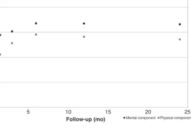
Figure 5. Mean SF-36 general health scores over time. Mean change from preoperation to 24 months was 17.9 SD 19.5 ( P < 0.001) for physical component summary, and 13.5 SD 22.2 ( P < 0.001) for men-tal component summary.
100 80 60 40 20 0 0 5 10 15 20
Mental component Physical component25
Follow-up (mo)
Score
TABLE 1.
Radiographical Results: Coronal Cobb Angles
Preoperation
n, Mean (SD) Postoperation n, Mean (SD) n, Mean (SD)24 mo
Change Pre- to Postoperation Mean (SD), Percent Change, P Change Postop-eration to 24 mo Mean (SD), P Total population 91, 20.9 (10.4) 86, 13.5 (9.2) 68, 15.2 (10.6) − 7.4 (8.2), 35.4%, < 0.001 1.6 (5.2), 0.019 Supplemental fi xation Stand alone 14, 19.4 (5.9) 14, 15.4 (8.6) 13, 18.9 (11.4) − 5.1 (4.0), 26.2%, 0.001 3.9 (4.5), 0.025* Lateral plate 7, 18.7 (8.0) 6, 10.3 (6.0) 4, 13.7 (9.2) − 8.5 (6.7), 45.4%, 0.027* 3.0 (5.3), 0.431* Unilateral pedicle screws 27, 15.5 (7.0) 25, 13.2 (7.5) 21, 14.3 (8.6) − 2.3 (5.1), 14.8%, 0.035 1.1 (3.4), 0.178 Bilateral pedicle screws 43, 25.2 (12.0) 43, 13.6 (10.7) 31, 14.6 (11.7) − 11.2 (9.0), 44.4%, < 0.001 1.0 (6.3), 0.385 Fixation technique †
Percutaneous 19, 25.2 (12.5) 19, 16.9 (12.5) 16, 18.1 (13.3) − 8.4 (6.9), 33.3%, < 0.001 1.8 (3.1), 0.037 Open 24, 25.2 (11.7) 24, 11.0 (8.4) 15, 10.8 (8.7) − 13.3 (9.9), 52.7%, < 0.001 0.2 (8.4), 0.937
*Small patient population ( < 10).
1858 www.spinejournal.com October 2013 aged 40 years or older undergoing primary surgery for
adult scoliosis with a minimum of 2-year follow-up. Patient-reported outcome data demonstrated improvement in dis-ability and function. However, the overall complication rate was 49%. Major complications occurred in 26% of patients, including pulmonary embolism, retroperitoneal hematoma, pseudarthrosis, sacral fracture, and deep infection. Minor complications including transient brachial plexus or peroneal nerve injury, pneumothorax, atrial fi brillation, splenic lac-eration, dural tear, pleural effusion, and urinary tract infec-tion occurred in 31% of patients. Pateder et al 39 reported a
2.4% 30-day mortality rate in 407 patients undergoing adult deformity surgery.
The overall complication rate of this study was 24% with 12% considered major, and there were no mortalities related to the procedure. The lower complication rate in this study is likely explained by the lateral approach to the anterior spine, avoiding many of the associated complications with traditional anterior surgery. In XLIF, the abdominal vascu-lature is not mobilized, the ureter is not manipulated, and the peritoneal cavity is not retracted. In Daubs’ series, the anterior approach was associated with a 10.9% incidence of iliac vein tears. 14 , 38 Pateder et al 39 evaluated the risk of
pul-monary embolism after deformity surgery and reported a 2.4% incidence. Neither complication occurred in this series. Furthermore, less invasive approaches to the posterior spine with percutaneous fi xation have consistently demonstrated a lower complication profi le than their open counterparts. 20 , 40 – 42
In particular, less invasive posterior approaches result in less blood loss and a lower incidence of postoperative wound infections. 43 – 45 Moreover, early ambulation after surgery has
been shown to reduce the incidence of perioperative compli-cations. 46 As a minimally disruptive procedure with relatively
short hospital stay, patients in this cohort were more likely to ambulate quickly after surgery.
Postoperative anterior thigh pain and lumbar plexus inju-ries have received attention as lateral transpsoas approaches have gained popularity. Pumberger et al 47 analyzed
post-operative neurological defi cits in 235 patients after lateral interbody fusion. They reported sensory defi cits in 1.6%, psoas mechanical defi cits in 1.6%, and lumbar plexus-related defi cits in 2.9% of patients. In this study, and as reported by Isaacs et al , 20 36 patients (33.6%) reported
postoperative weakness (primarily hip fl exion); however, nearly all cases were transient and only 2% had greater than 2 motor grades. The authors postulate that the transient hip fl exion weakness after XLIF is a consequence of psoas mus-cle penetration and retraction rather than a neural injury and recommend counseling patients about this possible side effect. Moreover, the use of directional, discrete-threshold, intraoperative neural monitoring while traversing the psoas muscle is recommended. This study suggested that the dura-tion of the XLIF procedure was a predictor of postoperative lower extremity weakness, suggesting that shorter periods of psoas muscle and lumbar plexus retraction are important to reduce these risks.
Correction of the coronal deformity is one goal of surgery in the DS patient population. A systematic review by Yadla
et al 7 of 49 publications reporting coronal correction in more
than 2000 adult patients with DS reported the average post-operative correction of coronal deformity as 40.7% (range: 1%–87%). In 2010, Good et al 3 reported a 43%
improve-ment in coronal Cobb angle at 2 years in 24 patients with adult scoliosis treated with open anterior/posterior surgery. In 2009, Maeda et al 48 compared coronal correction of adult
DS in patients treated with a circumferential approach using either bone morphogenic protein or iliac crest bone graft; correction was 50.6% and 42.5%, respectively. The current study of XLIF in the treatment of DS found a 35% correction of the coronal Cobb angle.
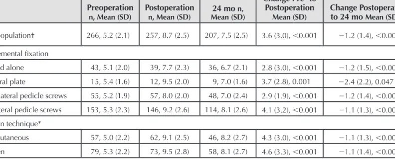
TABLE 2.
Radiographical Results: Disc Height
Preoperation
n, Mean (SD) Postoperation n, Mean (SD) Mean (SD)24 mo n,
Change Pre- to Postoperation
Mean (SD) Change Postoperation to 24 mo Mean (SD), P Total population† 266, 5.2 (2.1) 257, 8.7 (2.5) 207, 7.5 (2.5) 3.6 (3.0), < 0.001 − 1.2 (1.4), < 0.001 Supplemental fi xation
Stand alone 43, 5.1 (2.0) 39, 7.7 (2.3) 36, 6.7 (2.1) 2.8 (3.0), < 0.001 − 1.2 (1.5), < 0.001 Lateral plate 15, 5.4 (1.6) 12, 9.5 (2.0) 9, 7.0 (1.6) 3.7 (2.8), 0.001 − 2.4 (2.2), 0.047 Unilateral pedicle screws 55, 5.2 (1.9) 57, 8.0 (2.0) 48, 7.0 (2.4) 2.9 (1.9), < 0.001 − 1.2 (1.4), < 0.001 Bilateral pedicle screws 153, 5.3 (2.3) 146, 9.2 (2.6) 114, 8.1 (2.6) 4.1 (3.2), < 0.001 − 1.1 (1.3), < 0.001 Fixation technique*
Percutaneous 57, 5.0 (2.2) 62, 9.1 (2.5) 46, 8.2 (2.7) 4.3 (3.0), < 0.001 − 1.1 (1.3), < 0.001 Open 79, 5.3 (2.2) 73, 9.5 (2.8) 58, 8.1 (2.7) 4.6 (3.3), < 0.001 − 1.1 (1.4), < 0.001
*To remove the effect of unilateral versus bilateral fi xation, only patients with bilateral fi xation were included in the open versus percutaneous analysis. † Small patient population ( < 10).
In this study, coronal Cobb correction differed signifi cantly on the basis of choice of supplemental fi xation after XLIF, with the greatest corrections achieved in patients with open bilateral posterior pedicle fi xation. These fi ndings are likely refl ective of the ability in open posterior procedures to per-form posterior element releases and to better contour the rod and apply segmental compression and distraction to realign the spine. Similarly, bilateral posterior fi xation afforded a greater initial increase in mean disc height after surgery with less loss of height over time.
Although coronal correction alone does not correlate with clinical outcomes, 49 , 50 correction of sagittal balance has been
shown to be an important predictor of quality of life. 51 , 52 This
study supports prior studies that have suggested that XLIF is able to correct lordosis in patients with hypolordosis, 27 , 31 , 53
although this correction, too, may be impacted by choice of supplemental fi xation.
The revision rate for pseudarthrosis in DS treated with anterior and/or posterior procedures has been reported to be between 0% and 19%. 39 , 48 , 54 – 56 In this study, the rate of
revi-sion surgery for a diagnosis of pseudarthrosis was 2%. The low rate of pseudarthrosis observed after XLIF in this study is corroborated by previous studies reporting high rates of radiographical fusion. 11 , 14 , 22 , 25 This low rate of pseudarthrosis
after XLIF probably refl ects the ability to perform a thorough discectomy and place large interbody implants sitting on the endplate apophyseal ring.
Although this prospective study is the largest published series on XLIF for the treatment of scoliosis, certain limita-tions are evident. Treatment alternatives were not random-ized but left to surgeon preference, with resulting variability in the extent of treated levels and types of fi xation. The sites chosen to participate in this study were selected on the basis of their experience with XLIF and DS. These are high-volume
TABLE 3.
Summary of Revisions
Cause of Revision Revision (mo)Time to Initial Procedure Revision Procedure
Pseudarthrosis 8 Stand-alone XLIF L1–L5. L4–L5 corpectomy, L2–L5 decompression, and T11–L6 posterior fusion. Pseudarthrosis 6 L2–L5 XLIF, L5–S1 interbody fusion, T10–S1 bilateral pedicle screw fi xation. L4–S1 ALIF, removal of all posterior fiL5–S1 laminectomy, bilateral pedicle xation,
screw fi xation L4–S1.
Adjacent segment 5 L2–L4 XLIF, with unilateral L2 and L4 pedicle screws and rod. L4–L5 XLIF, with unilateral pedicle screw fi xation. Adjacent segment 2 L3–L5 XLIF, with unilateral L3 and L5 pedicle screw/rod. L2–L3 XLIF, L2–L5 laminectomy/foraminotomy, and insertion of unilateral
pedicle screws.
Adjacent segment 1 L1–L5 XLIF, with bilateral fi xation T10–L5. L5–S1 ALIF removal of T10 pedicle screws.
Adjacent segment 17 Stand-alone XLIF L1-L5. ALIF L5–S1.
Decompression, addition of
supplemental fi xation 19
L2–L5 XLIF, with unilateral L2 and L5 pedicle screws and rod.
L1–L5 laminectomy, hardware removal, L1– L5 decompression, addition of unilateral supplemental fi xation.
Removal of
supplemental fi xation 20 L1–L5 XLIF, with L5–S1 interbody fusion. L1–L5 unilateral pedicle screw fi xation. L1–L3 removal of pedicle screws and rods. Addition of
supplemental fi xation 1 Stand-alone XLIF L2–L5. L3–L5 pedicle screw fi xation. Addition of
supplemental fi xation 1 Stand-alone XLIF L2–L5. L2–L5 addition of bilateral posterior fi xation. Removal of
supplemental fi xation/
decompression 5
L3–L5 XLIF, with bilateral pedicle screw
fi xation. Hardware removal, L5–S1 decompression, S1 facet cyst removal. Revision of hardware,
osteotomy 3 L1–L5 XLIF, with bilateral L1–S1 pedicle screw fi xation.
T3–L1 addition of supplemental fi xation, revision of fi xation L1–L2, osteotomy T12–L2.
Decompression, addition of
supplemental fi xation 9 Stand-alone XLIF L2-L5.
L5–S1 foraminotomy with supplemental posterior fi xation, L1–L2
microdiscectomy.
1860 www.spinejournal.com October 2013 clinical practices, and the results of this study are refl ective
of such practices. Furthermore, with a 24-month follow-up compliance of less than 85%, there is a subset of patients with unknown outcomes. Although these conditions limit the con-clusions that might be drawn regarding the varied surgical constructs used, these data do refl ect real-world clinical expe-rience with the use of XLIF in the treatment of DS.
CONCLUSION
In the setting of DS, this study demonstrates that XLIF results in favorable clinical and radiographical results, while hav-ing a lower complication rate than has been reported with traditional surgical reconstruction in this patient population. Results from this study suggest that both Cobb correction and complication rate are signifi cantly affected by choice of fi xation after XLIF, with bilateral pedicle screws offering the greatest correction but a higher complication profi le.
11. Dakwar E , Cardona RF , Smith DA , et al. Early outcomes and safety of the minimally invasive, lateral retroperitoneal trans-psoas approach for adult degenerative scoliosis . Neurosurg Focus 2010 ; 28 : E8 .
12. Madhok R , Kanter AS . Extreme-lateral, minimally invasive, trans-psoas approach for the treatment of far-lateral lumbar disc hernia-tion . J Neurosurg Spine 2010 ; 12 : 347 – 50 .
13. Oliveira L , Marchi L , Coutinho E , et al. The use of rh-BMP2 in standalone extreme lateral interbody fusion (XLIF): clinical and radiological results after 24 months follow-up . World Spinal Col-umn J 2010 ; 1 : 19 – 25 .
14. Ozgur BM , Aryan HE , Pimenta L , et al. Extreme lateral interbody fusion (XLIF): a novel surgical technique for anterior lumbar inter-body fusion . Spine J 2006 ; 6 : 435 – 43 .
15. Pimenta L , Diaz RC , Guerrero LG . Charite lumbar artifi cial disc retrieval: use of a lateral minimally invasive technique. Technical note . J Neurosurg Spine 2006 ; 5 : 556 – 61 .
16. Rodgers WB , Cox CS , Gerber EJ . Minimally invasive treatment (XLIF) of adjacent segment disease after prior lumbar fusions . Internet J Minim Invasive Spinal Technol 2009 : 3 : 1 – 8 .
17. Aharinejad S , Bertagnoli R , Wicke K , et al. Morphometric analysis of vertebrae and intervertebral discs as a basis of disc replacement . Am J Anat 1990 ; 189 : 69 – 76 .
18. Champain S , Benchikh K , Nogier A , et al. Validation of new clinical quantitative analysis software applicable in spine orthopaedic stud-ies . Eur Spine J 2006 ; 15 : 982 – 91 .
19. Charosky S , Guigui P , Blamoutier A , et al. Complications and risk factors of primary adult scoliosis surgery: a multicenter study of 306 patients . Spine 2012 ; 37 : 693 – 700 .
20. Isaacs RE , Hyde J , Goodrich JA , et al. A prospective, nonrandom-ized, multicenter evaluation of extreme lateral interbody fusion for the treatment of adult degenerative scoliosis: perioperative out-comes and complications . Spine 2010 ; 35 : S322 – 30 .
21. Diaz R , Phillips F , Pimenta L . XLIF for lumbar degenerative sco-liosis: outcomes of minimally invasive surgical treatment out to 3 years postoperatively . Spine J 2006 ; 6 : 75S .
22. Anand N , Rosemann R , Khalsa B , et al. Mid-term to long-term clinical and functional outcomes of minimally invasive correc-tion and fusion for adults with scoliosis . Neurosurg Focus 2010 ; 28 : E6 .
23. Ozgur BM , Agarwal V , Nail E , et al. Two-year clinical and radio-graphic success of minimally invasive lateral transpsoas approach for the treatment of degenerative lumbar conditions . SAS J 2010 ; 4 : 41 – 6 .
24. Tormenti MJ , Maserati MB , Bonfi eld CM , et al. Complications and radiographic correction in adult scoliosis following combined transpsoas extreme lateral interbody fusion and posterior pedicle screw instrumentation . Neurosurg Focus 2010 ; 28 : E7 .
25. Wang MY , Mummaneni PV . Minimally invasive surgery for thora-columbar spinal deformity: initial clinical experience with clinical and radiographic outcomes . Neurosurg Focus 2010 ; 28 : E9 . 26. Meyerding H . Spondylolisthesis; surgical fusion of lumbosacral
portion of spinal column and interarticular facets; use of autog-enous bone grafts for relief of disabling backache . J Int Coll Surg 1956 ; 26 : 566 – 91 .
27. Mundis GM , Akbarnia BA , Phillips FM . Adult deformity correc-tion through minimally invasive lateral approach techniques . Spine 2010 ; 35 : S312 .
28. Bridwell KH , Glassman S , Horton W , et al. Does treatment (non-operative and (non-operative) improve the two-year quality of life in patients with adult symptomatic lumbar scoliosis: a prospec-tive multicenter evidence-based medicine study . Spine 2009 ; 34 : 2171 – 8 .
29. Li G , Passias P , Kozanek M , et al. Adult scoliosis in patients over sixty-fi ve years of age: outcomes of operative versus nonopera-tive treatment at a minimum two-year follow-up . Spine 2009 ; 34 : 2165 – 70 .
30. Elowitz EH , Yanni DS , Chwajol M , et al. Evaluation of indirect decompression of the lumbar spinal canal following minimally invasive lateral transpsoas interbody fusion: radiographic and out-come analysis . Minim Invasive Neurosur 2011 ; 54 : 201 – 6 .
Key Points
XLIF is associated with similar clinical and radio-graphical results as are reported in the literature after traditional open surgical procedures, while having a substantially lower complication rate.
Improvement of coronal and sagittal alignment is achieved with XLIF.
Greater reduction of deformity and lower rate of subsidence were observed when XLIF was com-bined with bilateral pedicle screw fi xation.
Higher perioperative complications were ob-served when pedicle screw fi xation was applied in an open fashion.
References
1. Tribus CB . Degenerative lumbar scoliosis: evaluation and manage-ment . J Am Acad Orthop Surg 2003 ; 11 : 174 – 83 .
2. Winter RB , Lonstein JE . Adult scoliosis . AAOS Instr Cours Lec 1983 ; 32 : 170 – 91 .
3. Good CR , Lenke LG , Bridwell KH , et al. Can posterior-only sur-gery provide similar radiographic and clinical results as combined anterior (thoracotomy/thoracoabdominal)/posterior approaches for adult scoliosis ? Spine 2010 ; 35 : 210 – 8 .
4. Gupta MC . Degenerative scoliosis. Options for surgical manage-ment . Orthop Clin North Am 2003 ; 34 : 269 – 79 .
5. Schlenk RP , Kowalski RJ , Benzel EC . Biomechanics of spinal defor-mity . Neurosurg Focus 2003 ; 14 : e2 .
6. Dorward IG , Lenke LG . Osteotomies in the posterior-only treat-ment of complex adult spinal deformity: a comparative review . Neurosurg Focus 2010 ; 28 : E4 .
7. Yadla S , Maltenfort MG , Ratliff JK , et al. Adult scoliosis surgery outcomes: a systematic review . Neurosurg Focus 2010 ; 28 : E3 . 8. Zimmerman RM , Mohamed AS , Skolasky RL , et al. Functional
outcomes and complications after primary spinal surgery for sco-liosis in adults aged forty years or older: a prospective study with minimum two-year follow-up . Spine 2010 ; 35 : 1861 – 6 .
9. Billinghurst J , Akbarnia BA . Extreme lateral interbody fusion— XLIF . Curr Orthop Pract 2009 ; 20 : 238 – 51 .
10. Caputo AM , Michael KW , Chapman TM Jr , et al. Clinical out-comes of extreme lateral interbody fusion in the treatment of adult degenerative scoliosis . Sci World J 2012 ; 2012 : 680643 .
31. Marchi L , Oliveira L , Amaral R , et al. Lateral interbody fusion for treatment of discogenic low back pain: minimally invasive surgical techniques . Adv Orthop 2012 ; 2012 : 282068 .
32. Rodgers WB , Gerber EJ , Patterson JR . Fusion after minimally dis-ruptive anterior lumbar interbody fusion: analysis of extreme lateral interbody fusion by computed tomography . SAS J 2010 ; 4 : 63 – 6 . 33. Rodgers WB , Lehmen JA , Gerber EJ , et al. Grade 2
spondylolis-thesis at L4–5 treated by XLIF: safety and midterm results in the “worst case scenario. ” Scientific World J 2012 ; 2012 : 356712 . 34. Smith WD , Dakwar E , Le TV , et al. Minimally invasive surgery for
traumatic spinal pathologies: a mini-open, lateral approach in the thoracic and lumbar spine . Spine 2010 ; 35 : S338 – 46 .
35. Uribe JS , Dakwar E , Le TV , et al. Minimally invasive surgery treatment for thoracic spine tumor removal: a mini-open, lateral approach . Spine 2010 ; 35 : S347 – 54 .
36. Voyadzis JM , Anaizi AN . Minimally invasive lumbar transfacet screw fi xation in the lateral decubitus position after extreme lateral interbody fusion: a technique and feasibility study . J Spinal Disord Tech 2013 ; 26 : 98 – 106 .
37. Fujita T , Kostuik JP , Huckell CB , et al. Complications of spinal fusion in adult patients more than 60 years of age . Orthop Clin North Am 1998 ; 29 : 669 – 78 .
38. Daubs MD , Lenke LG , Cheh G , et al. Adult spinal deformity sur-gery: complications and outcomes in patients over age 60 . Spine 2007 ; 32 : 2238 – 44 .
39. Pateder DB , Gonzales RA , Kebaish KM , et al. Short-term mortal-ity and its association with independent risk factors in adult spinal deformity surgery . Spine 2008 ; 33 : 1224 – 8 .
40. Khoo LT , Fessler RG . Microendoscopic decompressive laminot-omy for the treatment of lumbar stenosis . Neurosurgery 2002 ; 51 : S146 – 54 .
41. Perez-Cruet MJ , Foley KT , Isaacs RE , et al. Microendoscopic lum-bar discectomy: technical note . Neurosurgery 2002 ; 51 : S129 – 36 . 42. Schwender JD , Holly LT , Rouben DP , et al. Minimally invasive
transforaminal lumbar interbody fusion (TLIF): technical feasibility and initial results . J Spinal Disord Tech 2005 ; 18 ( suppl ): S1 – 6 . 43. O’Toole JE , Eichholz KM , Fessler RG . Surgical site infection
rates after minimally invasive spinal surgery . J Neurosurg Spine 2009 ; 11 : 471 – 6 .
44. Rodgers WB , Gerber EJ , Rodgers JA . Lumbar fusion in octo-genarians: the promise of minimally invasive surgery . Spine 2010 ; 35 : S355 – 60 .
45. Smith JS , Shaffrey CI , Sansur CA , et al. Rates of infection after spine surgery based on 108,419 procedures: a report from the Scoliosis Research Society Morbidity and Mortality Committee . Spine 2011 ; 36 : 556 – 63 .
46. Kamel HK , Iqbal MA , Mogallapu R , et al. Time to ambulation after hip fracture surgery: relation to hospitalization outcomes . J Gerontol 2003 ; 58 : 1042 – 5 .
47. Pumberger M , Hughes AP , Huang RR , et al. Neurologic defi cit following lateral lumbar interbody fusion . Eur Spine J 2012 ; 21 : 1192 – 9 .
48. Maeda T , Buchowski JM , Kim YJ , et al. Long adult spinal defor-mity fusion to the sacrum using rhBMP-2 versus autogenous iliac crest bone graft . Spine 2009 ; 34 : 2205 – 12 .
49. Di SM , Lolli F , Bakaloudis G , et al. Dynamic stabilization for degenerative lumbar scoliosis in elderly patients . Spine 2010 ; 35 : 227 – 34 .
50. Schwab FJ , Smith VA , Biserni M , et al. Adult scoliosis: a quan-titative radiographic and clinical analysis . Spine 2002 ; 27 : 387 – 92 .
51. Glassman SD , Bridwell K , Dimar JR , et al. The impact of positive sagittal balance in adult spinal deformity . Spine 2005 ; 30 : 2024 – 9 . 52. Glassman SD , Berven S , Bridwell K , et al. Correlation of
radio-graphic parameters and clinical symptoms in adult scoliosis . Spine 2005 ; 30 : 682 – 8 .
53. Marchi L , Abdala N , Oliveira L , et al. Stand-alone lateral interbody fusion for the treatment of low-grade degenerative spondylolisthe-sis . Scientific World J 2012 ; 2012 : 456346 .
54. Bess RS , Lenke LG , Bridwell KH , et al. Comparison of thoracic pedicle screw to hook instrumentation for the treatment of adult spinal deformity . Spine 2007 ; 32 : 555 – 61 .
55. Scheufl er KM , Cyron D , Dohmen H , et al. Less invasive surgical correction of adult degenerative scoliosis, part I: technique and radiographic results . Neurosurgery 2010 ; 67 : 696 – 710 .
56. Wu CH , Wong CB , Chen LH , et al. Instrumented posterior lumbar interbody fusion for patients with degenerative lumbar scoliosis . J Spinal Disord Tech 2008 ; 21 : 310 – 5 .