Review article
Non-Coding RNAs in Bacteria
Amanda C. Garcia 1, Vera Lúcia Pereira dos Santos 1, Teresa Cavalcanti 2, Luiz Martins Collaço 2, Hans Graf 1
1 Departamento de Medicina Interna, Universidade Federal do Paraná, PR, Brasil.
2 Departamento de Patologia, Universidade Federal do Paraná, PR, Brasil.
Abstract
Genes encoding regulatory RNAs known as short RNAs (sRNAs) or non-coding sRNAs (ncRNAs), modulate physiological responses through different mechanisms, through RNA-RNA interaction or RNA-protein interaction. These molecules transcribed in trans and in cis relative to the target RNA. They are located between the coding regions of proteins, i.e., in the intergenic regions of the genome and show signals of promoters and termini sequences generally Rho-independent. The size of the ncRNAs genes ranges from ~ 50 to ~ 500 nucleotides and several transcripts are processed by RNase with smaller end products, which modulate physiological responses through different mechanisms, by RNA-RNA interaction or RNA-protein interactions and some interactions may be stabilized by the Hfq chaperone. The Riboswitches constitute another class of ncRNAs, located in the 5'UTR region of an mRNA that promote transcriptional regulation through their interaction with a linker molecule. Recently, in prokaryotes, CRISPR (Clustered Regularly Interspaced Short Palindromic Repeats) regions have described, which repeats of sequences of palindromic bases are. Each replicate consists of short segments of "spacer DNA" from exposures prior to a bacteriophage virus or exogenous plasmid. The CRISPR system consists of an immune system of resistance to exogenous molecules.
Keywords: ncRNA, cis-encoded ncRNA, trans-encoded ncRNA, riboswitch, CRISPR.
Introduction
Non-coding RNAs can be classified as short non-messenger RNAs (snmRNAs), small non-coding RNAs (ncRNAs), untranslated RNAs (untranslated RNAs) RNAs), or non-protein-encoding RNAs (npcRNAs) [1]. Short RNAs (small RNAs or sRNAs or non-coding RNAs or ncRNAs are molecules that modulate physiological responses through different mechanisms through RNA-RNA interaction or RNA-protein interaction). Some interactions can be stabilized by the chaperone Hfq [2-4], which occurs commonly in the class of trans-encoded ncRNAs where the formation of the ncRNA:Hfq:mRNA complex may act positively or negatively on post-transcriptional regulation [5].
The most studied ncRNAs are the cis-encoded RNAs and the trans-encoded RNAs, the first transcripts being in cis and antisense relative to the target mRNA and the second transcripts in genomic regions distant from the target mRNA [6]. Therefore, only the cis-encoded RNAs present perfect base pairing with the targets. The Riboswitches constitute another class of ncRNAs, located in the 5'UTR region of an mRNA, which promote transcriptional regulation through its interaction with a linker molecule [7-9]. Recently, in prokaryotes, CRISPR (Clustered Regularly Interspaced Short Palindromic Repeats) regions that are repeats of sequences of palindromic bases have described [6, 10]. Each replicate consists of short segments of "spacer DNA" from exposures prior to a bacteriophage virus or exogenous plasmid. The CRISPR \ cas system consists of an immune system of resistance to exogenous molecules [11].
2. RNAs NOT ENCODING IN PROCARIOTS
The scientific and technological advance provided the discovery of the functions of those considered as main RNAs: messenger RNA (mRNA), molecule of information transfer during protein synthesis; RNA ribosomal (rRNA) component of the structure of protein synthesis and RNA transporter (tRNA) capable of transporting amino acids and interacting with proteins [21]). Several structural and functional studies of RNA have performed and allowed the description of new classes of RNA, with varied functions and in the Archea, Bacteria and
Eukaria domains (Table1) [22].
While genomics considers and analyzes genomes as coding sequences for mRNAs, rRNAs and tRNAs, RNomica further investigates the genes encoding RNAs that are untranslated but are functional RNAs and are involved in different cellular processes [23, 24]. Non-coding RNAs are involved in several cellular processes, such as chromosome replication in cell division (diF), transcription regulation (6S RNA), RNA processing (mRNA), mRNA stability and translation (antisense sequence - spot42), protein stability (tmRNA) and transport (4.5S or ffs) [25]. There are ncRNAs involved in oxidative stress (oxyS), stationary phase transcription (dsrA, rprA), related to the control of plasmid copy number (RNAI, RNAIII), carbon storage (csrBC) and carbon transport (gcvB) [24].
The class of ncRNAs with regulatory function is involved in several regulatory mechanisms such as gene expression in the modulation of outer membrane surface proteins (Omp) [26]. Some ncRNAs may bind to proteins in order to modulate their activities, as is the case with the 6S RNA that forms a complex with RNA polymerase (RNAP) [2, 24, 27]. These molecules are transcribed in trans or in cis relative to target RNA [4, 28]. The genes are located between the protein-coding regions, that is, in the intergenic regions of the genome and exhibit signals of promoters and terminator sequences, generally Rho-independent [29-32].
Fig. 1 - Structure of a ncRNAs gene in bacteria, where it has a CTTGAC (N) (-35) and CTATAT (-10) promoter region, ribosome binding site (RBS), an AUG coding region at the 5'UTR. It can vary in size from 50 to 500 base pairs, and in the 3'UTR region it has a terminator sequence (Rho-independent).
Jager and collaborators (2012) [40] demonstrated that the interaction of ncRNA162 ̸ target RNA in Archaea can occur with cis-encoded or trans-encoded action, through two distinct domains. As in Bacteria, ncRNAs in Archaea are involved in many biological processes, such as metabolic regulation, adaptation to environmental conditions, stress response, regulation of morphology, and cellular behavior. In Methanosarcina mazei GO1, Babski et al. (2014) [41] identified ncRNAs that align in the 5'UTR region of target mRNAs and in Sulfolobus solfataricus ncRNAs that align in the 3'UTR region of target mRNAs. Already for Pyrobaculum sp. and Haloferax volcanii, there is evidence that ncRNAs can align with target mRNAs at both the 5'UTR end and the 3'UTR end. In addition, they have observed that the nfRNA TRF down-regulates translation in response to binding to RBS.
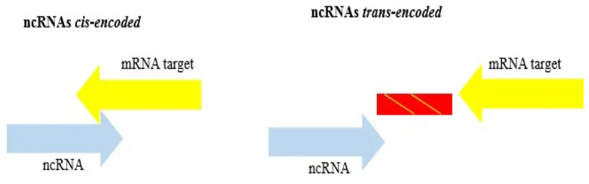
Fig. 2 – Sense of transcription ncRNAs and pairing mode with target RNAs. Genes of cis-encoded ncRNAs are located close to the target mRNA encoding genes while trans-encoded ncRNAs genes are located far from their target mRNAs. After transcription, the cis-encoded ncRNAs make short and perfect pairing with their targets and trans-encoded ncRNAs make long and imperfect pairing.
The mechanism of action of ncRNAs varies according to their function (Figures 3 and 4). In the post-transcriptional step the cis-encoded ncRNAs can cause mRNA degradation (Figure 3a), translation inhibition (Figures 3b), cleavage of the target mRNA (Figures 3c) (5'UTR Overlaping) and transcription termination (Figures 2d) [42]. The interaction of the trans-encoded ncRNA with target mRNA may result in inhibition of translation (Figures 4a) and degradation of mRNA by the negative interaction of 5'UTR inhibiting the ribosome binding site, or in the degradation paired with RNase (Figures 4b). The ncRNA trans-encoded with its target mRNA may form an inhibitory structure that could somehow block the site of the ribosome binding site, thereby impeding translation (Figures 4.c) [42]. Many cis-encoded RNAs with antisense orientation can form binary
Fig. 3 –Mechanisms of cis-encoded action in which, in the genome, the ncRNA is located close to the gene of its target mRNA. The cis-encoded ncRNAs represented in yellow and their target in blue. 3a. Degradation of mRNA. 3b. Inhibition of translation. 3c. Cleavage of mRNA and 3d. Transcription termination.
Fig. 4 – Mechanisms of trans-encoded action in which, in the genome, the ncRNA is located far from the gene of its target mRNA. The trans-encoded ncRNAs are represented in red and their target mRNA in blue. Complementarity of base pairs is limited. 3a. Inhibition of translation. 3b. Degradation of mRNA. 3c. Allows translation.
2.1 cis-encoded ncRNAs
initiation of replication, plasmid conjugation, transposition, mRNA degradation and some cell metabolism pathways. These events continue to be the subject of investigation by different researchers [4, 28, 43, 45].
The cis-encoded ncRNAs are encoded at the same locus of their target mRNA, but in the antisense sense of duplex, thus being fully complementary during the interaction. The mechanism of post-transcriptional response of gene regulation involves a high degree of sequence complementarity, and this was considered an indication that interaction of the Hfq protein would not be required [23, 45]. However, some researchers reported interference of Hfq in the cis-encoded ncRNA pairing [46-48]. In general, these ncRNAs act by complementing the mRNA ribosome-binding site, inhibiting, in turn, the translation [38, 45]. An example of cis-encoded typical ncRNA is the 5'ureB of Helicobacter pylori, located 5'-antisense to the ureB gene that makes up the ureAB operon [49, 50]. This ncRNA contains 292 bp and negatively regulates ureAB operon expression by blocking translation in the 5 'portion of ureB (Figure 4). The ureAB genes of H. pylori are located in the cluster of two
ureAB-ureIEFGH operons and encode the UreA and UreB subunits of apoenzyme urease. This enzyme is essential for the survival of H. pylori at low pH since its reaction produces NH3 and HCO3 into the environment, thus allowing homeostasis for bacterial growth [49]. The diversity of the cis-encoded ncRNA and their regulatory roles vary according to organisms as. For example, Salmonella enteric serovar Typhimurium possesses the cis-encoded ncRNA lesR-1 whose function is to control replication in eukaryotic cells [51] and Salmonella serovar Typi
possesses the cis-encoded ncRNA AmgR involved in virulence in rats [52] and AsdA which regulates intracellular replication [53].
The same diversity observed with respect to regulatory strategies. In Escherichia coli the base pairing between the cis-encoded ncRNA GadY and its gadXW target mRNA causes the cleavage of the duplex between gadX and gadW, providing increased levels of the gadX
transcript. The GadX product acts as a transcription factor for the GadAGadB operon during the synthesis of glutamate decarboxylase and this process consists of an acid stress deflating system in E. coli [6, 42].
The SymR-SymE system in E. coli consists of two genes, the cis-encoded ncRNA symR
Peng et al. (2000) [54] identified in the Brucella abortus 2308 the cis-encoded ncRNA BsrH that positively regulates the expression of the hemH gene, thus evidencing the importance of the regulatory expression that ncRNA BsrH exerts on its target mRNA. The cis-encoded antisense ncRNAs are also common in the replication mechanism of plasmids. For example, in the replication control of the ColE1 plasmid, which uses ncRNA instead of proteins to initiate replication at the site of origin, two partially complementary RNAs are transcribed from opposite strands. The larger RNA, with 250-500 nucleotides (RNAII), is transcribed from the sense strand and forms a stable hybrid with template DNA. This hybrid is then processed by the RNAse H to generate a primer for the DNA polymerase. The smaller RNA, with 68-108 nucleotides (RNAI), is transcribed from the antisense strand and is complementary to the 5 'RNAII region. It functions as a negative regulator of primer formation by forming the duplex RNAI/RNAII that prevents the formation of hybrid RNAII/DNA template. The concentration of RNAI is proportional to the number of plasmids per cell, thus constituting a negative feedback loop that regulates plasmid replication in response to metabolic changes [37, 55].
2.2 trans-encoded ncRNAs
As previously mentioned, the mode of action of trans-encoded ncRNAs differs from cis-encoded ncRNAs by the limited sharing of base pairs with their target mRNAs [42, 50, 56]. This type of ncRNA is encoded in trans and may have target mRNAs at different locations in the genome. The transcribed ncRNA generally requires the chaperone Hfq protein to stabilize the target ncRNA-RNA interaction due to imperfect base pairing and thus avoids its eventual degradation by RNase [39, 42, 56, 57]. The most studied chaperone is the Hfq protein that in
E. coli interacts with 40% of the ncRNAs [6]. Schoroeder et al. (2016) [58] report that almost 50% of all bacterial species possess trans-encoded ncRNAs that require the chaperone protein Hfq, one exception being Listeria monocytogenes in which most trans-encoded ncRNAs are independent of Hfq.
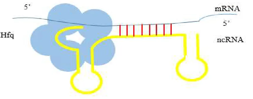
binds to the ncRNA and target mRNA and the other distal one that binds to the poly U tail (Figure 5) [62].
Fig. 5 - Probable model of interaction of Hfq protein with the ncRNA / mRNA pair.
Peng and collaborators (2015) [54] have reported, through fluorescence resonance energy transfer (FRET) studies, structural models in the Hfq interacts with PAPI, PNPase and RNaseE. Soper et al. (2010) [63] described three regulatory ncRNAs DsrA, RprA, and ArcZ in E. coli
that regulate positively the translation of RpoS sigma factor when ncRNA and rpoS mRNA pairing occurs. They identified the formation of an inhibitory clamp in the 5'UTR region and demonstrated that binding to Hfq is important for the stability of the RNA: RNA complex. In the negative regulation, base pairing occurs between the ribosome binding sequence (RBS) and ncRNA, blocking the ribosome binding, or degradation by RNases.
In E. coli, regulation of OmpC protein expression involves the Micf ncRNA and a 5 'UTR regulation of 22 antisense nucleotides to ompC mRNA [64]. This interaction also involves Hfq and results in inhibition of translation. In Salmonella typhimurium is the Micc ncRNA, associated with the Hfq protein, which silences the ompD mRNA through the duplex of 12 base pairs RNA/RNA in the protein-coding region. MicC does not inhibit translation initiation in the downstream position, but it accelerates RNaseE-dependent activity [65, 66].
In Salmonella enterica serovar Typhimurium the trans-encoded ncRNA IsrM, controls the pathogenic factor SPI-1 [67]. In contrast, RybB-1 and RybB-2 are associated with regulation of oxidative stress response [68]. In Salmonella enterica serovar Typi the trans-encoded
protein, whereas in Neisseria meningitidis the trans-encoded ncRNA Nrrf has property of regulation of iron homeostasis [70-72].
Other examples of trans-encoded ncRNA: AbcR-1 and AbcR-2 in Brucella abortus are related to virulence in rats and are important for the survival of macrophages [73] and Mrc7 in the Mycobacterium tuberculosis species is involved in the regulation of the secretion system of TAT [74].
3. RIBOSWITCHES
Riboswitches are structured elements of non-coding RNA considered cis-encoded
elements of RNA, located mostly in the 5'UTR region of a target mRNA and less frequent at the 3 'end UTR [7, 42, 50]. However, Loh et al. (2009) [75] described a case of riboswitch that controls in trans the expression of the virulence regulating protein PrfA in Listeria monocytogenes. They have the ability to control gene expression at the level of transcription and translation and to acquire different conformations in response to environmental signals, such as high temperatures and the binding of small molecules, such as metabolites or metal ions [9, 42, 76]. Recently, a wide variety of riboswitches have identified in prokaryotes. Bacillus subtilis has 2% of all its genes regulated by riboswitches that do not bind to intracellular metabolites, such as Flavin mononucleotide (FMN), thymine pyrophosphate, S-adenosyl-methionine (SAM), lysine and guanine (Figure 6) [43, 50, 77, 78].
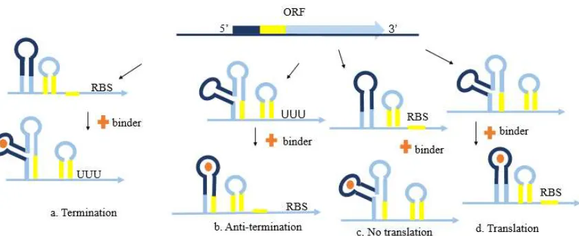
The structure of the riboswitch is composed of two parts, the aptamer region that serves as the binding site for a ligand and the expression platform that assumes a suitable conformation for the transduction (Figure 7) [42, 79]. The binding of the binding molecule causes conformational changes in the native riboswitch structure, which can regulate transcription and translation processes [7]. The latter sequence of the expression platform forms a splicing with the aptamer domain, whether or not it is bound to the linker and may possibly signal a transcription term or control the helical structure at the ribosome-binding site [7, 80].
prevents translation (Figure 7c). In contrast, binding of a linker may cause RBS exposure and promote translation (Figure 7d).
Fig. 6 – Riboswitch Structural Arrangement and Binding Regulatory Function. The aptamer region (pink) and an expression platform (yellow) on the 5’ UTR of the respective mRNA (blue) form
Riboswitch. Binding molecule has a regulatory function in the processes of transcription (a and b) and translation.
Riboswitches may play a role in biological cell systems because of the large number of gene families involved. Corbino et al. (2005) [82] found methA motifs in Agrobacterium tumefaciens similar to the S-adenosylmethionine decoding riboswitch (SAM). The SAM-II
riboswitch class presents a great structural diversity, of low conservation, being able to alter the conformational structure of the mRNA [7, 83, 84, 85].
The structure of the riboswitch may contain several motifs that control gene expression by detecting specific metabolite concentrations, which makes these structures promising targets for antibiotics. Suresch et al. (2016) [86] have shown that S-adenosylmethionine riboswitch-III, present in anaerobic bacteria, is involved in the methionine and SAM biosynthetic pathway regulation process. In the presence or absence of the ligand, S-adenosylmethionine riboswitch-III exerts a double function, which facilitates the conformational change between the partially and fully folded state, forming a stable duplex structure, which strengthens the interactions between the Shine-Dalgarno nucleotides (SD) and anti-Shine-Dalgarno (aDS).
proposed that methionine biosynthesis is probably the only means of using cobalamin in this strain of cyanobacteria.
4. The ncRNAs that Motivate Protein Activity
The first ncRNAs characterized in the E. coli enterobacterium include 4.5S, 6S, tmRNA (RNA transfer-messenger) and Spot42 [42, 88]. In E. coli, Spot42 RNA was discovered almost 40 years ago as a small and unstable RNA molecule encoded by the spf gene also present in
Vibrionaceae, a class of γ-proteobacteria. Salmonicidal Alivivium encodes a Spot42 RNA ncRNA with 84% identity to that of E. coli. Deletion of the spf gene results in a 25% decrease in the action of RNA polymerase I (Pol I) [89]. This ncRNA optimizes carbon uptake and metabolism by binding to target mRNAs related to this metabolism, such as galactose operon
galK. The ncRNA Spot42 is an example of ncRNA that acts traditionally by the ncRNA/mRNA interaction, but there are cases, such as the CsrB and the 6SRNA that regulate target protein activity and usually have specific recognition sequences [39].
[92]. In Pseudomonas aeruginosa Csr (Rms) controls several systems like Rhl and several virulence factors [92].
Hindley identified 6S RNA in 1967 in Escherichia coli [25]. Since then, 6S RNA predicted through computational tools and experimentally analyzed in several bacterial species [93]. Wehner et al. (2014) [94] analyzed 1611 bacterial genomes and determined a set of 1,750 RNA 6S genes, 1,367 new. In the Rfam database, the 6S RNA described by two entries, the first RF00013 with 153 sequences, and the second RF01685 with 89 sequences.
It has been demonstrated in E. coli that 6S RNA (ssrS gene) can act in the transcription process by associating with sigma 70 factor dependent RNA polymerase (6S RNA:RNAP), modulating its function. This ncRNA sequestrates almost all of the RNAP holoenzyme in late stationary phase and thereby contributes to transcriptional adaptation of stationary phase growth [87, 93, 94].
Regarding the position of 6S RNA in the genome, a synthetic pattern observed for the
Bacteria domain and this pattern is most widely observed in Gamma-Proteobacteria. Among the common synaptic genes are: ygfA (5-formyltetrahydrofolate cyclo ligase), zapA (ZapA protein), AAA (+) (ATPase superfamily) and peptidase_M24 (PF00557 family of metallopeptidases) [95, 96]. P. Gutiérrez, et al. (2005) [92] identified the ZapA protein in
Bacillus subtilis together with the tubulin FtsZ protein.
5. CRISPR (Clustered Regularly Interspaced Short Palindromic Repeats)
In 2007, Barrangou [97] and colleagues demonstrated in bacterial cells the change in resistance to phage infection by removing or adding spacer sequences similar to the sequences of invading phages. This strategy defined an adaptive immune defense system that uses short RNA and called Clustered Regularly Interspaced Short Palindromic Repeats (CRISPR). In the genome, the CRISPR locus contains hundreds of spacer sequences (~ 26-72 nucleotides) flanked by repeats of 21 to 47 nucleotides. They are usually associated with proteins (cas), forming the CRISPR system, which confers RNA-mediated resistance against nucleic acids from, for example, bacteriophage, plasmids or mobile genetic elements [1, 43, 98, 99]. The CRISPR locus is present in ~ 40% of bacterial genomes and in 90% of archaeal genomes [42, 100, 101].
1]. Cas proteins provide the enzymatic machinery required to acquire new spacers and create recognition marks of invading elements [1029].
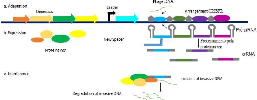
The arrangement and processing of CRISPR RNAs involves phases (Figure 7) [102]. Adhesion: invasive DNA is integrated into the CRISPR locus, resulting in new replicates in the matrix (Figure 7a); Expression: transcription of the transcript containing the phage RNA (new spacer) and its processing by the Cas proteins to form crRNA (matrix crRNA composed of single spacer sequences flanked by short repeats) and adjacent coded Cas proteins (Figure 7b); Interference: it is the formation of the complex CrRNAs Cas Cas proteins and later, the invasion happens to the target DNA and its respective degradation (Figure 7.c).
Fig. 7 - Array model of CRISPR system\Cas. The. Adaptation: DNA phage integrated into the CRISPR matrix and formation of the new spacer. B. Expression: Processing of cas proteins. W. Interference: Invasion to target DNA and its respctive degradation.
CRISPR may expressed in species such as Legionella pneumophila, where sRNA is the Cas2-dependent crRNA involved in the stress response [103-105]. In Listeria monocytogenes
the RliB-CRISPR sRNA also makes use of the CRISPR\Cas system in antiviral resistance [106,107]. Francisella novicida possesses the ncRNAs, Cas9-dependent crRNA, tracrRNA and scaRNA, with CRISPR\Cas property in the regulation of viral endogenous factors [105]. According to Marraffini (2016) [108] the strain Streptococcus pyogenes has the CRISPR \ Cas9 system whose function is to intermediate the anti-phagocytic resistance. In Campylobacter jejuni PT14 the CRISPR\Cas14 system protects against invasion/presence of bacteriophage [109].
system is integrated into the genome of the host cell, thus generating immunological memory, a natural ability based on the information contained in the DNA. Such a system properly manipulated in the formation of new bases of information storage in living organisms, something that has investigated in recent years.
6. Prediction of ncRNAs and ats Targets
Bioinformatics is dedicated to the development of computer programs for the treatment of
biological data and the identification of gene sequences, the prediction of three-dimensional protein configuration, the identification of enzyme inhibitors, the promotion of protein grouping, the establishment of phylogenetic trees and the analysis of expression experiments gene [111]. It provides tools for the development of Genomics, Transcriptomics, Proteomics and Metabolomics [112].
Regarding the prediction of ncRNAs, several computational approaches have developed for the identification of genes in intergenic regions of prokaryotic genomes [113, 114]. Many of these are based on the search for transcriptional signals, conserved promoter sequences, rho-independent terminator, transcription factor site, such as sRNAPredict [116], putative prediction algorithm analysis using TransTermHP database [116, 117] or TRANSFER [118]. sRNAPredict3 and SIPHT are recent computational versions of prediction of ncRNAs in bacteria) [16]. sRNAscanner and sRNAfinder [119] were developed to overcome the limitation of the prediction of available transcription signals in all genomic sequences and proved to be efficient. The nocoRNAc (non-coding RNA characterization) is a computational tool developed to study the interactions between ncRNA-mRNA in conjunction with the prediction of ncRNAs in bacterial genomes. This program uses transcription terminator signals predicted through the TransTermHP tool, the promoters are identified by the Stress Induced Duplex Destabilization
(SIDD) model and verifies possible regions to be destabilized [120, 121]. Thus, regions of the genome flanked by promoter sequence and Rho-independent terminator sequence, candidate to encode ncRNAs, identified. In a distinct approach, the Cufflinks tool locates regions of the genome with considerable levels of transcription and free ORFs [122].
ncRNAs in bacteria [14,127]. In the case of secondary structures, the RNAFold performs statistical statistical analysis of the RNA folding, through the perturbation of the thermodynamic parameters, for its prediction [119, 128, 129].
Regarding target prediction, the development of models is very important as it integrates bioinformatics for prediction and experimental validation for confirmation of mRNA targets. The classification of ncRNAs provides information on the complementarity of bases (perfect or imperfect) with their target mRNAs and eventual binding to proteins, altering their activity [130-134]. The targetRNA2 targeting mRNA prediction tool is one of the most widely used currently [135]. It utilizes various features including conserving ncRNA in other bacteria, the secondary structure of both the ncRNA and each target candidate, and the energy of hybridization between the interactions. It has the ability to integrate data from RNA-seq material when available. Another computational approach used to predict target mRNAs, the IntaRNA tool [136], is also considered quite efficient and rapid in predicting the interactions between ncRNA ̸ mRNA. It uses energy-free hybridization and has integration with the CopraRNA tool, which predicts ncRNAs by comparing the query-sequence with available sequences in the program [136, 137]. Other approaches include direct detection by means of microarrays, northern blotting, [3, 30, 111, 138, 139].
Pnek et al. (2008) [140] have identified predicted ncRNA in Streptomyces based on the study of sequence conservation in intergenic regions, localization of the transcription terminator factor, and genomic arrangement of syngenic genes to ncRNAs. They detected expression of 20 ncRNAs by microarray and RT-PCR, used a computational approach to determine secondary structure and identified 6S ncRNA. Voss et al. (2009) [141] used a cyanobacteria model to predict ncRNAs from transcriptome and proteome data and identified the Yfr2a-Yfr2c ncRNA, a conserved structure among cyanobacteria. To predict the existence of the 5'-operon-leader, the Rho-independent 3'-transcription terminator and riboswitches, these authors used the computational tools TransTermHP, ClustalW, RNAz, and RNAfold for validation using Northern Blot [142]. Modi et al. (2011) [137] performed the functional characterization of ncRNAs in E. coli
M. tuberculosis contain different sets of ncRNAs in the intergenic regions and have suggested that this characteristic may be the basis of the observed physiological differences between the two species. Schroeder et al. (20) observed that in the genus Rickettsia, ncRNAs are the major post-transcriptional regulators involved in virulence, survival, plasmid expression, primary and secondary metabolism, and presumably encode trans-encoded ncRNAs involved in the pathogen interaction and host.
Conclusions and perspectives
Knowledge about the post-transcriptional regulation that ncRNAs exert on a given target mRNA related to its position and matched in ncRNA: mRNA, and which sRNA class being worked on. There are different classes of sRNAs and how these can regulate several mechanisms is forming regulatory network, which plays a fundamental role in the regulatory circuit of the genome under study. The advancement of computational technologies aiming at providing a knowledge about the role and functionality of non-coding RNA in the various domains of life. Which have developed research for prediction and validation that help the understanding of what considered genomic trash today it known of the great importance that ncRNAs exert despite size are excellent builders and foundations that make up the genomic machinery.
Referências
[1] C.H. Hoe, et al., Bacterial sRNAs: Regulation in stress. Int. J. Med. Microbiol, 45 (6) (2013) 1203-1212.
[2] C. Pichon, B. Felden, Proteins that interact with bacterial small RNA regulators. Fems. Microbiol. Rev, 31 (5) (2007) 614-625.
[3] J. Livny, M.K. Waldor, Identification of small RNAs in diverse bacterial species. Curr. Opin. Microbiol. 10 (2) (2007) 1096-1101.
[4] J.M. Liu, A. Camilli, A broadening world of bacterial small RNAs. Curr. Opin. Microbiol, 13 (2010) 18-23.
[5] M.A. Faner, A.L. Feig, Identifying and characterizing Hfq–RNA interactions. Methods, 63 (2) (2013) 144–159.
[7] W.C. Winkler, Riboswitches and the role of noncoding RNAs in bacterial metabolic control. Current Opinion in Chemical Biology, 9 (6) (2005) 594–602.
[8] M.M. Meyer, et al., Challenges of ligand identification for riboswitch candidates. RNA Biology, 8 (1) (2011) 5-10.
[9] B. Izar, M.A. Mraheil, T, Hain, Identification and Role of Regulatory Non-Coding RNAs in
Listeria monocytogenes. Int. J. Mol. Sci, 12 (8) (2011) 5070-5079.
[10] Brosius, J.; Tiedge, H. Rnomenclature. RNA Biol, 1 (2) (2004) 81-83.
[11] Marraffini, L. A.; Sontheimer, E. J. CRISPR interference limits horizontal gene transfer in staphylococci by targeting DNA. Science, 322 (5909) (2008) 1843-1845.
[12] E. Rivas, et al., Computational identification of noncoding RNAs in E. coli by comparative genomics. Curr. Biol, 11 (17) (2001) 1369-73.
[13] Pichon, C.; Felden, B. Intergenic sequence inspector: searching and identifying bacterial RNAs. Bioinformatics, 19 (13) (2003) 1707–1709.
[14] Gruber, A. R.; et al., RNAz 2.0: improved noncoding RNA detection. Pac. Symp. Biocomput, (2010) 69–79.
[15] Livny, J., et al., Identification of Pseudomonas aeruginosa sRNAs and prediction of sRNA-encoding genes in 10 diverse pathogens using the bioinformatic tool sRNAPredict2. Nucleic Acids Res. 34 (12) (2006) 3484–3493.
[16] Livny, J., et al., High-Throughput, Kingdom wide prediction and annotation of bacterial non-coding RNAs. PLoS One, 3 (9) (2008) e3197.
[17] Sharma, C. M., Vogel, J. Experimental approaches for the discovery and characterization of regulatory small RNA. Curr. Opin. Microbiol, 12 (5) (2009) 536-546.
[18] Shinhara, A., et al., Deep sequencing reveals as-yet-undiscovered small RNAs in Escherichia coli. BMC. Genomics, 12 (428) (2011) 1471-2164.
[19] Pichon, C.; et al., An in silico model for identification of small RNAs in whole bacterial genomes: characterization of antisense RNAs in pathogenic Escherichia coli and Streptococcus agalactiae strains. Nucleic. Acids. Research, 40 (7) (2012) 2846-61.
[20] Schroeder, C. L. C., et al., Bacterial small RNAs in the Genus Rickettsia. BMC Genomics, 16 (1075) (2015) 1-18.
[21] M.M Cox, J.A Doudna, M. O’donnell, M. Biologia Molecular – Princípios e Técnicas. Artmed, (1) (2012).
[23] A. Hüttenhofer, P. Schattner, P. Polacek, Non-coding RNAS: hope or hype? Trends genet, 21 (5) 2005 289-97.
[24] Ulvé, V. M., et al., Identification of chromosomal alpha-proteobacterial small RNAs by comparative genome analysis and detection in Sinorhizobium meliloti strain 1021. BMC Genomics, 8 (467) (2007)1-16.
[25] K. Mikulík, Structure and functional properties of prokaryotic small noncoding RNAs. Folia. Microbiol. (Praha), 48 (4) (2003) 443-468.
[26] Massé, E., Majdalani, N., Gottesman, S. Regulatory roles for small RNAs in bacteria. Curr. Opin. Microbiol., 6 (2) (2003) 120-124.
[27] Gottesman, S., Storz, G. Bacterial Small RNA Regulators: Versatile Roles and Rapidly Evolving Variations. Cold Spring Harb Perspect Biol., 3 (12) (2011) a003798.
[28] Storz G.; Altuvia, S.; Wassarman, K. M. An abundance of RNA regulators. Annu. Rev. Biochem, 74 (2005) 199-217.
[29] Ermolaeva, M. D.; et al., Prediction of Transcription Terminators in Bacterial Genomes. Mol. Biol, 301 (2000) 27-33.
[30] Wassarman, K. M., et al., Identification of novel small RNAs using comparative genomics and microarrays. Genes Dev, 15 (13) (2001) 1637–1651.
[31] Argaman, L., et al., Novel small RNA-encoding genes in the intergenic region of Escherichia coli. Current Biology, 11 (12) (2001) 941–950.
[32] Ciampi, M. S. Rho-dependent terminators and transcription termination. Microbiology, 152 (2006) 2515–2528.
[33] K.M. Wassarman, P.J. Kiley, Global approaches for finding small RNA and small open reading frame functions. J Bacteriol, 192 (1) (2010) 26-8.
[34] K.B. Arnvig, T. Cortes, D.B. Young, Noncoding RNA in Mycobacteria. Microbiol Spectrum, 2 (2) (2014) 1-24.
[35] Updegrove, T.B.; Zhang, A.; Storz, G. Hfq: the flexible RNA matchmaker. Curr Opin Microbiol, 30 (2016) 133-138.
[36] C.M. Sharma, J. Vogel, Experimental approaches for the discovery and characterization of regulatory small RNA. Curr. Opin. Microbiol, 12 (5) (2009) 536-546.
[37] E. Massé, S. Gottesman, A small RNA regulates the expression of genes involved in iron metabolism in Escherichia coli. Proc. Natl. Acad. Sci, 99 (7) (2002) 4620-4625.
[39] F. Repoila, F. Darfeuille, Small regulatory non-coding RNAs in bacteria: physiology and mechanistic aspects, Biol Cell, 101 (2) (2009) 117-131.
[40] D. Jäger., et al., An archaeal sRNA targeting cis- and trans-encoded mRNAs via two distinct domains. Nucleic Acids Res, 40 (2012) (21) 10964-79.
[41] J. Babski, et al., Small regulatory RNAs in Archaea. RNA Biol. 11 (5) (2014) 484-493.
[42] L.S. Waters, G. Storz, Regulatory RNAs in bacteria. Cell, 136 (4) (2009) 615-628.
[43] G. Oliva, T. Sahr, C. Buchrieser, Small RNAs, 5' UTR elements and RNA-binding proteins in intracellular bacteria: impact on metabolism and virulence. FEMS. Microbiol. Rev., 39 (3) (2015) 331-49.
[44] E.G. Wagner, R.W. Simons, Antisense RNA control in bacteria, phages, and plasmids. Annu. Rev. Microbiol., 48 (1994) 713-42.
[45] S. Brantl, Regulatory mechanisms employed by cis-encoded antisense RNAs. Current Opinion in Microbiology, 10 (2) (2007) 102–109.
[46] A. Sittka, et al., Deep sequencing analysis of small noncoding RNA and mRNA targets of the global post-transcriptional regulator, Hfq. PLoS Genet, 22 (4) (2008) 8-e1000163.
[47] R. Lorenz, et al., ViennaRNA Package 2.0. Algorithms, Mol. Biol, 6 (26) (2011).
[48] C.C. Rossi, et al., A computational strategy for the search of regulatory small RNAs in
Actinobacillus pleuropneumoniae. RNA, 22 (9) (2016) 1373-1385.
[49] Y. Wen, J. Feng, G. Sachs, Helicobacter pylori 5'ureB-sRNA, a cis-encoded antisense small RNA, negatively regulates ureAB. J. Bacteriol., 195 (3) (2013) 444-452.
[50] Y. Han, et al., Regulation of pathogenicity by noncoding RNAs in bacteria. Future. Microbiol, 8 (5) (2013) 579-591.
[51] J. Gonzalo-Asensio, et al., A novel antisense RNA from the Salmonella virulence plasmid pSLT expressed by non-growing bacteria inside eukaryotic cells. PLoS One, 8 (10) (2013) e77939.
[52] E.J. Lee, E.A. Groisman, An antisense RNA that governs the expression kinetics of a multifunctional virulence gene. Mol. Microbiol, 76 (4) (2010) 1020-1033.
[53] I. Dadzie, et al., Identification and characterization of a cis-encoded antisense RNA associated with the replication process of Salmonella enterica serovar Typhi. PLoS. One, 8 (4) (2013) e61308.
[54] X. Peng, H. Dong, Q. Wu, A new cis-encoded sRNA, BsrH, regulating the expression of hemH gene in Brucella abortus 2308. FEMS. Microbiol, Lett, 362 (2) (2015) 1-7.
[56] T. Azhikina, et al., Role of Small Noncoding RNAs in Bacterial Metabolism. Biochemistry, 80 (13) (2015) 1633-1646.
[57] J. Richards, J.G. Belasco, A new window onto translational repression by bacterial sRNAs. Mol. Cell, 32 (6) (2008) 751-753.
[58] C.L. Schroeder, et al., Identification and Characterization of Novel Small RNAs in Rickettsia prowazekii. Front. Microbiol., 7 (2016) 1-14 .
[59] G.R. Richards, C.K. Vanderpool, Molecular call and response: the physiology of bacterial small RNAs. Biochim. Biophys. Acta, 1809 (10) (2011) 525-31.
[60] G.M. Cech, et al., The Escherichia Coli Hfq Protein: An Unattended DNA-Transactions Regulator. Front. Mol. Biosci, 3 (2016) 1-6.
[61] T.M. Link, P. Valentin-Hansen, R.G. Brennan, R. G. Structure of Escherichia coli Hfq bound to polyriboadenylate RNA. PNAS, 106 (46) (2009) 19292-19297.
[62] E. Holmqvist, et al., Global RNA recognition patterns of post-transcriptional regulators Hfq and CsrA revealed by UV crosslinking in vivo. EMBO. J, 35 (9) (2016) 991-1011.
[63] T. Soper, et al., Positive regulation by small RNAs and the role of Hfq. Proc. Natl. Acad. Sci, 107 (21) (2010) 9602-9607.
[64] S. Chen, et al., MicC, a second small-RNA regulator of Omp protein expression in Escherichia coli. J. Bacteriol., 186 (20) (2004) 6689-6697.
[65]V. Pfeiffer, et al., Coding sequence targeting by MicC RNA reveals bacterial mRNA silencing downstream of translational initiation. Nat. Struct. Mol. Biol., 16 (8) (2009) 840-846.
[66] Z. Wroblewska, M. Olejniczak, Hfq assists small RNAs in binding to the coding sequence of ompD mRNA and in rearranging its structure. RNA, 22 (7) (2016) 979-994.
[67] H. Gong, et al., A Salmonella small non-coding RNA facilitates bacterial invasion and intracellular replication by modulating the expression of virulence factors. PLoS. Pathog, 7 (9) (2011) e1002120.
[68] P.F. Calderón, et al., The small RNA RyhB homologs from Salmonella typhimurium participate in the response to S-nitrosoglutathione-induced stress. Biochem. Biophys. Res Commun, 450 (1) (2014) 641-5.
[69] J.M. Leclerc, C.M. Dozois, F. Daigle, Role of the Salmonella enterica serovar Typhi Fur regulator and small RNAs RfrA and RfrB in iron homeostasis and interaction with host cells. Microbiology, 159 (3) (2013) 591-602.
[71] J.R. Mellin, et al., Novel and iron-regulated small RNA, NrrF, is required for indirect fur-mediated regulation of the sdhA and sdhC genes in Neisseria meningitidis. J. Bacteriol, 189 (10) (2007) 3686-94.
[72] J.R. Mellin, et al., Role of Hfq in iron-dependent and -independent gene regulation in Neisseria meningitidis. Microbiology, 156 (8) (2010) 2316-2326.
[73] C.C. Caswell, et al., Identification of two small regulatory RNAs linked to virulence in
Brucella abortus 2308. Mol. Microbiol., 85 (2) (2012) 345-360.
[74] L. Solans, et al., The PhoP-dependent ncRNA Mcr7 modulates the TAT secretion system in
Mycobacterium tuberculosis. PLoS. Pathog, 10 (5) (2014) e1004183.
[75] E. Loh, et al., A trans-acting riboswitch controls expression of the virulence regulator PrfA in Listeria monocytogenes. Cell, 139 (4) (2009) 770-9.
[76] N.A. Smith, A.L. Eamens, M.B. Wang, The presence of high-molecular-weight viral RNAs interferes with the detection of viral small RNAs. RNA, 16 (5) (2010) 1062-1067.
[77] A.S. Mironov, et al., Sensing small molecules by nascent RNA: a mechanism to control transcription in bacteria. Cell, 11 (5) (2002) 747-756.
[78] T.M. Henkin, Riboswitch RNAs: using RNA to sense cellular metabolism. Genes Dev., 22 (24) (2008) 3383-3390.
[79] P.C. Anthony, et al., Folding energy landscape of the thiamine pyrophosphate riboswitch aptamer. PNAS, 109 (5) (2012) 1485-1489.
[80] Z. Weinberg, et al., Identification of 22 candidate structured RNAs in bacteria using the CMfinder comparative genomics pipeline. Nucleic. Acids. Res, 35 (14) (2007) 4809-4819.
[81] J.E. Barrick, et al., New RNA motifs suggest an expanded scope for riboswitches in bacterial genetic control. PNAS, 101 (17) (2004) 6421-6426.
[82] K.A. Corbino, et al., Evidence for a second class of S-adenosylmethionine riboswitches and other regulatory RNA motifs in alpha-proteobacteria. Genome Biol., 6 (8) (2005) R70.
[83] V. Epshtein, A.S. Mironov, E. Nudler, The riboswitch-mediated control of sulfur metabolism in bacteria. Proc. Natl. Acad. Sci, 100 (9) (2003) 5052-5056.
[84] B.A. McDaniel, et al., Transcription termination control of the S box system: direct measurement of S-adenosylmethionine by the leader RNA. Proc. Natl. Acad. Sci, 100 (6) (2003) 3083-8.
[85] D. Lalaouna, et al., Regulatory RNAs and target mRNA decay in prokaryotes. Biochim. Biophys. Acta, 1829 (6-7) (2013) 742-747.
[87] A.A. Pérez, D.A. Rodionov, D.A. Bryant, Identification and Regulation of Genes for Cobalamin Transport in the Cyanobacterium Synechococcus sp. Strain PCC 7002. J. Bacteriol, 198 (19) (2016) 2753-2761.
[88] S. Gottesman, G. Storz, Bacterial Small RNA Regulators: Versatile Roles and Rapidly Evolving Variations. Cold Spring Harb Perspect Biol. 3 (12) (2011) a003798.
[89] A.E. Trotochaud, K.M. Wassarman, 6S RNA function enhances long-term cell survival. J. Bacteriol., 186 (15) (2004) 4978-4985.
[90] C.A. Vakulskas, et al., Antagonistic control of the turnover pathway for the global regulatory sRNA CsrB by the CsrA and CsrD proteins. Nucleic. Acids. Res., 44 (16) (2016) 7896-7910.
[91] M.Y. Liu.; H. YANG.; T. Romeo, The product of the pleiotropic Escherichia coli gene CsrA modulates glycogen biosynthesis via effects on mRNA stability. J. Bacteriol, 177 (10) (1995) 2663-2672.
[92] P. Gutiérrez, et al., Solution structure of the carbon storage regulator protein CsrA from Escherichia coli. J. Bacteriol., 187 (10) (2005) 3496-3501.
[93] B. Steuten, et al., Regulation of transcription by 6S RNAs: insights from the Escherichia coli and Bacillus subtilis model systems, RNA Biol., 11 (5) (2014) 508-521.
[94] S. Wehner, et al., Dissemination of 6S RNA among bacteria, RNA Biol., 11 (11) (2014) 1467-1478.
[95] I.F. de Oliveira et al., Characterization of ftsZ mutations that render Bacillus subtilis resistant to MinC, PLoS. One, 5 (8) (2010) e12048.
[96] R. Barrangou, L.A. Marraffini, CRISPR-Cas systems: Prokaryotes upgrade to adaptive immunity. Mol. Cell, 54 (2) (2014) 234-244.
[97] D. Rath, et al., The CRISPR-Cas immune system: biology, mechanisms and applications. Biochimie, 117 (2015) 119-28.
[98] E. Evguenieva-Hackenberg, G. Klug, New aspects of RNA processing in prokaryotes. Curr. Opin. Microbiol., 14 (5) (2011) 587-92.
[99] L.A. Marraffini, E.J. Sontheimer, CRISPR interference limits horizontal gene transfer in staphylococci by targeting DNA. Science, 322 (5909) (2008) 1843-1845.
[100] M. Babu, et al., A dual function of the CRISPR-Cas system in bacterial antivirus immunity and DNA repair. Mol. Microbiol., 79 (2) (2011) 484-502.
[101] C. Richter, J.T. Chang, P.C. Fineran, Function and regulation of clustered regularly interspaced short palindromic repeats (CRISPR) / CRISPR associated (Cas) systems. Viruses, 4 (10) (2012) 2291-311.
[103] F.F. Gunderson, N.P. Cianciotto, The CRISPR-associated gene Cas2 of Legionella pneumophila is required for intracellular infection of amoebae. M. Bio, 4 (2) (2013) e00074-13.
[104] F.F. Gunderson, et al., Nuclease activity of legionella pneumophila Cas2 promotes intracellular infection of amoebal host cells, Infect Immun, 83 (3) (2015) 1008-18.
[105] A. Toledo-Arana, et al., The Listeria transcriptional landscape from saprophytism to virulence. Nature, 459 (7249) (2009) 950-956.
[106] N. Sesto, et al., PNPase dependent CRISPR System in Listeria. PLoS. Genet, 10(1) (2014) e1004065
[107] T.R. Sampson, et al., A CRISPR/Cas system mediates bacterial innate immune evasion and virulence. Nature, 497 (7448) (2013) 254-257.
[108] A.L. Marraffini, The CRISPR-Cas system of Streptococcus pyogenes. Function and applications. In: FERRETTI, J. J.; STEVENS, D. L.; FISCHETTI, V. A. Streptococcus pyogenes: Basic Biology to Clinical Manifestations. Oklahoma City (OK): University of Oklahoma Health Sciences Center, (2016) 1-17.
[109] S.P. Hooton, K.J. Brathwaite, I.F. Connerton, The Bacteriophage Carrier State of Campylobacter jejuni Features Changes in Host Non-coding RNAs and the Acquisition of New Host-derived CRISPR Spacer Sequences. Front. Microbiol, 7 (355) (2016) 1-8.
[110] G. Amitai, R. Sorek, CRISPR-Cas adaptation: insights into the mechanism of action. Nat. Rev. Microbiol, 14 (2) (2016) 67-76.
[111] N.D.D. Araújo, et al., A era da bioinformática: seu potencial e suas implicações para as ciências da saúde. The efects and applications of bioinformatics on the biomedical area. Estud. Biol., 30 (70-72) (2008) 143-148.
[112] E. Binneck, As ômicas: integrando a bioinformação. Revista. Biotecnologia, 32 (2004).
[113] S. Altuvia, Identification of bacterial small non-coding RNAs; experimental approaches. Curr Opin Microbiol, 10 (3) (2007) 257-261.
[114] J. Sridhar, et al., sRNAscanner: a computational tool for intergenic small RNA detection in bacterial genomes. PLoS. One, 5 (8) (2010) e11970,.
[115] J. Livny, et al., sRNAPredict: an integrative computational approach to identify sRNAs in bacterial genomes. Nucleic Acids Res. 33 (2005) 4096–4105.
[116] C.L. Kingsford, K. Ayanbule, S.L. Salzberg, Rapid, accurate, computational discovery of Rho-independent transcription terminators illuminates their relationship to DNA uptake. Genome Biol, 8 (2) (2007) R22.
[118] E. Wingender, et al., TRANSFAC: an integrated systemfor gene expression regulation. Nucleic. Acids. Res, 28 (1) (2000) 316–319.
[119] J. Sridhar, et al., sRNAscanner: a computational tool for intergenic small RNA detection in bacterial genomes. Plos. One, 5 (8) (2010) e11970.
[120] A. Herbig, K. Nieselt, nocoRNAc: characterization of non-coding RNAs in prokaryotes. BMC. Bioinformatics, 12 (40) (2011) 1-13.
[121] J. Sridhar, P. Gunasekaran, Computational small RNA prediction in bacteria. Bioinform. Biol. Insights, 7 (2013) 83-95.
[122] C. Trapnell, et al.,Transcript assembly and quantification by RNA-Seq reveals unannotated transcripts and isoform switching during cell differentiation. Nat. Biotechnol, 28 (5) (2010) 511-5.
[123] D. Gautheret, A. Lambert, Direct RNA motif definition and identification from multiple sequence alignments using secondary structure profiles. J. Mol. Biol, 313 (5) (2001) 1003-1011.
[124] E.P. Nawrocki, D.L. Kolbe, S.R. Eddy, Infernal 1.0: inference of RNA alignments. Bioinformatics, 25(10) (2009) 1335-1337.
[125] A. Coventry, D.J. Kleitman, B. Berger, MSARI: multiple sequence alignments for statistical detection of RNA secondary structure. PNAS, 101 (33) (2004) 12102-12107.
[126] J.S. Pedersen, et al., Identification and classification of conserved RNA secondary structures in the human genome. PLoS. Comput. Biol, 4 (2006) e33.
[127] J. Fallmann, et al., Recent advances in RNA folding. J Biotechnol, 10 (261) (2017) 97-104.
[128] D.M. Layton, R. Bundschuh, A statistical analysis of RNA folding algorithms through thermodynamic parameter perturbation. Nucleic. Acids. Res, 33 (2) (2005) 519-524.
[129] R. Lorenz, et al., ViennaRNA Package 2.0. Algorithms. Mol. Biol, 6 (26) (2011).
[130] B. Tjaden, et al., Target prediction for small, noncoding RNAS in bacteria. Nucleic. Acids. Res, 34 (9) (2006) 2791-2802.
[131] Tjaden, B. Targetrna: a tool for predicting targets of small RNA action in bacteria. nucleic. Acids. Res, 36 (2008) 109-113.
[132] Cao, S.; Xu, X.; Chen, S. J. Predicting structure and stability for rna complexes with intermolecular loop-loop base-pairing. RNA, 20 (6) (2014) 835-845.
[133] W. LI, et al., Predicting sRNAs and their targets in bacteria. Genomics Proteomics Bioinformatics. 10 (5) (2012) 276-284.
[135] P.R. Wright, et al., CopraRNA and IntaRNA: predicting small RNA targets, networks and interaction domains. Nucleic. Acids. Res, 42 (2014) 119-123.
[136] A. Busch, A.S. Richter, R. Backofen, IntaRNA: efficient prediction of bacterial sRNA targets incorporating target site accessibility and seed regions. Bioinformatics, 24 (24) (2008) 2849-2856.
[137] S.R. Modi, et al., Functional characterization of bacterial sRNAs using a network biology approach. PNAS, 108 (2011) 15522-15527.
[138] P.M. Clark, et al., Argonaute CLIP-Seq reveals miRNA targetome diversity across tissue types. Sci. Rep., 4 (5947) (2014) 1-11.
[139] J. Pánek, et al., Biocomputational prediction of small non-coding RNAS in streptomyces. BMC. Genomics, 9 (217) (2008) 1-11.
[140] B. Voss, et al., Biocomputational prediction of non-coding RNAs in model cyanobacteria. BMC. Genomics. 10 (123) (2009) 1-15.
[141] M. Brenes-Álvarez, et al., Identification of conserved and potentially regulatory small RNAS in Heterocystous cyanobacteria. Front. Microbiol., 7 (48) (2016) 1-11.
[142] J.S. Khoo, et al., Computational discovery and RT-PCR validation of novel Burkholderia conserved and Burkholderia pseudomallei unique sRNAs. BMC. Genomics. 13 (7) (2012) 1-14.