A flexible genome-scale resource of SARS-CoV-2 coding sequence clones
Dae-Kyum Kim1,2,3, Jennifer J. Knapp1,2,3, Da Kuang 1,2,3, Patricia Cassonnet4,5,6, Payman Samavarchi-Tehrani3, Hala Abdouni3, Ashyad Rayhan 1,2,3, Dayag Sheykhkarimli1,2,3, Étienne Coyaud7, Sylvie van der Werf4,5,6, Caroline Demeret 4,5,6, Anne-Claude Gingras2,3, Brian Raught8, Yves Jacob4,5,6,*, Frederick P. Roth1,2,3,9,*
1 Donnelly Centre, University of Toronto, Ontario, Canada
2 Department of Molecular Genetics, University of Toronto, Toronto, Ontario, Canada
3 Lunenfeld-Tanenbaum Research Institute, Sinai Health System, Toronto, Ontario, Canada
4 Unité de Génétique Moléculaire des Virus à ARN, Département Virologie, Institut Pasteur,
Paris, France.
5 UMR3569, Centre National de la Recherche Scientifique, Paris, France.
6 Université de Paris, Paris, France.
7 Univ. Lille, Inserm, CHU Lille, U1192 - Protéomique Réponse Inflammatoire Spectrométrie de
Masse - PRISM, F-59000 Lille, France
8 Department of Medical Biophysics, Princess Margaret Cancer Centre, University of Toronto,
Toronto, Ontario, Canada.
9 Department of Computer Science, University of Toronto, Toronto, Ontario, Canada
* Correspondence: [email protected] (Y.J), [email protected] (F.P.R.)
Abstract
The world is facing a major health crisis, the global pandemic of COVID-19 caused by the
SARS-CoV-2 coronavirus, for which no approved antiviral agents or vaccines are currently
available. Here we describe a collection of codon-optimized coding sequences for SARS-CoV-2 cloned into Gateway-compatible entry vectors, which enable rapid transfer into a
variety of expression and tagging vectors. The collection is freely available via Addgene. We hope that widespread availability of this SARS-CoV-2 resource will enable many subsequent
Introduction
The world is facing a major health crisis: A global pandemic of the coronavirus disease
COVID-19, a severe respiratory illness caused by a novel virus from the family Coronaviridae
(SARS-CoV-2), has infected millions and caused over 100,000 deaths (1). COVID-19
manifestation in patients can range from asymptomatic (no symptoms) to severe pneumonia
and death (2). Early analysis of the outbreak in China outlines symptoms that commonly include fever, dry cough, shortness of breath and myalgia (3). Person-to-person spread
through respiratory droplets has been identified as a major source of transmission of the virus
(4). To limit contagion, various measures from social distancing to nationwide lockdowns, have been imposed to contain and control the transmission of SARS-CoV-2 (5). Despite these
measures, the number of confirmed COVID-19 cases has continued to rise (1), highlighting the need for an effective vaccine and antiviral agents. Furthermore, the extrapolations concerning
the evolution of the pandemic are particularly alarming (6). It is therefore of intense and
pressing interest to better understand this virus and its interaction with host cells on a molecular level.
Shortly after the outbreak, the complete genome of one SARS-CoV-2 strain was published (7). Using the genome sequence as a reference, Chan et al. identified 12 viral open reading frames (ORFs) (8), including ORF1ab, a large polyprotein which is post-translationally processed into 16 proteins. More recently, Wu et al. discovered two additional viral ORFs (ORF9Bwu and ORF10wu) with unclear functions (7). Progress on molecular characterization
has been made on several viral proteins (9, 10), providing valuable insights into host-virus interaction. However, more research is necessary. The Gateway system offers efficient and
high-throughput transfer of the CDSs into a large selection of Gateway-compatible destination
vectors used for protein expression in many biological systems, e.g. Escherichia coli,
Saccharomyces cerevisiae, insect, or mammalian cells (11). Broad availability of a collection of SARS-CoV-2 coding sequences (CDSs) has the potential to enable many downstream biochemical and structural studies and thus a better understanding of processes within the
viral life cycle, possibly yielding scalable assays for screening drug candidates that could
Methods and Results
Based on the published annotation of the genome sequence of the HKU-SZ-005b (8) and Wuhan-Hu-1 (7) isolates of SARS-CoV-2(8), we requested the synthesis of ORF sequences
(GenScript, IDT), including termination codons and attB recombination sequences, with optimization of codon usage to reduce GC content and optimize expression in human and insect cells. ORF9Bwu, an alternative ORF within the N gene from the SARS-COV-2 (7), was
subsequently amplified by Polymerase Chain Reaction (PCR) from the viral N gene (see
Supplementary Table 1 ). These sequences were then incorporated into Gateway Entry plasmids: either pDONR207 (Invitrogen) or pDONR223 (12).
To enable C-terminal fusion constructs, we also generated an equivalent set of
Gateway-compatible clones without termination codons. These clones were made by either
PCR-amplifying the whole plasmid with primers that eliminated the stop codon, or by amplifying CDS regions from the first collection (see Supplementary Table 1 for primers), using
downstream primers with complementarity regions that were internal to each stop codon, and
which simultaneously incorporated the flanking sequences necessary for incorporation into a Gateway Entry plasmid (pDONR207 and 223).
Each SARS-CoV-2 CDS bacterial clone was isolated from a single colony, and its inserted
CDS was confirmed by full-length Sanger sequencing. All clones with a pDONR223 backbone
were sequenced with M13F and M13R primers (TCAG DNA sequencing facility, Toronto, Canada). Clones with a pDONR207 backbone were sequenced with customized forward and
reverse primers. Primer sequences are available in Supplementary Table 1.
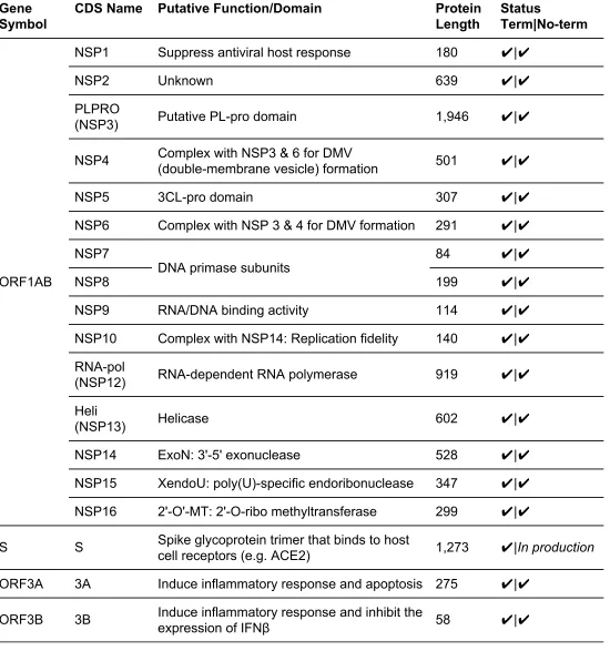
A total of 54 clones are currently included in the Gateway-compatible collection (Table 1),
covering 28 out of 29 total annotated CDSs in the SARS-CoV-2 genome. (NSP11 was omitted because of its 36 base pair length which makes it incompatible with the Gateway cloning
system (13).) All 28 of these CDS regions are available in clones with termination codons,
while 26 of 28 are currently available without termination codons.
summarizes all CDSs in the collection, together with their nucleotide and amino acid lengths, annotated functions and direct links to Addgene.
We hope that this SARS-CoV-2 CDS-clone collection will be a valuable resource for many
applications, including study of how coronaviruses can exploit host cellular processes for the
viral replication cycle, e.g., (15), and understanding virus-host protein-protein interactions (16, 17), production of recombinant virus proteins for structural studies (18), mapping of protein
subcellular localization using N-terminal fluorescent reporters (19), or development of vaccines
or other therapeutics (20, 21).
Acknowledgement
This work was supported by Canadian Institute for Health Research (CIHR) Foundation Grant
(F.P.R.), the Canada Excellence Research Chairs Program (F.P.R.), the LabEx Integrative Biology of Emerging Infectious Diseases (grant 10-LABX-0062) (Y.J.) and PREPARE, E-U
GRANT N° 602525 (Y.J.). D.-K.K. was supported by a Banting Postdoctoral Fellowship
through the Natural Sciences and Engineering Research Council (NSERC) of Canada and by the Basic Science Research Program through the National Research Foundation (NRF) of
Korea funded by the Ministry of Education (2017R1A6A3A03004385).
Conflict of Interest
Table 1. The genome-scale SARS-CoV-2 coding sequence clone collection. Production status for
clones with (Term) or without termination codon (No-term) is included. Check marks (✔) indicates that clones are currently available on Addgene.
Gene Symbol
CDS Name Putative Function/Domain Protein
Length
Status
Term|No-term
ORF1AB
NSP1 Suppress antiviral host response 180 ✔|✔
NSP2 Unknown 639 ✔|✔
PLPRO
(NSP3) Putative PL-pro domain 1,946 ✔|✔
NSP4 Complex with NSP3 & 6 for DMV
(double-membrane vesicle) formation 501 ✔|✔
NSP5 3CL-pro domain 307 ✔|✔
NSP6 Complex with NSP 3 & 4 for DMV formation 291 ✔|✔
NSP7
DNA primase subunits 84 ✔|✔
NSP8 199 ✔|✔
NSP9 RNA/DNA binding activity 114 ✔|✔
NSP10 Complex with NSP14: Replication fidelity 140 ✔|✔
RNA-pol
(NSP12) RNA-dependent RNA polymerase 919 ✔|✔
Heli
(NSP13) Helicase 602 ✔|✔
NSP14 ExoN: 3'-5' exonuclease 528 ✔|✔
NSP15 XendoU: poly(U)-specific endoribonuclease 347 ✔|✔
NSP16 2'-O'-MT: 2'-O-ribo methyltransferase 299 ✔|✔
S S Spike glycoprotein trimer that binds to host
cell receptors (e.g. ACE2) 1,273 ✔|In production
E E Envelope protein pentamer 75 ✔|✔
M M Membrane protein 222 ✔|✔
ORF6 6 Antagonize STAT1 function and IFN
signalling, and induce DNA synthesis 61 ✔|✔
ORF7A 7A Induce inflammatory response and apoptosis 121 ✔|✔
ORF7B 7B Induce inflammatory response 43 ✔|✔
ORF8 8 Induce apoptosis and DNA synthesis 121 ✔|✔
N N Facilitate viral RNA packaging 419 ✔|In production
ORF9B 9B Induce apoptosis 98 ✔|✔
ORF9Bwu 9Bwu Unknown 73 ✔|✔
References
1. World Health Organization (2020) COVID-19 Situation Reports. World Health Organization. 2. Huang,C., Wang,Y., Li,X., Ren,L., Zhao,J., Hu,Y., Zhang,L., Fan,G., Xu,J., Gu,X., et al.
(2020) Clinical features of patients infected with 2019 novel coronavirus in Wuhan, China.
Lancet, 395, 497–506.
3. World Health Organization (2020) Report of the who-china joint mission on coronavirus disease 2019 (covid-19).
4. Yu,P., Zhu,J., Zhang,Z., Han,Y. and Huang,L. (2020) A familial cluster of infection associated with the 2019 novel coronavirus indicating potential person-to-person transmission during the incubation period. J. Infect. Dis., 10.1093/infdis/jiaa077.
5. Cohen,J. and Kupferschmidt,K. (2020) Countries test tactics in “war” against COVID-19.
Science, 367, 1287–1288.
6. Ferguson,N., Laydon,D., Nedjati Gilani,G., Imai,N., Ainslie,K., Baguelin,M., Bhatia,S.,
Boonyasiri,A., Cucunuba Perez,Z., Cuomo-Dannenburg,G., et al. (2020) Report 9: Impact of non-pharmaceutical interventions (NPIs) to reduce COVID19 mortality and healthcare demand. 10.25561/77482.
7. Wu,F., Zhao,S., Yu,B., Chen,Y.-M., Wang,W., Song,Z.-G., Hu,Y., Tao,Z.-W., Tian,J.-H., Pei,Y.-Y., et al. (2020) A new coronavirus associated with human respiratory disease in China. Nature, 579, 265–269.
8. Chan,J.F.-W., Kok,K.-H., Zhu,Z., Chu,H., To,K.K.-W., Yuan,S. and Yuen,K.-Y. (2020) Genomic characterization of the 2019 novel human-pathogenic coronavirus isolated from a patient with atypical pneumonia after visiting Wuhan. Emerg. Microbes Infect., 9,
221–236.
9. Walls,A.C., Park,Y.-J., Tortorici,M.A., Wall,A., McGuire,A.T. and Veesler,D. (2020) Structure, Function, and Antigenicity of the SARS-CoV-2 Spike Glycoprotein. Cell, 10.1016/j.cell.2020.02.058.
10. Zhang,L., Lin,D., Sun,X., Curth,U., Drosten,C., Sauerhering,L., Becker,S., Rox,K. and Hilgenfeld,R. (2020) Crystal structure of SARS-CoV-2 main protease provides a basis for design of improved α-ketoamide inhibitors. Science, 10.1126/science.abb3405.
11. Walhout,A.J.M., Temple,G.F., Brasch,M.A., Hartley,J.L., Lorson,M.A., van den Heuvel,S. and Vidal,M. (2000) [34] GATEWAY recombinational cloning: Application to the cloning of large numbers of open reading frames or ORFeomes. In Thorner,J., Emr,S.D.,
Abelson,J.N. (eds), Methods in Enzymology. Academic Press, Vol. 328, pp. 575–IN7. 12. Rual,J.-F., Hirozane-Kishikawa,T., Hao,T., Bertin,N., Li,S., Dricot,A., Li,N., Rosenberg,J.,
13. ThermoFisher Gateway Recombination and Seamless Cloning Support. ThermoFisher. 14. Kamens,J. (2015) The Addgene repository: an international nonprofit plasmid and data
resource. Nucleic Acids Res., 43, D1152–7.
15. de Wilde,A.H., Snijder,E.J., Kikkert,M. and van Hemert,M.J. (2018) Host Factors in Coronavirus Replication. In Tripp,R.A., Tompkins,S.M. (eds), Roles of Host Gene and Non-coding RNA Expression in Virus Infection. Springer International Publishing, Cham, pp. 1–42.
16. Lasso,G., Mayer,S.V., Winkelmann,E.R., Chu,T., Elliot,O., Patino-Galindo,J.A., Park,K., Rabadan,R., Honig,B. and Shapira,S.D. (2019) A Structure-Informed Atlas of
Human-Virus Interactions. Cell, 178, 1526–1541.e16.
17. Gordon,D.E., Jang,G.M., Bouhaddou,M., Xu,J., Obernier,K., O’Meara,M.J., Guo,J.Z., Swaney,D.L., Tummino,T.A., Huttenhain,R., et al. (2020) A SARS-CoV-2-Human
Protein-Protein Interaction Map Reveals Drug Targets and Potential Drug-Repurposing.
bioRxiv, 10.1101/2020.03.22.002386.
18. Edavettal,S.C., Hunter,M.J. and Swanson,R.V. (2012) Genetic construct design and recombinant protein expression for structural biology. Methods Mol. Biol., 841, 29–47. 19. Tanz,S.K., Castleden,I., Small,I.D. and Millar,A.H. (2013) Fluorescent protein tagging as a
tool to define the subcellular distribution of proteins in plants. Front. Plant Sci., 4, 214. 20. McDonald,W.F., Huleatt,J.W., Foellmer,H.G., Hewitt,D., Tang,J., Desai,P., Price,A.,
Jacobs,A., Takahashi,V.N., Huang,Y., et al. (2007) A West Nile virus recombinant protein vaccine that coactivates innate and adaptive immunity. J. Infect. Dis., 195, 1607–1617. 21. Jing,L., Haas,J., Chong,T.M., Bruckner,J.J., Dann,G.C., Dong,L., Marshak,J.O.,
McClurkan,C.L., Yamamoto,T.N., Bailer,S.M., et al. (2012) Cross-presentation and