Review
1
Programming of Cell Resistance to Genotoxic and
2
Oxidative Stress
3
Ilya O. Velegzhaninov
1,2*, Vitaly A. Ievlev
3, Yana I. Pylina
1, Dmitry M. Shadrin
1and
4
Olesya M. Vakhrusheva
25
1 Institute of Biology of Komi Science Centre of Ural Branch of RAS, Syktyvkar 167982, Russia;
6
[email protected] (Y.I.P.); [email protected] (D.M.S.);
7
2 Vyatka State University, Kirov 610000 Russia; [email protected]
8
3 St. Olaf College, Northfield, MN 55057, USA; [email protected]
9
* Correspondence: [email protected]; Tel.: +7-909-121-9693
10
Abstract:
Different organisms, cell types, and even similar cell lines can dramatically differ in
11
resistance to genotoxic stress. This testifies to the wide opportunities for genetic and epigenetic
12
regulation of stress resistance. These opportunities could be used to increas the effectiveness of
13
cancer therapy, develop new varieties of plants and animals, and search for new pharmacological
14
targets to enhance human radioresistance, for example, for -manned deep space expeditions.
15
Based on the comparison of transcriptomic studies in cancer cells, in this review we propose that
16
there is a high diversity of genetic mechanisms of development of genotoxic stress resistance. This
17
review focused onpossibilities and limitations of the proposed regulation of the resistance of normal
18
cells whole organisms to genotoxic and oxidative stress by overexpressing of stress-response genes.
19
Moreover, the existing experimental data on the effect of such overexpression on the resistance of
20
cells and organisms to various genotoxic agents has been analyzed and systematized. We suggest
21
that the recent advances in the development of multiplex and highly customizable gene
22
overexpression technology that utilizes the mutant Cas9 protein and the wealth of available data on
23
gene functions and their signal networks open new opportunities for research in this field.
24
Keywords:
cell programming;
stress
resistance; gene overexpression; radiation; oxidative stress;
25
chemical genotoxins; malignant transformation; diversity of mechanisms
26
27
1. Introduction
28
Genotoxic stress, including oxidative stress, causes DNA damage. The evolutionary
29
conservative cellular mechanisms of DNA-damage prevention and response (DNA repair, defense
30
against reactive oxygen species, cell cycle checkpoints and apoptosis) protect cells from mutations
31
and tissues from marinization [1,2] On the one hand genotoxic stress can induce carcinogenesis, on
32
the other hand it is used to treat cancer. The advancement of knowledge on regulation of
stress-33
resistance in cells and organisms is extremely important for increasing the effectiveness of cancer
34
treatment. In particular, the creation of new in vitro models of upregulated cell resistance to genotoxic
35
and oxidative stresses allows to expand the spectrum of in vivo models for studies of genetic
36
regulation of carcinogenesis. In addition, it was suggested multiple times that gene therapy of normal
37
tissues surrounding tumor can be used for increasing their resistance to genotoxins. This can help to
38
minimize the negative side effects of cancer treatment by chemotherapy and radiation therapy [3–5].
39
This technology can also be used for gene therapy and gene prophylaxis of diseases associated with
40
increased sensitivity to DNA-damaging agents [6]. Understanding the mechanisms of cellular stress
41
resistance, and especially resistance to oxidative stress is one of the most important tasks in studies
42
of lifespan extension [7,8]. Knowledge of stress-resistance is also important when creating new
43
genetically modified varieties of plants and breeds of animals [9]. Additionally, the problem of
44
prolonged exposure of astronauts to cosmic ionizing radiation is a great challenge that needs to be
45
addressed in order to make deep space expeditions possible [10,11]. One of the possible solutions is
46
a pharmacological or geno-therapeutic enhancement of human radioresistance.Lastly, cell cultures
47
with multiple enhanced stress resistance can find application in recombinant therapeutic protein
48
production [12]. To achieve all the objectives listed above, excluding the last one, it is necessary to
49
ensure that tissue function and cells’ ability to elicit apoptotic and cell cycle responses are both not
50
affected as a result of genetic engineering interventions. Ideally, an increase in resistance to genotoxic
51
stress should lead to a decrease in the frequency of somatic mutations and neotransformations at the
52
organismal level.
53
The functions of many stress-response genes have been well studied. Signal-cascade networks
54
of gene activation in response to various damaging agents have also been elucidated. Such
55
knowledge can help identifying potential gene targets and their combinations for transcriptional
56
activation to increase resistance to gen
о
toxic and oxidative stress. However, without an array of
57
experimental data, it is not possible to accurately predict the results of such activations. Moreover, it
58
is difficult to predict the biological consequences of overexpression of the same gene to varying
59
degrees. The discovery of the CRISPR/Cas adaptive immunity and the development of methods for
60
its application for genome [13] and epigenome editing [14–18] significantly expands the possibilities
61
for further studies of stress resistance programming. In particular, relatively simple and adjustable
62
multiplex overexpression of genes by nuclease-null Cas9 (dCas9) can successfully activate
multi-63
subunit molecular complexes or entire signal cascades. Moreover, this technology provides activation
64
of genes in endogenous context, covering splice variants [16]. These advantages distinguish it from
65
the previously dominant gene overexpression technology which relies on introduction of cDNA into
66
the cell under a constantly active or inducible promoter. To date, there are very few works in the
67
literature that used CRISP/dCac9 for gene overexpression. Most of the articles are devoted to
68
optimization of the technology and its application in various fields of biological science. However,
69
this technology has already begun to prove its high potential. For example, it was shown the
70
possibilities of reactivation of silenced tumor supressors in vitro [19] and regulation of tumor
71
phenotypes in vivo [20]. In this regard, this review discusses the current state of knowledge about
72
modulation of resistance of normal and cancer cells as well as whole organisms to genotoxic and
73
oxidative stress by genes overexpression. To assess the potential of genetic regulation of
stress-74
resistance the review also discusses transcriptomic studies in cancer cells with different levels of
75
radioresistance.
76
2. The diversity of mechanisms of stress resistance in cancer cells
77
Mechanisms for development of genotoxic and oxidative stress resistance in tumor cells are well
78
described in a variety of reviews [21–24]. Clearly, cells lacking the capacity for apoptosis or
79
irreversible cell cycle arrest will exhibit a resistant phenotype due to their continued ability to
80
proliferate even under severe genotoxic stress conditions. Continued exposure to genotoxins in a
81
combination with abnormal response to DNA damage can lead to further loss of control mechanisms
82
and can increase resistance to stress in tumor cells. For example, this can happen through the
83
missense mutations in tumor suppressors. It can also be induced by a shift in a balance between
84
homologous (HR) and non-homologous end-joining (NHEJ) in double strand break DNA repair [22].
85
In addition, resistance to genotoxic stress is associated with the activation of oncogenes
N-ras
,
K-ras
86
[25,26],
MET
[27],
YAP
[28]. Radioresistance is also associated with the activity of the
Sox2
and
Oct3/4
87
genes that induce pluripotency and stem cell-like properties in cancer cells [29].
88
Due to the risk of carcinogenesis the mechanisms described above cannot be used as practical
89
targets for induction of cellular stress-resistance. However, stress resistance of tumor cells is often
90
formed by the mechanisms that are not associated with initiation of malignant transformation. As
91
mentioned above, when components of genome stability machinery are altered it could lead to an
92
increase in mutation rate in tumors, and result in an increased genetic heterogeneity of cells. This
93
heterogeneity facilitates rapid selection of subpopulations of cells that are resistant to stress [23]. The
94
possibility of this selection-based mechanism of resistance has been repeatedly confirmed in direct
95
selection experiments [30–32]. However, there is also evidence that stress-resistance can be induced
96
selection or independently of it often results from overexpression of the genes encoding transporter
98
proteins which support enhanced drug efflux [24]. In many cases, overactivation of DNA damage
99
recognition and repair as well as detoxification of free radicals are also observed. For example,
Rad51
100
gene which is involved in homologous recombination is overexpressed in a variety of human cancer
101
types. This often leads to chemo-resistance of these tumors [34]. An inverse correlation was observed
102
between the expression of the excision repair gene
ERCC1
and the sensitivity to platinum treatment
103
of various types of tumors [35]. An enhancement of excision repair activity in lung cancer cells can
104
also be associated with a SIRT1 dependent increase in XPA sensitivity to DNA damage [36].
105
Expression of the antioxidant defense gene -
MnSOD
- correlates with resistance to doxorubicin and
106
mitomycin C in gastric carcinoma cells [37].
RPA1
gene which is involved in DNA replication and
107
repair is overexpressed as a result of selection of a radioresistant clone in esophageal carcinoma cell
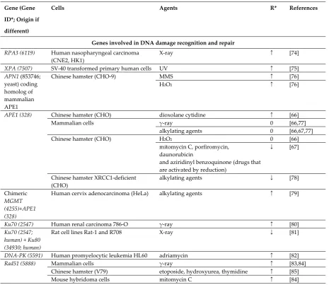
108
line TE-1. Inhibition of RPA1 in that radioresistant clone restored the normal sensitivity to ionizing
109
radiation [38].
110
There are many other examples of a established link between genotoxic stress resistance and
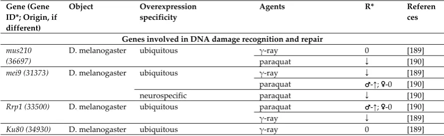
111
overexpression of genes involved in DNA repair, xenobiotic detoxification or efflux. However, the
112
diversity of possible mechanisms of resistance seems to be evenlarger. This is supported by the
113
studies comparing transcriptomes of similar cell lines that differ in sensitivity to genotoxic agents.
114
For example, a comparison of ten microarray studies performed on cancer cells with different degrees
115
of resistance to ionizing radiation did not identify any commonly overexpressed genes [39–48]. We
116
could not find a gene that would be significantly overexpressed in three or more comparison pairs.
117
Approximately 95 percent of the total number of overexpressed genes were observed in only one
118
study and were absent in others (Figure 1).
119
Thus, the diversity of pathways leading to resistance in cancer cells, allows us to suggest a wide
120
range of possibilities for increasing resistance of normal cells to genotoxic and oxidizing agents. We
121
suppose, that if we exclude all targets that affect cell cycle control, apoptosis, proliferation and
122
differentiation, we can enhance stress-resistance without the risk of increasing malignancy.
123
Moreover, the increased efficiency of cellular defense systems should in theory lead to a decrease in
124
carcinogenesis. This assumption is supported by the fact that the activity of DNA repair systems
125
inversely correlates with the risk of neotransformation[49]. In addition, a decrease in alkylating
126
agent-induced carcinogenesis has been repeatedly demonstrated upon overexpression of the gene 0
6-127
methylguanine-DNA methyltransferase (
MGMT
), which is responsible for DNA damage recognition
128
and repair [50–55].
129
3. Genotoxic stress resistance in experimental models with gene overexpression
130
Change in gene transcription is only one of the existing ways of readjusting the mechanisms of
131
stress resistance. Another way of establishing stress resistance is pharmacological targeting of
132
proteins and signaling cascades which seem more acceptable for clinical applications. However,
133
accumulation of experimental data on the effects of overexpression of individual genes and their
134
combinations is required to develop pathways of stress-resistance regulation which might help
135
finding new pharmacological targets. The literature on the effects of overexpression of
stress-136
responsive genes on the resistance of cells and organisms to genotoxins is overwhelmingly broad.
137
However, we attempted to systematically analyze such published experimental studies to reveal any
138
patterns and/or commonalities. Being mindful of the scale and the variety of the published studies,
139
in our analysis we chose a simple algorithm of grouping the target genes by their function. The
140
resulting lists of reviewed published reports are presented in Tables 1 and 2 for in vitro and in vivo
141
studies, respectively. One interesting, but not totally surprising, finding of our analysis was that most
142
studies driven by a targeted hypothesis (about involvement of a particular gene in stress resistance
143
based on previous experimental evidence) found that overexpression of the gene did increase stress
144
resistance. On the other hand it seems that in case of randomly selected targets, the predominant
145
outcome would be sensitization to stress, likely due to a disruption of normal gene activity regulation.
146
As suggested above, the two most promising gene categories for inferring resistance by
147
responsible for efflux and detoxification of xenobiotics. Overexpression of these genes tends to be the
149
most successful strategy to enhance resistance to genotoxic stresses without the risk of increasing the
150
frequency of neoplastic transformations. However, overexpression of these targets does not always
151
lead to an expected/desiredoutcome. Firstly, an increase in survival can mask the decrease in DNA
152
repair quality. For example, overexpression of the gene encoding DNA polymerase
β
in CHO cells
153
lead to an increase in survival after treatment with cisplatin, melphalan or mechlorethamine.
154
However, it also dramatically increased the frequency of mutations in surviving cells. It has been
155
repeatedly shown that this is due to the fact that DNA polymerase
β
is the most error prone
156
eukaryotic DNA polymerase [56–59]. Therefore, the required outcome and endpoints to be used
157
should be carefully selected. Secondly, the effect of overexpression of various single elements of a
158
repair or detoxification system/pathway can sometimes produce an effect that is opposite of the
159
expected one. At the cellular level, the two main groups of reasons for this are a) the imbalance
160
between the elements of the protective systems, and b) the absence of the expected relationship
161
between the level of gene transcription and the activity ofgene product. The latter primarily applies
162
to all proteins whose activity depends on post-translational modifications. The mismatch between
163
the mRNA levels and the protein function may also arise when a gene encodes only one subunit of a
164
multisubunit protein complexes. Forexample, stability of the DNA repair protein XPC depends on
165
the levels of HR23A and HR23B proteins [60], therefore overexpression of
XPC
gene may not be
166
sufficient to enhance nucleotide excision repair. Consistent with this, an averaged quantitative
167
relationship between the levels of mRNA and corresponding protein tends be weak [61]. However,
168
estimations of this correlation are still the subject of discussion and differ widely in the range from
169
0.21 to 0.9 [62]. In exceptional cases, for example in the case of ribosomal proteins, mRNA can be a
170
repressor of translation of its own product. This phenomenon is known to occur for the RpS3 protein
171
which is involved in stress responses [63].
172
173
Figure 1. Genes that are overexpressed in radioresistant cancer cells in comparison with parental or
174
similar but radiosensitive cells. The results of ten studies performed with microarrays were used.
175
Only 15 of the 337 overexpressed genes are repeated twice in different studies: a – C-JUN; b - CXCL10,
176
IFI44, IFIH1, IFITM1, STAT1, DDX60, HERC6, IFI27, PLSCR1, IFIT1, IFI35, IFIT3; c - ISG15; d - ERP70.
177
Numbers in parenthesis is the quantity of transcripts analyzed. * - Genes that are involved in
178
apoptosis, DNA repair, cell cycle control, cell proliferation and other mechanisms of stress response.
179
The imbalance of protective systems resulting from overexpression of individual genes may be
181
caused by several different mechanisms. First, it can be driven by the imbalance in productivity of
182
successive stages of a single cascade. For example, a wide range of modified bases in S. cerevisiae is
183
excised using MAG1 (3-methyladenine DNA glycosylase). The abasic sites generated by MAG1 are
184
processed normally by the major yeast APN1-encoded AP endonuclease. Disproportionately high
185
expression of
MAG1
compared to the AP endonuclease increases spontaneous mutation by up to
600-186
fold in S. cerevisiae and by 200-fold in Escherichia coli [64]. CHO cells with overexpressed
MPG
gene
187
are more sensitive to alkylating agent N-methyl-N'-nitro-N-nitroso-guanidine (MNNG) which is also
188
associated with excessive accumulation of abasic sites [65].
189
Secondly, there are situations when an increase in resistance to one agent is accompanied by
190
sensitization to others. For example, overexpression of
APE1
increases the resistance of CHO cells to
191
dioxolane cytidine [66], but it sensitizes cells to agents which are activated by reduction reactions.
192
The latter takes place because the product of
APE1
gene has a RedOx function in addition to AP
193
endonuclease activity [67]. Another mechanism is a shift in balance between the two competing
194
processes. For example, overexpression of
XRCC1
required for base excision repair (BER) slows
gap-195
filling, because of the competion of BER with nucleotide excision repair for the PCNA protein [68].
196
The listed nuances of regulation of resistance to genotoxic stress explain the opposite outcomes
197
observed during the overexpression of the same genes in different experiments (Tables 1 and 2). The
198
same opposite outcomes are observed on the level of functional groups of gene, which obtained using
199
PANTHER classification system [69,70]. The classification shows that researchers mainly chose the
200
genes encoding nucleic acid binding proteins and proteins that catalyzes a redox reactions. This is
201
expected, since the many proteins of these groups are involved in DNA repair and oxidative stress
202
defence, respectively. At the same time, if we divide the experiments based on the direction of the
203
effect on stress-resistance, the ratio of the functional groups does not change significantly (Fig. 2).
204
This means that we can not say that in fact overexpression of the genes of one of these functional
205
groups more effectively increases the stress resistance than the overexpression of the genes of the
206
other group.At the level of the whole organism, potential disruptions of functional interactions
207
between cells, tissues, organs and organ systems are added to the intra-cellular mechanisms of
208
imbalance listed above. But improvements in survival, decrease in frequency of mutations, fewer
209
incidence of cancer and some others desirable outcomes are still observed as a result of
210
overexpression of stress-responsive genes in a number of studies, which holds promise (Table 2).
211
In addition to the above, there are, apparently, many other factors that can radically change the
212
influence of overexpression of certain genes on cellular stress-resistance. This is supported by the cell
213
line specific effect of overexpression of the proto-oncogene
HER2/neu
in human breast and ovarian
214
cancer cells. In six different cell lines, overexpression led to either a decrease, or an increase in
215
sensitivity to chemotherapeutic agents of different classes [71]. These experimental data provide
216
additional evidence in favor of the need for further studies of genetic regulation of stress resistance
217
in normal and cancerous cells as well as stress-resistance of an organism as a whole.
218
219
Figure 2. The functional classification of overexpressed genes using PANTHER protein classes.
221
Human orthologues of genes listed in table 1 were divided into two groups, depending on the effect
222
of their overexpression on the resistance of cells (“In vitro”). The same division was performed for
223
orthologues of genes listed in Table 2 (“In vivo”). Each groups was classified using PANTHER
224
classification system using Protein class ontology [69,70]. * - total number of hits of analyzed genes to
225
“PANTHER protein class” classification.
226
4. Prospects
227
The decrease in stress-resistance of cells in the variety of experiments described above is largely
228
due to the multicomponent nature of stress response mechanisms that the studied genes participate
229
in. Numerous experimental data that support the high efficiency of overexpression of the
MGMT
230
gene support this assumption (Tables 1 and 2). Product of this gene solely performs recognition and
231
repair of damaged DNA bases, in contrast to most other elements of cell protective systems that
232
operate in cooperation with many other gene products [72]. Considering the accumulated detailed
233
knowledge of such interactions, development of multiplex gene activation systems with mutant
234
RNA-guided Cas9 protein opens up the widest opportunities for studying regulation of stress
235
resistance. Multiplex activation using one large [73] or a number of small [16] plasmids, using
236
activators with different degrees of efficiencies, allows selecting the appropriate range of activation.
237
To some extent, the level of superactivation of individual genes can be adjusted by selecting sgRNA
238
for sequences located at different distances from the transcription start site.
239
Table 1. Effect of overexpression of stress responsive genes on resistance to genotoxic agents in vitro
240
Gene (Gene
ID*; Origin if
different)
Cells Agents R* References
Genes involved in DNA damage recognition and repair
RPA3 (6119) Human nasopharyngeal carcinoma
(CNE2, HK1)
X-ray ↑ [74]
XPA (7507) SV-40 transformed primary human cells UV ↑ [75]
APN1 (853746; yeast) coding homolog of mammalian APE1
Chinese hamster (CHO-9) MMS ↑ [76]
H2O2 ↑ [76]
APE1 (328) Chinese hamster (CHO) dioxolane cytidine ↑ [66]
Mammalian cells γ-ray 0 [66,77]
alkylating agents 0 [66,67,77]
Chinese hamster (CHO) H2O2 0 [66]
mitomycin C, porfiromycin, daunorubicin
and aziridinyl benzoquinone (drugs that are activated by reduction)
↓ [67]
Chinese hamster XRCC1-deficient (CHO)
alkylating agents ↓ [78]
Chimeric
MGMT (4255)+APE1 (328)
Human cervix adenocarcinoma (HeLa) alkylating agents ↑ [79]
Ku70 (2547) Human renal carcinoma 786-O γ-ray ↑ [80]
Ku70 (2547; human) + Ku80 (34930; human)
Rat cell lines Rat-1 and R708 X-ray ↓ [81]
DNA-PK (5591) Human promyelocytic leukemia HL60 adriamycin ↑ [82]
Rad51 (5888) Mammalian cells γ-ray ↑ [83,84]
Chinese hamster (V79) etoposide, hydroxyurea, thymidine ↑ [85]
Prpf19 (27339) Human umbilical vein/vascular endothelium cells (HUVECs)
bleomycin, DL-buthionine-sulfoximine ↑ [11]
ALC1 (9557) Human osteosarcoma U2OS cells phleomycin ↓ [86]
Lig III (3980) Human cervix adenocarcinoma (HeLa S3)
MNNG ↑ [87]
DNA pol β (5423)
Chinese hamster (CHO) cisplatin, melphalan, mechlorethamine
↑↓ [56]
Mouse embryo fibroblast (MEF) MMS ↑0↓ [59]
Tag (947137; E.coli) coding methyladenine DNA
glycosylase I
Chinese hamster (V79) MMS, MNU, EMS ↑ [88,89]
MNU, ENU 0 [89]
Murine fibroblast (NIH3T3) and murine H1 melanoma cells (B78)
MNU, MNNG, DMS, temozolomlde 0 [90]
AlkA (947371
;E.coli) coding methyladenine DNA
glycosylase II
Chinese hamster (V79 and Irs1) DMS, EMS, MMS ↑ [91]
MPG (4350) Chinese hamster (V79 and Irs1) DMS, EMS, MMS ↑ [91]
Chinese hamster (CHO) MMS ↓ [92]
bis-chloroethylnitrosourea, melphalan 0 [93]
DMS, EMS, MMS 0 [94]
MMS, MNNG ↓ [65]
Mouse embryo fibroblast (MEF) temozolomide ↓ [95,96]
FPG (946765; E. coli) coding homolog of mammalian OGG1
Chinese hamster (CHO and V79) γ-ray ↑ [97]
Chinese hamster (CHO) aziridine ↑ [98]
dOGG1 (31806) Drosophila S2 cells paraquat, H2O2 ↓ [99]
S-nitroso-N-acetylpenicillamine ↑ [99]
OGG1 (4968; human)
Chinese hamster (AA8 and AS52) potassium bromate or [R]-1-[(10-chloro- 4-oxo-3-phenyl-4H-benzo[a]quinolizine-1-yl)-carbonyl]-2-pyrrolidinemethanol plus light
↑ [100]
ERCC1 (2067; human)
Chinese hamster (AA8) melphalan, cisplatin ↓ [101]
UV 0 [101]
NTH (947122; E.coli)
Chinese hamster (XRS7) γ-ray 0 [102]
H2O2 ↑ [102]
bleomycin ↓ [102]
Ogt (945853; E.coli)
Mammalian cells alkylating agents ↑ [103–105]
Ada (946710; E.coli) and its truncated and modified versions
Mammalian cells alkylating agents ↑ [103–116]
Chinese hamster lung fibroblasts dibromoalkanes ↓ [104]
Chinese hamster (V79) MMS, HN2 0 [113]
Chinese hamster (CHO) UV, ENU 0 [111]
MGMT (4255) and its modified versions
Mammalian cells alkylating agents ↑ [111,117–
124]
Chinese hamster (CHO) UV, ENU 0 [111]
alkB (946708; E.coli)
Human cervix adenocarcinoma (HeLa) MMS, DMS ↑ [125]
Genes involved in detoxification and efflux of free radicals and xenobiotics
SOD1 (6647) Human lymphoblastoid cells (TK6) γ-ray 0 [126]
Human primary lung fibroblasts (HPLF)
γ-ray ↑ [127]
Astrocytes of mice xanthine oxidase with hypoxanthine, menadione
↑ [128]
Brain neurons of mice S-nitroso-N-acetylpenicillamine, spermine-NONOate, diethylamine-NONOate
↑ [129]
H2O2 0 [129]
menadione ↓ [129]
Human glioma cells (U118-9) γ-ray ↑ [131]
SOD2 (6648) Human lung adenocarcinoma cisplatin ↑ [132]
Human cells γ-ray ↑ [126,127,133,
134] Human lymphoblastoid cells (TK6) paraquat ↑ [126] Human hepatocellular carcinoma cells
(HLE)
X-ray ↑ [135]
Human gastric carcinoma cells doxorubicin ↑ [37]
ALDH3A1 (218)
Human adenocarcinoma cells (MCF7) 4-hydroxyperoxycyclophosphamide, doxorubicin, etoposide, 5-fluorouracil, γ -ray, H2O2
↑ [136]
CAT (847) Normal human keratinocytes UV ↑ [130]
Mouse aortic endothelial cells (MAECs)
benzo(a)pyrene ↑ [137]
TRX (41737) Drosophila S2 Cells H2O2 ↑ [138]
MTII (17750) Chinese hamster ovary cells (K1-2) Cadmium chloride, MNU, MNNG ↑ [139]
γ-ray, bleomycin, MMS,
N-hydroxyethyl-N-hloroethylnitrosourea
0 [139]
Mouse C127 cisplatin, melphalan, chlorambucil ↑ [140] 5-fluorouracil, vincristine 0 [140]
Mouse β-cell streptozotocin ↑ [128]
MTI (17748) Mouse embryo fibroblasts (NIH/3T3) tert-butyl hydroperoxide ↑ [141]
Chinese hamster (V79) Amsacrine, menadione, arsenite, TPA ↑ [142]
Zn(II) ↑ [143]
alkylating agents 0 [143]
Genes involved in control of proliferation and cell cycle
CCND1 (595) Human adenocarcinoma cells (MCF7) γ-ray ↓ [144]
p21 (1026) Glioma cells (T-98G, U-251MG with mutant p53 allele and U-87MG with wild-type p53). Medulloblastoma cells MED-3.
γ-ray ↑ [145]
Genes involved in regulation of apoptosis
BCL2 (596) Mice thymocytes Ionizing radiation (not specified) ↑ [146]
Rat 6 fibroblast (R6) UV ↑ [147]
Human bladder cancer cells BIU87 adriamycin ↑ [148] Mouse embryo fibroblasts (NIH/3T3) γ-ray ↑ [149] Human breast cancer cells
(MDA-MB-231)
γ-ray ↑ [149]
Human non-small cell lung carcinoma (H1299)
Ionizing radiation (not specified) ↓ [150]
Genes with other function
USP22 (23326) Human lung carcinoma cells (A549) cisplatin ↑ [151]
IGF1R (3480) Mammalian cells γ-ray ↑ [152–156]
Sirt1 (23411) Hepatocellular carcinoma cells (SK-Hep1)
doxorubicin ↑ [157]
Human skin fibroblasts (HS27) UV ↑ [158]
Human endometrial carcinoma cells (HHUA)
cisplatin ↑ [159]
Human gastric cancer cells (SGC7901) adriamycin, cisplatin, fluorouracil ↑ [160] Normal human foreskin fibroblasts
(HCA2)
Endonuclease induced DBS 0 [161]
Sirt2 (22933) Normal human foreskin fibroblasts (HCA2)
Endonuclease induced DBS 0 [161]
NAMPT (10135)
Human prostate adenocarcinoma cells (LNCaP)
H2O2 ↑ [162]
VASH1 (22846) Human umbilical vein/vascular endothelium cells (HUVECs)
H2O2 ↑ [163]
Sirt6 (51548) Normal human foreskin fibroblasts (HCA2)
Endonuclease induced DBS, paraquat, neocarzinostatin
↑ [161]
Sirt7 (51547) Mouse embryo fibroblasts (NIH/3T3) doxorubicin ↑ [164]
Normal human foreskin fibroblasts (HCA2)
Endonuclease induced DBS ↑ [161]
BRCC3 (79184) Nasopharyngeal carcinoma cells (CNE2) X-ray ↑ [165]
STAT1 (6772) Human head and neck squamous cell carcinoma cells (SCC-61)
X-ray ↑ [45]
SLC25A11 (67863)
Mouse motoneuron-like cells (NSC34) H2O2, ethacrynic acid, sodium
nitroprusside
↑ [167]
ICAM-3 (3385) Human lung carcinoma cells (H1299) γ-ray ↑ [40]
AKR1C3 (8644) Human prostate cells (DU145) 6 MV photons ↑ [168]
Pin1 (5300) Cervix epidermoid carcinoma (Me180) cisplatin ↑ [169]
PVT1 (5820) Human cancer cell lines cisplatin ↑ [170,171]
WRAP53 (55135)
Human osteosarcoma cells (U2OS) γ-ray ↑ [172]
TRF2 (7014) Human fibroblasts (MRC-5) H2O2 ↑ [173]
Normal human foreskin fibroblasts (HCA2)
Endonuclease induced DBS ↑ [174]
MYC (4609) Normal human foreskin fibroblasts γ-ray ↓ [175]
TEIF (57410) Human cervix adenocarcinoma (HeLa) H2O2 ↑ [176]
PARP1 (142) Rat ovarian tumor cells (O-342) γ-ray, MNNG ↓ [177]
cisplatin 0 [177]
Chinese hamster (C060) γ-ray ↓ [178]
Chinese hamster (CHO) UV, MMS ↑ [179]
HOTAIR (100124700)
Human ovarian carcinoma cells (2780) cisplatin ↑ [180]
RPS3 (42761; Drosophila)
Human bone marrow cells from Fanconi anemia patients
mitomycin C ↑ [181]
Drosophila S2 cells paraquat, H2O2 ↓ [99]
S-nitroso-N-acetylpenicillamine ↑ [99]
RPS3 (6188) Human skin fibroblasts UV ↑ [182]
CAIII (54232; rat)
Mouse embryo fibroblasts (NIH/3T3) H2O2 ↑ [183]
constitutively active PI3K p110 (170911)
Rat embryo fibroblasts (MR4) and human papilloma cells (RT4)
γ-ray ↑ [26]
p53 (7157) Multidrug resistant human
osteosarcoma cells (U-2OSR2 and KHOSR2)
taxol, cisplatin, doxorubicin ↓ [184]
Human non–small cell lung cancer (A549, H1299) and colon cancer cell lines (HCT116 p53+/+, HCT116 p53−/−)
bleomycin ↓ [185]
Human non–small cell lung cancer (A549; H1299; H358)
cisplatin, paclitaxel ↓ [186]
Human colon cancer cells (HT29) γ-ray ↓ 0 [187]
SMAR1 (54971) Human adenocarcinoma cells (MCF7) Irradiation by 89SrCl2 ↓ [188]
Gene ID* - EntrezGene ID for the organism from which the cDNA originated. When listed experiments
241
performed in different species the human EntrezGene ID are specified.R* - resistance estimated based on
242
survival, growth inhibition, DNA damage and mutagenesis andpoints. MNU – N-methyl-N-nitrosourea; ENU
243
– N-ethyl-N-nitrosourea; MMS – methylmethanesulphonate; EMS – ethylmethanesulfonate; MNNG –
N-methyl-244
N'-nitro-N-nitrosoguanidine; DMS – dimethylsulfate
245
Table 2. Effect of overexpression of stress responsive genes on resistance to genotoxic agents in vivo
246
Gene (Gene ID*; Origin, if different)
Object Overexpression specificity
Agents R* Referen
ces
Genes involved in DNA damage recognition and repair mus210
(36697)
D. melanogaster ubiquitous γ-ray 0 [189]
paraquat ↓ [190]
mei9 (31373) D. melanogaster ubiquitous γ-ray ↓ [189]
paraquat ♂-↑; ♀-0 [190]
neurospecific paraquat ↓ [190]
Rrp1 (33500) D. melanogaster ubiquitous paraquat ♂-↑; ♀-0 [190]
γ-ray ↓ [189]
paraquat ♂-↑; ♀-0 [190]
Brca2 (37916) D. melanogaster ubiquitous γ-ray 0 [189]
spnB (41746) D. melanogaster ubiquitous γ-ray 0 [189]
dPrp19 (37123) D. melanogaster ubiquitous paraquat, cisplatin ♀-↑ [191]
Ada (946710; E.coli) and its truncated and modified versions
Mice ubiquitous dimethylnitrosamine,
diethylnitrosamine
↑ [192]
hepatic MNU, nitrosodimethylamine
↑ [193]
MGMT (4255)
and its modified versions
Mice bone marrow alkylating agents ↑ [118,119,
123,194] ubiquitous but predominantly
in the thymus
alkylating agents ↑ [52,54,55 ,195– 197]
epidermal alkylating agents ↑ [50,198]
lung 4-(methylnitrosamino)- 1-(3-pyridyl)-1-butanone
↑ [53]
Genes involved in detoxification and efflux of free radicals and xenobiotics
Gclc (53581) D. melanogaster ubiquitous paraquat ↑ [199]
SOD1 (6647) D. melanogaster motorneurons paraquat ↑ [200]
γ-ray ↑ [200]
ubiquitous paraquat 0 [201]
Mice ubiquitous benzo(a)pyrene ↑ [202,203]
SOD2 (36878) D. melanogaster ubiquitous 100% O2 0 [204]
EC-SOD (6649)
Mice alveolar type II and nonciliated distal bronchial epithelial cells
4-MV photons ↑ [205]
CAT (847) D. melanogaster ubiquitous H2O2 ↑ [206]
Mice heart-specific doxorubicin ↑ [207]
ubiquitous benzo(a)pyrene ↑ [202,203]
proton irradiation ↑ [208,209]
MTII (17750) Mice ubiquitous streptozotocin ↑ [128]
Genes involved in control of proliferation and cell cycle
Mnk (35288) D. melanogaster neurospecific paraquat ↓ [190]
dGADD45 (35646)
D. melanogaster ubiquitous γ-ray ↓ [189]
neurospecific paraquat ♂-↑; ♀-0 [210]
γ-ray 0 [210]
Genes involved in regulation of apoptosis BCL2 (596;
human)
Mice ubiquitous X-ray ↑ [211]
Genes with other function WRNexo
(42208)
D. melanogaster neurospecific paraquat ↓ [190]
ubiquitous γ-ray 0 [189]
Per (31251) D. melanogaster neurospecific paraquat ↑ [212]
CLOCK (38872)
D. melanogaster neurospecific paraquat ↑ [212]
Cyc (40162) D. melanogaster neurospecific paraquat ↓ [212]
IGF1R_(3480; human)
KSN nude mice tumor generated by transgenic HeLa cells
X-ray ↑ [152]
Sirt1 (93759) Mice heart-specific paraquat ↑ [213]
VASH1 (22846; human)
Mice intratracheally infected with adenovirus vector encoding human VASH1
paraquat ↑ [163]
dFOXO (41709)
D. melanogaster pericerebral fat body paraquat ↑ [214]
Gene ID* - EntrezGene ID for the organism from which the cDNA originated. When listed experiments
247
performed in different species the human EntrezGene ID are specified. R* - resistance estimated based on
248
survival, growth inhibition, DNA damage, mutagenesis or neoplastic transformation andpoints.
249
Acknowledgments: We thank Dmitry Klokov for critically reading and correcting the manuscript. The work
251
was supported by a Grant of The President of The Russian Federation (МК-2929.2017.4)
252
Conflicts of Interest: The authors declare no conflict of interest.
253
References
254
1. Slupphaug, G.; Kavli, B.; Krokan, H. E. The interacting pathways for prevention and repair of oxidative
255
DNA damage. Mutat. Res.2003, 531, 231–251.
256
2. Ciccia, A.; Elledge, S. J. The DNA damage response: making it safe to play with knives. Mol. Cell2010, 40,
257
179–204, doi:10.1016/j.molcel.2010.09.019.
258
3. Rafferty, J. A.; Hickson, I.; Chinnasamy, N.; Lashford, L. S.; Margison, G. P.; Dexter, T. M.; Fairbairn, L. J.
259
Chemoprotection of normal tissues by transfer of drug resistance genes. Cancer Metastasis Rev.1996, 15, 365–
260
383.
261
4. Allay, J. A.; Koç, O. N.; Davis, B. M.; Gerson, S. L. Retroviral-mediated gene transduction of human
262
alkyltransferase complementary DNA confers nitrosourea resistance to human hematopoietic progenitors.
263
Clin. Cancer Res. Off. J. Am. Assoc. Cancer Res.1996, 2, 1353–1359.
264
5. Hickson, I.; Fairbairn, L. J.; Chinnasamy, N.; Dexter, T. M.; Margison, G. P.; Rafferty, J. A. Protection of
265
mammalian cells against chloroethylating agent toxicity by an O6-benzylguanine-resistant mutant of
266
human O6-alkylguanine-DNA alkyltransferase. Gene Ther.1996, 3, 868–877.
267
6. Frosina, G. Gene prophylaxis by a DNA repair function. Mol. Aspects Med. 2007, 28, 323–344,
268
doi:10.1016/j.mam.2007.02.002.
269
7. Moskalev, A. A.; Shaposhnikov, M. V.; Plyusnina, E. N.; Zhavoronkov, A.; Budovsky, A.; Yanai, H.; Fraifeld,
270
V. E. The role of DNA damage and repair in aging through the prism of Koch-like criteria. Ageing Res. Rev.
271
2013, 12, 661–684, doi:10.1016/j.arr.2012.02.001.
272
8. Kudryavtseva, A. V.; Krasnov, G. S.; Dmitriev, A. A.; Alekseev, B. Y.; Kardymon, O. L.; Sadritdinova, A. F.;
273
Fedorova, M. S.; Pokrovsky, A. V.; Melnikova, N. V.; Kaprin, A. D.; Moskalev, A. A.; Snezhkina, A. V.
274
Mitochondrial dysfunction and oxidative stress in aging and cancer. Oncotarget 2016, 7, 44879–44905,
275
doi:10.18632/oncotarget.9821.
276
9. Abdallah, N. A.; Prakash, C. S.; McHughen, A. G. Genome editing for crop improvement: Challenges and
277
opportunities. GM Crops Food2015, 6, 183–205, doi:10.1080/21645698.2015.1129937.
278
10. Cucinotta, F. A.; Kim, M.-H. Y.; Chappell, L. J.; Huff, J. L. How Safe Is Safe Enough? Radiation Risk for a
279
Human Mission to Mars. PLoS ONE2013, 8, doi:10.1371/journal.pone.0074988.
280
11. Voglauer, R.; Chang, M. W.-F.; Dampier, B.; Wieser, M.; Baumann, K.; Sterovsky, T.; Schreiber, M.; Katinger,
281
H.; Grillari, J. SNEV overexpression extends the life span of human endothelial cells. Exp. Cell Res.2006,
282
312, 746–759, doi:10.1016/j.yexcr.2005.11.025.
283
12. Zhao, X.; Guo, J.; Yu, Y.; Yi, S.; Yu, T.; Fu, L.; Hou, L.; Chen, W. Overexpression of survivin and cyclin D1
284
in CHO cells confers apoptosis resistance and enhances growth in serum-free suspension culture. Biotechnol.
285
Lett.2011, 33, 1293–1300, doi:10.1007/s10529-011-0577-9.
286
13. Rabiner, L. R.; Juang, B.-H. Fundamentals of speech recognition. 1993.
287
14. Gilbert, L. A.; Larson, M. H.; Morsut, L.; Liu, Z.; Brar, G. A.; Torres, S. E.; Stern-Ginossar, N.; Brandman, O.;
288
Whitehead, E. H.; Doudna, J. A.; Lim, W. A.; Weissman, J. S.; Qi, L. S. CRISPR-Mediated Modular
RNA-289
Guided Regulation of Transcription in Eukaryotes. Cell2013, 154, 442–451, doi:10.1016/j.cell.2013.06.044.
290
15. Maeder, M. L.; Linder, S. J.; Cascio, V. M.; Fu, Y.; Ho, Q. H.; Joung, J. K. CRISPR RNA–guided activation of
291
16. Chavez, A.; Scheiman, J.; Vora, S.; Pruitt, B. W.; Tuttle, M.; P R Iyer, E.; Lin, S.; Kiani, S.; Guzman, C. D.;
293
Wiegand, D. J.; Ter-Ovanesyan, D.; Braff, J. L.; Davidsohn, N.; Housden, B. E.; Perrimon, N.; Weiss, R.; Aach,
294
J.; Collins, J. J.; Church, G. M. Highly efficient Cas9-mediated transcriptional programming. Nat. Methods
295
2015, 12, 326–328, doi:10.1038/nmeth.3312.
296
17. Hilton, I. B.; D’Ippolito, A. M.; Vockley, C. M.; Thakore, P. I.; Crawford, G. E.; Reddy, T. E.; Gersbach, C. A.
297
Epigenome editing by a CRISPR-Cas9-based acetyltransferase activates genes from promoters and
298
enhancers. Nat. Biotechnol.2015, 33, 510–517, doi:10.1038/nbt.3199.
299
18. Klann, T. S.; Black, J. B.; Chellappan, M.; Safi, A.; Song, L.; Hilton, I. B.; Crawford, G. E.; Reddy, T. E.;
300
Gersbach, C. A. CRISPR–Cas9 epigenome editing enables high-throughput screening for functional
301
regulatory elements in the human genome. Nat. Biotechnol.2017, 35, 561–568, doi:10.1038/nbt.3853.
302
19. Garcia-Bloj, B.; Moses, C.; Sgro, A.; Plani-Lam, J.; Arooj, M.; Duffy, C.; Thiruvengadam, S.; Sorolla, A.;
303
Rashwan, R.; Mancera, R. L.; Leisewitz, A.; Swift-Scanlan, T.; Corvalan, A. H.; Blancafort, P. Waking up
304
dormant tumor suppressor genes with zinc fingers, TALEs and the CRISPR/dCas9 system. Oncotarget2016,
305
7, 60535–60554, doi:10.18632/oncotarget.11142.
306
20. Braun, C. J.; Bruno, P. M.; Horlbeck, M. A.; Gilbert, L. A.; Weissman, J. S.; Hemann, M. T. Versatile in vivo
307
regulation of tumor phenotypes by dCas9-mediated transcriptional perturbation. Proc. Natl. Acad. Sci.2016,
308
113, E3892–E3900.
309
21. Gillet, J.-P.; Gottesman, M. M. Mechanisms of Multidrug Resistance in Cancer. In Multi-Drug Resistance in
310
Cancer; Zhou, J., Ed.; Humana Press: Totowa, NJ, 2010; Vol. 596, pp. 47–76 ISBN 978-1-60761-415-9.
311
22. Bouwman, P.; Jonkers, J. The effects of deregulated DNA damage signalling on cancer chemotherapy
312
response and resistance. Nat. Rev. Cancer2012, 12, 587–598, doi:10.1038/nrc3342.
313
23. Holohan, C.; Van Schaeybroeck, S.; Longley, D. B.; Johnston, P. G. Cancer drug resistance: an evolving
314
paradigm. Nat. Rev. Cancer2013, 13, 714–726, doi:10.1038/nrc3599.
315
24. Al-Dimassi, S.; Abou-Antoun, T.; El-Sibai, M. Cancer cell resistance mechanisms: a mini review. Clin. Transl.
316
Oncol.2014, 16, 511–516, doi:10.1007/s12094-014-1162-1.
317
25. Bernhard, E. J.; Stanbridge, E. J.; Gupta, S.; Gupta, A. K.; Soto, D.; Bakanauskas, V. J.; Cerniglia, G. J.;
318
Muschel, R. J.; McKenna, W. G. Direct evidence for the contribution of activated N-ras and K-ras oncogenes
319
to increased intrinsic radiation resistance in human tumor cell lines. Cancer Res.2000, 60, 6597–6600.
320
26. Gupta, A. K.; Bakanauskas, V. J.; Cerniglia, G. J.; Cheng, Y.; Bernhard, E. J.; Muschel, R. J.; McKenna, W. G.
321
The Ras radiation resistance pathway. Cancer Res.2001, 61, 4278–4282.
322
27. De Bacco, F.; Luraghi, P.; Medico, E.; Reato, G.; Girolami, F.; Perera, T.; Gabriele, P.; Comoglio, P. M.;
323
Boccaccio, C. Induction of MET by Ionizing Radiation and Its Role in Radioresistance and Invasive Growth
324
of Cancer. JNCI J. Natl. Cancer Inst.2011, 103, 645–661, doi:10.1093/jnci/djr093.
325
28. Fernandez-L, A.; Squatrito, M.; Northcott, P.; Awan, A.; Holland, E. C.; Taylor, M. D.; Nahlé, Z.; Kenney,
326
A. M. Oncogenic YAP promotes radioresistance and genomic instability in medulloblastoma through
IGF2-327
mediated Akt activation. Oncogene2012, 31, 1923–1937, doi:10.1038/onc.2011.379.
328
29. Ghisolfi, L.; Keates, A. C.; Hu, X.; Lee, D.; Li, C. J. Ionizing radiation induces stemness in cancer cells. PloS
329
One2012, 7, e43628.
330
30. Koike, K.; Abe, T.; Hisano, T.; Kubo, T.; Wada, M.; Kohno, K.; Kuwano, M. Overexpression of multidrug
331
resistance protein gene in human cancer cell lines selected for drug resistance to epipodophyllotoxins. Jpn.
332
31. Calcagno, A. M.; Ambudkar, S. V. Molecular Mechanisms of Drug Resistance in Single-Step and Multi-Step
334
Drug-Selected Cancer Cells. In Multi-Drug Resistance in Cancer; Zhou, J., Ed.; Humana Press: Totowa, NJ,
335
2010; Vol. 596, pp. 77–93 ISBN 978-1-60761-415-9.
336
32. Breen, L.; Keenan, J.; Clynes, M. Generation of lung cancer cell line variants by drug selection or cloning.
337
Methods Mol. Biol. Clifton NJ2011, 731, 125–133, doi:10.1007/978-1-61779-080-5_11.
338
33. Pisco, A. O.; Brock, A.; Zhou, J.; Moor, A.; Mojtahedi, M.; Jackson, D.; Huang, S. Non-Darwinian dynamics
339
in therapy-induced cancer drug resistance. Nat. Commun.2013, 4, doi:10.1038/ncomms3467.
340
34. Klein, H. L. The consequences of Rad51 overexpression for normal and tumor cells. DNA Repair2008, 7,
341
686–693, doi:10.1016/j.dnarep.2007.12.008.
342
35. Martin, L. P.; Hamilton, T. C.; Schilder, R. J. Platinum Resistance: The Role of DNA Repair Pathways. Clin.
343
Cancer Res.2008, 14, 1291–1295, doi:10.1158/1078-0432.CCR-07-2238.
344
36. Choi, J. Y.; Park, J.-M.; Yi, J. M.; Leem, S.-H.; Kang, T.-H. Enhanced nucleotide excision repair capacity in
345
lung cancer cells by preconditioning with DNA-damaging agents. Oncotarget2015, 6, 22575.
346
37. Hur, G.-C.; Cho, S. J.; Kim, C.-H.; Kim, M. K.; Bae, S. I.; Nam, S. Y.; Park, J.-W.; Kim, W. H.; Lee, B. L.
347
Manganese superoxide dismutase expression correlates with chemosensitivity in human gastric cancer cell
348
lines. Clin. Cancer Res.2003, 9, 5768–5775.
349
38. Zhang, D. J.; Xiang, J.; Wang, X.; Wang, J.; Xiao, J. C.; Xu, W.; Xu, H.; Xin, Y.; Zhang, L. Z.; Pei, D. S.; Zheng,
350
J. N.; Gu, Y. M. RPA1 expression in esophageal carcinoma and its influence on radiosensitivity of
351
esophageal carcinoma TE-1 cells. Panminerva Med.2015, 57, 183–189.
352
39. Kitahara, O.; Katagiri, T.; Tsunoda, T.; Harima, Y.; Nakamura, Y. Classification of sensitivity or resistance
353
of cervical cancers to ionizing radiation according to expression profiles of 62 genes selected by cDNA
354
microarray analysis. Neoplasia N. Y. N2002, 4, 295–303, doi:10.1038/sj.neo.7900251.
355
40. Chung, Y. M.; Kim, B.-G.; Park, C.-S.; Huh, S. J.; Kim, J.; Park, J. K.; Cho, S. M.; Kim, B. S.; Kim, J. S.; Yoo, Y.
356
D.; Bae, D.-S. Increased expression of ICAM-3 is associated with radiation resistance in cervical cancer. Int.
357
J. Cancer2005, 117, 194–201, doi:10.1002/ijc.21180.
358
41. Fukuda, K.; Sakakura, C.; Miyagawa, K.; Kuriu, Y.; Kin, S.; Nakase, Y.; Hagiwara, A.; Mitsufuji, S.; Okazaki,
359
Y.; Hayashizaki, Y.; Yamagishi, H. Differential gene expression profiles of radioresistant oesophageal cancer
360
cell lines established by continuous fractionated irradiation. Br. J. Cancer 2004, 91, 1543–1550,
361
doi:10.1038/sj.bjc.6602187.
362
42. Guo, W.-F.; Lin, R.-X.; Huang, J.; Zhou, Z.; Yang, J.; Guo, G.-Z.; Wang, S.-Q. Identification of differentially
363
expressed genes contributing to radioresistance in lung cancer cells using microarray analysis. Radiat. Res.
364
2005, 164, 27–35.
365
43. Hanna, E.; Shrieve, D. C.; Ratanatharathorn, V.; Xia, X.; Breau, R.; Suen, J.; Li, S. A novel alternative
366
approach for prediction of radiation response of squamous cell carcinoma of head and neck. Cancer Res.
367
2001, 61, 2376–2380.
368
44. Guo, Y.; Zhu, X.-D.; Qu, S.; Li, L.; Su, F.; Li, Y.; Huang, S.-T.; Li, D.-R. Identification of genes involved in
369
radioresistance of nasopharyngeal carcinoma by integrating gene ontology and protein-protein interaction
370
networks. Int. J. Oncol.2012, 40, 85–92, doi:10.3892/ijo.2011.1172.
371
45. Khodarev, N. N.; Beckett, M.; Labay, E.; Darga, T.; Roizman, B.; Weichselbaum, R. R. STAT1 is
372
overexpressed in tumors selected for radioresistance and confers protection from radiation in transduced
373
sensitive cells. Proc. Natl. Acad. Sci. U. S. A.2004, 101, 1714–1719.
374
46. Souchek, J. J.; Baine, M. J.; Lin, C.; Rachagani, S.; Gupta, S.; Kaur, S.; Lester, K.; Zheng, D.; Chen, S.; Smith,
375
resistance reveals cholesterol biosynthesis as a novel target for radiosensitisation. Br. J. Cancer2014, 111,
377
1139–1149, doi:10.1038/bjc.2014.385.
378
47. Higo, M.; Uzawa, K.; Kouzu, Y.; Bukawa, H.; Nimura, Y.; Seki, N.; Tanzawa, H. Identification of candidate
379
radioresistant genes in human squamous cell carcinoma cells through gene expression analysis using DNA
380
microarrays. Oncol. Rep.2005, 14, 1293–1298.
381
48. Ogawa, K.; Utsunomiya, T.; Mimori, K.; Tanaka, F.; Haraguchi, N.; Inoue, H.; Murayama, S.; Mori, M.
382
Differential gene expression profiles of radioresistant pancreatic cancer cell lines established by fractionated
383
irradiation. Int. J. Oncol.2006, 28, 705–713.
384
49. Sevilya, Z.; Leitner-Dagan, Y.; Pinchev, M.; Kremer, R.; Elinger, D.; Rennert, H. S.; Schechtman, E.;
385
Freedman, L. S.; Rennert, G.; Paz-Elizur, T.; Livneh, Z. Low Integrated DNA Repair Score and Lung Cancer
386
Risk. Cancer Prev. Res. (Phila. Pa.)2014, 7, 398–406, doi:10.1158/1940-6207.CAPR-13-0318.
387
50. Becker, K.; Gregel, C. M.; Kaina, B. The DNA repair protein O6-methylguanine-DNA methyltransferase
388
protects against skin tumor formation induced by antineoplastic chloroethylnitrosourea. Cancer Res.1997,
389
57, 3335–3338.
390
51. Sekiguchi, M.; Nakabeppu, Y.; Sakumi, K.; Tuzuki, T. DNA-repair methyltransferase as a molecular device
391
for preventing mutation and cancer. J. Cancer Res. Clin. Oncol.1996, 122, 199–206.
392
52. Zaidi, N. H.; Pretlow, T. P.; O’Riordan, M. A.; Dumenco, L. L.; Allay, E.; Gerson, S. L. Transgenic expression
393
of human MGMT protects against azoxymethane-induced aberrant crypt foci and G to A mutations in the
394
K-ras oncogene of mouse colon. Carcinogenesis1995, 16, 451–456.
395
53. Liu, L.; Qin, X.; Gerson, S. L. Reduced lung tumorigenesis in human methylguanine
DNA--396
methyltransferase transgenic mice achieved by expression of transgene within the target cell. Carcinogenesis
397
1999, 20, 279–284.
398
54. Allay, E.; Reese, J. S.; McGuire, E. A.; Koc, O. N.; Sedransk, N.; Gerson, S. L. Potentiation of
399
lymphomagenesis by methylnitrosourea in mice transgenic for LMO 1 is blocked by O 6-alkylguanine
400
DNA-alkyltransferase. Oncogene1997, 15, 2127–2132.
401
55. Qin, X.; Zhou, H.; Liu, L.; Gerson, S. L. Transgenic expression of human MGMT blocks the hypersensitivity
402
of PMS2-deficient mice to low dose MNU thymic lymphomagenesis. Carcinogenesis1999, 20, 1667–1673.
403
56. Canitrot, Y.; Cazaux, C.; Fréchet, M.; Bouayadi, K.; Lesca, C.; Salles, B.; Hoffmann, J.-S. Overexpression of
404
DNA polymerase β in cell results in a mutator phenotype and a decreased sensitivity to anticancer drugs.
405
Proc. Natl. Acad. Sci.1998, 95, 12586–12590.
406
57. Canitrot, Y.; Frechet, M.; Servant, L.; Cazaux, C.; Hoffmann, J. S. Overexpression of DNA polymerase beta:
407
a genomic instability enhancer process. FASEB J. Off. Publ. Fed. Am. Soc. Exp. Biol.1999, 13, 1107–1111.
408
58. Chan, K.; Houlbrook, S.; Zhang, Q.-M.; Harrison, M.; Hickson, I. D.; Dianov, G. L. Overexpression of DNA
409
polymerase results in an increased rate of frameshift mutations during base excision repair. Mutagenesis
410
2007, 22, 183–188, doi:10.1093/mutage/gel070.
411
59. Luo, Q.; Lai, Y.; Liu, S.; Wu, M.; Liu, Y.; Zhang, Z. Deregulated expression of DNA polymerase β is involved
412
in the progression of genomic instability. Environ. Mol. Mutagen.2012, 53, 325–333, doi:10.1002/em.21697.
413
60. Okuda, Y.; Nishi, R.; Ng, J. M. Y.; Vermeulen, W.; van der Horst, G. T. J.; Mori, T.; Hoeijmakers, J. H. J.;
414
Hanaoka, F.; Sugasawa, K. Relative levels of the two mammalian Rad23 homologs determine composition
415
and stability of the xeroderma pigmentosum group C protein complex. DNA Repair2004, 3, 1285–1295,
416
doi:10.1016/j.dnarep.2004.06.010.
417
61. Wilhelm, M.; Schlegl, J.; Hahne, H.; Gholami, A. M.; Lieberenz, M.; Savitski, M. M.; Ziegler, E.; Butzmann,
418
Mollenhauer, M.; Slotta-Huspenina, J.; Boese, J.-H.; Bantscheff, M.; Gerstmair, A.; Faerber, F.; Kuster, B.
420
Mass-spectrometry-based draft of the human proteome. Nature2014, 509, 582–587, doi:10.1038/nature13319.
421
62. Fortelny, N.; Overall, C. M.; Pavlidis, P.; Freue, G. V. C. Can we predict protein from mRNA levels? Nature
422
2017, 547, E19–E20, doi:10.1038/nature22293.
423
63. Kim, H. D.; Kim, T.-S.; Joo, Y. J.; Shin, H.-S.; Kim, S.-H.; Jang, C.-Y.; Lee, C. E.; Kim, J. RpS3 translation is
424
repressed by interaction with its own mRNA. J. Cell. Biochem.2010, n/a-n/a, doi:10.1002/jcb.22537.
425
64. Glassner, B. J.; Rasmussen, L. J.; Najarian, M. T.; Posnick, L. M.; Samson, L. D. Generation of a strong
426
mutator phenotype in yeast by imbalanced base excision repair. Proc. Natl. Acad. Sci.1998, 95, 9997–10002.
427
65. Coquerelle, T.; Dosch, J.; Kaina, B. Overexpression of N-methylpurine-DNA glycosylase in Chinese hamster
428
ovary cells renders them more sensitive to the production of chromosomal aberrations by methylating
429
agents—a case of imbalanced DNA repair. Mutat. Res. Repair1995, 336, 9–17.
430
66. Schild, L. J.; Brookman, K. W.; Thompson, L. H.; Wilson, D. M. Effects of Ape1 overexpression on cellular
431
resistance to DNA-damaging and anticancer agents. Somat. Cell Mol. Genet.1999, 25, 253–262.
432
67. Prieto-Alamo, M. J.; Laval, F. Overexpression of the human HAP1 protein sensitizes cells to the lethal effect
433
of bioreductive drugs. Carcinogenesis1999, 20, 415–419.
434
68. Tsai, Y.-C.; Wang, Y.-H.; Liu, Y.-C. Overexpression of PCNA Attenuates Oxidative Stress-Caused Delay of
435
Gap-Filling during Repair of UV-Induced DNA Damage. J. Nucleic Acids 2017, 2017, 1–12,
436
doi:10.1155/2017/8154646.
437
69. Muruganujan, A.; Mi, H.; Casagrande, J. T.; Thomas, P. D. Large-scale gene function analysis with the
438
PANTHER classification system. Nat. Protoc.2013, 8, 1551, doi:10.1038/nprot.2013.092.
439
70. Mi, H.; Huang, X.; Muruganujan, A.; Tang, H.; Mills, C.; Kang, D.; Thomas, P. D. PANTHER version 11:
440
expanded annotation data from Gene Ontology and Reactome pathways, and data analysis tool
441
enhancements. Nucleic Acids Res.2017, 45, D183–D189, doi:10.1093/nar/gkw1138.
442
71. Pegram, M. D.; Finn, R. S.; Arzoo, K.; Beryt, M.; Pietras, R. J.; Slamon, D. J. The effect of HER-2/neu
443
overexpression on chemotherapeutic drug sensitivity in human breast and ovarian cancer cells. Oncogene
444
1997, 15, 537–547, doi:10.1038/sj.onc.1201222.
445
72. Sancar, A.; Lindsey-Boltz, L. A.; Ünsal-Kaçmaz, K.; Linn, S. Molecular Mechanisms of Mammalian DNA
446
Repair and the DNA Damage Checkpoints. Annu. Rev. Biochem. 2004, 73, 39–85,
447
doi:10.1146/annurev.biochem.73.011303.073723.
448
73. Shao, S.; Chang, L.; Sun, Y.; Hou, Y.; Fan, X.; Sun, Y. Multiplexed sgRNA Expression Allows Versatile Single
449
Nonrepetitive DNA Labeling and Endogenous Gene Regulation. ACS Synth. Biol. 2017,
450
doi:10.1021/acssynbio.7b00268.
451
74. Qu, C.; Zhao, Y.; Feng, G.; Chen, C.; Tao, Y.; Zhou, S.; Liu, S.; Chang, H.; Zeng, M.; Xia, Y. RPA3 is a potential
452
marker of prognosis and radioresistance for nasopharyngeal carcinoma. J. Cell. Mol. Med. 2017,
453
doi:10.1111/jcmm.13200.
454
75. Cleaver, J. E.; Charles, W. C.; McDowell, M. L.; Sadinski, W. J.; Mitchell, D. L. Overexpression of the XPA
455
repair gene increases resistance to ultraviolet radiation in human cells by selective repair of DNA damage.
456
Cancer Res.1995, 55, 6152–6160.
457
76. Tomicic, M.; Eschbach, E.; Kaina, B. Expression of yeast but not human apurinic/apyrimidinic endonuclease
458
renders Chinese hamster cells more resistant to DNA damaging agents. Mutat. Res. Repair1997, 383, 155–
459
77. Herring, C. J.; Deans, B.; Elder, R. H.; Rafferty, J. A.; MacKinnon, J.; Barzilay, G.; Hickson, I. D.; Hendry, J.
461
H.; Margison, G. P. Expression levels of the DNA repair enzyme HAP1 do not correlate with the
462
radiosensitivities of human or HAP1-transfected rat cell lines. Br. J. Cancer1999, 80, 940.
463
78. Sossou, M. APE1 overexpression in XRCC1-deficient cells complements the defective repair of oxidative
464
single strand breaks but increases genomic instability. Nucleic Acids Res. 2005, 33, 298–306,
465
doi:10.1093/nar/gki173.
466
79. Hansen, W. K.; Deutsch, W. A.; Yacoub, A.; Xu, Y.; Williams, D. A.; Kelley, M. R. Creation of a Fully
467
Functional Human Chimeric DNA Repair Protein COMBINING O 6-METHYLGUANINE DNA
468
METHYLTRANSFERASE (MGMT) AND AP ENDONUCLEASE (APE/REDOXEFFECTOR FACTOR 1 (Ref
469
1)) DNA REPAIR PROTEINS. J. Biol. Chem.1998, 273, 756–762.
470
80. Qi, D.; Hu, Y.; Zhang, Y.; Peng, T.; Ji, W. Effect of Ku70 expression on radiosensitivity in renal carcinoma
471
786-O cells. Cancer Cell Int.2014, 14, 44, doi:10.1186/1475-2867-14-44.
472
81. Kasten, U.; Borgmann, K.; Burgmann, P.; Li, G.; Dikomey, E. Overexpression of Human Ku70/Ku80 in Rat
473
Cells Resulting in Reduced DSB Repair Capacity with Appropriate Increase in Cell Radiosensitivity but
474
with No Effect on Cell Recovery. Radiat. Res.1999, 151, 532, doi:10.2307/3580029.
475
82. Shen, H.; Schultz, M.; Kruh, G. D.; Tew, K. D. Increased expression of DNA-dependent protein kinase
476
confers resistance to adriamycin. Biochim. Biophys. Acta1998, 1381, 131–138.
477
83. Vispé, S.; Cazaux, C.; Lesca, C.; Defais, M. Overexpression of Rad51 protein stimulates homologous
478
recombination and increases resistance of mammalian cells to ionizing radiation. Nucleic Acids Res.1998, 26,
479
2859–2864.
480
84. Rukść, A.; Birmingham, E. C.; Baker, M. D. Altered DNA repair and recombination responses in mouse cells
481
expressing wildtype or mutant forms of RAD51. DNA Repair 2007, 6, 1876–1889,
482
doi:10.1016/j.dnarep.2007.07.006.
483
85. Lundin, C.; Schultz, N.; Arnaudeau, C.; Mohindra, A.; Hansen, L. T.; Helleday, T. RAD51 is Involved in
484
Repair of Damage Associated with DNA Replication in Mammalian Cells. J. Mol. Biol.2003, 328, 521–535,
485
doi:10.1016/S0022-2836(03)00313-9.
486
86. Ahel, D.; Horejsi, Z.; Wiechens, N.; Polo, S. E.; Garcia-Wilson, E.; Ahel, I.; Flynn, H.; Skehel, M.; West, S. C.;
487
Jackson, S. P.; Owen-Hughes, T.; Boulton, S. J. Poly(ADP-ribose)-Dependent Regulation of DNA Repair by
488
the Chromatin Remodeling Enzyme ALC1. Science2009, 325, 1240–1243, doi:10.1126/science.1177321.
489
87. Ho, E. L. Y. Repair of single-strand DNA interruptions by redundant pathways and its implication in
490
cellular sensitivity to DNA-damaging agents. Nucleic Acids Res.2003, 31, 7032–7040, doi:10.1093/nar/gkg892.
491
88. Klungland, A.; Fairbairn, L.; Watson, A. J.; Margison, G. P.; Seeberg, E. Expression of the E. coli
3-492
methyladenine DNA glycosylase I gene in mammalian cells reduces the toxic and mutagenic effects of
493
methylating agents. EMBO J.1992, 11, 4439.
494
89. Klungland, A.; Bjørås, M.; Hoff, E.; Seeberg, E. Increased removal of 3-alkyladenine reduces the frequencies
495
of hprt mutations induced by methyl- and ethylmethanesulfonate in Chinese hamster fibroblast cells.
496
Nucleic Acids Res.1994, 22, 1670–1674.
497
90. Imperatori, L.; Damia, G.; Taverna, P.; Garattini, E.; Citti, L.; Boldrini, L.; D’Incalci, M. 3T3 NIH murine
498
fibroblasts and B78 murine melanoma cells expressing the Escherichia coli N3-methyladenine-DNA
499
glycosylase I do not become resistant to alkylating agents. Carcinogenesis1994, 15, 533–537.
500
91. Habraken, Y.; Laval, F. Increased resistance of the Chinese hamster mutant irsl cells to monofunctional
501
alkylating agents by transfection of the E. coli or mammalian N3-methyladenine-DNA-glycosylase genes.
502
92. Calléja, F.; Jansen, J. G.; Vrieling, H.; Laval, F.; van Zeeland, A. A. Modulation of the toxic and mutagenic
504
effects induced by methyl methanesulfonate in Chinese hamster ovary cells by overexpression of the rat
N-505
alkylpurine-DNA glycosylase. Mutat. Res. Mol. Mech. Mutagen.1999, 425, 185–194.
506
93. Bramson, J.; O’Connor, T.; Panasci, L. Effect of alkyl-N-purine DNA glycosylase overexpression on cellular
507
resistance to bifunctional alkylating agents. Biochem. Pharmacol.1995, 50, 39–44.
508
94. Ibeanu, G.; Hartenstein, B.; Dunn, W. C.; Chang, L.-Y.; Hofmann, E.; Coquerelle, T.; Mitra, S.; Kaina, B.
509
Overexpression of human DNA repair protein N-methylpurine-DNA glycosylase results in the increased
510
removal of N-methylpurines in DNA without a concomitant increase in resistance to alkylating agents in
511
Chinese hamster ovary cells. Carcinogenesis1992, 13, 1989–1995.
512
95. Trivedi, R. N.; Almeida, K. H.; Fornsaglio, J. L.; Schamus, S.; Sobol, R. W. The role of base excision repair in
513
the sensitivity and resistance to temozolomide-mediated cell death. Cancer Res. 2005, 65, 6394–6400,
514
doi:10.1158/0008-5472.CAN-05-0715.
515
96. Trivedi, R. N.; Wang, X. -h.; Jelezcova, E.; Goellner, E. M.; Tang, J. -b.; Sobol, R. W. Human Methyl Purine
516
DNA Glycosylase and DNA Polymerase Expression Collectively Predict Sensitivity to Temozolomide. Mol.
517
Pharmacol.2008, 74, 505–516, doi:10.1124/mol.108.045112.
518
97. Laval, F. Expression of the E. coli fpg gene in mammalian cells reduces the mutagenicity of gamma-rays.
519
Nucleic Acids Res.1994, 22, 4943–4946.
520
98. Cussac, C.; Laval, F. Reduction of the toxicity and mutagenicity of aziridine in mammalian cells harboring
521
the Escherichia coli fpg gene. Nucleic Acids Res.1996, 24, 1742–1746.
522
99. Radyuk, S. N.; Michalak, K.; Rebrin, I.; Sohal, R. S.; Orr, W. C. Effects of ectopic expression of Drosophila
523
DNA glycosylases dOgg1 and RpS3 in mitochondria. Free Radic. Biol. Med. 2006, 41, 757–764,
524
doi:10.1016/j.freeradbiomed.2006.05.021.
525
100. Hollenbach, S.; Dhénaut, A.; Eckert, I.; Radicella, J. P.; Epe, B. Overexpression of Ogg1 in mammalian cells:
526
effects on induced and spontaneous oxidative DNA damage and mutagenesis. Carcinogenesis1999, 20, 1863–
527
1868.
528
101. Bramson, J.; Panasci, L. C. Effect of ERCC-1 overexpression on sensitivity of Chinese hamster ovary cells to
529
DNA damaging agents. Cancer Res.1993, 53, 3237–3240.
530
102. Harrison, L.; Skorvaga, M.; Cunningham, R. P.; Hendry, J. H.; Margison, G. P. Transfection of the
531
Escherichia coli nth Gene into Radiosensitive Chinese Hamster Cells: Effects on Sensitivity to Radiation,
532
Hydrogen Peroxide, and Bleomycin Sulfate. Radiat. Res.1992, 132, 30, doi:10.2307/3578330.
533
103. Harris, L. C.; Margison, G. P. Expression in mammalian cells of the Escherichia coli O6
alkylguanine-DNA-534
alkyltransferase gene ogt reduces the toxicity of alkylnitrosoureas. Br. J. Cancer1993, 67, 1196–1202.
535
104. Abril, N.; Margison, G. P. Mammalian Cells Expressing Escherichia coli O 6 -Alkylguanine-DNA
536
Alkyltransferases Are Hypersensitive to Dibromoalkanes. Chem. Res. Toxicol. 1999, 12, 544–551,
537
doi:10.1021/tx980250h.
538
105. von Hofe, E.; Fairbairn, L.; Margison, G. P. Relationship between O6-alkylguanine-DNA alkyltransferase
539
activity and N-methyl-N’-nitro-N-nitrosoguanidine-induced mutation, transformation, and cytotoxicity in
540
C3H/10T1/2 cells expressing exogenous alkyltransferase genes. Proc. Natl. Acad. Sci. U. S. A.1992, 89, 11199–
541
11203.
542
1