Abdominal aortic aneurysms:
case report
Camille Hadida, DC, FCCS(C)*
Moez Rajwani, BSc, DC**
* Private practice, Thornhill, Ontario. ** Private practice, North York, Ontario. © JCCA 1998.
occur in any artery or vein, the most common and serious aneurysms occur in the aorta.4 An aneurysm is defined as a focal dilation of the aorta. The AAA has been referred to as the most common potentially life-threatening finding that may routinely be first detected in a chiropractor’s office.1 Risk factors for AAA include male sex, increasing age, tobacco use, family history and probably hypertension.8
Case Report
A 71-year-old male was referred to a chiropractic clinic with subacute low back pain. The patient reported an inter-mittent history of low back pain over the past 1½ months. All low back movements were reported as painful. The pain was described as a dull achy sensation in the
lum-A 71-year-old male presented to a chiropractic clinic with subacute low back pain. While the pain appeared to be mechanical in nature, radiographic evaluation revealed an abdominal aortic aneurysm, which required the patient to have vascular surgery. This case report illustrates the importance of the history and physical examination in addition to a thorough knowledge of the features of abdominal aortic aneurysms. The application of spinal manipulative therapy in patients with (AAA) is also discussed.
(JCCA 1998; 42(4):216–221)
K E Y W O R D S: chiropractic, abdominal aneurysm.
Introduction
The literature sites several cases of abdominal aortic aneu-rysms (AAA) in patients presenting to a chiropractic office with low back pain.1,2,3,4,5 The morbidity and/or mortality of AAA is undisputed. Furthermore, it is generally ac-cepted that AAA’s greater than 5 cm are best managed with surgical intervention.6 Conversely, according to Ouriel et al.7 a major limitation of using aneurysm diam-eter as the sole indication for operative intervention may relate to the variability of patient body size and normal aortic size. Larger patients have larger aortas, and it is probable that an aneurysm of a given diameter may be associated with a substantially different potential for rup-ture in a large versus a small patient. While aneurysms can
Un homme de 71 ans, souffrant d’une lombalgie
subaiguë, s’est présenté à une clinique de chiropratique. Même si la douleur semblait d’origine mécanique, le cliché radiographique a révélé un anévrisme de l’aorte abdominale, ce qui nécessitait une intervention
chirurgicale vasculaire. Le présent compte rendu souligne l’importance de l’anamnèse et de l’examen physique, outre une connaissance approfondie des symptômes de l’anévrisme de l’aorte abdominale. Le traitement par manipulations vertébrales dans les cas d’anévrisme de l’aorte abdominale fait également l’objet de discussion.
(JACC 1998; 42(4):216–221)
C Hadida, M Rajwani
bosacral region. The pain was sharp at times, with no pre-cipitating factors. The pain radiated into the posterior left leg. Standing aggravated the pain, as did sitting and driv-ing for long periods of time. There was no bowel or blad-der dysfunction. Prior to his presentation, the patient had been in bed for a period of two weeks following a diagno-sis of lumbosacral sprain and prescription of bed rest and heat application by his medical doctor. The patient de-scribed a similar episode approximately one-year prior. That episode subsided after a two-week use of extra strength Tylenol. A review of systems revealed a prior history of prostate hypertrophy, high blood pressure, and cardiovascular disease and bypass surgery 15 years ago. Current medications included Cardizem.
Physical examination revealed reflexes of 2+ bilaterally in the upper and lower extremities. Heel and toe walk and cerebellar testing were unremarkable. Sensory and motor testing were also unremarkable. His blood pressure was 140/85 (controlled). Peripheral pulses were present.
Orthopedic testing revealed a 50% limitation in lumbar flexion and extension. All other ranges were unremark-able. No nerve root tension signs were present. Straight leg raise was 30 degrees on the left and 70 degrees on the right, both causing lumbosacral discomfort. Sacroiliac provoca-tion tests were positive including posteroanterior com-pression, Yeoman’s and Patrick Fabere’s tests. Lumbar Kemp’s test elicited pain in the right L5–S1 region. Mus-cular hypertonicities were noted in the lumbar paraspinals, psoas and quadratus lumborum muscles. The radicular pain could not be recreated through physical examination. Chiropractic examination including static joint play and motion palpation revealed lumbar facet joint dysfunction in the L4–L5 and L5–S1 regions.
A differential diagnosis of lumbar spinal stenosis, lum-bosacral sprain, facet syndrome, and lumbar myofascial strain was made. X-rays were recommended to the patient, who was initially reluctant to comply. He agreed to having x-rays on the basis that any further treatment would be denied without them.
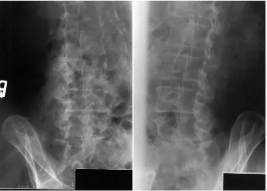
Radiographic examination of the lumbar spine revealed extensive calcification throughout the aorta and an aneu-rysm about its bifurcation. The patient was referred to his medical doctor with these findings and a recommendation for diagnostic ultrasound study and referral to a cardiovas-cular surgeon. He underwent vascardiovas-cular surgery two months after his initial presentation.
At follow-up two weeks post surgery, the low back pain had reduced to a mild discomfort and the leg pain had subsided.
Etiology and epidemiology
The pathogenesis of AAA has been frequently associated with atherosclerosis.1,3 However, Ernst6 feels that this view is restricted, as atherosclerosis may only represent a secondary response to pre-existing vessel wall injury. Others have attributed AAA to arteriosclerosis.2,9 Arterio-sclerosis is a group of diseases characterized by thickening and loss of elasticity of the arterial walls. Atherosclerosis is a form of arteriosclerosis in which atheromas containing cholesterol, lipoid material, and lipophages are formed within the intima and inner media of a large and medium sized artery.10 Hypertension is considered to be a predis-posing factor to AAA.4 Infection (syphilitic and mycotic), inflammation, trauma, auto-immune disease, cystic me-dial necrosis, Marfan’s syndrome, genetic predisposition and hemodynamic mechanical factors have also been im-plicated in the pathogenesis of AAA.3,6,11,12
Abdominal aortic aneurysms are frequently found in healthy older adults who are radiographed for back pain.1 AAA’s are most often found between the renal arteries and the iliac bifurcation.1,3 The most common location of AAA’s is at the bifurcation of the aorta into the common iliac arteries.3 Occasionally, the ascending arch and descending thoracic aorta are affected.4 AAA commonly occurs in adults 60 years of age or older, and has been reported to be four times more frequent in males.1 Some authors suggest that AAA may occur in individuals as early as 50 years of age.13
Clinical presentation
C Hadida, M Rajwani
might be suspected.14 The patient may report abdominal, lumbar spine, costovertebral angle, and suprapubic or leg pain.15 Clinical signs of potential rupture include pain, a pulsatile mass in the abdomen, and hypotension.16 This presentation may be considered a surgical emergency. Adjacent structures may be affected by aneurysms result-ing in their respective symptomatology includresult-ing: com-pression of a ureter, erosion of the anterior vertebral body, occlusion of blood supply to the spinal cord, rupture into the peritoneal cavity, and emboli.4,9
Observation may reveal a pulsating abdominal mass.3 Due to the proposed association between atherosclerosis and hypertension, assessing a patient’s blood pressure should be performed routinely in high-risk patients. Ap-proximately 90% of AAA may be detected through pal-pation of pulsations felt in the horizontal and sagittal planes.17 However, the ability to palpate an aneurysm de-pends on the size of the lesion, as well as the thickness of the abdominal wall and the skill of the examiner.2 A bruit or thrill may be detected on auscultation.18 Frame et al.8 evaluated the cost effectiveness of screening by physical examination versus abdominal ultrasound (US) for AAA in men 60–80 years of age. They concluded that a one-time screen via abdominal palpation in this population may be cost effective but of small benefit. A one-time screen via US was considered cost effective with moderate benefit. In either case, repeat screening was not cost effective. Wolf et al.19 found that ultrasonographic screening of individu-als undergoing lower extremity arterial evaluation was cost-effective for a high-risk population.
Natural history
According to Ernst,6 the true natural history of AAA can only be determined by prospective randomized analyses comparing treated versus untreated aneurysms, and these do not exist. As a result, available data on natural history is derived from observations relating aneurysmal size and rate of expansion to the risk of rupture. According to Imperato,17 aneurysms measuring greater than 5 cm have a tendency to continue growing and to rupture. At 6 cm there is a 25% chance of death due to rupture in one year and more than 50% chance of rupture in 5 years. At measures greater than 6 cm the chance of death due to rupture is 50% within the first year. The risk of rupture within 2 years is 75%, and within 5 years is 90%.17 Clinically, the most common catastrophe resulting from AAA is acute aortic
dissection.14 A dissecting abdominal aortic aneurysm usu-ally presents with severe pain and associated symptoms such as syncope, hemiplegia and paraplegia.4,20 Aortic dissection occurs 50% more often than rupture of aortic aneurysms.15
Radiographic findings
Stites and Canterbury1 caution chiropractors against the reliance on plain film findings in assessing abdominal aor-tic aneurysms. The finding of an elongated and tortuous abdominal aorta in the same population (adults over 60 years of age) may cause clinicians to incorrectly assume an aortic abdominal aneurysm is present due to the dilated nature of these tortuous aortas. An even more grave conse-quence of relying on plain film findings follows the fact that a vessel wall is poorly visualized with such studies, making measurements subject to error. Plain film radio-graphs may reveal the calcified rim of an aortic aneurysm, however, aneurysms may be present and not detected radiographically if the walls of the abdominal aorta are not extensively calcified.2 Generally, radiographic signs of an abdominal aortic aneurysm include a thin curvilinear rim of continuous or discontinuous calcification. There may be erosion of a vertebral body or bodies on a lateral view.16 The anteroposterior view may demonstrate a soft tissue mass with calcification off to the left side of the spine. When it is possible to visualize the vessel wall on plain films, any measurement exceeding 3.8 cm warrants further investigation using diagnostic ultrasound (US) or com-puted tomography (CT).1,16 These imaging modalities are considered to yield the most reliable information regarding the presence and size of an aneurysm.14,17
Treatment
While surgical intervention has been reported to be an effective treatment, as many as 62% of patients with rup-tured aneurysms die prior to reaching a hospital.6,21 Ab-dominal aneurysms are usually repaired surgically via prosthetic grafts.1 This procedure is considered successful with few complications, including perigraft hematoma, infection and pseudoaneurysm.12 Surgical intervention following a rupture usually has a high mortality rate (80%), demonstrating the importance of preventative sur-gical intervention in candidates at risk.
with AAA. According to Thorkeldsen,5 spinal manipula-tive therapy is not the appropriate treatment for AAA. While this is rational, it does not address the question of contraindication in patients who present with concurrent mechanical low back pain. The literature sites one case report of a 66-year-old Caucasian male who presented with low back pain and was subsequently treated with a course of manipulative therapy.2 Two weeks following discharge the patient returned with persistent back pain and radiographic evaluation revealed an aneurysm of the abdominal aorta measuring 7 cm in diameter. While the paper did not discuss the applicability of SMT in patients with AAA, it demonstrated a case where spinal manipula-tion did not result in any harm to the patient. Conversely,
Vernon et al.3 discuss two cases of AAA presenting to a chiropractor’s office, a 59-year-old male and a 63-year-old female. In both cases, information was missing regarding the type of care given, including the utilization of SMT prior to the discovery of the aneurysms. According to Weston,22 with large and symptomatic AAA, SMT is ab-solutely contraindicated, and may be performed on pa-tients with AAA under 5 cm in diameter. In these papa-tients, modified manipulative techniques with minimized tor-sional stress on the lumbar spine should be utilized.
While some authors2 clearly state that AAA is an abso-lute contraindication to SMT, these statements are often not supported by a rationale or scientific evidence. Weston22 does rationalize that manipulation may cause
C Hadida, M Rajwani
harm by producing a rapid stretch to the weakened ab-dominal aorta. Since fewer than half of all cases of AAA present with symptoms,2,14 it is likely that many patients with AAA are being treated with SMT for mechanical low back pain. Furthermore, the chiropractic literature does not cite cases where SMT was a direct cause of AAA rupture.
At this time, a clinical trial involving SMT in patients with AAA may not be considered safe or ethical, due to the general view that SMT is contraindicated. However, in-creased contribution to the chiropractic literature, of cases where lumbar SMT was performed on patients later found to have AAA, would provide enough data to warrant a retrospective study. Such studies should explore the appli-cability of SMT in patients with AAA, the cause of back pain (if any), gender, age at time of presentation, size of AAA at time of discovery, type, frequency and duration of treatment provided, response to treatment, and any con-founding factors such as atherosclerosis, hypertension, or underlying cardiovascular history. This type of research may provide insight into the level of contraindication (i.e. as it may relate to the size of the aneurysm).
Conclusion
The abundance of “silent” abdominal aortic aneurysms presented in the literature emphasizes the importance of considering ominous causes of back pain when forming a differential diagnosis. Most authors discuss the impor-tance of a thorough history and physical examination, ap-propriate knowledge of differential diagnoses, and early diagnosis and referral in patients suspected of having AAA. While a general view exists that SMT is contraindi-cated in AAA, the literature does not confirm complica-tions in patients with AAA who have undergone SMT. Chiropractors are urged to document and submit case stud-ies where patients with AAA have been treated with SMT, and any noted complications.
References
1 Stites J, Canterbury R. Aneurysm of the abdominal aorta. ACA J Chiropractic. Nov 1989; 65–67.
2 Harger BL. Abdominal aortic aneurysm: a case report. ACA J Chiropractic. Dec 1993; 69–71.
3 Vernon LF, Peacock JR, Esposito AP. Abdominal aortic aneurysms presenting as low back pain: a report of two cases. JMPT 1986; 9(1):47–50.
4 Boline PD, Anderson AV. Abdominal aortic aneurysm. AJCM 1989; 2(3):114–116.
5 Thorkeldsen A. Abdominal aortic aneurysm: a case report. European J Chiropractic 1993; 41:95–100.
6 Desforges JF. Abdominal aortic aneurysms. New England J Med 1993; 328(16):1167–1172.
7 Oureil K, Green RM, Donaayre C, Shortell CK, Elliot J, DeWeese JA. An evaluation of new methods of expressing aortic aneurysm size: Relationship to rupture. J Vasc Surg 1992; 15(1):12–25.
8 Frame PS, Fryback DG, Patterson C. Screening for abdominal aortic aneurysm in men ages 60–80 years. A cost-effectiveness analysis. Annals Internal Medicine 1993; 119(5):411–416.
9 Robbins SL, Cotran RS, Kuna VK. Pathological basis of bone disease, 3rd edition: WB Saunders Co 1984; 529. 10 Dorland’s Pocket Medical Dictionary, 24th edition. WB
Saunders Co. Philadelphia, 1982; p 46 and 67.
11. LaRoy LL, Cormier PJ, Matalon TAS, Patel SK, Turner DA, Silver B. Imaging of abdominal aortic aneurysms. American J Roentgenology 1989; 152:785–792.
12 Levine E. The retroperitoneum. In: Putmen CE, Ravin CE, eds. Textbook of diagnostic imaging. WB Saunders, Philadelphia 1988;1309–1310.
13 Muluk SC, Gertler JP, Brewster DC, Cambria RP, LaMuraglia GM, et al. Presentation and patterns of aortic aneurysms in young patients. J Vasc Surg. 1994; 20:808–8. 14 Erskin JM. Current medical diagnosis and treatment.
Lange Medical Books, 1981; 258.
15 Miller CD. When to suspect aortic dissection: what treatment? Cardiovascular Med 1984; 10:811–818. 16 Yochum TR, Rowe LJ. Essentials of skeletal radiology.
Vol 2, 1st edition. Williams and Wilkins, Baltimore 1987; 1020–1023.
17 Imperato AM. Aneurysm of the abdominal aorta. Hospital Medicine. Aug 1988; 41–64.
18 Krupp MA, Schroeder SA, Tierney IM. Current medical diagnosis and treatment. Lange Medical Books, 1987; 255. 19 Wolf YG, Otis SM, Schwend RB, Bernstein EF. Screening
for abdominal aortic aneurysms during lower extremity arterial evaluation in the vascular laboratory. J Vasc Surg. 1995; 22:417–423.
20 Bates B. A guide to physical examination, 3rd edition. JJB Lippincott Co., Philadelphia, 1983; 66–76.
21 Ingoldby CJH, Wujanto R, Mitchell JE. Impact of vascular surgery on community mortality from ruptured aortic aneurysms. Br J Surg 1986; 73:551–553.