Open Access
Research article
Hepatocyte and keratinocyte growth factors and their receptors in
human lung emphysema
Marcel Bonay*
†1,2, Anne Boutten
†1,3, Véronique Leçon-Malas
3,
Joëlle Marchal
1, Paul Soler
1, Michel Fournier
5, Guy Leseche
6,
Monique Dehoux
1,3and Bruno Crestani
1,4Address: 1INSERM U 700, Faculté Xavier Bichat, Paris, France, 2Service de Physiologie-Explorations Fonctionnelles, Hôpital Bichat-Claude Bernard,
Assistance Publique-Hôpitaux de Paris, Université Paris 7, Paris, France, 3Service de Biochimie A, Hôpital Bichat-Claude Bernard, Assistance
Publique-Hôpitaux de Paris, Université Paris 7, Paris, France, 4Service de Pneumologie Hôpital Bichat-Claude Bernard, Assistance
Publique-Hôpitaux de Paris, Université Paris 7, Paris, France, 5Service de Pneumologie, Hôpital Beaujon, Assistance Publique-Hôpitaux de Paris, Université
Paris 7, Paris, France and 6Service de Chirurgie Thoracique, Hôpital Beaujon, Assistance Publique-Hôpitaux de Paris, Université Paris 7, Paris,
France
Email: Marcel Bonay* - marcel.bonay@bch.aphp.fr; Anne Boutten - anne.boutten@bch.aphp.fr; Véronique Leçon-Malas - anne.boutten@bch.aphp.fr; Joëlle Marchal - marcel.bonay@bch.aphp.fr; Paul Soler - anne.boutten@bch.aphp.fr;
Michel Fournier - bruno.crestani@bch.aphp.fr; Guy Leseche - marcel.bonay@bch.aphp.fr; Monique Dehoux - monique.dehoux@bch.aphp.fr; Bruno Crestani - bruno.crestani@bch.aphp.fr
* Corresponding author †Equal contributors
Abstract
Background: Hepatocyte and keratinocyte growth factors are key growth factors in the process of alveolar repair. We hypothesized that excessive alveolar destruction observed in lung emphysema involves impaired expression of hepatocyte and keratinocyte growth factors or their respective receptors, c-met and keratinocyte growth factor receptor. The aim of our study was to compare the expression of hepatocyte and keratinocyte growth factors and their receptors in lung samples from 3 groups of patients: emphysema; smokers without emphysema and non-smokers without emphysema.
Methods: Hepatocyte and keratinocyte growth factor proteins were analysed by immunoassay and western blot; mRNA expression was measured by real time quantitative polymerase chain reaction.
Results: Hepatocyte and keratinocyte growth factors, c-met and keratinocyte growth factor receptor mRNA levels were similar in emphysema and non-emphysema patients. Hepatocyte growth factor mRNA correlated negatively with FEV1 and the FEV1/FVC ratio both in emphysema patients and in smokers with or without emphysema. Hepatocyte and keratinocyte growth factor protein concentrations were similar in all patients' groups.
Conclusion: The expression of hepatocyte and keratinocyte growth factors and their receptors is preserved in patients with lung emphysema as compared to patients without emphysema. Hepatocyte growth factor mRNA correlates with the severity of airflow obstruction in smokers. Published: 10 October 2005
BMC Pulmonary Medicine 2005, 5:13 doi:10.1186/1471-2466-5-13
Received: 06 July 2005 Accepted: 10 October 2005 This article is available from: http://www.biomedcentral.com/1471-2466/5/13
© 2005 Bonay et al; licensee BioMed Central Ltd.
Background
Emphysema is one of the most important cause of respi-ratory insufficiency with increasing mortality and morbid-ity [1]. Lung emphysema is defined pathologically by the destruction of alveolar walls with abnormal permanent enlargement of the air spaces distal to the terminal bron-chiole. Precise mechanims contributing to the destruction of alveolar walls remain unclear. Cigarette smoking may induce emphysema by stimulating neutrophils and alveo-lar macrophages to produce proteases leading to the deg-radation of the alveolar extracellular matrix and oxidant injury contributing to alveolar destruction [2]. T lym-phocytes contribute to the recruitment and activation of these inflammatory cells and may be involved in the apoptosis and destruction of alveolar epithelium [3-5]. Recent evidence of increased apoptosis of alveolar epithe-lial and endotheepithe-lial cells in emphysematous lung suggests that primary alterations of the alveolar epithelium and endothelium might participate in the pathogenesis of the disease [6-8].
The alveolar epithelium is essential for maintenance of the integrity of the alveolar spaces. Functional restoration of the alveolar epithelium after an injury requires the pro-liferation and migration of type 2 pneumocytes and their differentiation into type 1 pneumocytes, a tightly regu-lated phenomenon. Growth factors have been shown to control both phases of the process [9-12]. Among these factors, the hepatocyte growth factor (HGF), a het-erodimeric protein obtained through the cleavage of an inactive precursor, called 90-kD proHGF, and the kerati-nocyte growth factor (KGF, also named fibroblast growth factor-7, FGF-7) have been identified as key factors in the process of alveolar repair, both in acute or chronic condi-tions [13-15]. HGF and KGF respectively act through spe-cific receptors c-met, a membrane bound tyrosine kinase [16] and FGFR2IIIb, also named KGF-R [17]. HGF and KGF productions by lung fibroblasts from emphysema have been shown to be reduced when compared with con-trols [18]. Recently, Shigemura et al reported that decreased HGF expression due to a failure in sustained endogenous production after injury was associated with
emphysema-related histopathologic and physiological changes in a rat model of elastase-induced emphysema [19]. In this animal model, HGF could have a therapeutic effect [20,19].
We hypothesized that a defective expression of growth fac-tors involved in human alveolar epithelium repair such as HGF and KGF or their specific receptors might participate in the pathophysiology of lung emphysema. We therefore evaluated the expression of HGF, KGF, and their receptors c-met and KGF-R in lung biopsies from patients with emphysema and from non-emphysema patients, accord-ing to their smokaccord-ing status.
Methods
This study was approved by the ethics committee of Saint Germain-en-Laye hospital.
Patients
Lung samples were obtained during surgery in adult patients (>18 years) from Beaujon university hospital (Clichy, France) and from Bichat university hospital (Paris, France).
Patients with lung emphysema
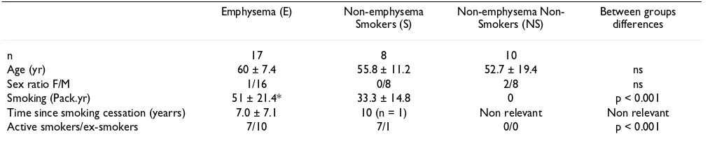
Seventeen patients with radiographically defined emphy-sema (E group) were included. All patients were active or ex-smokers (Table 1). CT-scan, pulmonary function tests and α1-AT deficiency were systematically documented. Patients with α1-AT deficiency were excluded. They underwent bullectomy (n = 3), lobectomy (n = 4), lung transplantation (n = 3) or lung volume reduction (n = 7). Pulmonary function tests demonstrated mild to severe air-flow obstruction and lung distension (Table 2). Tissue samples were taken from the resected parenchyma in a macroscopically emphysematous region. Nine patients with emphysema were receiving corticosteroids, either oral (n = 2) and/or inhaled (n = 8). In all patients, lung emphysema was suspected on CT-scan and confirmed by the pathological examination of lung resection samples. The severity of emphysema was approached through pul-monary function abnormalities.
Table 1: Clinical characteristics of patients with and without emphysema
Emphysema (E) Non-emphysema Smokers (S)
emphysema Non-Smokers (NS)
Between groups differences
n 17 8 10
Age (yr) 60 ± 7.4 55.8 ± 11.2 52.7 ± 19.4 ns
Sex ratio F/M 1/16 0/8 2/8 ns
Smoking (Pack.yr) 51 ± 21.4* 33.3 ± 14.8 0 p < 0.001
Time since smoking cessation (yearrs) 7.0 ± 7.1 10 (n = 1) Non relevant Non relevant
Active smokers/ex-smokers 7/10 7/1 0/0 p < 0.001
Non-emphysema patients
Normal tissue was obtained from 18 non-emphysema patients. Eight patients were smokers (non-emphysema smoker, S) and 10 were non-smokers (non-emphysema non-smoker, NS). S patients were undergoing surgery for the resection of a localised primary lung carcinoma (n = 7) or a benign lesion (n = 1). NS patients were undergoing surgery for the resection of a localised primary lung carci-noma (n = 3), lung metastases (n = 2) or a benign lesion (n = 5). Tissue samples were taken at a site distant from the pathological process and without macroscopical and microscopical evidence of emphysema. S patients had mild to moderate alterations of pulmonary functions tests (table 2). Increased cumulative tobacco exposure was observed in the E group as compared with the S group (Table 1). No difference was observed between groups for age and sex ratio (Table 1). Two patients without emphy-sema (S group) received inhaled corticosteroids.
Processing of lung samples
Lung tissue fragments (about 0.2 cm3) were immediately frozen in liquid N2 and stored at -80°C until RNA and protein analysis. The histopathology of biopsies was eval-uated on paraffin-embedded sections to verify the features of emphysema or normal lungs. The concentration of pro-teins in biopsies was evaluated from 100 mg of lung samples homogenised with 0.5 ml PBS containing 200 µM phenylmethylsulfonyl fluoride, 1 mg/ml leupeptin and 1 mg/ml aprotinin. The homogenates were centri-fuged at 10,000 g for 10 min at 4°C to remove tissue fragments and the supernatants were collected and stored at -80°C until measurement.
Quantitative analysis of mRNA expression
Total RNA was extracted from frozen lung tissue and reverse transcribed. Each sample was analysed by reverse transcriptase-real-time polymerase chain reaction (RT-PCR) with specific primers (table 3) to quantify the
expression of mRNA of HGF, KGF, c-met and KGF-R as described previously [21].
HGF and KGF concentration in lung homogenates The proteins HGF and KGF (Quantikine®, R&D Systems; respective detection limits were 40 and 15 pg/ml for HGF, KGF) were measured in lung homogenates from 26 out of 35 patients when enough lung sample was available (13 E, 5 S, 8 NS).
HGF western blotting
Lung homogenates from 4 patients (2 patients with emphysema and 2 patients without emphysema) were examined by Western blotting as previously described [22].
Statistical analysis
Data were analysed by Statview software (Abacus Con-cepts, Inc.) and displayed as mean ± SD. Between-group differences were first assessed by non-parametric analysis of variance (Kruskal-Wallis test). In the case of global sig-nificant difference, between two groups comparisons were assessed by the non-parametric Mann-Whitney U-test. Correlations were assessed by the Spearman's rank order test. Categorical data were analysed using the Chi-squared test. A p value < 0.05 was regarded as significant.
Results
Lung expression of KGF and KGF receptor
KGF mRNA (figure 1A) and KGF protein (figure 1C) were detected in the lungs of all patients. No difference was observed according to the presence of emphysema, and the smoking status.
KGF-R mRNA (figure 1B) was detected in the lungs of all patients. A considerable variability was observed in KGF-R transcript levels in patients with or without emphysema. No difference was observed according to the presence of emphysema and the smoking status.
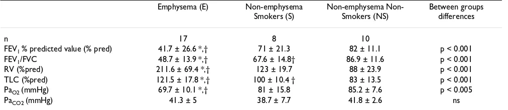
Table 2: Pulmonary function tests of patients with and without emphysema.
Emphysema (E) Non-emphysema Smokers (S)
emphysema Non-Smokers (NS)
Between groups differences
n 17 8 10
FEV1 % predicted value (% pred) 41.7 ± 26.6 *,† 71 ± 21.3 82 ± 11.1 p < 0.001
FEV1/FVC 48.7 ± 13.9 *,† 67.6 ± 14.8† 86.9 ± 11.6 p < 0.001
RV (%pred) 211.6 ± 69.4 *,† 123 ± 19.7 88 ± 23.9 p < 0.001
TLC (%pred) 121.5 ± 17.8 *,† 100 ± 10.4 † 83 ± 13.5 p < 0.001
PaO2 (mmHg) 69.7 ± 10.1 *,† 81 ± 15.8 85.2 ± 7.6 p < 0.005
PaCO2 (mmHg) 41.3 ± 5 38.7 ± 7.7 41.8 ± 2.6 ns
*: vs S, p < 0.01; †: vs NS, p < 0.01; as assessed by Mann-Whitney U-test. ns: non significant
KGF mRNA, KGF protein and KGF-R mRNA did not cor-relate with age, cumulative tobacco exposure, period since smoking cessation, use of inhaled corticosteroids, pulmo-nary function tests and arterial blood gases.
Lung expression of HGF and HGF receptor
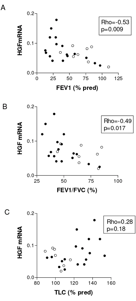
Although a considerable variability of HGF mRNA expres-sion was observed, no difference was found between emphysema and non-emphysema groups (figure 2A). In emphysema patients, HGF mRNA correlated positively with total lung capacity (TLC) (Rho = 0.51, p = 0.04, n = 17) and negatively with forced expiratory volume in one second (FEV1) (Rho = -0.53, p = 0.03, n = 17) and FEV1/ FVC (forced vital capacity) (Rho = -0.54, p = 0.03, n = 17). When all smokers were studied together (smokers with or without emphysema), again, significant correlations between HGF mRNA and FEV1 (Rho = -0.53, p = 0.009, n = 25), and FEV1/FVC (Rho = -0.49, p = 0.017, n = 25) were found. However, the correlation between HGF mRNA and TLC was no more significant (Rho = 0.28, p = 0.18, n = 24) (figure 3). There was no correlation between HGF mRNA and cumulative tobacco exposure.
HGF protein was detected in lung homogenates from all patients as assessed by immunoassay. HGF concentration was not different between groups (figure 2C). HGF pro-tein correlated positively with residual volume (RV) (Rho = 0.43, p = 0.04, n = 24), TLC (Rho = 0.45, p = 0.03, n = 24) and negatively with FEV1/FVC (Rho = -0.45, p = 0.03, n = 24). Although, no difference of HGF protein concen-trations was found between patients' groups, a significant correlation between HGF protein concentrations and cumulative tobacco exposure was observed (Rho = 0.49, p = 0.01, n = 26). As HGF immunoassay measured both proHGF and mature HGF, a western blot analysis was performed to characterize which form of HGF was present in lung homogenates. Western blot (figure 4) demon-strated that HGF was present mainly in the cleaved mature form (presence of the 69-kD alpha chain) both in the non-emphysematous and the emphysematous lungs.
HGF-R mRNA was detected in all patients. We found no difference between groups (figure 2B). Strong correlations were observed between HGF-R mRNA and KGF-R mRNA (Rho = 0.82, p < 0.0001, n = 35) and between HGF mRNA and KGF mRNA (Rho = 0.61, p = 0.004, n = 35) when all patients were taken together.
Discussion
The involvement of KGF and HGF in lung repair has been widely documented. Numerous studies in vitro and in vivo have demonstrated that KGF and HGF have protec-tive effects in experimental lung injury [15,23]. To our knowledge, this is the first study of KGF and HGF lung expression in human emphysema. Proteases, oxidant injury [2], chronic inflammation [3,5] and apoptosis [6-8] all contribute to the excessive alveolar wall destruction observed in lung emphysema. We hypothezised that a defect of the lung repair process might be associated with the pathophysiology of lung emphysema. Our results show that lung KGF mRNA and KGF protein are not altered in emphysema. In bleomycin-induced lung injury in rats, KGF and HGF increase in the lung [24]. A few clin-ical studies have assessed KGF concentrations in acute lung injury. Verghese et al reported that KGF was not increased in lung edema fluid whereas HGF was increased and associated with higher mortality [25]. Stern et al reported that KGF and HGF were increased in bronchoal-veolar lavage fluid in acute respiratory distress syndrome and associated with a poor prognosis [13]. Recently, Danan et al observed the highest KGF concentrations in tracheal aspirates from premature infants who survived without bronchopulmonary dysplasia, leading to the con-clusion that KGF may prevent injury to lung epithelium and enhanceits repair [26].
This study has some methodological limitations. A lim-ited number of patients was studied in each group, espe-cially in non emphysema groups which were mostly composed of lung biopsies obtained at a site distant from localised carcinoma. Furthermore, the patients could only Table 3: Primers and PCR cycling conditions.
Primers Sequences Denaturation annealing Cycles PCR products
HGF: Forward Reverse
5'-CAGAGGGACAAAGGAAAAGAA-3' 5'-GCAAGTGAATGGAAGTCCTTTA-3'
94°C, 15s 58°C, 60s 40 167 bp
KGF: Forward Reverse
5'-GAACAAGGAAGGAAAACTCTATGCAA-3' 5'-AAGTGGGCTGTTTTTTGTTCTTTCT-3'
94°C, 15s 60°C, 60s 40 201 bp
HGF-R: Forward Reverse
5'-GTTTACTTGTTGCAAGGGAGAAGACT-3' 5'-TAGGGTGCCAGCATTTTAGCA-3'
94°C, 15s 58°C, 60s 40 88 bp
KGF-R: Forward Reverse
5'-TTAAGCAGGAGCATCGCATTG-3' 5'-AACATCCAGGTGGTACGTGTGAT-3'
94°C, 15s 60°C, 60s 40 151 bp
Ubiquitin-c: Forward Reverse
5'-CACTTGGTCCTGCGCTTGA-3' 5'-TTTTTTGGGAATGCAACAACTTT-3'
Expression of KGF (A) and KGF-R (B) mRNA in lung tissue from emphysema and non-emphysema patients
Figure 1
Expression of KGF (A) and KGF-R (B) mRNA in lung tissue from emphysema and non-emphysema patients. Results are expressed as a ratio to Ubiquitin in arbitrary units. Individual and mean values (bar) are pre-sented. No difference between groups was found. E: emphy-sema patients; S and NS: non-emphyemphy-sematous smoker and non-smoker patients. KGF concentrations in lung homoge-nates were measured by immunoassay (C). The detection limit was 15 pg/ml. The results are displayed per 1 mg of lung tissue.
KGF mRNA
E S NS
0.0 0.1 0.2 0.3 1.1 1.2
KGF/Ubiquitin
ratio
KGF-R mRNA
E S NS
0 1 2
KG
F-R/
Ubi
q
u
iti
n
ra
ti
o
KGF protein
E S NS
0 500 1000 1500
KG
F
(pg/ml)
A
B
C
Expression of HGF (A) and c-met (B) mRNA in lung tissue from emphysema and non-emphysema patients
Figure 2
Expression of HGF (A) and c-met (B) mRNA in lung tissue from emphysema and non-emphysema patients. Results are expressed as a ratio to Ubiquitin in arbitrary units. Individual and mean values (bar) are pre-sented. No difference between groups was found. E: emphy-sema patients; S and NS: non-emphyemphy-sematous smoker and non-smoker patients. HGF concentrations in lung homoge-nates were measured by immunoassay (C). The detection limit was 40 pg/ml. The results are displayed per 1 mg of lung tissue.
HGF mRNA
E S NS
0.0 0.1 0.2
H
G
F/U
bi
quit
in
rat
io
HGF-R mRNA
E S NS
0 1 2 3 4 5
HGF-R/Ubiquitin
ra
tio
HGF protein
E S NS
0 10000 20000 30000 40000
HGF
(pg/ml)
A
B
be evaluated at one time point in the course of their dis-ease. Inclusion of smokers without emphysema allowed the differentiation of emphysema-related and tobacco-related events. Because only one tissue sample from surgically resected material was available for examination, the expression of HGF, KGF and their receptors reflects regional disease activity and may be unrepresentative of the entire lung. Indeed, it is well known that emphysema affects different lung regions to a varying extent. Moreo-ver, we evaluated HGF and KGF in lung homogenates only. Future studies should address the expression of HGF and KGF in a more cell-specific fashion.
In our study, although HGF mRNA lung expression was similar in emphysema and non emphysema patients, a correlation was found between HGF mRNA and the deterioration of pulmonary function tests in emphysema patients. The correlation between airflow obstruction and HGF mRNA level was similarly observed when all smok-ers with or without emphysema were studied, suggesting that emphysema was not a main determinant of HGF mRNA level in the lung. This strong correlation between airflow obstruction and HGF mRNA in smokers suggests that the increase of HGF mRNA was not related to the presence of emphysema but rather to the degree of airflow obstruction. This observation is supported by the correla-tion between HGF protein in lung homogenates and the FEV1/FVC ratio in our population. These results are in agreement with the observations of Sauleda et al, who reported that HGF protein concentrations were increased in broncho-alveolar lavage of patients with chronic obstructive pulmonary disease as compared to smokers and non-smoker controls [27]. Interestingly, the increased
Correlation between HGF mRNA and pulmonary function in smoker patients
Figure 3
Correlation between HGF mRNA and pulmonary function in smoker patients. The ratio of lung HGF to Ubiquitin mRNA was correlated with: (A) forced expiratory volume in one second (FEV1 % predicted), (B) FEV1/FVC (forced vital capacity), but not with (C) total lung capacity (TLC % predicted). Full circle (•): emphysema patients; open circle (o): smoker patients without emphysema.
0 25 50 75 100 125
0.0 0.1 0.2
Rho=-0.53 p=0.009
FEV1 (% pred)
HG
F
m
R
NA
25 50 75 100
0.0 0.1 0.2
Rho=-0.49 p=0.017
FEV1/FVC (%)
H
G
F
m
RNA
80 100 120 140 160 0.0
0.1 0.2
Rho=0.28 p=0.18
TLC (% pred)
H
G
F
m
RNA
A
B
C
Western blot analysis of HGF in lung tissue
Figure 4
lung expression of other growth factors (fibroblast growth factors 1 and 2 and their receptors) has already been reported in chronic obstructive pulmonary disease [28].
The mechanisms underlying the correlation between air-flow obstruction and HGF mRNA in smokers are unclear. Although speculative, we can propose that the mechanical constraints applied to alveolar tissue secondary to airflow obstruction may stimulate HGF production by alveolar epithelial cells, since Yamamoto et al showed that mechanical stretch induced HGF in alveolar type II cells in vitro [29]. Furthermore, airway inflammation could con-tribute to increase local HGF expression by neutrophils [22] and macrophages [30]. Interestingly, Aharinejad et al have shown that serum HGF concentrations increased at the time of lung graft rejection, a situation associated with airflow obstruction [31].
As HGF and KGF are key factors in the process of alveolar repair [15], we suggest that their production might be not adapted to the degree of alveolar injury. Indeed, in view of the chronic lung injury observed in emphysema, one could expect an increase of HGF and KGF expression as observed in acute lung injury in rats [24] and in humans [25,13]. However the direct assessment of HGF content in lung homogenates is technically difficult. Indeed, HGF is a heparin binding growth factor. High concentrations of inactive precursor of HGF (proHGF) are probably bound to proteoglycans of the extracellular matrix and may not be assayed in the lung homogenates by immunoassay. In this study, western blot analysis showed that HGF was only in the mature active form, both in lung biopsies from emphysema and non-emphysema patients.
Recently, HGF has been shown to stimulate pulmonary regeneration and to improve pulmonary function in ani-mal models of elastase-induced lung emphysema [20,19]. Preserved expression of HGF-R could allow therapeutic use of growth factors in lung emphysema. Further studies are needed to assess the therapeutic potential of HGF and KGF in lung emphysema.
Conclusion
The main results of our study are that: i) KGF and HGF lung expression is preserved in emphysema patients, ii) HGF-R and KGF-R mRNA are consistently expressed in the lung of emphysema patients and are not modified by the smoking status, iii) HGF mRNA correlates with the sever-ity of airflow obstruction in smokers.
List of abbreviations
α1-AT: α1-antitrypsinE: emphysema
FEV1: forced expiratory volume in one second
FGF: fibroblast growth factor
FVC: forced vital capacity
HGF: hepatocyte growth factor
KGF: keratinocyte growth factor
mRNA: messenger ribonucleic acid
NS: non-smoker without emphysema
RT-PCR: reverse transcriptase-real-time polymerase chain reaction
RV: residual volume
S: smoker without emphysema
SD: standard deviation
TLC: total lung capacity
Competing interests
The author(s) declare that they have no competing interests.
Authors' contributions
MB and AB equally participated in the design of the study, conducted the majority of the research experiments and drafted the manuscript.
VL participated in the majority of the research experiments.
MF participated in the design of the study.
JM and PS conducted some experiments.
GL participated in the design of the study.
MD and BC conceived of the study, participated in its design, and in drafting the manuscript.
All authors read and approved the final version of the manuscript.
Acknowledgements
Part of this work was supported by the legs Poix, Chancellerie des Univer-sités de Paris.
Publish with BioMed Central and every scientist can read your work free of charge
"BioMed Central will be the most significant development for disseminating the results of biomedical researc h in our lifetime."
Sir Paul Nurse, Cancer Research UK
Your research papers will be:
available free of charge to the entire biomedical community
peer reviewed and published immediately upon acceptance
cited in PubMed and archived on PubMed Central
yours — you keep the copyright
Submit your manuscript here:
http://www.biomedcentral.com/info/publishing_adv.asp
BioMedcentral
References
1. Barnes PJ: Chronic obstructive pulmonary disease. N Engl J Med 2000, 343:269-280.
2. Shapiro SD: The macrophage in chronic obstructive pulmo-nary disease. Am J Respir Crit Care Med 1999, 160:S29-32. 3. Finkelstein R, Fraser RS, Ghezzo H, Cosio MG: Alveolar
inflamma-tion and its relainflamma-tion to emphysema in smokers. Am J Respir Crit Care Med 1995, 152:1666-1672.
4. Retamales I, Elliott WM, Meshi B, Coxson HO, Pare PD, Sciurba FC, Rogers RM, Hayashi S, Hogg JC: Amplification of inflammation in emphysema and its association with latent adenoviral infection. Am J Respir Crit Care Med 2001, 164:469-473.
5. Turato G, Zuin R, Miniati M, Baraldo S, Rea F, Beghe B, Monti S, For-michi B, Boschetto P, Harari S, Papi A, Maestrelli P, Fabbri LM, Saetta M: Airway inflammation in severe chronic obstructive pul-monary disease: relationship with lung function and radio-logic emphysema. Am J Respir Crit Care Med 2002, 166:105-110. 6. Kasahara Y, Tuder RM, Cool CD, Lynch DA, Flores SC, Voelkel NF:
Endothelial cell death and decreased expression of vascular endothelial growth factor and vascular endothelial growth factor receptor 2 in emphysema. Am J Respir Crit Care Med 2001,
163:737-744.
7. Kasahara Y, Tuder RM, Taraseviciene-Stewart L, Le Cras TD, Abman S, Hirth PK, Waltenberger J, Voelkel NF: Inhibition of VEGF receptors causes lung cell apoptosis and emphysema. J Clin Invest 2000, 106:1311-1319.
8. Aoshiba K, Yokohori N, Nagai A: Alveolar wall apoptosis causes lung destruction and emphysematous changes. Am J Respir Cell Mol Biol 2003, 28:555-562.
9. Kim HJ, Sammak PJ, Ingbar DH: Hepatocyte growth factor stim-ulates migration of type II alveolar epithelial cells on the pro-visional matrix proteins fibronectin and fibrinogen. Chest
1999, 116:94S-95S.
10. Mason RJ, Leslie CC, McCormick-Shannon K, Deterding RR, Naka-mura T, Rubin JS, Shannon JM: Hepatocyte growth factor is a growth factor for rat alveolar type II cells. Am J Respir Cell Mol Biol 1994, 11:561-567.
11. Ohmichi H, Matsumoto K, Nakamura T: In vivo mitogenic action of HGF on lung epithelial cells: pulmotrophic role in lung regeneration. Am J Physiol 1996, 270:L1031-9.
12. Panos RJ, Patel R, Bak PM: Intratracheal administration of hepa-tocyte growth factor/scatter factor stimulates rat alveolar type II cell proliferation in vivo. Am J Respir Cell Mol Biol 1996,
15:574-581.
13. Ware LB, Matthay MA: Keratinocyte and hepatocyte growth factors in the lung: roles in lung development, inflammation, and repair. Am J Physiol Lung Cell Mol Physiol 2002, 282:L924-40. 14. Stern JB, Fierobe L, Paugam C, Rolland C, Dehoux M, Petiet A,
Dom-bret MC, Mantz J, Aubier M, Crestani B: Keratinocyte growth fac-tor and hepatocyte growth facfac-tor in bronchoalveolar lavage fluid in acute respiratory distress syndrome patients. Crit Care Med 2000, 28:2326-2333.
15. Crestani B, Dehoux M, Hayem G, Lecon V, Hochedez F, Marchal J, Jaf-fre S, Stern JB, Durand G, Valeyre D, Fournier M, Aubier M: Differ-ential role of neutrophils and alveolar macrophages in hepatocyte growth factor production in pulmonary fibrosis. Lab Invest 2002, 82:1015-1022.
16. Weidner KM, Sachs M, Birchmeier W: The Met receptor tyrosine kinase transduces motility, proliferation, and morphogenic signals of scatter factor/hepatocyte growth factor in epithe-lial cells. J Cell Biol 1993, 121:145-154.
17. Dionne CA, Crumley G, Bellot F, Kaplow JM, Searfoss G, Ruta M, Bur-gess WH, Jaye M, Schlessinger J: Cloning and expression of two distinct high-affinity receptors cross-reacting with acidic and basic fibroblast growth factors. Embo J 1990, 9:2685-2692. 18. Plantier L, Marchand-Adam S, Marchal-Somme J, Leseche G, Fournier
M, Dehoux M, Aubier M, Crestani B: Defect of hepatocyte growth factor production by fibroblasts in human pulmonary emphysema. Am J Physiol Lung Cell Mol Physiol 2005, 288:L641-7. 19. Shigemura N, Sawa Y, Mizuno S, Ono M, Ohta M, Nakamura T,
Kaneda Y, Matsuda H: Amelioration of pulmonary emphysema by in vivo gene transfection with hepatocyte growth factor in rats. Circulation 2005, 111:1407-1414.
20. Ishizawa K, Kubo H, Yamada M, Kobayashi S, Suzuki T, Mizuno S, Nakamura T, Sasaki H: Hepatocyte growth factor induces
ang-iogenesis in injured lungs through mobilizing endothelial progenitor cells. Biochem Biophys Res Commun 2004, 324:276-280. 21. Marchand-Adam S, Marchal J, Cohen M, Soler P, Gerard B, Castier Y, Leseche G, Valeyre D, Mal H, Aubier M, Dehoux M, Crestani B:
Defect of hepatocyte growth factor secretion by fibroblasts in idiopathic pulmonary fibrosis. Am J Respir Crit Care Med 2003,
168:1156-1161.
22. Grenier A, Chollet-Martin S, Crestani B, Delarche C, El Benna J, Bout-ten A, Andrieu V, Durand G, Gougerot-Pocidalo MA, Aubier M, Dehoux M: Presence of a mobilizable intracellular pool of hepatocyte growth factor in human polymorphonuclear neutrophils. Blood 2002, 99:2997-3004.
23. Ray P, Devaux Y, Stolz DB, Yarlagadda M, Watkins SC, Lu Y, Chen L, Yang XF, Ray A: Inducible expression of keratinocyte growth factor (KGF) in mice inhibits lung epithelial cell death induced by hyperoxia. Proc Natl Acad Sci U S A 2003,
100:6098-6103.
24. Adamson IY, Bakowska J: Relationship of keratinocyte growth factor and hepatocyte growth factor levels in rat lung lavage fluid to epithelial cell regeneration after bleomycin. Am J Pathol 1999, 155:949-954.
25. Verghese GM, McCormick-Shannon K, Mason RJ, Matthay MA:
Hepatocyte growth factor and keratinocyte growth factor in the pulmonary edema fluid of patients with acute lung injury. Biologic and clinical significance. Am J Respir Crit Care Med 1998,
158:386-394.
26. Danan C, Franco ML, Jarreau PH, Dassieu G, Chailley-Heu B, Bour-bon J, Delacourt C: High concentrations of keratinocyte growth factor in airways of premature infants predicted absence of bronchopulmonary dysplasia. Am J Respir Crit Care Med 2002, 165:1384-1387.
27. Sauleda J, Noguera A, Blanquer D, Fuster A, Pons AR, Pons J, Agusti A: Growth factors in chronic obstructive pulmonary disease [abstract]. Eur Respir J 2004, 24 (suppl):320s.
28. Kranenburg AR, De Boer WI, Van Krieken JH, Mooi WJ, Walters JE, Saxena PR, Sterk PJ, Sharma HS: Enhanced expression of fibrob-last growth factors and receptor FGFR-1 during vascular remodeling in chronic obstructive pulmonary disease. Am J Respir Cell Mol Biol 2002, 27:517-525.
29. Yamamoto H, Teramoto H, Uetani K, Igawa K, Shimizu E: Stretch induces a growth factor in alveolar cells via protein kinase. Respir Physiol 2001, 127:105-111.
30. Morimoto K, Amano H, Sonoda F, Baba M, Senba M, Yoshimine H, Yamamoto H, Ii T, Oishi K, Nagatake T: Alveolar macrophages that phagocytose apoptotic neutrophils produce hepatocyte growth factor during bacterial pneumonia in mice. Am J Respir Cell Mol Biol 2001, 24:608-615.
31. Aharinejad S, Taghavi S, Klepetko W, Abraham D: Prediction of lung-transplant rejection by hepatocyte growth factor. Lan-cet 2004, 363:1503-1508.
Pre-publication history
The pre-publication history for this paper can be accessed here: