R E S E A R C H
Open Access
Thoracic index in adults with asthma: a
study of validity and reliability
Yannely Serrano-villar
1*and Eliana-isabel Rodríguez-grande
2Abstract
Background:The Thoracic Index (TI) is a useful tool for evaluating costal mobility as a component of respiritory mechanics in adults with asthma. In a review of the literature, however, few studies were found that reported the psychometrics of this test. The goal of this study is to evaluate the reproducibility and validity of the TI in adults with asthma.
Methods:A cross-sectional study was conducted to evaluate the diagnostic tests. Measurements were done randomly by two independent evaluators. The variables measured included thoracic mobility (TI and
photogrammetric analysis), sociodemographic and anthropometric variables, and other variables related to the disease. TI reliability included the determination of the intra- and inter-evaluator agreement and reproducibility using the Bland and Altman limits of agreement method and the Interclass Correlation Coefficient (ICC). The convergent validity was established using Pearson’s correlation coefficient. The level of significance wasp< 0.05. Results:Twenty-six adults with stable asthma participated in this study. The limits of the intra- and inter-evaluator agreement were found to be acceptable and good, respectively, with an average of differences close to zero in both cases. The intra-evaluator reproducibility was between poor and acceptable (TI between 0.57 and 0.93), while the inter-evaluator reproducibility was between acceptable and good (TI between 0.62 and 0.86). The convergent validity between the TI and photogrammetric analysis was between moderate and high (r between 0.55 and 0.73). Conclusions:The TI is a reliable and valid measurement that can be used to evaluate costal mobility in adults with asthma. In a clinical setting, it can contribute to a nonbiased measurement, and in a research environment, it is useful for documenting the results of interventions, reducing the probability that the results will be affected by any variability in measurement.
Keywords:Asthma, Thoracic index, Reproducibility, Validity, Costal mobility, Respiratory mechanics
Background
Asthma is considered the world’s most common chronic respiratory disease. It affects 334 million people of all ages and is the 14th most important disorder in the world in terms of its extent and duration of disability [1]. It is characterized by a chronic inflammation of the airways, limiting the expiratory airflow, which produces intermittent hyperinflation and adaptations in the thor-acic cage to move the trapped air, with reduced costal mobility [2–4].
The evaluation of thoracic mobility allows us to quan-tify the functional consequences and the degree to which
asthma is controlled [3]. Photogrammetry is one of the tools available for this evaluation, providing a kinematic analysis of respiratory movements, but it requires special equipment and training, and the computerized analysis of images requires additional time per patient, increasing its cost and minimizing its clinical applicability [5–7]. Thoracic perimetry is used in a clinical context, expressed as the Thoracic Index (TI), because it is a low-cost and easy-to-apply technique. In a review of the literature, however, no studies were found that evaluated the TI’s psychometric properties in adults with stable asthma [8–11].
Having reliable and valid measuring tools in a clinical setting allows the clinician to provide an objective evalu-ation and diagnosis, and to undertake appropriate * Correspondence:[email protected]
1Universidad Industrial de Santander Facultad de Salud, Bucaramanga,
Santander, Colombia
Full list of author information is available at the end of the article
interventions to improve the respiratory mechanics compromised by asthma [2]. They are also useful in a re-search context for demonstrating the results of phy-siotherapeutic interventions, decreasing the likelihood that the effects obtained will be influenced by any vari-ability in the measurements [9].
For these reasons, it is necessary to evaluate the psy-chometric properties of the tests used in clinical prac-tice, using tests and measurements of proven validity and reproducibility as test comparators. The reliability and validity of the TI are tested against a photogrammet-ric analysis of the breathing cycle. This will allow an im-proved evaluation of the respiratory mechanics in asthma. It will also allow the control of the disease to be monitored and the provision of an evidentiary basis for intervention programs to control symptoms, prevent complications, and improve the functionality and quality of life of this population [12,13].
The objective of this study was to evaluate the intra-and inter-evaluator reliability of TI measurements, intra-and the convergent validity between the TI and photogram-metric analysis in a population of asthmatic adults.
Method
A study was conducted to evaluate the reliability and valid-ity of diagnostic tests using a cross-sectional sample [14].
Subjects
The subject group included adults with stable asthma. People with comorbidities such as the following were ex-cluded: cardiac disease, uncontrolled arterial hyperten-sion, post-operative from lung biopsy, spinal injury, ocular injury, tracheotomy, upper respiratory tract sur-gery or trauma, hemodynamic instability, pregnancy, or respiratory infections [15] with musculoskeletal or neurological sequels or diseases that compromised thor-acic mobility and muscular control [16], with a lack of voluntary force during spirometry, defined as the peak expiratory flow (PEF) or forced expiratory flow (FEF) at 25%, below the 60% normal value [17–19]. The measure-ments were taken at the Movement Analysis Laboratory at the School of Physiotherapy at the Universidad Indus-trial de Santander.
Evaluators
Two physical therapists with clinical experience of be-tween 5 and 13 years participated in this study. They standardized their verbal instructions and manual con-tact with the subjects and were trained in applying the tests to avoid classification bias.
Procedure
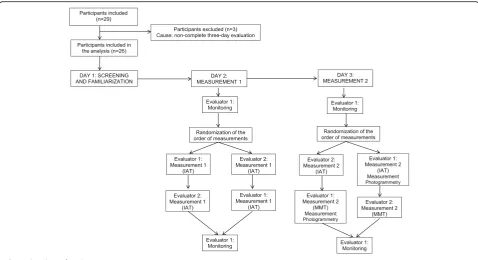
The protocol consisted of measurements that were taken on three days (between 2 and 8 days apart). The screening
and familiarization with the object of the study were carried out on the first day, including the anthropometric and spi-rometric measurements, the evaluation of the sociodemo-graphic variables and the variables related to the disease and its monitoring, as well as familiarization with the vari-ables of thoracic mobility. The TI was measured independ-ently by the two evaluators on the second day. The measurement of the TI by the evaluators was repeated on the third day. An evaluator also conducted the photogram-metry on the third day. The thoracic mobility variables were measured in random order, and an evaluator moni-tored the participants at the beginning and end of each ses-sion. The two evaluators were blinded to previous measurements and to the measurements of the other evalu-ator, and the evaluations were carried out at the same time of day. The subjects were asked to continue their medical treatment for the duration of the study (Fig.1).
Measurements Spirometry
A Spirobank G brand MIR SRL spirometer was used; the technical procedures recommended by the American Thoracic Society and the European Respiratory Society were followed [17, 20]. The subjects exhaled as hard as they could at least three times. Their peak expiratory flow (PEF) was measured, as was their forced expiratory flow, (FEF)25%. If the PEF was under 60% of the predicted value
[17,18,20], the participant was excluded from the study. The volume exhaled at the end of the first second (FEV1)
was also noted, as was the forced vital capacity (FVC) and the ratio FEV1/FVC.
Anthropometric variables
Body weight was measured using a portable digital scale. Height was measured to an accuracy of 1 mm using a non-stretchable metal tape measure. Recommendations for the anthropometric procedures were followed as stated in the National Health and Nutrition Examination Survey manual published by the Centers for Disease Control and Prevention [21]. Height was measured in meters (m) and weight in kilograms (Kg), and their values were used to calculate the body mass index (BMI, weight/height2) [22,23].
Monitoring variables
Variables of thoracic mobility
Thoracic mobility was measured using the TI and photo-grammetric analysis. The evaluation was conducted with the participants in a sitting position in a backless chair with their feet flat on the floor and arms resting on adjustable-height armrests. The thorax was uncovered for the men and partially uncovered for the women. Thoracic circum-ference was measured using a non-stretchable metal tape measure at three levels in the rib cage. Reflective markers were placed on the anatomical structures at each level, in-cluding the axillary level, the third anterior intercostal space in the front, and the fifth thoracic spinous process in the back; at the level of the xiphoid, the xiphoid process in the front and the tenth thoracic spinous process in the back; at the umbilical level, the umbilical scar in the front and the third thoracic spinous process in the back. The procedure was standardized, putting zero on the metal tape measure at the corporal midpoint at the levels of the respective re-flective markers. The participants were requested to take a maximally deep breath and then to exhale maximally. The tape measure was adjusted at the end of the inhalation and again at the end of the exhalation, and the result in centi-meters was obtained by finding the difference between the diameter at the maximal inhalation and the diameter at the maximal exhalation [9,11,28].
Measurements were taken twice at each level; the higher of the two measurements and the average of the two mea-surements were recorded.
For the photogrammetric analysis, reflective markers were placed at the same levels as for the TI measurement,
and photographic images were taken of the participants at maximum inhalation and maximum exhalation. As with the measurements, two photographs were taken, and each one was processed using Software for Postural Evaluation (SAPO). Thoracic-abdominal displacement in centimeters was obtained at the axillary, xiphoidal, and abdominal levels for the maximum and average values of each meas-urement [29].
Statistical analysis
Having taken two measurements at each level, assuming an explanatory power of 80% and a significance level of 5% with a loss percentage of 20% [30], it was determined that between 25 and 35 people would be sufficient to as-sess the psychometric properties.
results of Carter et al. as follows: little (r= 0.0–0.25), low (r= 0.26–0.49), moderate (r= 0.50–0.69), high (r= 0.70– 0.89), and very high (r= 0.90–1.00).
Results Participant flow
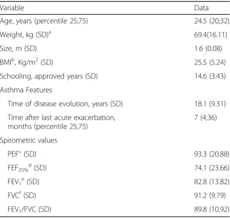
Twenty-nine adults bwith stable asthma were included in the study; three of them did not complete the re-quired evaluations because they did not attend the three measurement days, so 26 were included in the definitive analysis (Fig. 1); 16/26 (61.54%) were women. Table 1 describes the sample in terms of the general spirometric characteristics and characteristics related to the disease. The spirometric pattern was determined to be normal for all the participants based on FEV1and FVC
exceed-ing 80% of the predicted value, and the FEV1/FVC
rela-tion over 70% of the predicted value [20].
TI reliability
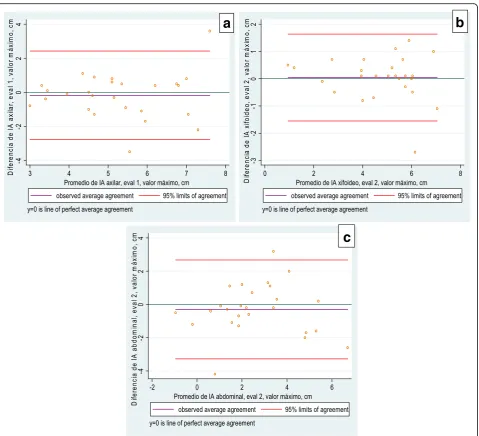
The analysis of the intra-evaluator agreement showed ac-ceptable limits of agreement for two evaluators, with the average of the differences between them close to zero, using both the maximum value and the average (Fig. 2). In the intra-observer analysis, the Bland Altman plots showed a limits of agreement narrowest at the xiphoid level (Evalu-ator 1: 1.892 and 1.576; Evalu(Evalu-ator 2: 1.006 and 1.206) and greater difference at the abdominal level (Evaluator 1: 3.519 and 2.888; Evaluator 2: 3.038 and 2.415).
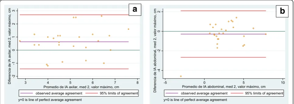
In the inter-observer analysis the Bland-Altman plots showed a smaller difference at the xiphoid level (meas-urement 1: 1.787 and 1.917; meas(meas-urement 2: 1.773 and 2.342) and greater at the abdominal level (measurement 1: 3.638 and 3.100; measurement 2: 2.659 and 2.090). Only 3.23% of the data were outside the limits of agree-ment, representing one participant at the xiphoid level and one at the abdominal level, both on the first meas-urement day. The following figures illustrate a similar data distribution, without significant bias (Figs.3and 4). In general, there was good intra-and inter-evaluator agreement at the three levels of measurement [9].
TI reproducibility
The intra-evaluator reproducibility for measurement at the axillary level was between poor and acceptable (ICC between 0.57 and 0.68), analyzing the maximum and average values, respectively. At the xiphoid level it was good (ICC between 0.85 and 0.93) and at the abdominal level it was between acceptable and good (ICC between 0.70 and 0.82), with wider confidence intervals at the ax-illary and abdominal levels. The reproducibility between the evaluators was acceptable at the axillary level both for the maximum and average values (ICC between 0.62 and 0.66), and good at the xiphoid and abdominal levels (ICC between 0.78 and 0.86), with wider confidence in-tervals at the axillary level (Tables2and3).
Convergent validity between the TI and thoracic kinematics
The correlation using the maximum value was moderate at the axillary and xiphoid levels, and high at the ab-dominal level (r axillary: 0.621; xiphoid: 0.668; abdom-inal: 0.733), with narrower confidence intervals at the abdominal level. The correlation using the average value was moderate at the axillary level (r: 0.55) and high at the xiphoid and abdominal levels (r: 0.757 and 0.696, re-spectively), with narrower confidence intervals at the xiphoid level (Fig.5).
Discussion
Within the established reliability, the limits to agreement were narrower at all the measurement levels than those re-ported by Malaguti et al. [9], who evaluated the reproduci-bility of thoracic cirtometry in people with COPD. This result can be attributed to the standardization of all the conditions within the protocol. The limits were considered acceptable both for the intra-evaluator and inter-evaluator agreements, using the maximum and average values with an average of differences close to zero, thus demonstrating control of measurement bias, possibly resulting from the session where the evaluators were trained and the partici-pants were familiarized with the measurements.
Table 1Characteristics of the sample (N= 26)
Variable Data
Age, years (percentile 25;75) 24.5 (20;32)
Weight, kg (SD)a 69.4(16.11)
Size, m (SD) 1.6 (0.08)
BMIb, Kg/m2(SD) 25.5 (5.24)
Schooling, approved years (SD) 14.6 (3.43)
Asthma Features
Time of disease evolution, years (SD) 18.1 (9.31)
Time after last acute exacerbation, months (percentile 25;75)
7 (4;36)
Spirometric values
PEFc(SD) 93.3 (20.88)
FEF25%d(SD) 74.1 (23.66)
FEV1e(SD) 82.8 (13.82)
FVCf(SD) 91.2 (9.79)
FEV1/FVC (SD) 89.8 (10.92)
a
Standard Deviation
b
Body Mass Index
c
Peak Expiratory Flow
d
Forced Expiratory Flow at 25% of FVC
e
Forced Expiratory Volume during the first second
f
In both studies, the limits were narrowest at the xiph-oid level and widest apart at the abdominal level. Mala-guti et al. [9] attributed the greater variability at the abdominal level to differences in the breathing patterns of each participant at maximum inhalation. In addition, when analyzing the variability at the abdominal level in the seated position, the distribution of the pulmonary volumes should be taken into consideration. Lee et al. [32] concluded that subtle changes in the trunk position of healthy adults can alter the configuration and move-ment of the thoracic wall, as well as the distribution of the respiratory volume, changes that can be attributed to modifications in muscular activation [33].
These modifications in the neck and trunk muscles are re-lated to the double function of these muscles in the
respiratory cycle and postural control [34, 35], especially at the abdominal level. Postural adjustments in a seated pos-ition can influence breathing patterns due to the alternation of these functions. Small postural changes can favor the pre-dominance of either the postural or the respiratory function [33,36]. Thus, despite having a standardized seated posture for every participant, small postural adjustments may have influenced the variability of the measurements.
Romei et al. [6] found that costal kinematics in healthy adults are significantly affected by the trunk position. A gradually increasing inclination of the trunk leads to a progressive reduction in rib cage displacement and an increasing abdominal contribution leads to tidal volume. These findings may also explain the variable TI measure-ments for individual subjects, and indicate aspects of
a
b
c
intra-subject measurements that can generate random errors outside the evaluator’s control.
In addition, the measurement of costal mobility has been used to determine the effectiveness of physiothera-peutic interventions for people with asthma. Burianová et al. [37] evaluated the effect of physiotherapeutic treat-ment on thoracic mobility and reported a statistically significant improvement with total increments of be-tween 1.5 cm and 2.1 cm for men at the level of the fourth rib and xiphoid level, respectively, and of 2 cm for women at both levels. The findings of this study, however, suggest that changes under 2.1 cm cannot be considered clinically significant, as they can represent randomization given the variability of measurements for either the evaluated subject or the evaluator rather than the effect of the intervention, which is highly relevant in the clinical practice of physiotherapy.
With regard to reproducibility, a wide IC of 95% was found at every level, which should be analyzed in light of
the participants’pathological condition. In asthma, hyper-inflation takes place during the crisis. When the disease recedes, breathing patterns may vary from person to per-son [38]. Thus, greater variations in costal mobility may appear in asthma patients than in people with COPD, which could explain the lesser reproducibility at the axil-lary level in this study compared to Malaguti et al. [9].
The intra-evaluator reproducibility of between accept-able and good may be attributaccept-able to the influence of sex over mobility. Romei et al. [6] showed that the ab-dominal contribution to tidal volume is less among women, so including subjects of both sexes in this study may have introduced an additional source of variability. The participants’ posture during the evaluation consti-tutes another influential factor. Verschekelen et al. [39] described a greater contribution of the superior costal level when respiratory maneuvers were carried out at the vital capacity level in a standing position compared to measurements at the vital capacity level in a supine
a
b
Fig. 4Level of agreement between evaluators, maximum value.aAxillary level.bAbdominal level
a
b
position. These findings could also explain the variability in the results at the axillary level.
The inter-evaluator reproducibility in this project was similar to that of Malaguti et al. [9]. The ICCs were ac-ceptable at the axillary level and good at the xiphoid and abdominal levels, with 95% IC. Muscular activation of the upper limbs may have contributed to lesser reproducibility at the axillary level. In healthy subjects, elevating the arm during everyday activities increases ventilatory activity, as the muscles implicated in the positioning of the arm de-crease their participation in respiration, thus affecting the mechanics of the ventilatory effort [40]. These adjust-ments may apply to the present study, which could ex-plain the acceptable reproducibility at the axillary level.
The intra- and inter-evaluator reproducibility was lower at the axillary level, which is considered below the clinically acceptable level. This level is based on the ap-plication of the measurements in clinical trials. In these studies the variability of group means is related to sam-ple size. For that reason a 0.70 reliability threshold is appropriate [41].
On the other hand, the use of 0.7 threshold in the clin-ical scenario, would be limited due that the random vari-ability of patient is greater that in clinical trial conditions. Additionally, intra- and inter-evaluator re-producibility was classified as clinically acceptable in xiphoid and abdominal levels. However, at the xiphoid level there was better inter-evaluator reproducibility than intra-evaluator, a finding that we consider a ran-dom effect because no clinical reasons are identified for such differences; and in any case, these differences do not represent changes in the decision to use or not the TI in the clinical or research context.
With regard to the external convergence construct val-idity, the analysis using the maximum value and the aver-age of the two instances of the TI measurement and photogrammetry showed a correlation of between moder-ate and high at every level. The confidence intervals were wide at the three levels.
This positive correlation between the kinematic and TI analyses is based on the fact that both tests were evaluating the same construct: costal mobility, and the maneuvers used in the two cases are similar. Costal mo-bility results from the distensimo-bility of tissue, and it could be inferred that both methods measure this biomechan-ical property at the three levels. Previous studies [9] have suggested that distensibility is greatest at the abdominal level, which could explain the high correlation observed in this study at that level.
The lesser correlation at the costal level (axillary and xiphoid) can be understood beginning with the modifi-cations to the distribution of air in this area, responding to individual breathing patterns and the consequences of intermittent hyperinflation. In asthma, hyperinflation is considered intermittent, as it appears during crises and disappears during inter-crisis periods [38]. With regard to the type of pattern used, it was considered relevant to evaluate the correlation by allowing each subject to spontaneously maximize his/her respiration, which allowed for modified distributions of the volume of air in each test.
The consequences of hyperinflation with regard to the distribution of air in the rib cage have been studied by other authors [3], and it has been established that the volume of trapped air moves principally to the upper costal level, thus reducing distensibility [33]. This, to-gether with the pathologic increased time taken to empty a lung of air and the local limitation to respiratory flow, produces hyperinflation in the other regions of the lung in patients with asthma [42].
The results of this study suggest that TI reliability and validity are similar when using measurements of the maximum and average values. The maximum value cor-responds to a greater effort by a person to move lung volumes and capacities into and out of the rib cage,
Table 3Inter-evaluator TI reproducibility (N = 26)
Measurement level
Inter-evaluator TI reproducibility in Measurement 1
Inter-evaluator TI reproducibility in Measurement 2
ICC [2,k] IC 95% ICC [2,k] IC 95%
Maximum value
Axillary 0.644 0.324–0.827 0.629 0.261–0.825
Xiphoid 0.845 0.684–0.927 0.806 0.618–0.908
Abdominal 0.787 0.583–0.898 0.850 0.696–0.929
Average value
Axillary 0.664 0.376–0.835 0.634 0.243–0.832
Xiphoid 0.865 0.724–0.937 0.853 0.668–0.935
Abdominal 0.793 0.592–0.901 0.841 0.680–0.925
The data are the reproducibility between the evaluators on each measurement day
ICCInterclass Correlation Coefficient,ICConfidence Interval Table 2Intra-evaluator TI reproducibility (N= 26)
Measurement level
Evaluator 1 Evaluator 2
ICC [2,k] IC 95% ICC [2,k] IC 95%
Maximum value
Axillary 0.596 0.279–0.796 0.571 0.237–0.783
Xiphoid 0.856 0.708–0.933 0.885 0.760–0.947
Abdominal 0.784 0.577–0.897 0.704 0.446–0.855
Average value
Axillary 0.688 0.422–0.846 0.631 0.324–0.817
Xiphoid 0.858 0.711–0.933 0.935 0.863–0.970
Abdominal 0.820 0.641–0.914 0.718 0.469–0.862
while the average value illustrates the typical form of costal mobility when the subject is requested to exert a greater-than-baseline respiratory effort [43].
For the above reasons, the TI can be used in clinical practice with people who can perform maximally at just one attempt, as well as those who require two or more attempts to obtain a result. In either case, a standardized protocol that includes every level of measurement should be considered, with an emphasis on explaining the maneuver and the commands or requests that are most effective at eliciting maximum force.
Some factors in the evaluation of the TI are outside the control of the evaluator, for example, sex, dynamic pos-tural adjustments, breathing pattern, and muscular activa-tion during the test [6], but they should be considered in the clinical setting when the effects of the disease or the results of the evaluation and the physiotherapeutic man-agement are being analyzed. The evaluation of the effects of an intervention on costal mobility should include all these factors and should always be analyzed keeping in mind that statistically significant changes should be reflected in an improvement of the clinical condition.
Within the limits of the study, it was found that the TI’s psychometric properties were established for adults with stable asthma, for which reason the re-sults are limited to people with these characteristics. It is recommended that the psychometric properties of these evaluations be assessed with other age groups, at different phases of the disease, and with other pathologies.
The TI measurements and measurements of costal kine-matics were taken after requesting maximum respiratory force. However, lung volumes and capacities were not measured objectively using tools such as plethysmogra-phy. Thus, it was not possible to standardize the exact quantifiable volume when the measurements were taken. It is recommended that lung volumes and capacities be
quantified using plethysmography to decrease variability in TI measurement in future research.
Conclusions
Based on our review of the literature, this is the first study to evaluate the reliability and validity of perimetry in adults with stable asthma. Many works have demon-strated changes to respiratory mechanics in people with asthma, so this kind of evaluation should be routine for asthma patients.
The quality of the measurement techniques deter-mines the quality of the research results and the deci-sions for the clinical management of these patients, and studies of reliability and validity help avoid errors in interpreting variables before and after interventions. In this study, the good reliability can be attributed to the standardization of the test and the use of a special ses-sion for familiarization. Nonetheless, it is important to remember that some aspects could not be controlled by the evaluator such as the subject’s sex and his/her dy-namic postural adjustments.
A moderate to high correlation was found between the costal mobility variables, and the validity of the external convergent construct for the TI was established. This correlation stems from the fact that the same construct (costal mobility) was evaluated for both tests, and it de-pends on the distensibility of tissues. The elements that affect this correlation and that are not susceptible to control are individual breathing patterns and the conse-quences of the pathology for respiratory mechanics.
It can be concluded that the TI is a valid and reprodu-cible measurement that can be used by health profes-sionals during the physical examination of the thorax to evaluate thoracic mobility in adults with asthma, thus broadening their analysis of the respiratory mechanics in each case. This test can also be applied in controlled
a
b
clinical trials to determine the effectiveness of thera-peutic interventions for optimizing respiratory force in people suffering from this pathology.
Abbreviations
BMI:Body mass index; CI95%: Confidence intervals at 95%; FEF: Forced expiratory flow; FEV1: Volume exhaled at the end of the first second; FVC: Forced vital capacity; ICC: Interclass correlation coefficient; PEF: Peak expiratory flow; TI: Thoracic index
Acknowledgements
This study was supported by the Neurociencias y Comportamiento UIS-UPB Research Group.
Availability of data and materials
Data relating to this study are from available from the corresponding author on request.
Patient consent
The participants who voluntarily agreed to allow photographs to be taken for publication signed an informed consent form.
Authors’contributions
All the authors contributed to this study’s conception and design. All the authors are accountable for every aspect of this work, for the integrity of the data, and for the accuracy of the data analysis. All the authors read and approved the final manuscript.
Ethics approval and consent to participate
This project was approved by the Scientific Research Ethics Committee of the School of Health of Universidad Industrial de Santander with code 7083, and was carried out in accordance with the Good Clinical Practice rules and the Ethics Principles for Medical Research Involving Human Beings as defined in the latest revision of the Declaration of Helsinki. All of the participants voluntarily agreed to be enrolled in the study and signed an informed consent form.
Consent for publication
All the authors of this manuscript have read and agreed to its content and are accountable for every aspect of its accuracy and integrity in accordance with the ICMJE criteria that the article is original, has not already been published in a journal, and is not currently under consideration by another journal. Furthermore, written informed consent for the publication of their clinical details and clinical images was obtained from the patients. A copy of the consent form is available for review by the Editor of this journal.
Competing interests
The authors declare that they have no competing interests.
Publisher’s Note
Springer Nature remains neutral with regard to jurisdictional claims in published maps and institutional affiliations.
Author details
1Universidad Industrial de Santander Facultad de Salud, Bucaramanga,
Santander, Colombia.2Sede Quinta Mutis, Escuela de Medicina y Ciencias de la Salud, Universidad del Rosario, Carrera 24N # 63D-69, Bogotá, Colombia.
Received: 12 October 2017 Accepted: 10 April 2018
References
1. Bateman ED, Hurd SS, Barnes PJ, Bousquet J, Drazen JM, FitzGerald M, et al. Global strategy for asthma management and prevention: GINA executive summary. Eur Respir J. 2008;31(1):143–78.
2. Laghi F, Tobin MJ. Disorders of the respiratory muscles. Am J Respir Crit Care Med. 2003;168(1):10–48.
3. Gorini M, Iandelli I, Misuri G, Bertoli F, Filippelli M, Mancini M, et al. Chest wall hyperinflation during acute bronchoconstriction in asthma. Am J Respir Crit Care Med. 1999;160(3):808–16.
4. Pedrolongo P, Gatti EM, Jamami M, Di Lorenzo BA, Costa D. Relação da medida da amplitude tóraco-abdominal de adolescentes asmáticos e saudáveis com seu desempenho físico. 2011;1:107–14. 24.
5. Cihak-TR-2005-32.pdf [Internet]. [cited 2017 Oct 9]. Available from:http:// cmp.felk.cvut.cz/ftp/articles/sara/Cihak-TR-2005-32.pdf.
6. Romei M, Mauro AL, D’Angelo MG, Turconi AC, Bresolin N, Pedotti A, et al. Effects of gender and posture on thoraco-abdominal kinematics during quiet breathing in healthy adults. Respir Physiol Neurobiol. 2010;172(3):184–91. 7. Davidson J, dos Santos AMN, Garcia KMB, Yi LC, João PC, Miyoshi MH, et al.
Photogrammetry: an accurate and reliable tool to detect thoracic musculoskeletal abnormalities in preterm infants. Physiotherapy. 2012;98(3):243–9.
8. Bockenhauer SE, Chen H, Julliard KN, Weedon J. Measuring thoracic excursion: reliability of the cloth tape measure technique. J Am Osteopath Assoc. 2007;107(5):191–6.
9. Malaguti C, Rondelli RR, de Souza LM, Domingues M, Dal Corso S. Reliability of chest wall mobility and its correlation with pulmonary function in patients with chronic obstructive pulmonary disease. Respir Care. 2009; 54(12):1703–11.
10. Caldeira V d S, Starling CCD, Britto RR, Martins JA, Sampaio RF, Parreira VF. Precisão e acurácia da cirtometria em adultos saudáveis. J Bras Pneumol. 2007;33(5):519–26.
11. Custers JWH, Arets HGM, Engelbert RHH, Kooijmans FTC, van der Ent CK, Helders PJM. Thoracic excursion measurement in children with cystic fibrosis. J Cyst Fibros. 2005;4(2):129–33.
12. Plaza Moral V. GEMA4.0. Guía española para el manejo del asma. Arch Bronconeumol. 2015;51(Supplement 1):2–54.
13. Apter AJ. Advances in adult asthma diagnosis and treatment and health outcomes, education, delivery, and quality in 2011: what goes around comes around. J Allergy Clin Immunol. 2012;129(1):69–75.
14. Orozco LC. Confiabilidad o de la consistencia, reproducibilidad, acuerdo y algo más. In: Medición en salud Diagnóstico y evaluación de resultados: un manual critico más allá de lo básico. primera edición. Bucaramanga: División de publicaciones UIS; 2010. p. 73–103.
15. Puente Maestú L, García de Pedro J. Lung function tests in clinical decision-making. Arch Bronconeumol. 2012;48(5):161–9.
16. Troosters T, Casaburi R, Gosselink R, Decramer M. Pulmonary rehabilitation in chronic obstructive pulmonary disease. Am J Respir Crit Care Med. 2005; 172(1):19–38.
17. Miller MR, Hankinson J, Brusasco V, Burgos F, Casaburi R, Coates A, et al. Standardisation of spirometry. Eur Respir J. 2005;26(2):319–38. 18. L Maestu, J Garcia. Lung function tests in clinical decision-making. 2012.
48(5):161–169.
19. Schlegelmilch RM, Kramme R. Pulmonary Function Testing. In: Springer Handbook of Medical Technology [Internet]. Berlin: Springer; 2011. p. 95– 117. Available from: https://link.springer.com/chapter/10.1007/978-3-540-74658-4_8. [cited 2017 Oct 9].
20. Pellegrino R, Viegi G, Brusasco V, Crapo RO, Burgos F, Casaburi R, et al. Interpretative strategies for lung function tests. Eur Respir J. 2005;26(5):948–68. 21. Hernandez AG. Tratado de nutricion / Nutrition Treatise: Composicion Y
Calidad Nutritiva De Los Alimentos / composition and nutritional quality of foods. Ed Médica Panamericana. 2010;2:820.
22. CDC. CDC Works 24/7 [Internet]. Centers for Disease Control and Prevention. 2017. Available from:https://www.cdc.gov/index.htm. [cited 2017 Jun 8]. 23. NHANES - National Health and Nutrition Examination Survey Homepage
[Internet]. 2017. Available from:https://www.cdc.gov/nchs/nhanes/index. htm. [cited 2017 Oct 9].
24. Heyward VH. Evaluación de la aptitud física y prescripción del ejercicio. Ed Médica Panamericana. 2008;5:15–33.
25. Pryor JA, Prasad AS. Physiotherapy for respiratory and cardiac problems: adults and paediatrics. London: Elsevier Health Sciences; 2008. p. 648. 26. Pulsioximetría (medición de la saturación de oxígeno) [Internet]. [cited 2017
Oct 9]. Available from:https://www.elsevierclinicalskills.es/procedimientos/ 1129/pulsioximetr%C3%83%C2%ADa-medici%C3%83%C2%B3n-de-la-saturaci%C3%83%C2%B3n-de-ox%C3%83%C2%ADgen.
27. Heyward VH. Evaluación y prescripción del ejercicio. Editorial Paidotribo; 2006. p. 284.
28. [AQUI - epg4-76 ok.pdf [Internet]. [cited 2017 Oct 9]. Available from:http:// biblioteca.univap.br/dados/INIC/cd/epg/epg4/epg4-76%20ok.pdf 29. Linder W. Digital Photogrammetry [Internet]. Berlin, Heidelberg: Springer
30. Microsoft Word - pearson.doc - pearson2.pdf [Internet]. [cited 2017 Oct 9]. Available from:http://test.fisterra.com/gestor/upload/guias/pearson2.pdf 31. L Orozco. Medición en salud. Diagnóstico y evaluación de resultados. Un
manual crítico más allá de lo básico. 1 edición. Bucaramanga: Publicaciones UIS; 2010. p. 17–25.
32. Lee L-J, Chang AT, Coppieters MW, Hodges PW. Changes in sitting posture induce multiplanar changes in chest wall shape and motion with breathing. Respir Physiol Neurobiol. 2010;170(3):236–45.
33. Courtney R. The functions of breathing and its dysfunctions and their relationship to breathing therapy. Int J Osteopath Med. 2009;12(3):78–85. 34. Lesmes JD. Evaluación clínico-funcional del movimiento corporal humano.
Ed Médica Panamericana. 2007;1:72–95.
35. Kendall FP, McCreary EK, Provance PG. Muscles, testing and function: with posture and pain. USA: Williams & Wilkins; 1993. p. 451.
36. Segizbaeva MO, Pogodin MA, Aleksandrova NP. Effects of body positions on respiratory muscle activation during maximal inspiratory maneuvers. Adv Exp Med Biol. 2013;756:355–63.
37. Burianová K, Vařeková R, Vařeka I. The effect of 8 week pulmonary rehabilitation programme on chest mobility and maximal inspiratory and expiratory mouth pressure in patients with bronchial asthma. Acta Gymnica. 2008;38(3):55–60.
38. Jardim JR, Mayer AF, Camelier A. Músculos respiratorios y rehabilitación pulmonar en asmáticos. Arch Bronconeumol. 2002;38(4):181–8. 39. Verschakelen JA, Demedts MG. Normal thoracoabdominal motions.
Influence of sex, age, posture, and breath size. Am J Respir Crit Care Med. 1995;151(2 Pt 1):399–405.
40. Brown PI, Johnson MA, Sharpe GR. Determinants of inspiratory muscle strength in healthy humans. Respir Physiol Neurobiol. 2014;196:50–5. 41. Frost MH, Reeve BB, Liepa AM, Stauffer JW, Hays RD. What Is Sufficient
Evidence for the Reliability and Validity of Patient-Reported Outcome Measures? vol. 2; 2007. p. S94–S105.
42. Filippelli M, Duranti R, Gigliotti F, Bianchi R, Grazzini M, Stendardi L, et al. Overall contribution of chest wall hyperinflation to breathlessness in asthma. Chest. 2003;124(6):2164–70.