R E S E A R C H A R T I C L E
Open Access
Effects of computerized cognitive training
on neuroimaging outcomes in older adults:
a systematic review
Lisanne F. ten Brinke, Jennifer C. Davis, Cindy K. Barha and Teresa Liu-Ambrose
*Abstract
Background:Worldwide, the population is aging and the number of individuals diagnosed with dementia is rising
rapidly. Currently, there are no effective pharmaceutical cures. Hence, identifying lifestyle approaches that may prevent, delay, or treat cognitive impairment and dementia in older adults is becoming increasingly important. Computerized Cognitive Training (CCT) is a promising strategy to combat cognitive decline. Yet, the underlying mechanisms of the effect of CCT on cognition remain poorly understood. Hence, the primary objective of this systematic review was to examine peer-reviewed literature ascertaining the effect of CCT on both structural and functional neuroimaging measures among older adults to gain insight into the underlying mechanisms by which CCT may benefit cognitive function.
Methods:In accordance with PRISMA guidelines, we used the following databases: MEDLINE, EMBASE, and CINAHL. Two
independent reviewers abstracted data using pre-defined terms. These included: main study characteristics such as the
type of training (i.e., single- versus multi-domain), participant demographics (age≥50 years; no psychiatric conditions),
and the inclusion of neuroimaging outcomes. The Physiotherapy Evidence Database (PEDro) scale was used to assess quality of all studies included in this systematic review.
Results:Nine studies were included in this systematic review, with four studies including multiple MRI sequences. Results
of this systematic review are mixed: CCT was found to increase and decrease both brain structure and function in older adults. In addition, depending on region of interest, both increases and decreases in structure and function were associated with behavioural performance.
Conclusions:Of all studies included in this systematic review, results from the highest quality studies, which were two
randomized controlled trials, demonstrated that multi-domain CCT could lead to increases in hippocampal functional connectivity. Further high quality studies that include an active control, a sample size calculation, and an appropriate training dosage, are needed to confirm these findings and their relation to cognition.
Keywords:Computerized cognitive training, Neuroimaging, Brain structure, Brain function, Older adults
Background
With our ageing population, the incidence of dementia is rising rapidly. Currently, over 47 million people world-wide are diagnosed with dementia and this number is expected to triple by 2050 [1]. In 2010 it was estimated that the worldwide cost of dementia was 604 billion US dollars [1]. Thus it is imperative to find strategies that promote cognitive healthy aging to minimize the projected
societal, health, and economic burden by reducing or delaying the potential progression to mild cognitive impair-ment or deimpair-mentia.
Currently, there is no pharmaceutical cure for dementia. As such, identifying lifestyle approaches that may prevent, delay, or even treat cognitive impairment and dementia in older adults is becoming increasingly important [2]. Even when an effective pharmacological therapy is available, lifestyle approaches (i.e., exercise, nutrition, and cognitive training) can be used in conjunction as lifestyle interven-tions result in multidimensional benefits [3]. In recent years, there is growing interest in complex mental activity * Correspondence:teresa.ambrose@ubc.ca
Aging, Mobility, and Cognitive Neuroscience Laboratory, Department of Physical Therapy, Djavad Mowafaghian Centre for Brain Health, University of British Columbia, 2215 Wesbrook Mall, Vancouver, BC V6T 1Z3, Canada
as a strategy to promote healthy cognitive aging. Complex mental activity comprises all activities that are cognitively challenging for an individual [4], such as memory and executive functioning training, or dance. A meta-analysis of human cohort studies provides robust evidence that complex patterns of mental activity in early, mid-life, and late-life stages is associated with a significant reduction in dementia incidence [5]. Furthermore, they found an asso-ciation between increased levels of complex mental activ-ity in late life and lower dementia rates, independent of other predictors. Finally, it showed a dose-response rela-tionship between the amount of complex mental activities in late life and dementia risk [5].
Computerized cognitive training (CCT) is one example of complex mental activity that could be used to promote healthy cognitive aging. CCT is defined as cognitive train-ing on an individual electronic device (e.g., computer, laptop, tablet/iPad) that requires a physical response such as a button press, and excludes training that primarily re-quires an individual to perform two tasks simultaneously, in order to compare performance with single-task con-ditions (i.e., dual-task training). Notably, CCT is an ap-proach that could be used by those who are limited in their ability to physically participate in other strategies, such as exercise. A meta-analyses shows that CCT improved overall cognitive performance in older adults [6]. Specifically it showed improvements in verbal and non-verbal memory, working memory, processing speed, and visuospatial skills [6]. Recent randomized controlled trials (RCT’s) of CCT in older adults showed that both two and three months of training resulted in improved global cognition compared with an active control group [7, 8]. Additionally, an RCT showed that CCT resulted in improvements in memory and processing speed which were still visible twelve months post-training [7], and shows that CCT is able to maintain its bene-fits. Playing a real-time strategy video game for 23.5 h improved performance in executive functions, indicat-ing transfer of trainindicat-ing after participatindicat-ing in complex mental activities [9]. Thus, current evidence suggests that CCT is a promising strategy for promoting healthy cognitive aging.
Cognitive training is based on the notion that the brain, even with age, can change for the better, if given the ap-propriate environmental stimuli, thoughts, and emotions [10]. This capacity of the brain is called“neuroplasticity”. In the same way that physical training improves physical abilities, cognitive training (or brain training) may induce neuroplastic changes in the brain, resulting in improved cognitive abilities. One of the fundamental principles of neuroplasticity is the concept of synaptic plasticity– the notion that individual connections within the brain are constantly being removed or recreated, largely dependent upon how they are used [11]. Cognitive training aims to
harness this principle of neuroplasticity by using guided practice on a set of tasks related to memory, attention, or other cognitive processes.
To gain more insight in what potential neuroplastic changes CCT may induce; incorporating different neuro-imaging techniques in studies could be a good approach to help demonstrate these changes in the brain. For example, synaptic plasticity as a result of stimulation by CCT could potentially be captured by functional connectivity, measured with resting-state functional magnetic resonance imaging (rsfMRI), by strengthen-ing connections within and between networks [12]. To date, it is not well established how CCT impacts re-gional brain volume, functional activity, and functional or structural connectivity in older adults. Although work has been done among younger adults illustrating changes in functional activity in the middle frontal gyrus and superior and inferior parietal cortices after working memory training [13], these findings don’t necessarily translate to an older adult population. Therefore, gaps remain in understanding the under-lying mechanisms of training-induced neuroplasticity in older adults. Addressing this knowledge void, this systematic review aims to ascertain the mechanisms by which CCT exerts an impact on brain structure and function by using different neuroimaging techniques such as volumetric magnetic resonance imaging (MRI), task-based functional MRI (fMRI), rsfMRI, and diffu-sion tensor imaging (DTI). Through understanding the underlying neural mechanisms of CCT, our goal is to provide knowledge on how to design improved and tar-geted interventions that help combat or prevent cogni-tive decline throughout life.
Methods
Search strategy
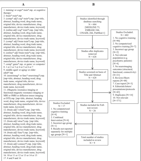
In accordance with the Preferred Reporting Items for Systematic reviews and Meta-Analyses (PRISMA) state-ment [14], we conducted a comprehensive search of MEDLINE, EMBASE, and CINAHL databases to identify all the studies that investigated neuroimaging outcomes resulting from CCT interventions. We limited our search to adults aged 55 years and older with and with-out cognitive impairment, who have not been diagnosed with dementia. We did not limit the search based on publication date, as CCT is a relative novel research topic. The final search (see Fig. 1a for search strategy) was done on July 7 (2016) and included a check for recent publications in PubMed.
Study selection
55 years and older). Study designs included in this sys-tematic review were RCT’s and quasi-experimental studies. Studies that used samples of younger and older adults but reported group results separately were in-cluded in this systematic review. We inin-cluded studies
that focused on both single- and multi-domain CCT programs. We considered single-domain CCT training as training that targeted a specific cognitive ability, such as working memory. In contrast, multi-domain CCT was considered training that consisted of a series of tasks
A
B
that targeted multiple cognitive abilities (e.g., executive functions and memory). We excluded studies that did not focus on CCT or studies that used CCT in combin-ation with other types of intervention (e.g., non-CCT, exercise), reviews and short reports. A full list of exclu-sion criteria and the excluexclu-sion pathway is displayed in Fig. 1b. Critical review of titles and abstracts resulted in 26 articles for full-text review.
Data extraction and quality assessment
We developed a list of data extraction items. This list in-cluded reference, study sample, study design, MRI magnet, neuroimaging outcomes, cognitive function measured, training program/task, cognitive domain trained, descrip-tion of training, training frequency and duradescrip-tion, total hours of training, supervised/home-based training, and control group. Two authors [LTB and CKB] independently extracted the data from the included studies. Discrepancies were discussed and solved by two authors [JCD and TLA].
The Physiotherapy Evidence Database (PEDro) scale [15] was used to assess the quality of the included stud-ies. We [LTB and TLA] added three additional items to the PEDro scale to ensure a proper assessment of inter-vention studies using neuroimaging outcomes. These three items included were: 1) cognition measured to as-sist the interpretation of neuroimaging results; 2) sample size calculation; and 3) compliance reported (yes/no). To answer the items in the quality assessment, we used a‘+’ for items that were present and a‘-‘for items that were absent. The quality assessment was performed independ-ently by two authors [LTB and CKB]. Discrepancies were discussed and reviewed by two authors [JCD and TLA]. Consensus between two authors [LTB and CKB] was achieved after discussion (K=0.98). Because item one of the PEDro scale is related to external validity, it is not included in the overall PEDro score. Therefore, the max-imum quality assessment score calculated by the PEDro was 10 points (each‘+’indicates one point), and will be reported in the results. Studies with a PEDro score of 6/ 10 or higher were considered studies of moderate to high quality. The additional item list had a maximum score of three points and trends from this list will be descriptively discussed in the results.
Results
Overview of studies included
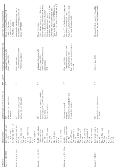
Of the 684 articles identified, nine were included in this systematic review (Table 1). These nine papers included four RCT’s [16–19] and five quasi-experimental studies [20–24]; all nine studies had a different study duration. Details of the interventions included are provided in Table 2. The results are categorized into four categories: 1) Volumetric structural imaging (n= 4) [16, 19, 20, 22]; 2) Task-based fMRI (n = 3) [18, 21, 22]; 3) Connectivity
(n = 7) [16, 17, 19, 20, 22–24]; and 4) Correlation
between imaging outcomes and cognitive function out-comes (n = 8) [16–22, 24], (Table 3). Results are
re-ported in order of study quality, starting with the highest quality.
Structural imaging (n= 4)
Four studies [16, 19, 20, 22] reported volumetric and cor-tical thickness outcomes (Table 3). A randomized con-trolled study (full factorial design) multi-domain cognitive training study using Cogpack [19], older adults with mild cognitive impairment (MCI) trained for a total of 78 h over a period of 6 months under supervision. Combined cognitive training with resistance training resulted in in-creased cortical thickness in the posterior cingulate cortex. However, in the same study they found that cognitive training alone led to a decrease in the posterior cingulate cortex thickness. However, there was no difference in de-crease in thickness compared with the control group.
In addition, a twelve-week supervised multi-domain CCT study [16] using the same program (CogPack) showed that 36 h of training resulted in an increase in grey matter density in the right post-central gyrus com-pared with a decrease in the active control group. Add-itionally, the training resulted in a difference in rate of thickness change over time in both the left fusiform gyrus and the supramarginal and post-central gyri.
In contrast, in an object-location learning paradigm study [20] participants performed training on three consecutive days where they had to learn the correct spatial location of buildings on a street map. On each training day, the training was followed by a cued recall and recognition task. Hippocampal volumes was mea-sured pre- and post-training. The authors found that the object-location learning paradigm did not lead to changes in hippocampal volume.
In another quasi-experimental study [22], participants performed an adaptive working memory training (n -Back) for twelve 45-min sessions over 4 weeks. Difficulty level of the training was based on individual perform-ance and increases over time. Results showed that the training did not result in changes in grey matter volume in the working memory network.
In summary, one RCT [19] found cortical thinning as a result of cognitive training alone. In contrast, another RCT [16] found an increase in grey matter density fol-lowing training. Finally, one study [22] found that cogni-tive training did not result in changes in grey matter, and one study [20] found that cognitive training did not lead to changes in hippocampal volume.
Task-based fMRI (n= 3)
Table 1 Characteristics of studies included (Continued) Stre nziok et al. [ 24 ] 20 14 Hea lthy olde r adu lts N =4 2 69.21 ± 4.93 years Tra ining group 1 N =1 4 69.70 ± 6.9 years Tra ining group 2 N =1 4 68.52 ± 5.6 years Tra ining group 3 N =1 4 69.41 ± 2.3 years Q uasi-experime ntal Pre -post Leng th of follow up: Not stated Not stated • Resting -stat e fMR I • DTI Reasoni ng/Proble m Solving (WAI S III Matrix Reasoni ng subtest, [ 48 ] Every day problems Test, [ 49 ] Word Se ries and Letter Se ries Test s) [ 50 ] Episodic Mem ory (W echsler Memo ry Scale Logical Mem ory Su btest) [ 51 , 52 ] Spatial Work ing Mem ory (I nformat ion-processing Visuo-Spatial D elayed Mat ch-to-Sam ple Test ) [ 53 , 54 ] Auditory W or king Memory (L ett er N umber Se q u encing subtest o f W AIS III) [ 48 ] Lövde n et al. [ 23 ] 2010 Hea lthy olde r adu lts b: Subsample CO GITO
study N=2
Table
2
Details
of
the
computerized
cognit
ive
training
intervention
for
the
studies
included
(Continued)
Perc
eptu
al
spee
d
(Ch
oice
react
ion
task
s,
Comparison
Task
s)
Antone
nko
et
al.
[
20
]
2016
Object-locati
on
Learn
ing
Paradig
m
Mem
ory
Object-loc
atio
n
Learnin
g
Par
adigm:
Lear
n
the
cor
rect
spa
tia
l
loca
tions
of
b
u
ild
ings
o
n
a
str
eet
ma
p.
Fi
ve
blocks
of
120
stimulus-location
p
airing
with
a
response
in
ter
va
l
o
f
3
s.
Each
block
was
followed
by
a
cued
recal
l
an
d
a
recogni
tion
task
3c
o
n
se
cu
ti
ved
ay
s
5
learn
ing
block
s/day
Unknow
n
Unsp
ecified
No
Con
trol
Hein
zel
et
al.
[
22
]
2014
n
-Back
train
ing
Executive
Fu
nction:
Work
ing
Mem
ory
Adap
tive
n
-bac
k
training,
3
runs
(12
block
s/run)
each
se
ssion.
D
ifficulty
le
vel
in
cre
ase
d
acco
rdi
n
g
to
in
di
vid
ua
l
p
e
rf
o
rm
anc
e
(hi
g
h
e
r
w
o
rk
in
g
m
e
m
o
ry
loa
d
,s
ho
rt
ened
interstim
u
lu
s
interva
l
(IS
I).
ISI
ran
g
e
d
from
150
0
to
5
00
ms
in
st
eps
o
f
5
0
0
ms.
4
we
eks
3×/wee
k
45
min/session
9 Supervis
ed
No
cont
Table
3
Results
for
Imaging
Outco
me
measures
(Continued)
tr
aining
ac
tiv
ation
in
inferior
an
d
right
mi
ddl
e
fr
ontal
gyrus
(
t
=
5
.91),
left
m
idd
le
frontal
gyrus
(
t
=
4.57)
and
left
thal
amus
(
t
=
5.37)
.
Vi
sual
detec
tion
single
task
:
no
chang
e
Dual
task
:
no
change
Div
ided
Fixed
Al
p
h
an
um
er
ic
si
ng
le
ta
sk
:
no
chang
e
Vi
sual
detec
tion
single
ta
sk
:
Decre
ased
post-training
activation
in
right
cerebe
llum
(
t=
4.73)
an
d
right
mid
dle
occipital
gyrus
(
t
=
4
.68)
when
perfor
ming
the
visua
l
dete
ction
tas
k.
Dual
task
(50/50):
S
mall
increase
in
post
-training
activation
in
right
an
d
left
middle
frontal
gy
rus
(areas
11,
47;
t
=
4.41
and
t
=
4.52
respectively). Divide
d
Vari
able
Alpha
nume
ric
single
task:
no
chang
e
Visual
detectio
n
sing
le
task:
no
chang
e
Dual
task:
Significant
increased
activation
in
right
middl
e
frontal
gyrus
(area
10
;
for
20%
atten
tion
allocat
ion
t
=
5.35
and
50
%
atten
tion
allocat
ion
t
=
4.78).
No
reduc
ed
post-t
raining
activ
ation
in
80
%
atten
tion
allocation
.
Visual
detectio
n
single
task
:
No
change
Dual
task
(cost
sco
re)
b:
Single
repeated:
No
improve
ments
in
dual
tasking
Divide
d
Fixed
:
Redu
ced
dua
l-task
cost
(F(
1,34)
=
6.97
,
p
<
.001
,
η
2=
.45)
Divide
d
Vari
able:
Redu
ced
dua
l-task
cost
and
w
er
e
able
to
modify
attentiona
l
priority
(F
(2,33)
=
5.17,
p
<
.001,
η
2 =.
34
)
activ
ation
an
d
react
ion
time
(
r
=.
5
6
,
p
<
.05).
D
ivided
Variabl
e:
Si
gnificant
neg
ative
corr
elati
on
(post
train
ing)
be
tween
activ
ation
of
right
superior
and
middl
e
front
al
gyrus
(B
rodman
n
area
10)
and
atte
ntiona
l
cos
t
(
r
=
−
.55,
p
<
measured via task-based fMRI (Table 3). An RCT [18] showed that 2200 min of cognitive training over a period of 5 weeks resulted in a significant increase in left anterior hippocampus activity compared with an active control group. The cognitive training consisted of seven games aimed to improve auditory processing speed and accuracy. Task difficulty was adjusted throughout the training based on individual performance. The active control group performed computer-based activities such as reading online newspapers and playing computer games targeting visuospatial abilities.
A two-week quasi-experimental study looked at fo-cused and divided (fixed and variable) attention training [21]. In the focused attention training, two tasks (i.e., alphanumeric task and a visual detection task) were per-formed back to back but separate so participants focused on one task at a time. In the divided attention training, participants performed two tasks at the same time with an equal amount of attention (fixed) or under different attention allocations (variable). Results showed that training a single alphanumeric task for 6 h over two weeks decreased activation in the inferior and right mid-dle frontal gyrus, in the left midmid-dle frontal gyrus and in the left thalamus. No differences in functional brain acti-vation were found after performing the single visual detection task or the in the dual task condition. Partici-pants who were assigned to training where they per-formed both the alphanumeric task and the visual detection task at the same time (i.e., dual task) did not show differences in performance during the alpha-numeric task in the scanner. However, participants showed decreased functional brain activation at post-training compared with pre-post-training in the cerebellum and right middle occipital gyrus during the single visual detection task. Additionally, participants showed a slight increase in activation in both the right and left middle frontal gyrus. Finally, participants who were assigned to the training group where they had to perform dual tasks under different attention allocation levels (i.e., 80%, 50%, or 20%), showed increased activation in the right middle frontal gyrus (area 10) for 20% and 50% attention alloca-tion when performing the dual task. No significant changes in functional brain activation were found during the 80% attention allocation task, neither during the alphanumeric single task, nor during the visual detection single task performance.
In an adaptive n-back training program [22], partici-pants performed 12 sessions of approximately 45 min each over 4 weeks. The difficulty level of the training was based on individual performance and was increased across training sessions by increasing working memory load and decreasing the interstimulus interval. Results of this study showed a non-significant time (2) by working memory load (3) interaction, with a significant main
effect of time. This main effect of time demonstrates a reduction in working memory network functional brain activity measured by the Blood Oxygen Level Dependent (BOLD) signal after 12 training sessions. Only decreases in the 1-back (and not 2-back or 3-back) condition were significant, which indicates this main effect of time is driven by the BOLD signal during the 1-back condition.
In summary, an RCT [18] showed that 2200 min of CCT resulted in increased in left anterior hippocampus activity compared with an active control group. One quasi-experimental study [21] showed that depending on the task and region of interest, all training conditions re-sulted in both increased and decreased activity. Finally, a second quasi-experimental study [22] found that 12 ses-sions ofn-back training resulted in a significant decrease in working memory activity; however decrease in activity was driven by performance on the 1-back condition.
Connectivity
Resting-state fMRI (n = 5)
Five studies [16, 17, 19, 22, 24] looked at changes in func-tional connectivity after CCT (Table 3). An RCT [19] ex-amined the effect of progressive resistance training (PRT), computerized multi-domain cognitive training (CCT), or a combined intervention on brain structure and function in older adults with mild cognitive impairment (MCI). The study duration was 26 weeks, with a total of 78 h of training. In the cognitive training groups (i.e., PRT + CCT, and CCT + Sham), the posterior cingulate cortex showed significant decreases in resting-state functional connectiv-ity with both the superior frontal lobe and the anterior cingulate cortex. In addition, increases in resting-state functional connectivity between the hippocampus and the left superior frontal lobe were found compared with groups without CCT.
A second RCT of 12 weeks of multimodal CCT [16] showed that 36 h of cognitive training resulted in de-creases in resting-state functional connectivity between the posterior cingulate and the right superior frontal gyrus, while the control group showed significant in-creases in resting-state functional connectivity. In con-trast, CCT resulted in increased resting-state functional connectivity between the right hippocampus and the left superior temporal gyrus compared with a decrease in connectivity in the control group.
increased resting-state functional connectivity with the left middle frontal gyrus, the left inferior frontal gyrus, the left superior frontal gyrus and the left parietal lobe. In con-trast, the control group showed significant decreases in resting-state functional connectivity over the 10 weeks (see Table 3 for connectivity decreases).
A quasi experiment investigating the effect of three different computer programs [24] found an increased resting-state functional connectivity in the dorsal net-work between the right superior parietal cortex (SPC) and left posterior inferior temporal lobe (ITL) in Rise Of Nation (RON) compared with a decrease in Space Fort-ress (SF). Finally, Brain Fitness (BF) resulted in signifi-cantly decreased resting-state functional connectivity between the right SPC and the left anterior ITL com-pared with an increase in RON.
Finally, a quasi-experimental study [22] looking at the effects of an adaptive n-back training program in older adults found that the 5-week training did not result in changes in task-based functional connectivity in the working memory network.
Structural connectivity (n = 4)
Four studies [16, 20, 23, 24] examined changes in struc-tural connectivity, using DTI, after CCT (Table 3). Whole brain diffusion tensor imaging (DTI) of an RCT of 12 weeks of multimodal CCT [16] showed that 36 h of cognitive training did not result in changes in struc-tural connectivity after training.
A quasi-experiment in healthy older adults looked at the effect of three different training protocols on brain structure [24]. The participants trained for 36 h over a period of 6 weeks; half of the training was supervised, and the other half was performed at their own homes. One training group performed BF, an auditory percep-tion game; the second training group performed SF, a complex skill acquisition game focused on visuomotor and working memory skills; and the third group per-formed RON, an off-the shelf real-time strategy game focused on for example attention, motor processing, working memory and reasoning. The authors found changes in the ventral and dorsal network. Axial diffu-sivity (AD) was increased in the right occipito-temporal white matter in the BF group, compared with a decrease in SF and RON.
Another quasi-experimental study [23] of approxi-mately 100 h of multi-domain cognitive training in both young and healthy older adults performed Diffusion Tensor Imaging (DTI) to look at the effects of training on structural connectivity in the brain. Result showed a significant decrease in MD in the genu of the corpus callosum compared with a passive control group who showed no changes in MD. They also found a significant
increase of fractional anisotropy (FA) in the genu of the corpus callosum compared with the control group.
Diffusion Tensor Imaging results from a third quasi-experimental study [20] that involved 3 consecutive days of training an object-location learning paradigm, showed that the 3-day training resulted in a significant decrease in mean diffusivity (MD) in the fornix at post-training compared with pre-training. No changes in MD were found in the hippocampus as a result of the training. In addition, the results showed an increase in FA in the for-nix, however this increase was not significant.
In summary, the seven [16, 17, 19, 20, 22–24] above mentioned rsfMRI and DTI studies showed both in-creases and dein-creases in functional and structural con-nectivity after CCT. The variety in study protocol (i.e., training type, duration) and the regions of interest chosen for neuroimaging analysis makes the comparison between studies difficult.
Correlation between imaging outcomes and cognitive
function outcomes (n= 8)
Eight studies [16–22, 24] assessed the association between cognitive performance and neuroimaging findings (Table 3). An RCT in older adults with a history of stroke [19] found that increases in posterior cingulate grey matter were associated with improvements in global cognition. Additionally, a cognitive training by time interaction showed that the increased connectivity between the hippocampus and the left superior frontal lobe was related to increased memory domain performance. However, this interaction takes into account all training groups that had a cognitive training component (i.e., also cognitive training combined with resistance training). The inclu-sion of the combination group might have influenced this interaction.
In contrast, an RCT looking at CCT in older adults with MCI [18] found no significant correla-tions between neuroimaging and cognitive results. However, the authors found a non-significant trend suggesting that, in both groups, increases in hippocampal activity might be related to improved memory scores on the RBANS.
after three weeks of training was associated with in-creases in global cognition after 3 months of training.
A quasi-experimental study [21] found that in partici-pants performing the alphanumeric task in the single task condition (i.e., focus on one task at the time), there was a significant positive correlation between both the right in-ferior and the middle frontal gyrus activation and reaction time. Thus shorter reaction time (i.e., better performance) was associated with a decrease in brain activation. In the divided variable condition (i.e., dual task with different at-tention allocation levels), there was a negative correlation between activation of the right superior and middle frontal gyrus and attentional cost post training. This correlation indicates that a better training performance (i.e., lower attentional cost during dual task performance) was associ-ated with higher levels of brain activation.
An RCT in older adults with a history of stroke [17] revealed that in the multimodal cognitive training group, resting-state functional connectivity between the left hippocampus and both the right frontal lobe and right frontal lobe, was associated with improved performance in memory executive function respectively. Additionally, increases in resting-state functional connectivity between the right hippocampus and the left frontal lobe and the left parietal lobe were associated with increases of memory and executive functioning. No significant associations between functional connectivity and behavioural perform-ance were found in the control group.
A quasi-experimental study looking at the effect of three different types of cognitive training on brain structure and function [24] found that in the BF training group an in-crease in thalamic AD was associated with an inin-crease in working memory performance. By comparing BF and SF, the authors found that an increase in occipito-temporal AD was associated with a decrease in everyday problem solving time. Additionally, they found an association between the increase in both the occipito-temporal AD and occipito-temporal-parietal AD and accuracy of spatial working memory tasks, indicating that a greater AD was associated with a smaller increase in accuracy on the memory task. Finally, looking at the contrast between SF and RON, functional connectivity decreases between the superior parietal cortex (SPC) and the posterior inferior temporal lobe (ITL) were related to better performance on every day problem solving tasks (i.e., decrease in time for task completion).
In another quasi-experimental study [20], participants training for 3 consecutive days on an object-location learning paradigm. The authors found that the previous mentioned increase in fornix FA on the post-test com-pared with pre-test was significantly associated with better recall performance. Thus, a higher increase in fornix FA over the course of the training resulted in a better recall performance on the object-location learning paradigm
task. Changes in fornix MD, hippocampal MD, and hippo-campal volume were not associated with recall perform-ance. Performance on the episodic memory control task was not associated with changes in fornix FA.
The last quasi-experimental study [22] looked at changes in short term memory (digit span) and found a non-significant trend between task-based functional acti-vation at baseline and improvement in digit span, which indicates that an increased activation might lead to in-creased short term memory performance.
In summary, eight [16–22, 24] of the nine studies [16–24] included demonstrated an association between changes in neuroimaging measures (volumetric or connectivity) and changes in behavioural outcomes. De-pending on the region of interest (i.e., both volumetric and connectivity), both increases and decreases in ac-tivity resulted in improved cognitive performance. One study [18] found no significant association between neu-roimaging and behavioural measures. One study [23] did not report the association between neuroimaging and cog-nition in older adults specifically.
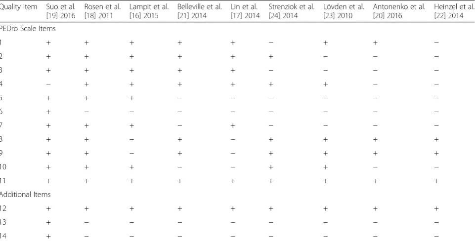
Quality assessment of the included studies
The quality of studies included in this systematic review varied substantially (Table 4). On average, the nine in-cluded studies met 7 of the 11 PEDro criteria. Two stud-ies of the highest quality [18, 19] meeting 9 of the 10 PEDro criteria; however, five [17, 20, 22–24] studies failed to meet five or more study quality criteria. Item 11 (i.e., included point measures and variability measures) was met for all nine studies. Item 8 (key outcome mea-sured for 85% of subjects) and nine (outcome data ana-lyzed by intention to treat) were met by seven of the nine studies [18–24]. Item 6, (i.e., blinding of all who ad-ministered the training) commonly received a negative response (i.e., one of the studies [19] blinded training administers). Frequent issues were failure to meet or re-port: 1) allocation concealment (n = 4) [20, 22–24]; 2)
blinding of all subjects (n= 6) [17, 20–24]; 3) blinding of
all who administered the training (n= 8) [16–18, 20–24]; 4)
blinding of assessors who measured at least one key outcome (n= 5) [20–24]; and 5) between-group
statis-tical comparisons for at least one key outcome (n= 4) [17, 20–22]. Item 9 (participants with available outcome measures received the treatment or control condition allocated) received 78% overall rater agreement between the authors [LTB and CKB], where the remaining ques-tions received a 100% overall rater agreement between the authors [LTB and CKB].
size calculation) and 14 (reported compliance) were not addressed by eight studies [16–18, 20–24].
Discussion
Findings from two high-quality studies examining the ef-fect of CCT on volumetric changes, suggest that multi-domain CCT programs with a duration ranging from 12 to 26 weeks could result in an increase in grey matter density [16], but in contrast could also result in a decrease in cortical thickness in the posterior cingulate [19]. This indicates that in a relatively short time span, multi-domain CCT might be able to alter brain structure. However, the overall heterogeneity of the findings between studies (i.e., potential functional improvements versus declines), which could be in part due to the differences in region of interest,
makes it difficult to draw definitive conclusions regarding the effect of CCT on brain structure.
Task-based functional brain activity decreased after training of a single task [21]; however, an increase in task-based brain activation was found in a more com-plex dual-task training [21] and a multi-domain CCT program [18]. This highlights that the CCT method (i.e., multi-domain versus single domain CCT) may play a critical role in task-based functional brain activity. Conversely, multi-domain CCT did not result in changes in structural connectivity [16], where an auditory perception-training program resulted in increased AD [24]. Resting-state functional connectivity was found to increase [16, 19] or decrease [16, 19, 24] depending on training type (e.g., single- versus multi-domain) and re-gion of interest. Below, we will discuss as to why we
Table 4Quality Assessment of Included Studies (N= 9)
Quality item Suo et al. [19] 2016
Rosen et al. [18] 2011
Lampit et al. [16] 2015
Belleville et al. [21] 2014
Lin et al. [17] 2014
Strenziok et al. [24] 2014
Lövden et al. [23] 2010
Antonenko et al. [20] 2016
Heinzel et al. [22] 2014
PEDro Scale Items
1 + + + + + − + + −
2 + + + + + + − − −
3 + + + + + − − − −
4 − + + + + + + − −
5 + + + − − − − − −
6 + − − − − − − − −
7 + + + − + − − − −
8 + + − + − + + + +
9 + + − + − + + + +
10 + + + − − + + − −
11 + + + + + + + + +
Additional Items
12 + + + + + + + + +
13 + − − − − − − − −
14 + − − − − − − − −
PEDro scoring system: receive a point (+) for each item that is met. When criteria were not met (−), no points were given
The maximum number of points is 10, which means excellent quality based on PEDro’s quality assessment
Additional Quality Assessment Items: Maximum score of 3 PEDro Scale
1. Eligibility criteria were specified (this item is not used to calculate the PEDro score) 2. Subjects were randomly allocated to groups
3. Allocation was concealed
4. The groups were similar at baseline regarding the most important prognostic indicators 5. There was blinding of all subjects
6. There was blinding of all therapists who administered the therapy 7. There was blinding of all assessors who measured at least one key outcome
8. Measures of at least one key outcome were obtained from more than 85% of the subjects initially allocated to groups
9. All subjects for whom outcome measures were available received the treatment or control condition as allocated or, where this was not the case, data for at
least one key outcome was analyzed by“intention to treat”
10. The results of between-group statistical comparisons are reported for at least one key outcome 11. The study provides both point measures and measures of variability for at least one key outcome Additional Items
12. Was cognition measured to assist the interpretation of neuroimaging results? 13. Was there a sample size calculation?
might see a discrepancy between single- and multi-domain CCT effects, and why this discrepancy might affect both structural and resting-state functional con-nectivity differently.
Task-based functional activity
Functional activation patterns in the brain change with aging as a result of neurophysiological changes. Compared with younger adults, functional activation patterns be-come less coordinated and localized in older adults, which result in loss of cognitive performance [25]. In the current review, three studies looked at functional activity in the brain while performing a task in the scanner. Activity levels in the brain while performing a task were both in-creased and dein-creased, depending on the type of training and region of interest. All three studies focused on differ-ent brain regions, which makes comparison difficult. However, results suggest that engaging in a more diverse or complex training (e.g., multi-domain CCT or dual-task training) might lead to an increased functional activation [18, 21] compared with training of a single task [21, 22]. In contrast, a short report focusing on transfer of training showed results that five weeks of training (i.e., letter mem-ory and updating tasks) resulted in increases in task-related functional activity in the striatum compared with a passive control group [26]. Though, besides the focus on different brain regions, the vast differences in study de-sign, such as the training duration, the presence or ab-sence of a control group, and the small number of studies ask for prudence for making assumptions.
Structural connectivity and type of training
DTI is an imaging technique used to determine the white matter microstructure of the brain by looking at how water molecules diffuse within the brain (i.e., the direction and amount of diffusion) [27, 28]. DTI is often quantified by measures such as FA and MD; which pro-vide information about the direction of diffusivity and molecular diffusion rate, respectively. Decreases in FA and increases in MD might indicate lower levels of mye-lin or the presence of axonal injury, as water molecules are able to diffuse more freely (i.e., isotropic) [29, 30]. However, rather than looking at one specific DTI scalar (e.g., FA, MD), scalars need to be combined with other neuroimaging measures (e.g., T2, PD, FLAIR) to give a more detailed and accurate picture of for example white matter abnormalities that might occur within the brain [30]. Studies have linked loss of white matter integrity, as measured with DTI, to be associated with age-related cognitive decline in otherwise healthy older adults [31]. In addition, a meta-analysis focusing on DTI in MCI and Alzheimer’s Disease found increased MD in both MCI and Alzheimer’s Disease, as well as decreased FA in
Alzheimers’ Disease compared with controls. More se-vere levels of Alzheimer’s Disease (i.e., lower scores on the Mini-Mental State Examination) were associated with reductions in FA [32].
Few studies looked at the effect of CCT on structural connectivity using DTI. One study of moderate-to-high quality (PEDro score of 7/10) found no changes in struc-tural connectivity after 12 weeks of multi-domain CCT, which could be due to the small sample size [16]. These findings are in contrast with a quasi-experimental study [22] that found that an average of 100 h of training over four months resulted in decreased MD and increased FA in the genu of the corpus callosum. These findings sug-gest that multi-domain CCT is able to alter white matter microstructure in the brain in older adults. This finding could be promising as disruptions in white matter organization are often paired with cognitive decline [33]. However, a limitation of this quasi-experimental study is the lack of an active control group. Thus, we need more high quality studies to replicate these findings and to examine how multi-domain CCT might be able to alter white matter microstructure.
Increases in AD in the right occipito-temporal white matter were found in a study examining the effect of an adaptive auditory perception computer game (i.e., single-domain). This increased AD was correlated with a lower score in everyday problem solving and spatial working memory accuracy [24]. However, due to the absence of an included control group, this study used contrasts between the three training groups to look at improve-ments between groups. Therefore, results will more likely provide information about the effect of the train-ing groups in relation to each other (i.e., which interven-tion shows the best results), than give informainterven-tion whether the intervention actually works.
Functional connectivity and type of training
Resting-state fMRI is used to map networks in the brain, such as the well-established Default Mode Network (DMN) and the Central Executive Network (CEN). These networks are activated in both the presence [34] or the ab-sence of a (cognitive) task [35, 36]. In patients with MCI or Alzheimer’s Disease, these functional networks in the brain are found to be disrupted [37, 38]. In addition, we can measure functional networks in the brain while per-forming a task with task-based fMRI.
left superior frontal lobe [19]. Additionally, a study with the same CCT program (i.e., COGPACK) found that multi-domain CCT resulted in increased resting-state functional connectivity between the right hippocampus and the left superior temporal gyrus after only three weeks of training [16]. These improvements in resting-state functional connectivity were significantly correlated with improved memory performance [19] and changes in glo-bal cognition at follow-up [16], respectively.
In accordance, an RCT of multi-domain CCT in older adults with a history of a stroke [17] found that CCT in-creased resting-state functional connectivity between the hippocampus and both the inferior frontal gyrus and the middle frontal gyrus. These increases in resting-sate func-tional connectivity were associated with significant positive changes in memory quotient and processing speed (Trail Making Test-A). Literature shows that resting-state func-tional connectivity between the hippocampus and the su-perior frontal lobe is reduced in MCI [37, 38]. Therefore, the current findings might indicate that multi-domain CCT could lead to improved cognitive performance through strengthening hippocampal functional networks and pre-venting memory loss that might be manifested by loss in hippocampal functional connectivity. However, the bio-logical underpinnings of this change in connectivity are still unclear. Current histological findings suggest training in-duced neuroplasticity could be a result of dendritic branch-ing, synaptogenesis or other factors such as angiogenesis [39]. Besides more human studies, we need to combine knowledge acquired from both human and animal (histo-logical analyses), to help understand how multi-domain CCT could result in these functional changes in the brain.
Immediate comparison between the results of a single-versus multi-domain program can be made within one quasi-experimental study [24]. Participants in this study were randomly assigned to one of three included cognitive training programs. Participants who were randomized in Brain Fitness, a training program considered more single-domain in nature, showed decreased resting-state func-tional connectivity between the superior parietal cortex and the inferior temporal lobe. In contrast, participants who were assigned to Rise of Nation, a more multi-domain training, showed increased resting-state functional connect-ivity between the superior parietal cortex and the inferior temporal lobe. This contrast could be due to the nature of the training (i.e., single-domain versus multi-domain), as another quasi-experimental study [22] of single-domain CCT showed no changes in task-based functional connect-ivity following training.
A recent study [40] comparing non-computerized single-domain and multi-domain training found that multi-domain cognitive training mainly resulted in in-creased memory proficiency, while single-domain train-ing primarily – but not only - enhances visuospatial
and attentional benefits. Results of the current system-atic review are in accordance with these findings, as the multi-domain CCT shows improvements in resting-state functional connectivity of hippocampus-frontal lobe and hippocampus-temporal lobe, which was asso-ciated with improvements in memory. Single-domain CCT did not result in similar findings. Gains in cogni-tion resulting from multi-domain were more prone to sustain compared to gains acquired in single-domain cognitive training. Thus, multi-domain cognitive train-ing might result in more widespread gains in cognitive functions, which maintain visible over a longer period of time compared to single-domain cognitive training.
Quality assessment
The quality of studies was heterogeneous. Commonly missed criteria, were those that focused on blinding of par-ticipants, blinding of individuals who delivered the CCT, and blinding of the assessors. These issues could result into bias (i.e., either positively or negatively) during training and follow-up measurements due to expectations of both study examiners (treatment delivery or assessors) and partici-pants. However, five [20–24] of the nine included studies were quasi-experimental and therefore the key characteris-tic of the more superior RCT, randomization into either an experimental or a control group, was lacking in these studies. The absence of a proper control group in these five quasi-experimental designs affects the interpret-ation of the results of the study; instead of whether a treatment works, quasi-experimental studies provide information on whether an intervention is more effect-ive than a standard or alternateffect-ive treatment.
Finally, of the three additionally included quality assess-ment criteria (i.e., item 12–14) two criteria (i.e., sample size calculation, compliance reported) were only met by one study [19]. The absence of sample size calculations and re-ported compliance in the remaining studies [16–18, 20–24], could result in a lack of power, which increases the chances of false negatives (i.e., type-II errors). This could mean that potential effects of CCT on neuroimaging parameters simply could not be detected due to a small sample size, and not because they were not present.
Limitations
addition, the heterogeneity of the findings in this systematic review might also be due to the large vari-ability in type of training (single- versus multi-domain) and the dosage and duration of training (i.e., days versus months). Thus, the heterogeneous nature of the study designs in this review makes it difficult to draw conclusions. To better understand the relevant mecha-nisms of CCT, neuroimaging outcomes need to be accom-panied with behavioural data. Furthermore, there are limited investigations regarding the transfer effects of CCT and the pattern of neuroplasticity associated with transfer. A high-quality study design, which includes for example an active control group, a literature-based training duration and dosage, and a sample size calcula-tion, would help increase the consistency and compar-ability of findings, which in turn would help increase the ability to draw appropriate conclusions.
Conclusions
This systematic review is an essential first step towards understanding the complex volumetric and functional changes, as well as changes in structural and functional connectivity that underlie CCT in older adults. However, the highly heterogeneous nature of the results in this systematic review, potentially due to the large variability in study design, indicates that more high-quality studies are needed to confirm and expand upon these findings. In addition, these studies do not provide information regarding the physiological and cellular mechanisms caus-ing these structural changes. More histological studies are needed to gain insight whether these CCT induced changes might be a result of for example neurogenesis or synaptic plasticity. Future studies should focus on multi-domain CCT, since this type of training has the potential to induce more widespread and long-lasting effects on cognition.
Abbreviations
AD:Axial diffusivity; BF: Brain fitness; BOLD: Blood oxygen level dependent; CCT: Computerized cognitive training; CEN: Central executive network; DMN: Default mode network; DTI: Diffusion tensor imaging; FA: Fractional anisotropy; fMRI: functional magnetic resonance imaging; ITL: Inferior temporal lobe; MCI: Mild cognitive impairment; MD: Mean diffusivity; MRI: Magnetic resonance imaging; PEDro: Physiotherapy evidence database; PRT: Progressive resistance training; RCT: Randomized controlled trial; RON: Rise of Nation; rsfMRI: resting-state functional magnetic resonance imaging; SF: Space fortress; SPL: Superior parietal cortex
Funding
This work was supported by funding from the Jack Brown and Family Alzheimer Research Foundation Society.
Availability of data and materials
All data supporting the conclusions of this article are included within the article.
Authors’contributions
LTB wrote the first draft of the manuscript. JCD and CKB provided helped with data extraction and drafting of tables. TLA and JCD conceived the study concept and design. TLA, JCD, and CKB wrote portions of the manuscript and critically reviewed the manuscript. All authors (TLA, JCD, CKB and LTB) have read and approved the manuscript.
Authors’information
LTB is a Mitacs Accelerate Doctoral Trainee. CKB is a Michael Smith Foundation for Health Research Postdoctoral Fellow. TLA is a Canada Research Chair in Physical Activity, Mobility and Cognitive Neuroscience.
Ethics approval and consent to participate
Not applicable.
Consent for publication
Not applicable.
Competing interests
The authors declare that they have no competing interests.
Publisher’s Note
Springer Nature remains neutral with regard to jurisdictional claims in published maps and institutional affiliations.
Received: 11 October 2016 Accepted: 30 June 2017
References
1. World Health Organization: Governments commit to advancements in dementia research and care. http://www.who.int/mediacentre/news/ releases/2015/action-on-dementia/en/. Accessed 1 Aug 2016.
2. Polidori MC, Nelles G, Pientka L. Prevention of dementia: focus on lifestyle. Int J Alzheimers Dis. 2010;2010:1–9.
3. Ngandu T, Lehtisalo J, Solomon A, Levalahti E, Ahtiluoto S, Antikainen R, Backman L, Hanninen T, Jula A, Laatikainen T, et al. A 2 year multidomain intervention of diet, exercise, cognitive training, and vascular risk monitoring versus control to prevent cognitive decline in at-risk elderly people (FINGER): a randomised controlled trial. Lancet. 2015;385(9984):2255–63.
4. Valenzuela M, Sachdev PS. Harnessing brain and cognitive reserve for the prevention of dementia. Indian J Psychiatry. 2009;51(Suppl 1):S16–21. 5. Valenzuela MJ, Sachdev P. Brain reserve and dementia: a systematic review.
Psychol Med. 2006;36(4):441–54.
6. Lampit A, Hallock H, Valenzuela M. Computerized cognitive training in cognitively healthy older adults: a systematic review and meta-analysis of effect modifiers. PLoS Med. 2014;11(11):e1001756.
7. Lampit A, Hallock H, Moss R, Kwok S, Rosser M, Lukjanenko M, et al. The timecourse of global cognitive gains from supervised computer-assisted cognitive training: a randomised, active-controlled trial in elderly with multiple dementia risk factors. J Prev Alzheimers Dis. 2014;1(1):33–9. 8. Smith GE, Housen P, Yaffe K, Ruff R, Kennison RF, Mahncke HW, Zelinski EM.
A cognitive training program based on principles of brain plasticity: results from the Improvement in Memory with Plasticity-based Adaptive Cognitive Training (IMPACT) study. J Am Geriatr Soc. 2009;57(4):594–603.
9. Basak C, Boot WR, Voss MW, Kramer AF. Can training in a real-time strategy video game attenuate cognitive decline in older adults? Psychol Aging. 2008;23(4):765–77.
10. Bruel-Jungerman E, Davis S, Laroche S. Brain plasticity mechanisms and memory: a party of four. Neuroscientist. 2007;13(5):492–505. 11. Citri A, Malenka RC. Synaptic plasticity: multiple forms, functions, and
mechanisms. Neuropsychopharmacology. 2008;33(1):18–41.
12. Guerra-Carrillo B, Mackey AP, Bunge SA. Resting-state fMRI: a window into human brain plasticity. Neuroscientist. 2014;20(5):522–33.
13. Olesen PJ, Westerberg H, Klingberg T. Increased prefrontal and parietal activity after training of working memory. Nat Neurosci. 2004;7(1):75–9. 14. Moher D, Liberati A, Tetzlaff J, Altman DG, Group P. Preferred reporting
items for systematic reviews and meta-analyses: the PRISMA statement. Ann Intern Med. 2009;151(4):264–9. W264
15. Maher CG, Sherrington C, Herbert RD, Moseley AM, Elkins M. Reliability of the PEDro scale for rating quality of randomized controlled trials. Phys Ther. 2003;83(8):713–21.
16. Lampit A, Hallock H, Suo C, Naismith SL, Valenzuela M. Cognitive training-induced short-term functional and long-term structural plastic change is related to gains in global cognition in healthy older adults: a pilot study. Front Aging Neurosci. 2015;7:14.
Resting-state functional magnetic resonance imaging study. J Int Med Res. 2014;42(3):659–68.
18. Rosen AC, Sugiura L, Kramer JH, Whitfield-Gabrieli S, Gabrieli JD. Cognitive training changes hippocampal function in mild cognitive impairment: a pilot study. J Alzheimers Dis. 2011;26(Suppl 3):349–57.
19. Suo C, Singh MF, Gates N, Wen W, Sachdev P, Brodaty H, Saigal N, Wilson GC, Meiklejohn J, Singh N, et al. Therapeutically relevant structural and functional mechanisms triggered by physical and cognitive exercise. Mol Psychiatry. 2016; 20. Antonenko D, Kulzow N, Cesarz ME, Schindler K, Grittner U, Floel A.
Hippocampal pathway plasticity is associated with the ability to form novel memories in older adults. Front Aging Neurosci. 2016;8:61.
21. Belleville S, Mellah S, de Boysson C, Demonet JF, Bier B. The pattern and loci of training-induced brain changes in healthy older adults are predicted by the nature of the intervention. PLoS One. 2014;9(8):e102710.
22. Heinzel S, Lorenz RC, Brockhaus WR, Wustenberg T, Kathmann N, Heinz A, Rapp MA. Working memory load-dependent brain response predicts behavioral training gains in older adults. J Neurosci. 2014;34(4):1224–33. 23. Lovden M, Bodammer NC, Kuhn S, Kaufmann J, Schutze H, Tempelmann C,
Heinze HJ, Duzel E, Schmiedek F, Lindenberger U. Experience-dependent plasticity of white-matter microstructure extends into old age. Neuropsychologia. 2010;48(13):3878–83.
24. Strenziok M, Parasuraman R, Clarke E, Cisler DS, Thompson JC, Greenwood PM. Neurocognitive enhancement in older adults: comparison of three cognitive training tasks to test a hypothesis of training transfer in brain connectivity. NeuroImage. 2014;85(Pt 3):1027–39.
25. Bishop NA, Lu T, Yankner BA. Neural mechanisms of ageing and cognitive decline. Nature. 2010;464(7288):529–35.
26. Dahlin E, Neely AS, Larsson A, Backman L, Nyberg L. Transfer of learning after updating training mediated by the striatum. Science. 2008;320(5882):1510–2. 27. Mori S. Introduction to diffusion tensor imaging. 1st ed. Amsterdam: Elsevier
Science; 2007.
28. Mori S, Zhang J. Principles of diffusion tensor imaging and its applications to basic neuroscience research. Neuron. 2006;51(5):527–39.
29. Soares JM, Marques P, Alves V, Sousa N. A hitchhiker’s guide to diffusion tensor imaging. Front Neurosci. 2013;7:31.
30. Alexander AL, Lee JE, Lazar M, Field AS. Diffusion tensor imaging of the brain. Neurotherapeutics. 2007;4(3):316–29.
31. Kerchner GA, Racine CA, Hale S, Wilheim R, Laluz V, Miller BL, Kramer JH. Cognitive processing speed in older adults: relationship with white matter integrity. PLoS One. 2012;7(11):e50425.
32. Sexton CE, Kalu UG, Filippini N, Mackay CE, Ebmeier KP. A meta-analysis of diffusion tensor imaging in mild cognitive impairment and Alzheimer’s disease. Neurobiol Aging. 2011;32(12):2322 e2325–18.
33. Bartzokis G. Alzheimer's disease as homeostatic responses to age-related myelin breakdown. Neurobiol Aging. 2011;32(8):1341–71.
34. Seeley WW, Menon V, Schatzberg AF, Keller J, Glover GH, Kenna H, Reiss AL, Greicius MD. Dissociable intrinsic connectivity networks for salience processing and executive control. J Neurosci. 2007;27(9):2349–56. 35. Raichle ME, MacLeod AM, Snyder AZ, Powers WJ, Gusnard DA, Shulman
GL. A default mode of brain function. Proc Natl Acad Sci U S A. 2001; 98(2):676–82.
36. Greicius MD, Krasnow B, Reiss AL, Menon V. Functional connectivity in the resting brain: a network analysis of the default mode hypothesis. Proc Natl Acad Sci U S A. 2003;100(1):253–8.
37. Bai F, Watson DR, Yu H, Shi Y, Yuan Y, Zhang Z. Abnormal resting-state functional connectivity of posterior cingulate cortex in amnestic type mild cognitive impairment. Brain Res. 2009;1302:167–74.
38. Wang Z, Liang P, Jia X, Qi Z, Yu L, Yang Y, Zhou W, Lu J, Li K. Baseline and longitudinal patterns of hippocampal connectivity in mild cognitive impairment: evidence from resting state fMRI. J Neurol Sci. 2011;309(1–2):79–85.
39. Zatorre RJ, Fields RD, Johansen-Berg H. Plasticity in gray and white: neuroimaging changes in brain structure during learning. Nat Neurosci. 2012;15(4):528–36.
40. Cheng Y, Wu W, Feng W, Wang J, Chen Y, Shen Y, Li Q, Zhang X, Li C. The effects of multi-domain versus single-domain cognitive training in non-demented older people: a randomized controlled trial. BMC Med. 2012;10:30.
41. Graham DP, Cully JA, Snow AL, Massman P, Doody R. The Alzheimer’s Disease Assessment Scale-Cognitive subscale: normative data for older adult controls. Alzheimer Dis Assoc Disord. 2004;18(4):236–40.
42. Randolph C, Tierney MC, Mohr E, Chase TN. The repeatable battery for the assessment of neuropsychological status (RBANS): preliminary clinical validity. J Clin Exp Neuropsychol. 1998;20(3):310–9.
43. Dwolatzky T, Whitehead V, Doniger GM, Simon ES, Schweiger A, Jaffe D, Chertkow H. Validity of a novel computerized cognitive battery for mild cognitive impairment. BMC Geriatr. 2003;3:4.
44. Sahakian BJ, Owen AM. Computerized assessment in neuropsychiatry using CANTAB: discussion paper. J R Soc Med. 1992;85(7):399–402.
45. Robbins TW, James M, Owen AM, Sahakian BJ, McInnes L, Rabbitt P. Cambridge neuropsychological test automated battery (CANTAB): a factor analytic study of a large sample of normal elderly volunteers. Dementia. 1994;5(5):266–81.
46. Wechsler D. A standardized memory scale for clinical use. Aust J Psychol. 1945;19:87–95.
47. Hachinski V, Iadecola C, Petersen RC, Breteler MM, Nyenhuis DL, Black SE, Powers WJ, DeCarli C, Merino JG, Kalaria RN, et al. National Institute of Neurological Disorders and Stroke-Canadian Stroke Network vascular cognitive impairment harmonization standards. Stroke. 2006;37(9):2220–41. 48. Wechsler D. Wechsler Adult Intelligence Scale. 3rd ed. San Antonio:
Psychological Corporation; 1997.
49. Willis SLM, M. Manual for the Everyday Problems Test. University Park: Pennsylvania State University; 1993.
50. Schaie KW. Manual for the Schaie-Thurstone adult mental abilities test (STAMAT). Palo Alto: Consulting Psychological Press; 1985.
51. Wechsler D. Wechsler memory scale–revised: manual. San Antonio: Psychology Corporation; 1987.
52. Wechsler D. Wechsler memory scale—revised. San Antonio: Psychological Corporation; 2009.
53. Greenwood PM, Lambert C, Sunderland T, Parasuraman R. Effects of apolipoprotein E genotype on spatial attention, working memory, and their interaction in healthy, middle-aged adults: results From the National Institute of Mental Health’s BIOCARD study. Neuropsychology. 2005;19(2):199–211. 54. Parasuraman R, Greenwood PM, Kumar R, Fossella J. Beyond heritability:
neurotransmitter genes differentially modulate visuospatial attention and working memory. Psychol Sci. 2005;16(3):200–7.
55. Floel A, Suttorp W, Kohl O, Kurten J, Lohmann H, Breitenstein C, Knecht S. Non-invasive brain stimulation improves object-location learning in the elderly. Neurobiol Aging. 2012;33(8):1682–9.
56. Helmstadter CL, M. & Lux S. Verbaler Lern- und Merkfähigkeitstest (VLMT), Manual. Göttingen: Belz-Test; 2001.
57. Cohen JD, Perlstein WM, Braver TS, Nystrom LE, Noll DC, Jonides J, Smith EE. Temporal dynamics of brain activation during a working memory task. Nature. 1997;386(6625):604–8.
58. Brickenkamp R. Test d2. The d2 test of attention. 9th ed. Goettingen: Hogrefe; 2002.
59. Benton AH, K. Multilingual aphasia examination. Iowa City: AJA Associates; 1989. 60. Stroop JR. Studies of Interference in serial verbal reactions. J Exp Psychol.
1935;18:643–62.
61. Raven J, Summer B, Birchfield M, Brosier G, Burciaga L, Bykrit B. Manual for raven’s progressive matrices and vocabulary scales. Research supplement no. 3: a compendium of North American normative and validity studies. Oxford: Oxford Psychologists Press; 1990.
62. Horn W: Leistungspruefsystem LPS, vol. Hogrefe, 2 edn. Goettingen: Germany; 1983.
• We accept pre-submission inquiries
• Our selector tool helps you to find the most relevant journal
• We provide round the clock customer support
• Convenient online submission
• Thorough peer review
• Inclusion in PubMed and all major indexing services
• Maximum visibility for your research
Submit your manuscript at www.biomedcentral.com/submit