Original Research Article.
Prevalence of Odontogenic Keratocyst in Kashmir Valley:
A Retrospective Analysis Over 10 Years
Afreen Nadaf
1*, Shahid Hassan
2, Ajaz Shah
31*Tutor, Department of Oral Pathology, 2Assistant Professor, Department of OMFS, 3Professor and Head, Department of OMFS,
Government Dental College and Hospital Srinagar, Jammu and Kashmir, India.
ABSTRACT
Objective: This study aimed to assess the demographic characteristics of odontogenic keratocyst in a tertiary dental healthcare center in Kashmir valley.
Materials and Methods: A retrospective analysis of data of 1460 cases obtained between 2007 to 2017 from clinical records of the patients visiting department of oral surgery, Govt dental college and hospital Srinagar was assessed. Information regarding age distribution, gender, anatomical location and recurrence was documented.
Results: In the present study the overall prevalence of the odontogenic keratocyst was 16.7%. The patients with odontogenic keratocysts varied in age from 9 to 65 years, and the mean age of patients was 28.9 years. 26 cases of odontogenic keratocyst were seen during third decade of life followed by 21 cases reported during the second decade of life. Males were found to be more commonly affected than females. The mandible (59.7%) was more often affected than the maxilla mainly mandibular molar to ramus area (n=41). The majority 46 (52.88%) of these lesions were asymptomatic and 41(47.12%) patients had symptoms such as swelling, pain, pus discharge, trismus or fever due to pericoronitis. 21% of the patients presented recurrence after the surgery mainly in second and third decade of life.
Conclusion: Odontogenic keratocysts comprised of 16.7% of
oral lesions. The lesions mainly affected the males and were seen in younger age groups. Hence these data should be taken into consideration to improve planning of individual treatment and follow-up.
Keywords: Odontogenic Keratocyst, Prevalence, Oral
Lesions.
*Correspondence to:
Dr. Afreen Nadaf, Tutor,
Department of Oral Pathology,
Government Dental College and Hospital Srinagar, Jammu and Kashmir, India. Article History:
Received: 09-11-2017, Revised: 03-12-2017, Accepted: 27-12-2017
Access this article online
Website:
www.ijmrp.com
Quick Response code
DOI:
10.21276/ijmrp.2018.4.1.019
INTRODUCTION
Odontogenic keratocyst (OKC) is defined as “a benign uni- or multicystic, intraosseous tumour of odontogenic origin, with a characteristic lining of parakeratinized stratified squamous epithelium and potential for aggressive, infiltrative behaviour.
Philipsen in 1956 coined the term “odontogenic keratocyst”
because of his thought that these were odontogenic cysts and not inflammatory in origin.1 In contrast there were observations which
showed that the odontogenic keratocyst behaved more as a neoplasm and not like a cystic lesion. Finally, in the World Health Organization classification (2005), the former odontogenic keratocyst was added to the benign odontogenic tumours
category and the new term “keratocystic odontogenic tumor” was proposed.2 Odontogenic cysts, conceptually inseparable
from odontogenic tumors were omitted from the 2005 classification and have been reincorporated into the 2017 classification as odontogenic cyst.3
Odontogenic Keratocyst (OKC) is the third most common cyst occurring in the oral cavity and constitutes about 5.4- 17.4 percent of all odontogenic cysts.4 They are most common gnathic lesions,
arising mainly in the mandible particularly in the third molar region, mandibular angle, ramus, with a mandible-maxilla ratio of 2:1.1. This neoplasia is predominantly found in males and people of white origin. It can appear at any age however, it is more frequent between the ages of 20 and 30.5 Occasionally they have been
found to arise in a peripheral location especially in the gingiva
which has earned them the term “peripheral odontogenic keratocysts.” 6 Odontogenic keratocyst arises as an extension of
resorbing factors (enzyme activity in osteolysis). It is considered that the main cause for the origin of this lesion is a lack of regulation and a mutation in the PTCH gene13.7,8
Diagnosis of odontogenic keratocyst is mainly done by histopathological analysis, biopsy and radiographic examination. The radiographic appearance of OKC may range from a small unilocular radiolucency to a large multilocular radiolucency. It has long been of particular interest because of its potential for local destructive behavior, its recurrence rate and its tendency for multiplicity particularly when associated with nevoid basal cell carcinoma syndrome.9 There seems to be regional variations in
the distribution of odontogenic cysts and tumors in the literature. Very few studies have been reported among Asians, especially from India. Epidemiological data on odontogenic keratocyst are lacking from Kashmir valley. Hence this study was undertaken to address the demographic characteristics of odontogenic keratocyst in a tertiary dental healthcare center in Kashmir valley.
MATERIALS AND METHODS
A retrospective analysis of data obtained between 2007 to 2017 from clinical records of the patients visiting department of Oral surgery, Govt dental college and hospital Srinagar and pathology was carried out.
1460 cases of odontogenic cysts and tumors were selected. The study included patients having detailed information on age, lesion location, gender and recurrence. The diagnosis was confirmed by revaluation of hemotoxylin and eosin stained specimens using the diagnostic criteria outlined in the latest WHO classification of the cysts and tumors. Cases with incomplete records were excluded from the study. The study data was reviewed by a single observer. The following variables were studied: anatomical location, age group, gender and recurrence. The collected data was reviewed, organized, tabulated and subjected to descriptive analyses and were represented as frequency and percentages according to the gender.
Table 1: Distribution of odontogenic keratocyst among different age groups
Age group Male Female Total
<10 1 0 1
10-20 11 10 21
21-30 14 12 26
31-40 8 5 13
41-50 6 6 12
51-60 5 4 9
>60 3 2 5
Total 48 39 87
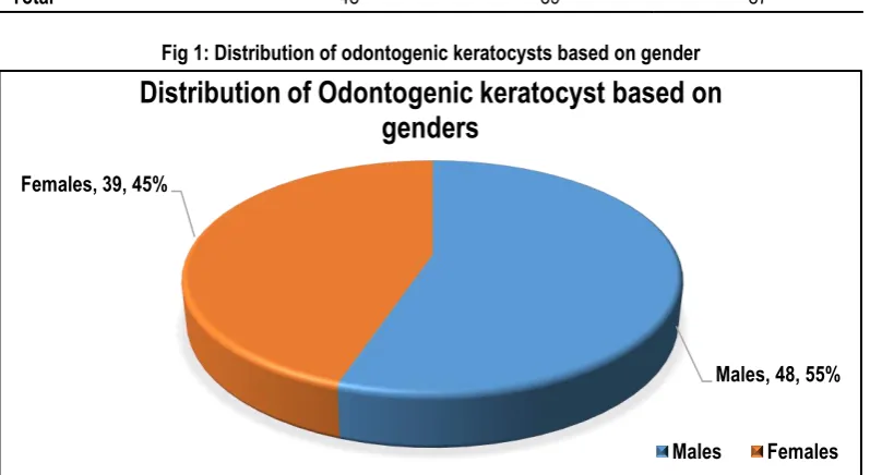
Fig 1: Distribution of odontogenic keratocysts based on gender
RESULTS
A total of 87 specimens out of a total of 1460 oral biopsies of odontogenic cysts and tumors evaluated histopathologically were designated as odontogenic keratocyst corresponding to overall prevalence of 16.7%. The age ranges from 9 to 65 years with mean age of 28.9 years. 26 cases of odontogenic keratocyst were seen during third decade of life followed by 21 cases reported during the second decade of life depicted in Table 1. The gender distribution showed a male predilection (n=48) which corresponds to a male: female ratio of about 1.23:1 (Table 2). Among 87 cases of odontogenic keratocyst, 41(47.12%) patients had symptoms such as swelling, pain, pus discharge, trismus or fever due to
pericoronitis. The remaining 46 (52.88%) patients were asymptomatic.
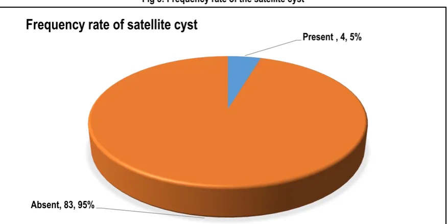
Mandible was the most common jaw involved (59.7%) mainly the mandibular posterior region mainly the mandibular molar to ramus area (n=41), followed by multiple location (n=13), maxillary extent location (n=10). The distribution based on different anatomic sites is shown in Table 3. Of the total 87 number of patients with odontogenic cysts, 21% of the patients presented recurrence after the surgery mainly in second and third decade of life. Of the 87 cases of odontogenic keratocyst 4% cases of satellite cyst were reported.
Males, 48, 55% Females, 39, 45%
Distribution of Odontogenic keratocyst based on
genders
Fig 2: Distribution of odontogenic keratocysts based on location
Fig 3: Frequency rate of the satellite cyst
DISCUSSION
Odontogenic keratocyst is a benign entity arising from remnants of the dental lamina with a relatively high prevalence. It has a potential aggressive behavior and a high recurrence rate, hence it requires special attention. Multiple authors have reported their invasive potential, due to its capacity to slowly grow in the anteroposterior marrow spaces, and can be transformed into a large lesion without causing notorious expansion.10 The
odontogenic keratocystic lesions experience geographical variations in different regions of the world and even the variation exists within the country. No previous study reported the patient demographics of odontogenic keratocyst for Kashmiri population. Hence study was conducted to assess the distribution of odontogenic keratocyst in Kashmir valley.
In the present retrospective study, 87 cases of odontogenic keratocyst were reported with an overall prevalance of 16.7%, presenting a lower frequency when compared with other studies
investigated by Siriwardena et al among Sri Lanka population, showing odontogenic keratocyst incidence of 25.7%, Servato et al and Luo et al with 31.7% , 38.73% incidence respectively.11-13 The
present finding is consistent with the studies conducted by Meningaud et al. and Tawfik et al.14,15 The study by Baghaei et al
reported the prevalence of odontogenic keratocyst of 18.6%.16
This variation among different population indicates the racial and environmental factors might probably influence on development of these lesions. The other probable reason could be due to the attitudes of the patients towards oral health. Many patients do not have the habit of routine regular dental check-up and only report to the clinics for symptomatic treatment and often engage in self-medications to relieve pain.
According to literature odontogenic keratocyst occurs over a wide age range with a peak incidence in the second and third decade of life and a gradual decline thereafter.17 Similar results were seen
5 6
41
5 7
10 13
0 10 20 30 40 50
1
Per
ce
nt
ag
e o
f OKC
Location of OKC
Distribution of odontogenic keratocyst based on location
Mandibular anteriors Mandibular posteriors Mandibular molar to ramus area
Maxillary anterior Maxillaryposterior Maxillary extent location
Multiple location
Present , 4, 5%
Absent, 83, 95%
in our study with a mean age 28.9. This outcome supports the age occurrence reported by other investigators on Turkish and Indian population.18,19 Studies by Myoung et al and Maurette et al
reported third decade of life as the most common age of co-occurence.20,21 A systematic review of odontogenic keratocyst
revealed a mean age of 36.5 years with a peak of incidence in the second and third decades of life.22 However, Kakarantza and
Nicolatou found a peak incidence in 5th and 6th decades of life.25
Gender distribution of odontogenic keratocyst in the present study showed a slightly higher male predominance with male –female ratio 1.23:1.The present finding is concordance with the findings reported by Sharma et al, Dong et al and Selvamani et al where male predominance is seen over females.17,24,25 Chirapathomsakul
et al contradict the literature data, presenting a higher frequency in the female gender with male to female ratio (1:1.2) among Thai population and Maurette et al found a male to female ratio of 1:2.1 in Brazilian patients.21,26
Odontogenic keratocyst have a tendency to occur in any part of the mandible and maxilla, but the majority, almost 70%, arise in the body of the mandible.27,28 The next common site is the
maxillary canine region.4,17 According to the pathology reports in
the present study, odontogenic keratocyst was chiefly located in the posterior part of the mandible mainly the ramus of the mandible (68.8%). This finding is concomitant to previous studies indicating posterior region of mandible as the main location of OKC.11,29 Goteti reported of odontogenic keratocyst location with a
mandible-to-maxilla ratio of 1.6:1.30 Odontogenic keratocyst of the
soft tissue have been reported in other anatomic sites like the buccal mucosa and masticatory muscles apart from the gingiva.6
Several researchers have reported that 50-90% of odontogenic keratocyst are symptomatic at the time of diagnosis.31 According
to the results of this study, 47.2% presented with symptoms chiefly swelling. trismus and pericoronitis which is in agreement with the results of other studies.20,32 Generally odontogenic
keratocyst of the jawbone do not produce a large expansion of the cortex as the cyst proliferates within the marrow spaces, thus growing in a longitudinal manner.33 This might be the reason some
cases might go unnoticed. While others expand uniformly on all sides, resulting in a large buccal swelling as the main clinical presentation.34
The presence of one or more daughter cysts adjacent to the cystic wall of the tumor was demonstrated in 4% of the lesions, which is considerably lower than the figure reported by Myoung et al, Pavellic et al (20.09%) and Zhao et al (15.79%).20,35,36 Figures for
the incidence of recurrence in reported series have varied from 12% to 60%.26,37 The odontogenic keratocyst is of particular
interest because it is clinically more aggressive than other forms of odontogenic cyst and tends to recur after surgery.
The recurrence rate observed in our study was 21%. Habibi et al and Patricia et al reported lower recurrence rate of 8.4% and 13.1%.31,38 In contrast Crowley et al found that 25% of their tumors
recurred 9 or more years after initial treatment.32 High recurrence
rate may be associated with daughter cyst formation or may be attributed to the fact that younger patients receive more conservative treatment.
In conclusion, overall our results showed prevalence of 16.7%, but showed a marked geographical variation compared to other studies. The variation could be due to differences in the criteria used for diagnosis or cultural differences between the geographic
areas which would require further research. Preservation of data available and the centralization of a tumor registry would benefit India very much. Thus, the knowledge of the clinical and histological behavior of these odontogenic keratocyst, gained through various epidemiological study, is needed to ensure early detection and prompt treatment for these lesions and prevention of morbidity.
REFERENCES
1. Wright JM. The odontogenic keratocyst: orthokeratinized variant. Oral Surg Oral Med Oral Pathol. 1981 Jun;51(6):609-18. 2. Yildirim G, Ataoglu H, Kalayci A, Özkan BT, Kucuk K, Esen A. Conservative Treatment Protocol for Keratocystic Odontogenic Tumour: A Follow-up Study of 3 Cases. J Oral Maxillofac Res. 2010 Oct 1;1(3):e7.
3. Wright JM, Vered M. Update from the 4th Edition of the World Health Organization Classification of Head and Neck Tumours: Odontogenic and Maxillofacial Bone Tumors. Head Neck Pathol. 2017 Mar;11(1):68-77.
4. Ali M, Baughman RA. Maxillary Odontogenic Keratocyst: A common and serious clinical misdiagnosis. J Am Dent Assoc. 2003 Jul; 134(7): 877- 83.
5. Madras J, Lapointe H. Keratocystic odontogenic tumour: reclassifi¬cation of the odontogenic keratocyst from cyst to tumour. J Can Dent Assoc. 2008 Mar;74(2):165-165h
6. Yamamoto K, Matsusue Y, Kurihara M, Takahashi Y, Kirita T. A keratocyst in the buccal mucosa with the features of keratocystic odontogenic tumor. Open Dent J. 2013 Nov 13;7:152-6.
7. Robles P, Roa I. Keratocystic odontogenic tumor: Clinicopathological aspects and treatment. J Oral Res 2014; 3(4): 249-256.
8. Vered M, Peleg O, Taicher S, Buchner A. The immunoprofile of odontogenic keratocyst (keratocystic odontogenic tumor) that includes expression of PTCH, SMO, GLI-1 and bcl-2 is similar to ameloblastoma but different from odontogenic cysts. J Oral Pathol Med. 2009 Aug;38(7):597-604.
9. Barnes L, Eveson JW, Reichart P, Sidransky D. World Health Organization Classification of Tumours. Pathology and genetics of head and neck tumours. Lyon: IARC Press, 2005.
10. Grasmuck EA, Nelson BL. Keratocystic odontogenic tumor. Head Neck Pathol. 2010 Mar;4(1):94-6.
11. Siriwardena BS, Tennakoon TM, Tilakaratne WM. Relative frequency of odontogenic tumors in Sri Lanka: Analysis of 1677 cases. Pathol Res Pract. 2012 Apr 15;208(4):225-30.
12. Servato JP, Prieto-Oliveira P, de Faria PR, Loyola AM, Cardoso SV. Odontogenic tumours: 240 cases diagnosed over 31 years at a Brazilian university and a review of international literature. Int J Oral Maxillofac Surg. 2013 Feb;42(2):288-93. 13. Luo HY, Li TJ. Odontogenic tumors: a study of 1309 cases in a Chinese population. Oral Oncol. 2009 Aug;45(8):706-11. 14. Meningaud JP, Oprean N, Pitak-Arnnop P, Bertrand JC. Odontogenic cysts: a clinical study of 695 cases. J Oral Sci. 2006 Jun;48(2):59-62.
15. Tawfik MA, Zyada MM. Odontogenic tumors in Dakahlia, Egypt: analysis of 82 cases. Oral Surg Oral Med Oral Pathol Oral Radiol Endod. 2010 Feb;109(2):67-73.
17. Sharma U, Rathore VPS, Kariya PB, V K Kishan. Keratocystic Odontogenic Tumor (Parakeratinised Oodontogenic Keratocyst) - A Review. J Adv Med Dent Scie Res 2016;4(2):50-55.
18. Koseoglu BG, Atalay B, Erdem MA. Odontogenic cysts: a clinical study of 90 cases. J Oral Sci. 2004 Dec;46(4):253-7. 19. Ramachandra P, Maligi P, Raghuveer H. A cumulative analysis of odontogenic cysts from major dental institutions of Bangalore city: A study of 252 cases. J Oral Maxillofac Pathol. 2011 Jan;15(1):1-5.
20. Myoung H, Hong SP, Hong SD, Lee JI, Lim CY, Choung PH, Lee JH, Choi JY, Seo BM, Kim MJ. Odontogenic keratocyst: Review of 256 cases for recurrence and clinicopathologic parameters. Oral Surg Oral Med Oral Pathol Oral Radiol Endod. 2001 Mar;91(3):328-33.
21. Maurette PE, Jorge J, de Moraes M. Conservative treatment protocol of odontogenic keratocyst: a preliminary study. J Oral Maxillofac Surg. 2006 Mar;64(3):379-83.
22. Sansare K, Raghav M, Mupparapu M, Mundada N, Karjodkar FR, Bansal S, et al. Keratocystic odontogenic tumor: systematic review with analysis of 72 additional cases from Mumbai India. Oral Surg Oral Med Oral Pathol Radiol Endod. 2013;115:128-39. 23. Kakarantza-Angelopoulou E, Nicolatou O. Odontogenic keratocysts: clinicopathologic study of 87 cases. J Oral Maxillofac Surg. 1990 Jun;48(6):593-9.
24. Dong Q, Pan S, Sun LS, Li TJ. Orthokeratinized odontogenic cyst: a clinicopathologic study of 61 cases. Arch Pathol Lab Med. 2010 Feb;134(2):271-5.
25. Selvamani M, Devi AY, Basandi PS, Madhushankari GS. Prevalence and clinicopathological comparison of kerotocystic odontogenic tumor and orthokeratinized odontogenic cyst in South Indian sample population: A retrospective study over 13 years. J Pharm Bioallied Sci. 2014 Jul;6(Suppl 1):S127-30.
26. Chirapathomsakul D, Sastravaha P, Jansisyanont P. A review of odontogenic keratocysts and the behavior of recurrences. Oral Surg Oral Med Oral Pathol Oral Radiol Endod. 2006 Jan;101(1):5-9.
27. Haring JI, Van Dis ML. Odontogenic keratocysts; a clinical, radiographic and histopathologic study. Oral Surg Oral Med Oral Pathol. 1988 Jul;66( 1):145-53.
28. Woolgar JA, Rippin JW, Browne RM. A comparative study of the clinical and histological features of recurrent and non-recurrent odontogenic keratocysts. J Oral Pathol. 1987 Mar;16(3):124-8. 29. Nalabolu GRK, Mohiddin A, Hiremath SKS, Manyam R, Bharath TS, Raju PR. Epidemiological study of odontogenic tumours: An institutional experience. J Infect Public Health. 2017 May - Jun;10(3):324-330.
30. Goteti SH. Odontogenic Tumors: A Review of 675 Cases in Eastern Libya. Niger J Surg. 2016 Jan-Jun;22(1):37-40.
31. Habibi A, Saghravanian N, Habibi M, Mellati E, Habibi M. Keratocystic odontogenic tumor: a 10-year retrospective study of 83 cases in an Iranian population. J Oral Sci. 2007;49(3):229-35. 32. Crowley TE, Kaugars GE, Gunsolley JC. Odontogenic keratocysts: a clinical and histologic comparison of the parakeratin and orthokeratin variants. J Oral Maxillofac Surg. 1992 Jan;50(1):22-6.
33. Makarla S, Bavle RM, Muniswamappa S, Narasimhamurthy S. A Large Extragnathic Keratocystic Odontogenic Tumour. Case Rep Pathol. 2015;2015:723010.
34. Shear M. The aggressive nature of the odontogenic keratocyst: is it a benign cystic neoplasm? Part 1. Clinical and early experimental evidence of aggressive behaviour. Oral Oncol. 2002 Apr;38(3):219-26.
35. Pavelic B, Katunaric M, Segovic S, Karadole MC, Katanec D, Saban A, Puhar I. The incidence of satellite cysts in keratocystic odontogenic tumors. Coll Antropol. 2014 Mar;38(1):269-73. 36. Zhao Y, Liu B, Cheng G, Wang SP, Wang YN. Recurrent keratocystic odontogenic tumours: report of 19 cases. Dentomaxillofac Radiol. 2012 Feb;41(2):96-102.
37. Brannon RB. The odontogenic keratocyst. A clinicopathologic study of 312 cases. Part I. Clinical features. Oral Surg Oral Med Oral Pathol. 1976 Jul;42(1):54-72.
38. Gonzalez-Alva P, Tanaka A, Oku Y, Yoshizawa D, Itoh S, Sakashita H, Ide F, Tajima Y, Kusama K. Keratocystic odontogenic tumor: a retrospective study of 183 cases. J Oral Sci. 2008 Jun;50(2):205-12.
[
Source of Support: Nil. Conflict of Interest: None Declared.
Copyright: © the author(s) and publisher. IJMRP is an official publication of Ibn Sina Academy of Medieval Medicine & Sciences, registered in 2001 under Indian Trusts Act, 1882. This is an open access article distributed under the terms of the Creative Commons Attribution Non-commercial License, which permits unrestricted non-commercial use, distribution, and reproduction in any medium, provided the original work is properly cited.