I
I
n
n
t
t
e
e
r
r
n
n
a
a
t
t
i
i
o
o
n
n
a
a
l
l
J
J
o
o
u
u
r
r
n
n
a
a
l
l
o
o
f
f
B
B
i
i
o
o
l
l
o
o
g
g
i
i
c
c
a
a
l
l
S
S
c
c
i
i
e
e
n
n
c
c
e
e
s
s
2018; 14(14): 2083-2093. doi: 10.7150/ijbs.25720
Review
Effect of Stromal Cells in Tumor Microenvironment on
Metastasis Initiation
Sen Guo, Chu-Xia Deng
Faculty of Health Sciences, University of Macau, Macau SAR, China
Corresponding author: Chu-Xia Deng, Faculty of Health Sciences, University of Macau, Macau SAR, China. cxdeng@umac.mo
© Ivyspring International Publisher. This is an open access article distributed under the terms of the Creative Commons Attribution (CC BY-NC) license (https://creativecommons.org/licenses/by-nc/4.0/). See http://ivyspring.com/terms for full terms and conditions.
Received: 2018.02.25; Accepted: 2018.06.02; Published: 2018.11.13
Abstract
The cellular environment where tumor cells reside is called the tumor microenvironment (TME),
which consists of borders, blood vessels, lymph vessels, extracellular matrix (ECM), stromal cells,
immune/inflammatory cells, secreted proteins, RNAs and small organelles. By dynamically
interacting with tumor cells, stromal cells participate in all stages of tumor initiation, progression,
metastasis, recurrence and drug response, and consequently, affect the fate of patients. During the
processes of tumor evolution and metastasis initiation, stromal cells in TME also experience some
changes and play roles in both the suppression and promotion of metastasis, while the overall
function of stromal cells is beneficial for cancer cell survival and movement. In this review, we
examine the effects of stromal cells in TME on metastasis initiation, including angiogenesis,
epithelial-mesenchymal transition (EMT) and invasion. We also highlight functions of proteins, RNAs
and small organelles secreted by stromal cells in their influences on multiple stages of tumor
metastasis.
Key words: TME, Stromal cells, Metastasis Initiation, Breast Cancer
Introduction
Tumor mass is very heterogeneous and
resembles a complicated organ more than a simple
accumulation of cells. The environment in which the
tumor exists is called the tumor microenvironment
(TME) and is composed of blood vessels, lymph
vessels, ECM, stromal cells, immune/inflammatory
cells, secreted proteins, RNAs and small organelles
(Figure 1A) [1]. TME plays indispensable roles in
tumor initiation, progression, metastasis, recurrence,
and drug resistance.
Metastasis can be separated into processes of
initiation, progression and virulence according to the
categories of metastatic genes. Initiation of metastasis
mainly includes the processes that occur in
preparation for malignant cells to invade and circulate
into vessels in TME. Those processes are angiogenesis,
epithelial-mesenchymal transformation (EMT) and
invasion/intravasation [2].
Angiogenesis is essential for tumor and stromal
cells to absorb nutrients and exchange air, and it
provides a tunnel for cells to move [3]. Through EMT,
tumor cells ordinarily become more stem-like,
aggressive, invasive and have stronger resistance to
multiple chemical therapies [4]. Invasion enables
tumor cells to intravasate into the circulatory system
and makes it possible to colonize at distant location
after circulation [5]. The process of intravasation is
essential for tumor cells to become circulating [5]. In
this article, we review the effects of stromal cells in
TME on metastasis initiation.
Functions of the Major Components in
Tumor Microenvironment
Stromal Cells in TME
In a tumor, non-transformed cells, which include
fibroblasts, mesenchymal stem cells, macrophages,
lymphocytes, endothelial cells, and pericytes,
participate in tumor progression and regression [1].
The targets that those cells have effects on and the
Ivyspring
mechanisms they are functioning through are
summarized in table 1.
Fibroblasts are the predominant cell type in TME
and are associated with all stages of the cancer. These
activated fibroblasts in tumors are called
myo-fibroblasts or Cancer-Associated-Fibroblasts
(CAFs). CAFs enhance tumorigenesis, angiogenesis
and metastasis by secreting growth factors and
cytokines and promoting TME remodeling through
the secretion of Matrix Metalloproteinases (MMPs),
ECM components and other enzymes (Table 1) [6].
For immune activity, CAFs suppress the activity of
cytotoxic T lymphocytes and recruit lymphocytes that
produce inflammatory signals to promote cancer
progression (Table 1). It has been reported that CAFs
can reconstitute anti-metastatic TME into a
pro-metastatic TME [7]. This might be due to the
function of stroma derived factors. Fibrinogen-like
protein 2 in TME activates CAFs and causes them to
become pro-tumorigenic, which promotes the
accumulation of myeloid-derived suppressor cells
(MDSCs) through the secretion of CXCL12. This then
affects tumorigenesis and cancer progression [8].
Additionally, CAFs facilitate resistance of anti-cancer
drugs or therapies and provide protection or
pro-proliferation factors in cancer cells (Figure 1B) [6].
Currently, more genes in fibroblasts inside TME have
been shown to be potential marks for the cancer
progression, like caveolin-1 [9], which can be
developed as therapy targets.
Table 1.
The Effects of Stromal Cells on Tumor in TME.
Cell Types Mechanisms Targets Effects on Tumor References
CAFs Secretion of Cytokines and Other
Factors Tumor Cells Drug-Resistance, Proliferation, Metastasis [6, 55, 107] Endothelial Cells Promote Angiogenesis
Secretion of ECM proteases and
components ECM Promote ECM Remodeling
Suppression of Immune Activities Cytotoxic T Lymphocytes Function in Immuno-Suppression Recruitment of T Lymphocytes Progression Promoting T
Lymphocytes Promote Cancer Progression Formation of Tumor Barriers Tumor Cells Provide Protection
MSCs Differentiation Fibroblast and Vascular
Pericytes Form Fibrovascular Network [12-14] Other Stromal Cells Maintain TME
Secretion of Cytokines Tumor Cells Depends on Conditions Transfer of Organelles Through
Nanotubes Tumor Cells and Stromal Cells Transfer of Proteins Through Exosomes Tumor Cells and Stromal Cells
Tumor Tropism Tumor Cells Promote Factors Delivery, TME Formation
TAMs Immune Suppression Cytotoxic Immune Cells Promote Cancer Progression [5, 15, 17, 74, 108, 109] Secretion of Cytokines (Including
Inflammatory Factors) Tumor Cells and Endothelial Cells Promote Angiogenesis, EMT, Invasion, and Intravasation Tropism Leading Tumor Cells Promote Intravasation
Secret ECM proteases and components ECM Promote ECM Remodeling T
Lympho-cytes CD8+ CD4+ Cytotoxic T Lymphocytes Tumor Cells Kill Tumor Cells [110-115] (Th1/Th2) Secrection of Heterogeneous Cytokines Lymphocytes (Mainly CD8+ T) Active Antitumor Immunity
Th17 Secrection of IL-17 Family Members Lymphocytes and tumor cells Regulate Antitumor Immunity and Angiogenesis
Treg Suppression of Excessive Immune
Activities Lymphocytes (Mainly CD8+ T) Mainly Suppress Antitumor Immunity B Lymphocytes Secretion of Antibodies Other Lymphocytes and
Tumor Cells Active T and NK Cells; Kill Tumor Cells [116-119] Suppression of Immune Activities T Lymphocytes and NKs Function in Immuno-Suppression
Secrection of IL-10 T Lymphocytes and Tumor
Cells Convert T into Treg; Regulate Proliferation and Metastasis of Tumor Cells
Endothelial Cells Line Vasculatures Blood Vessels Promote Angiogenesis [18,
120-122] Diameter Extending Blood Vessels Promote Extravasation
Abnormal Growth Blood Vessels Produce Hypoxia in TME, Regulate Proliferation and Therapy Resistance of Tumor Cells
ECM Remodeling ECM promote ECM Remodeling
Immune Responses Altering
(Lymphatic Vessels) Immune System Promote Lymphangiogenesis and cancer progression
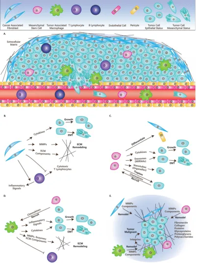
Figure. 1. Cells and Molecules (Grey) in TME
.
(A) TME and cells in TME:
CAFs, MSCs, TAMs, Lymphocytes, Endothelial cells, Pericytes, Tumor cells in
epithelial status and mesenchymal status.
(B) The roles of CAFs in TME:
Secret cytokines to affect tumor cells’ fates; Remodel ECM; Immunosuppression.
(C)
The roles of MSCs in TME:
Differentiate into other cell types; Secrete cytokines or miRNAs directly, or through exosomes; Transfer organelles through
nanotubes; Are recruited by tumor cells.
(D) The roles of TAMs in TME:
Are recruited by other cells; Secrete cytokines or inflammatory signals to affect tumor
cells’ fate; Remodel ECM; Immunosuppression.
(E) ECM in TME:
ECM have many molecules and is remodeled by CAFs, MSCs and TAMs, while it affects the fates
of tumors through integrins and other molecules.
Mesenchymal Stem Cells (MSCs) are a kind of
multipotent cell that can differentiate into multiple
mesenchymal cell lineages, including multipotent
stromal cells or mesenchymal stromal cells [10]. MSCs
have been reported to cure injured cells or tissues by
tropisms and attract the attention of different groups
to explore specific delivery vehicles for tumor therapy
[11, 12]. Residing in tumors, MSCs form a
fibrovascular network by differentiating into CAFs
and vascular pericytes [13]. While the function of
MSCs in cancer progression is diverse under different
conditions, any changes can switch the functions of
MSCs from tumor promotion into suppression
(Figure 1C) [14].
Macrophages play important roles in
inflammation, immunology, development and wound
healing [15]. They can also be recruited to TME by
tumor cells or MSCs through the secretion of specific
factors, then they become polarized and are referred
to as Tumor-Associated Macrophages (TAMs) [16].
TAMs suppress immune activity and promote cancer
progression, which is similar to their functions in
wound healing (Table 1) [17]. The balance of the
inflammation significantly influences the effects
TAMs have on tumors [15]. Also, macrophages
remodel ECM by organizing ECM and secreting ECM
components and enzymes (Figure 1D) [17].
In this article, we focus on three types of stromal
cells including CAFs, MSCs and TAMs. Additional
types of non-transformed cells in TME are
summarized in Table 1. Lymphocytes have
paradoxical functions in TME, although T cells and B
cells can be found in invasive tumor margins in
draining lymphoid organs or lymphoid structures
adjacent to TME [1]. Endothelial cells in TME
compose vasculatures and lymphatic vessels that
branch to the ends of the tumor, which is needed for
tumor growth and metastasis [18]. Similarly, pericytes
or perivascular stromal cells function in structures in
the support of blood vessels (Table 1) [19].
ECM in TME
ECM functions as a scaffold for the cells in TME
and plays a dynamic role in cancer progression,
especially as an essential regulator of invasive
processes [1]. ECM is a complicated system of
proteins, glycoproteins, proteoglycans and
polysaccharides, which contains multiple growth
factors and builds a tight interaction with cells in TME
[20]. In healthy tissue, ECM plays a tumor
suppressing role, while it becomes abnormal in
tumors and plays a tumor promoting role [21]. The
components functioning in tumor promoting roles in
ECM are induced in TME [21, 22] and they influence
tumor cells through interacting with integrins (Figure
1E) [23].
Recently, NK cells are reported to control
metastasis formation through influencing tumor
architecture by regulating the secretion of FN1, an
important ECM component [24]. This highlights the
function of interactions between stromal cells and
ECM in TME.
Secreted Factors in TME
As summarized in table 2, secreted factors in
TME function as signals between cells, or tools in
ECM remodeling, including cytokines, integrins,
proteases and microRNAs [25].
Cytokines are kind of small proteins that have
low molecular weights, which mediate the
communication between cells [26]. Cytokines in TME,
including tumor necrosis factor, interleukins, growth
factors and chemokines, regulate the progression of
cancer and determine the fate of stromal cells. This is
depended on the balance of their pro- and
anti-functions in inflammation, proliferation,
tumorigenesis, migration and apoptosis (Table 2) [27].
Integrins are integral membrane proteins and a
large family of cell surface receptors [28]. Integrins are
essential in the signaling and transfer of information
between cells or between a cell and the ECM. They are
also essential in maintaining cell-matrix adhesions.
Irregular cell-cell adhesions, caused by disrupted
integrins, are signs of cancer (Table 2). In TME, the
expressions of metastasis-promoting integrins are
enhanced, while those suppressing proliferation,
survival and migration are repressed [28]. Recently,
some groups are focusing on exosome derived
integrins as the researches on exosome are increasing
[29]. Also, integrins are reported to affect internal
activities of tumor, like nuclear alteration [30].
Extracellular proteolysis plays an essential role
in tissue homeostasis and TME [31, 32]. Among these
proteinases that mediate the proteolysis of ECM,
matrix metalloproteinases (MMPs) have the closest
relationship with cancer progression (Table 2) [33]. It
is already known that ECM degradation that is
mediated by MMPs promotes the invasion and
metastasis of cancer [33]. Furthermore, recent research
indicates that MMPs promote tumor growth,
angiogenesis and regulate apoptosis and some MMPs
function in tumor suppression (Table 2). Thus, MMPs
are also a family of proteins with paradoxical role in
TME [34].
Table 2.
The Effects of Secreted Factors on Tumor Cells in TME.
Factors Classifications (Examples) Mechanisms Targets Effects on Tumor References Cytokines Inflammatory
Factor Tumor Necrosis Factor (TNF-α) Induce NF-Apoptosis κB, TGF-β and Tumor and Stromal Cells Regulate Growth, Angiogenesis, and Invasion [124] Interleukin (IL-6) Induce JAK/STAT Pathway Tumor and
Endothelial Cells Promote EMT, Proliferation and Inhibit Apoptosis [125] Growth Factor TGF-β Immune and Inflammatory
Suppression, EMT Induction and Regulation of other Growth Factors
Tumor Cells and
Stromal Cells Early Stage: Tumor Suppressor; Late Stage: Promote Invasion and Metastasis [56, 78, 126]
VEGF Promote the Migration and
Proliferation of Endothelial Cells Endothelial Cells Promote EMT, Angiogenesis, and Intravasation [127-129] EGF Promote Growth, Recruit Tumor
Cells, Induce EMT TFs Tumor Cells and Stromal Cells Promote Proliferation, EMT and Intravasation [128, 129] FGF Promote Proliferation,
Angiogenesis Endothelial Cells Promote EMT, and Angiogenesis [127, 128, 130] PDGF Induce EMT TFs, Stimulate
VEGF and FGF while Stimulate Tsp-1
Tumor Cells, Fibroblasts, Endothelial Cells and Pericytes
Affect Intravasation and Immune Surveillance; Promote Angiogenesis, Fibroblast recruitment, Tumor growth and Metastasis
[131, 132]
HGF Induce EMT TFs Tumor Cells Promote EMT [55]
Chemokines (CXCL12 and
CXCR4) Chemotaxis; Recruit Stromal Cells or Tumor Cells in TME Tumor Cells and Stromal Cells Influence Growth, Proliferation and Migration of Tumor Cells and Stromal Cells
[133, 134]
Integrins Noncovalently Linked α and β Subunits (like α4β1and αvβ3, α5β1)
Regulate Cytoskeleton; Cell-Cell/Cell-Matrix Signal Transferring; Maintaining of Cell-Matrix Adhesions; Tissue Remodeling
Cell-Cell and Cell-Matrix Contact in TME
Mostly Promote Angiogenesis,
Intravasation and Metastasis [28, 83, 135]
Proteases Matrix Metalloproteinases
(MMPs) Degrade ECM, Basement Membrane and Cell-Cell/Cell-Matrix Junctions; Release Factors from ECM
ECM, and Junctions Promote Invasion, Metastasis, Growth and Angiogenesis while Regulate Apoptosis
[33, 34, 59]
urokinase-type Plasminogen
Activator (uPA) Activate Plasminogen into Plasmin Endothelial Cells Promote Intravasation [5, 83]
The Effects of Stromal Cells on
Metastasis Initiation
The Effects of Stromal Cells on Angiogenesis
Angiogenesis plays an initial role in the process
of metastasis, which provides a way for aggressive
tumor cells to leave primary tumors and move to
distant metastatic sites. In the process of angiogenesis,
stromal cells in TME play promoting roles. We
introduce these regulatory pathways involving
stromal cells in the following section [38].
Stromal cells affect the fate of TME through the
secretions of these angiogenesis-promoting factors
and angiogenesis-inhibiting factors. Among these
factors, VEGF is the most potent. VEGF promotes the
migration and proliferation of endothelial cells and
the permeability of vessels [39, 40]. In cells, activated
Ras and PI3K signal pathways trigger increased
transcription of VEGF and other factors that regulate
VEGF, like TGF-
β, PDGF and bFGF
[41].
Thrombospondin-1 (Tsp-1) is a potent
endogenous antiangiogenic protein. It functions by
binding to CD36 on the surface of endothelial cells
and makes them insensitive to VEGF and bFGF. Tsp-1
also inactivates MMP9 through binding; this
decreases the release of VEGF and bFGF from ECM.
Thus, in aggressive tumors, repression of Tsp-1 is
required and common [42-44]. The PI3K-Rho-ROCK-
Myc pathway is active in several types of human
breast cancer cells and it represses Tsp-1 via
phosphorylation. This pathway is called the
“angiogenic switch” due to its cell-autonomous
character [45]. Some tumor cells could overcome the
inhibitory function of Tsp-1 by producing increased
levels of VEGF [38].
as well as other growth factors that promote
angiogenesis by inhibiting the angiogenesis-
suppressing role of TSP1 [6].
Injection of tumor cells with MSCs into mice
produces twice the vessels than injecting tumor cells
alone. MSCs promote angiogenesis directly through
the secretion of VEGF and indirectly, through the
secretion of other growth factors, like TGF-
β, to
influence the effect of VEGF and the functions of other
cells [50]. In a recent clinical therapy, researchers
found that radiation therapy stimulates the secretion
of inflammatory mediators, like SDF-1
α
and PDGF-B,
from tumor cells. These signals increase the
recruitment of MSCs through the binding of CXCR4
or PDGFR-
β and enhancement of vasculogenesis by
triggering MSCs to differentiate into pericytes [51].
Hypoxia produced by tumors may recruit
TAMs to those hypoxic sites, and then induce TAMs
to produce HIF1-
α
and enhance its function on
transcription factors. This increases the production of
pro-angiogenesis factors like VEGF, basic Fibroblast
Growth Factor (bFGF), TNFα, and CXCL12
[3, 38].
Conversely, Colony-Stimulating Factor (CSF)
increases the production of metalloelastase via
macrophages
[52]. Metalloelastase cleaves
plasminogen into several small proteins, including the
antiangiogenic protein, angiostatin [53].
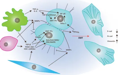
The Effects of Stromal Cells on EMT
EMT is the transition of tumor cells from
epithelial status to mesenchymal status and, therefore,
gives migrating and invasive abilities to these tumor
cells [54]. Stromal cells in TME also play crucial roles
in EMT, together with multiple factors that are
illustrated in Figure 2 and are discussed below.
In TME, CAFs, MSCs, and TAMs affect EMT
primarily through the secretion of growth factors like
TGF-
β, PDGF, EGF, VEGF, HGF or MMPs like MMP1,
MMP2, and MMP9 [4, 55].
There are several classes of growth factors
functioning in the process of EMT. The key regulators
among them are TGF-
β family members, which may
be secreted by stromal cells and tumor cells [56, 57].
TGF-
β induces EMT mainly through two pathways:
Smads dependent and Smads independent pathways.
In the Smads dependent pathway, TGF-
β induces
phosphorylation and heterodimer formation of
Smad2 and Smad3 by binding to membrane-bound
TGF-
β receptors
[58]. The Smad2/Smad3 complex
then interacts with Smad4 and is transferred into the
nucleus where they induce EMT Transcription Factors
(EMT TFs) such as snail, twist, and ZEB [58]. In the
Smads independent pathway, TGF-
β induces EMT by
activating RAS/ERK, JNK, p38 MAPK pathways that
are cooperated with integrins or simply through
integrin interactions [4].
Growth factors besides TGF-
β (like EGF, FGF,
HGF, VEGF and PDGF), also induce EMT via the
induction of EMT TFs and activation of the
RAS/RAF/MEK/ERK/MAPK pathway [55].
Matrix metalloproteinases (MMPs) also play
important roles in the process of EMT. MMPs can
degrade cell-matrix adhesions as well as cell-cell
junctions that assist the morphological transition from
epithelial to mesenchymal. MMPs also release EMT
regulators by degrading ECM into TME [59].
Besides secreting growth factors and MMPs,
TAMs also participate in this process through the
secretion of inflammatory factors like TNF-
α
and IL-6
[60-62]. Those inflammatory factors trigger EMT by
inducing TGF-
β or NF
-
κ
B pathways. Additionally,
TNF-
α
also induce EMT through the triggering of
ROS. IL-6 does it by activating the JAK/STAT3
pathway [63, 64]. While, enhanced expression levels
of COX2 in TAM is positively correlated to the
secretion of COX2, IL-6, PGE
2and MMP9, and
promotes the process of EMT of breast cancer cells by
activating the Akt pathway [65].
Recently, Cédric Blanpain reported that tumor
cells in the EMT process show progressive states from
epitheial to mesenchymal [66]. Further, they
determined the microenvironmental changes
company with the progressing EMT states. They also
found that the number and density of endothelial cells
and lymphatic cells (especially macrophages) were
increased in the progressing of EMT process. This
suggests interactions between blood vessel formation
and inflammatory with EMT [66]. Stromal cells also
can release MicroRNAs like miR21 and miR200 family
members through exosomes to communicate with
cancer cells and induce EMT [67-69].
The Effects of Stromal Cells on Invasion and
Intravasation
Invasion is a process in which tumor cells
migrate from one place to another by breaking the
ECM or basement membrane [70]. For example, the
migration of tumor cells from primary tumors into
sites near vessels is a typical invasion [71].
In the process of invasion, MMPs are secreted by
stromal cells or tumor cells and degrade the ECM and
basement membrane in the path of tumor cell
migration. MMPs also regulate the skeleton of tumor
cells that is related to cell motility and invasion [72].
Conversely, intravasation is the process by
which tumor cells migrate into blood or lymph vessels
through Trans-Endothelial Migration (TEM). There
are two kinds of intravasation: paracellular and
transcellular. Most tumor cells undergo paracellular
intravasation. In the process of paracellular TEM
(Figure 3), tumor cells migrate across the vessel walls
composed of endothelial cells by opening the
junctions between endothelial cells. In this process,
endothelial cells undergoing retraction to make space
for tumor cells [5]. In transcellular intravasation,
tumor cells migrate into the vessels directly across the
body of endothelial cells. Endothelial cells undergo
degradation of skeletons and contraction in this
process [73].
There are also multiple stromal cells and factors
that function in the process of intravasation in TME [5,
74]. Besides endothelial cells, Vessel Associated
Macrophages (VAMs) or Tumor Associated
Macrophages (TAMs) are crucial to this process.
VAMs attract tumor cells to invade toward vessels
through the secretion of EGF, meanwhile, tumor cells
secret SCF to recruit macrophages. These processes
build EGF/SCF paracrine interaction which depends
on the contact with each other [75]. TNF-
α, which is
secreted by macrophages, may trigger the retraction
of endothelial cells by binding to its receptors while it
also can make vessels more permeable for tumor cells
by inducing an apoptosis signal, along with other
apoptotic ligands [76].
Other stromal cells like MSCs and CAFs regulate
the process of intravasation through the secretion of
TGF-
β, PDGF, CXCL12/CXCR4 and MMPs
[5].
TGF-
β promotes EMT and invasion
[57] while
plays a paradoxical role in intravasation. Long-term
exposure of TGF-
β promotes the proliferation of
endothelial cells and inhibits the crossing of tumor
cells into vessels. Transient signals induced by TGF-
β
promote intravasation through the down-regulation
of CSF1, LHX2/PDGFβ and Twist
[5, 77, 78]. Similarly
to VEGF, TGF-
β also
promotes the opening of
junctions between endothelial cells by inhibiting the
complex of VE-Cadherin and
β
-catenin [74]. Besides
TGF-
β
itself, Latent TGF-
β Binding Protein 3 (LTBP3)
promotes the formation of intravasation involved
angiogenesis [79] and suggests that more
investigation on this factor is needed.
Urokinase-type Plasminogen Activator (uPA) is
the soluble portion of the protease system named
uPA/PAR. This soluble portion can be secreted by
CAFs, MSCs and macrophages [80-82]. Its function
depends on its binding to the PAR receptor and this
binding is promoted by MMP1. After uPA/PAR
binding, these proteins may cleave plasminogen into
plasmin, which cleaves CDCP1. Cleaved CDCP1
promotes the intravasation via the FAK/PI3K
pathway that cooperates with β
1-integrin [5, 83].
Angiogenesis is highly related to the process of
intravasation. Those angiogenic stimulators, like
VEGF, bFGF and PDGF, promote intravasation by
promoting angiogenesis to provide larger and more
permeable vessels. These factors also promote the
invasion of tumor cells toward vessels [74, 77]. There
are also several microRNAs that facilitate
intravasation of breast cancer cells, like miR10b and
miR200c [84, 85].
Conclusion and Future Aspects
Stromal cells are indispensable in tumors and
TME. They are recruited by tumor cells and affect
metastasis initiation through the regulation of tumor
cells and themselves.
In TME, CAFs mainly play promoting roles in
metastasis initiation (Table 1). This might be due to its
functions in immune suppression, ECM remodeling
and the secretion of pro-inflammatory and
pro-metastatic cytokines, enzymes and microRNAs
[67]. MSCs in TME affect metastasis initiation by
differentiating into other stromal cells or secreting
multiple factors. Currently, many labs are focused on
“cell therapy”, based on the research on MSCs. TAMs
are also potent stromal cells in TME. In metastasis
initiation, they function with at least three major
features. First, they secret inflammatory signals like
TNF-
α
and IL-6, then activate angiogenic or
EMT-promoting pathways. A recent report showed
that COX2 in TAMs is essential to their function with
this feature [65]. Second, they regulate the process of
intravasation by forming EGF/SCF paracrine
interaction with tumor cells. Third, TAMs also secret
multiple kinds of cytokines like IGF [86]. Other than
those three types, TAMs also transfer iron to tumor
and promote tumor cell proliferation [87], which open
a new door for research on TAMs.
Other than these types of stromal cells, there are
still several types of non-transformed cells in TME
like osteoblasts and osteoclasts in bone metastasis
[88], platelets and monocytes in angiogenesis and
tumor progression [89, 90], and neutrophils in
metastatic tumors [91]. The participation of stromal
cells in TME not only gives researchers more targets
and avenues, but also results in more uncontrolled
factors in cancer research.
Recently, a group put forward a method that
defined the sites of metastatic tumor
microenvironment by staining three parts: migrative
cancer cells, TAMs and endothelial cells [92]. They
named these sites TMEM (Tumor Microenvironment
of Metastasis) and found the number and status of
these sites correlate with the metastasis of the tumor.
Furthermore, they explored that TMEM mediated
neoadjuvant chemotherapy induced the metastasis of
breast cancer [93], which is crucial in the clinical
application and still a hot-point in current drug
development [94].
Exosomes in TME have been showed to
participate in multiple stages of cancer progression,
including metastasis initiation [95]. They are good
mediators of information transfer between cells and
their cargos include miRNAs, mRNAs, DNAs and
proteins [96]. Exosomes could also be released by
stromal cells and participate in the regulation of
cancer progression, especially those from MSCs
[97-99].
TME, which contributes to the fates of tumor cells.
Reports have shown that TAMs and CAFs undergo
metabolic reprograming of glucose, lipids and amino
acids in TME [100, 101]. While, tumor cells probably
enhance the aerobic glycolysis of TAMs and CAFs
and contribute to their activation [102, 103], and these
stromal cells should reshape the metabolism of TME
and convert nutrients into forms that are absorbable
for tumor cells [102]. Metabolism also changes the pH
value in TME. One group showed that acidification of
extracellular fluids promotes the activation of CAF
from MSC [104]. It would be a future aspect to explore
whether and how metabolism functions in the
metastasis of tumor cells in TME.
Currently, stromal cells could also be used in
combining therapies with immune therapy. Firstly,
immune therapy modifies TME to enrich preferred
phenotypes and increase immune effector cells, while
decreasing immune suppressive cells. Secondly,
immune therapy activates, triggers and regulates
stromal cells, while stromal cells release chemokines
or other factors to recruit or regulate immune cells
[105, 106]. These processes are all potential targets in
the future combination of therapies.
Acknowledgement
We thank the members of the Deng laboratory
for critical discussions. This work is supported by the
Chair Professor Grant (CPG2017-00026-FHS),
MYRG2016-00132-FHS and MYRG2016-00139 of
University of Macau and FDCT grants (065/2015/A2
and 094/2015/A3) to Chu-Xia Deng. The authors
declare no conflict of interest.
Competing Interests
The authors have declared that no competing
interest exists.
References
1. Balkwill FR, Capasso M, Hagemann T. The tumor microenvironment at a glance. J Cell Sci. 2012; 125: 5591-6.
2. Nguyen DX, Massague J. Genetic determinants of cancer metastasis. Nat Rev Genet. 2007; 8: 341-52.
3. Mittal K, Ebos J, Rini B. Angiogenesis and the tumor microenvironment: vascular endothelial growth factor and beyond. Semin Oncol. 2014; 41: 235-51. 4. Kalluri R, Weinberg RA. The basics of epithelial-mesenchymal transition. J
Clin Invest. 2009; 119: 1420-8.
5. Chiang SP, Cabrera RM, Segall JE. Tumor cell intravasation. Am J Physiol Cell Physiol. 2016; 311: C1-C14.
6. Kalluri R. The biology and function of fibroblasts in cancer. Nat Rev Cancer. 2016; 16: 582-98.
7. Murata T, Mekada E, Hoffman RM. Reconstitution of a metastatic-resistant tumor microenvironment with cancer-associated fibroblasts enables metastasis. Cell Cycle. 2017; 16: 533-5.
8. Zhu Y, Zhang L, Zha H, Yang F, Hu C, Chen L, et al. Stroma-derived Fibrinogen-like Protein 2 Activates Cancer-associated Fibroblasts to Promote Tumor Growth in Lung Cancer. Int J Biol Sci. 2017; 13: 804-14.
9. Shen XJ, Zhang H, Tang GS, Wang XD, Zheng R, Wang Y, et al. Caveolin-1 is a modulator of fibroblast activation and a potential biomarker for gastric cancer. Int J Biol Sci. 2015; 11: 370-9.
10. Spees JL, Lee RH, Gregory CA. Mechanisms of mesenchymal stem/stromal cell function. Stem Cell Res Ther. 2016; 7: 125.
11. Hata N, Shinojima N, Gumin J, Yong R, Marini F, Andreeff M, et al. Platelet-derived growth factor BB mediates the tropism of human mesenchymal stem cells for malignant gliomas. Neurosurgery. 2010; 66: 144-56; discussion 56-7.
12. Kidd S, Spaeth E, Dembinski JL, Dietrich M, Watson K, Klopp A, et al. Direct evidence of mesenchymal stem cell tropism for tumor and wounding microenvironments using in vivo bioluminescent imaging. Stem Cells. 2009; 27: 2614-23.
13. Spaeth EL, Dembinski JL, Sasser AK, Watson K, Klopp A, Hall B, et al. Mesenchymal stem cell transition to tumor-associated fibroblasts contributes to fibrovascular network expansion and tumor progression. PLoS One. 2009; 4: e4992.
14. Klopp AH, Gupta A, Spaeth E, Andreeff M, Marini F, 3rd. Concise review: Dissecting a discrepancy in the literature: do mesenchymal stem cells support or suppress tumor growth? Stem Cells. 2011; 29: 11-9.
15. Brady NJ, Chuntova P, Schwertfeger KL. Macrophages: Regulators of the Inflammatory Microenvironment during Mammary Gland Development and Breast Cancer. Mediators Inflamm. 2016; 2016: 4549676.
16. Shaked Y, McAllister S, Fainaru O, Almog N. Tumor dormancy and the angiogenic switch: possible implications of bone marrow- derived cells. Curr Pharm Des. 2014; 20: 4920-33.
17. Liguori M, Solinas G, Germano G, Mantovani A, Allavena P. Tumor-associated macrophages as incessant builders and destroyers of the cancer stroma. Cancers (Basel). 2011; 3: 3740-61.
18. Carmeliet P, Jain RK. Molecular mechanisms and clinical applications of angiogenesis. Nature. 2011; 473: 298-307.
19. Armulik A, Genove G, Betsholtz C. Pericytes: developmental, physiological, and pathological perspectives, problems, and promises. Dev Cell. 2011; 21: 193-215.
20. Mongiat M, Andreuzzi E, Tarticchio G, Paulitti A. Extracellular Matrix, a Hard Player in Angiogenesis. Int J Mol Sci. 2016; 17.
21. Robertson C. The extracellular matrix in breast cancer predicts prognosis through composition, splicing, and crosslinking. Exp Cell Res. 2016; 343: 73-81. 22. Insua-Rodriguez J, Oskarsson T. The extracellular matrix in breast cancer. Adv
Drug Deliv Rev. 2016; 97: 41-55.
23. Singh C, Shyanti RK, Singh V, Kale RK, Mishra JPN, Singh RP. Integrin expression and glycosylation patterns regulate cell-matrix adhesion and alter with breast cancer progression. Biochem Biophys Res Commun. 2018; 499: 374-80.
24. Glasner A, Levi A, Enk J, Isaacson B, Viukov S, Orlanski S, et al. NKp46 Receptor-Mediated Interferon-gamma Production by Natural Killer Cells Increases Fibronectin 1 to Alter Tumor Architecture and Control Metastasis. Immunity. 2018; 48: 107-19 e4.
25. Mbeunkui F, Johann DJ, Jr. Cancer and the tumor microenvironment: a review of an essential relationship. Cancer Chemother Pharmacol. 2009; 63: 571-82. 26. Landskron G, De la Fuente M, Thuwajit P, Thuwajit C, Hermoso MA. Chronic
inflammation and cytokines in the tumor microenvironment. J Immunol Res. 2014; 2014: 149185.
27. Zamarron BF, Chen W. Dual roles of immune cells and their factors in cancer development and progression. Int J Biol Sci. 2011; 7: 651-8.
28. Alphonso A, Alahari SK. Stromal cells and integrins: conforming to the needs of the tumor microenvironment. Neoplasia. 2009; 11: 1264-71.
29. Paolillo M, Schinelli S. Integrins and Exosomes, a Dangerous Liaison in Cancer Progression. Cancers (Basel). 2017; 9.
30. Madrazo E, Conde AC, Redondo-Munoz J. Inside the Cell: Integrins as New Governors of Nuclear Alterations? Cancers (Basel). 2017; 9.
31. Craddock RJ, Hodson NW, Ozols M, Shearer T, Hoyland JA, Sherratt MJ. Extracellular matrix fragmentation in young, healthy cartilaginous tissues. Eur Cell Mater. 2018; 35: 34-53.
32. Kumar S, Kulkarni R, Sen S. Cell motility and ECM proteolysis regulate tumor growth and tumor relapse by altering the fraction of cancer stem cells and their spatial scattering. Phys Biol. 2016; 13: 036001.
33. Kessenbrock K, Plaks V, Werb Z. Matrix metalloproteinases: regulators of the tumor microenvironment. Cell. 2010; 141: 52-67.
34. Noel A, Gutierrez-Fernandez A, Sounni NE, Behrendt N, Maquoi E, Lund IK, et al. New and paradoxical roles of matrix metalloproteinases in the tumor microenvironment. Front Pharmacol. 2012; 3: 140.
35. Manasa VG, Kannan S. Impact of microRNA dynamics on cancer hallmarks: An oral cancer scenario. Tumour Biol. 2017; 39: 1010428317695920.
36. Kuninty PR, Schnittert J, Storm G, Prakash J. MicroRNA Targeting to Modulate Tumor Microenvironment. Front Oncol. 2016; 6: 3.
37. Ding L, Ren J, Zhang D, Li Y, Huang X, Hu Q, et al. A novel stromal lncRNA signature reprograms fibroblasts to promote the growth of oral squamous cell carcinoma via LncRNA-CAF/interleukin-33. Carcinogenesis. 2018.
38. Watnick RS. The role of the tumor microenvironment in regulating angiogenesis. Cold Spring Harb Perspect Med. 2012; 2: a006676.
39. Huang H, Langenkamp E, Georganaki M, Loskog A, Fuchs PF, Dieterich LC, et al. VEGF suppresses T-lymphocyte infiltration in the tumor microenvironment through inhibition of NF-kappaB-induced endothelial activation. FASEB J. 2015; 29: 227-38.
40. Liu S, Jiang M, Zhao Q, Li S, Peng Y, Zhang P, et al. Vascular endothelial growth factor plays a critical role in the formation of the pre-metastatic niche via prostaglandin E2. Oncol Rep. 2014; 32: 2477-84.
42. Jeanne A, Schneider C, Martiny L, Dedieu S. Original insights on thrombospondin-1-related antireceptor strategies in cancer. Front Pharmacol. 2015; 6: 252.
43. Radziwon-Balicka A, Santos-Martinez MJ, Corbalan JJ, O'Sullivan S, Treumann A, Gilmer JF, et al. Mechanisms of platelet-stimulated colon cancer invasion: role of clusterin and thrombospondin 1 in regulation of the P38MAPK-MMP-9 pathway. Carcinogenesis. 2014; 35: 324-32.
44. Zhang X, Kazerounian S, Duquette M, Perruzzi C, Nagy JA, Dvorak HF, et al. Thrombospondin-1 modulates vascular endothelial growth factor activity at the receptor level. FASEB J. 2009; 23: 3368-76.
45. Kazerounian S, Lawler J. Integration of pro- and anti-angiogenic signals by endothelial cells. J Cell Commun Signal. 2018; 12: 171-9.
46. Hoeppner LH, Sinha S, Wang Y, Bhattacharya R, Dutta S, Gong X, et al. RhoC maintains vascular homeostasis by regulating VEGF-induced signaling in endothelial cells. J Cell Sci. 2015; 128: 3556-68.
47. Zhang M, Qiu L, Zhang Y, Xu D, Zheng JC, Jiang L. CXCL12 enhances angiogenesis through CXCR7 activation in human umbilical vein endothelial cells. Sci Rep. 2017; 7: 8289.
48. Miller EJ, Jecs E, Truax VM, Katzman BM, Tahirovic YA, Wilson RJ, et al. Discovery of Tetrahydroisoquinoline-Containing CXCR4 Antagonists with Improved in vitro ADMET Properties. J Med Chem. 2018.
49. Ziegler ME, Hatch MM, Wu N, Muawad SA, Hughes CC. mTORC2 mediates CXCL12-induced angiogenesis. Angiogenesis. 2016; 19: 359-71.
50. Coffelt SB, Marini FC, Watson K, Zwezdaryk KJ, Dembinski JL, LaMarca HL, et al. The pro-inflammatory peptide LL-37 promotes ovarian tumor progression through recruitment of multipotent mesenchymal stromal cells. Proc Natl Acad Sci U S A. 2009; 106: 3806-11.
51. Wang HH, Cui YL, Zaorsky NG, Lan J, Deng L, Zeng XL, et al. Mesenchymal stem cells generate pericytes to promote tumor recurrence via vasculogenesis after stereotactic body radiation therapy. Cancer Lett. 2016; 375: 349-59. 52. Tsuji T, Kelly NJ, Takahashi S, Leme AS, Houghton AM, Shapiro SD.
Macrophage elastase suppresses white adipose tissue expansion with cigarette smoking. Am J Respir Cell Mol Biol. 2014; 51: 822-9.
53. Aharinejad S, Paulus P, Sioud M, Hofmann M, Zins K, Schafer R, et al. Colony-stimulating factor-1 blockade by antisense oligonucleotides and small interfering RNAs suppresses growth of human mammary tumor xenografts in mice. Cancer Res. 2004; 64: 5378-84.
54. Liu Y, Liu B, Zhang GQ, Zou JF, Zou ML, Cheng ZS. Calpain inhibition attenuates bleomycin-induced pulmonary fibrosis via switching the development of epithelial-mesenchymal transition. Naunyn Schmiedebergs Arch Pharmacol. 2018.
55. Jing Y, Han Z, Zhang S, Liu Y, Wei L. Epithelial-Mesenchymal Transition in tumor microenvironment. Cell Biosci. 2011; 1: 29.
56. Morrison CD, Parvani JG, Schiemann WP. The relevance of the TGF-beta Paradox to EMT-MET programs. Cancer Lett. 2013; 341: 30-40.
57. Ma M, He M, Jiang Q, Yan Y, Guan S, Zhang J, et al. MiR-487a Promotes TGF-beta1-induced EMT, the Migration and Invasion of Breast Cancer Cells by Directly Targeting MAGI2. Int J Biol Sci. 2016; 12: 397-408.
58. Zhao M, Mishra L, Deng CX. The role of TGF-beta/SMAD4 signaling in cancer. Int J Biol Sci. 2018; 14: 111-23.
59. Nistico P, Bissell MJ, Radisky DC. Epithelial-mesenchymal transition: general principles and pathological relevance with special emphasis on the role of matrix metalloproteinases. Cold Spring Harb Perspect Biol. 2012; 4. 60. Balkwill F. Tumour necrosis factor and cancer. Nat Rev Cancer. 2009; 9: 361-71. 61. Wang S, Yan Y, Cheng Z, Hu Y, Liu T. Sotetsuflavone suppresses invasion and metastasis in non-small-cell lung cancer A549 cells by reversing EMT via the TNF-alpha/NF-kappaB and PI3K/AKT signaling pathway. Cell Death Discov. 2018; 4: 26.
62. Wu ST, Sun GH, Hsu CY, Huang CS, Wu YH, Wang HH, et al. Tumor necrosis factor-alpha induces epithelial-mesenchymal transition of renal cell carcinoma cells via a nuclear factor kappa B-independent mechanism. Exp Biol Med (Maywood). 2011; 236: 1022-9.
63. Julien S, Puig I, Caretti E, Bonaventure J, Nelles L, van Roy F, et al. Activation of NF-kappaB by Akt upregulates Snail expression and induces epithelium mesenchyme transition. Oncogene. 2007; 26: 7445-56.
64. Min C, Eddy SF, Sherr DH, Sonenshein GE. NF-kappaB and epithelial to mesenchymal transition of cancer. J Cell Biochem. 2008; 104: 733-44. 65. Gan L, Qiu Z, Huang J, Li Y, Huang H, Xiang T, et al. Cyclooxygenase-2 in
tumor-associated macrophages promotes metastatic potential of breast cancer cells through Akt pathway. Int J Biol Sci. 2016; 12: 1533-43.
66. Pastushenko I, Brisebarre A, Sifrim A, Fioramonti M, Revenco T, Boumahdi S, et al. Identification of the tumour transition states occurring during EMT. Nature. 2018.
67. Wang Z, Tan Y, Yu W, Zheng S, Zhang S, Sun L, et al. Small role with big impact: miRNAs as communicators in the cross-talk between cancer-associated fibroblasts and cancer cells. Int J Biol Sci. 2017; 13: 339-48. 68. Markopoulos GS, Roupakia E, Tokamani M, Chavdoula E, Hatziapostolou M,
Polytarchou C, et al. A step-by-step microRNA guide to cancer development and metastasis. Cell Oncol (Dordr). 2017; 40: 303-39.
69. Xia H, Hui KM. MicroRNAs involved in regulating epithelial-mesenchymal transition and cancer stem cells as molecular targets for cancer therapeutics. Cancer Gene Ther. 2012; 19: 723-30.
70. Truong D, Puleo J, Llave A, Mouneimne G, Kamm RD, Nikkhah M. Breast Cancer Cell Invasion into a Three Dimensional Tumor-Stroma Microenvironment. Sci Rep. 2016; 6: 34094.
71. Wyckoff JB, Wang Y, Lin EY, Li JF, Goswami S, Stanley ER, et al. Direct visualization of macrophage-assisted tumor cell intravasation in mammary tumors. Cancer Res. 2007; 67: 2649-56.
72. Chabottaux V, Ricaud S, Host L, Blacher S, Paye A, Thiry M, et al. Membrane-type 4 matrix metalloproteinase (MT4-MMP) induces lung metastasis by alteration of primary breast tumour vascular architecture. J Cell Mol Med. 2009; 13: 4002-13.
73. Khuon S, Liang L, Dettman RW, Sporn PH, Wysolmerski RB, Chew TL. Myosin light chain kinase mediates transcellular intravasation of breast cancer cells through the underlying endothelial cells: a three-dimensional FRET study. J Cell Sci. 2010; 123: 431-40.
74. Reymond N, d'Agua BB, Ridley AJ. Crossing the endothelial barrier during metastasis. Nat Rev Cancer. 2013; 13: 858-70.
75. Goswami S, Sahai E, Wyckoff JB, Cammer M, Cox D, Pixley FJ, et al. Macrophages promote the invasion of breast carcinoma cells via a colony-stimulating factor-1/epidermal growth factor paracrine loop. Cancer Res. 2005; 65: 5278-83.
76. Zervantonakis IK, Hughes-Alford SK, Charest JL, Condeelis JS, Gertler FB, Kamm RD. Three-dimensional microfluidic model for tumor cell intravasation and endothelial barrier function. Proc Natl Acad Sci U S A. 2012; 109: 13515-20. 77. Tsuji T, Ibaragi S, Hu GF. Epithelial-mesenchymal transition and cell
cooperativity in metastasis. Cancer Res. 2009; 69: 7135-9.
78. Giampieri S, Manning C, Hooper S, Jones L, Hill CS, Sahai E. Localized and reversible TGFbeta signalling switches breast cancer cells from cohesive to single cell motility. Nat Cell Biol. 2009; 11: 1287-96.
79. Deryugina EI, Zajac E, Zilberberg L, Muramatsu T, Joshi G, Dabovic B, et al. LTBP3 promotes early metastatic events during cancer cell dissemination. Oncogene. 2018.
80. Breznik B, Motaln H, Vittori M, Rotter A, Lah Turnsek T. Mesenchymal stem cells differentially affect the invasion of distinct glioblastoma cell lines. Oncotarget. 2017; 8: 25482-99.
81. Sugioka K, Mishima H, Kodama A, Itahashi M, Fukuda M, Shimomura Y. Regulatory Mechanism of Collagen Degradation by Keratocytes and Corneal Inflammation: The Role of Urokinase-Type Plasminogen Activator. Cornea. 2016; 35 Suppl 1: S59-S64.
82. Tian B, Chen X, Zhang H, Li X, Wang J, Han W, et al. Urokinase plasminogen activator secreted by cancer-associated fibroblasts induces tumor progression via PI3K/AKT and ERK signaling in esophageal squamous cell carcinoma. Oncotarget. 2017.
83. Casar B, Rimann I, Kato H, Shattil SJ, Quigley JP, Deryugina EI. In vivo cleaved CDCP1 promotes early tumor dissemination via complexing with activated beta1 integrin and induction of FAK/PI3K/Akt motility signaling. Oncogene. 2014; 33: 255-68.
84. Croset M, Goehrig D, Frackowiak A, Bonnelye E, Ansieau S, Puisieux A, et al. TWIST1 expression in breast cancer cells facilitates bone metastasis formation. J Bone Miner Res. 2014; 29: 1886-99.
85. Sigloch FC, Burk UC, Biniossek ML, Brabletz T, Schilling O. miR-200c dampens cancer cell migration via regulation of protein kinase A subunits. Oncotarget. 2015; 6: 23874-89.
86. Ireland L, Santos A, Campbell F, Figueiredo C, Hammond D, Ellies LG, et al. Blockade of insulin-like growth factors increases efficacy of paclitaxel in metastatic breast cancer. Oncogene. 2018.
87. Mertens C, Mora J, Oren B, Grein S, Winslow S, Scholich K, et al. Macrophage-derived lipocalin-2 transports iron in the tumor microenvironment. Oncoimmunology. 2018; 7: e1408751.
88. Wu JB, Yin L, Shi C, Li Q, Duan P, Huang JM, et al. MAOA-Dependent Activation of Shh-IL6-RANKL Signaling Network Promotes Prostate Cancer Metastasis by Engaging Tumor-Stromal Cell Interactions. Cancer Cell. 2017; 31: 368-82.
89. Qi C, Li B, Guo S, Wei B, Shao C, Li J, et al. P-Selectin-Mediated Adhesion between Platelets and Tumor Cells Promotes Intestinal Tumorigenesis in Apc(Min/+) Mice. Int J Biol Sci. 2015; 11: 679-87.
90. Sidibe A, Ropraz P, Jemelin S, Emre Y, Poittevin M, Pocard M, et al. Angiogenic factor-driven inflammation promotes extravasation of human proangiogenic monocytes to tumours. Nat Commun. 2018; 9: 355.
91. Felix K, Gaida MM. Neutrophil-Derived Proteases in the Microenvironment of Pancreatic Cancer -Active Players in Tumor Progression. Int J Biol Sci. 2016; 12: 302-13.
92. Robinson BD, Sica GL, Liu YF, Rohan TE, Gertler FB, Condeelis JS, et al. Tumor microenvironment of metastasis in human breast carcinoma: a potential prognostic marker linked to hematogenous dissemination. Clin Cancer Res. 2009; 15: 2433-41.
93. Karagiannis GS, Pastoriza JM, Wang Y, Harney AS, Entenberg D, Pignatelli J, et al. Neoadjuvant chemotherapy induces breast cancer metastasis through a TMEM-mediated mechanism. Sci Transl Med. 2017; 9.
94. Lv Y, Xu C, Zhao X, Lin C, Yang X, Xin X, et al. Nanoplatform Assembled from a CD44-Targeted Prodrug and Smart Liposomes for Dual Targeting of Tumor Microenvironment and Cancer Cells. ACS Nano. 2018.
95. Suchorska WM, Lach MS. The role of exosomes in tumor progression and metastasis (Review). Oncol Rep. 2016; 35: 1237-44.
96. Kahlert C, Kalluri R. Exosomes in tumor microenvironment influence cancer progression and metastasis. J Mol Med (Berl). 2013; 91: 431-7.
98. Zhang Z, Li X, Sun W, Yue S, Yang J, Li J, et al. Loss of exosomal miR-320a from cancer-associated fibroblasts contributes to HCC proliferation and metastasis. Cancer Lett. 2017; 397: 33-42.
99. Zheng P, Chen L, Yuan X, Luo Q, Liu Y, Xie G, et al. Exosomal transfer of tumor-associated macrophage-derived miR-21 confers cisplatin resistance in gastric cancer cells. J Exp Clin Cancer Res. 2017; 36: 53.
100. Shan T, Chen S, Chen X, Lin WR, Li W, Ma J, et al. Cancer-associated fibroblasts enhance pancreatic cancer cell invasion by remodeling the metabolic conversion mechanism. Oncol Rep. 2017; 37: 1971-9.
101. Rabold K, Netea MG, Adema GJ, Netea-Maier RT. Cellular metabolism of tumor-associated macrophages - functional impact and consequences. FEBS Lett. 2017.
102. Reina-Campos M, Moscat J, Diaz-Meco M. Metabolism shapes the tumor microenvironment. Curr Opin Cell Biol. 2017; 48: 47-53.
103. Dehne N, Mora J, Namgaladze D, Weigert A, Brune B. Cancer cell and macrophage cross-talk in the tumor microenvironment. Curr Opin Pharmacol. 2017; 35: 12-9.
104. Zhu H, Guo S, Zhang Y, Yin J, Yin W, Tao S, et al. Proton-sensing GPCR-YAP Signalling Promotes Cancer-associated Fibroblast Activation of Mesenchymal Stem Cells. Int J Biol Sci. 2016; 12: 389-96.
105. Giuliani M, Janji B, Berchem G. Activation of NK cells and disruption of PD-L1/PD-1 axis: two different ways for lenalidomide to block myeloma progression. Oncotarget. 2017; 8: 24031-44.
106. Nicodemus CF. Antibody-based immunotherapy of solid cancers: progress and possibilities. Immunotherapy. 2015; 7: 923-39.
107. Liao Z, Tan ZW, Zhu P, Tan NS. Cancer-associated fibroblasts in tumor microenvironment - Accomplices in tumor malignancy. Cell Immunol. 2018. 108. Poh AR, Ernst M. Targeting Macrophages in Cancer: From Bench to Bedside.
Front Oncol. 2018; 8: 49.
109. Knutsdottir H, Condeelis JS, Palsson E. 3-D individual cell based computational modeling of tumor cell-macrophage paracrine signaling mediated by EGF and CSF-1 gradients. Integr Biol (Camb). 2016; 8: 104-19. 110. Fridman WH, Pages F, Sautes-Fridman C, Galon J. The immune contexture in
human tumours: impact on clinical outcome. Nat Rev Cancer. 2012; 12: 298-306.
111. Campbell DJ, Koch MA. Treg cells: patrolling a dangerous neighborhood. Nat Med. 2011; 17: 929-30.
112. Hiraoka N, Onozato K, Kosuge T, Hirohashi S. Prevalence of FOXP3+ regulatory T cells increases during the progression of pancreatic ductal adenocarcinoma and its premalignant lesions. Clin Cancer Res. 2006; 12: 5423-34.
113. Gooden MJ, de Bock GH, Leffers N, Daemen T, Nijman HW. The prognostic influence of tumour-infiltrating lymphocytes in cancer: a systematic review with meta-analysis. Br J Cancer. 2011; 105: 93-103.
114. Lee GR. Phenotypic and Functional Properties of Tumor-Infiltrating Regulatory T Cells. Mediators Inflamm. 2017; 2017: 5458178.
115. Qi W, Huang X, Wang J. Correlation between Th17 cells and tumor microenvironment. Cell Immunol. 2013; 285: 18-22.
116. Shen M, Wang J, Ren X. New Insights into Tumor-Infiltrating B Lymphocytes in Breast Cancer: Clinical Impacts and Regulatory Mechanisms. Front Immunol. 2018; 9: 470.
117. Olkhanud PB, Damdinsuren B, Bodogai M, Gress RE, Sen R, Wejksza K, et al. Tumor-evoked regulatory B cells promote breast cancer metastasis by converting resting CD4(+) T cells to T-regulatory cells. Cancer Res. 2011; 71: 3505-15.
118. Schioppa T, Moore R, Thompson RG, Rosser EC, Kulbe H, Nedospasov S, et al. B regulatory cells and the tumor-promoting actions of TNF-alpha during squamous carcinogenesis. Proc Natl Acad Sci U S A. 2011; 108: 10662-7. 119. Horikawa M, Minard-Colin V, Matsushita T, Tedder TF. Regulatory B cell
production of IL-10 inhibits lymphoma depletion during CD20 immunotherapy in mice. J Clin Invest. 2011; 121: 4268-80.
120. Alitalo K. The lymphatic vasculature in disease. Nat Med. 2011; 17: 1371-80. 121. Goel S, Duda DG, Xu L, Munn LL, Boucher Y, Fukumura D, et al.
Normalization of the vasculature for treatment of cancer and other diseases. Physiol Rev. 2011; 91: 1071-121.
122. Swartz MA, Lund AW. Lymphatic and interstitial flow in the tumour microenvironment: linking mechanobiology with immunity. Nat Rev Cancer. 2012; 12: 210-9.
123. Harrell CR, Simovic Markovic B, Fellabaum C, Arsenijevic A, Djonov V, Volarevic V. Molecular mechanisms underlying therapeutic potential of pericytes. J Biomed Sci. 2018; 25: 21.
124. Qu Y, Zhao G, Li H. Forward and Reverse Signaling Mediated by Transmembrane Tumor Necrosis Factor-Alpha and TNF Receptor 2: Potential Roles in an Immunosuppressive Tumor Microenvironment. Front Immunol. 2017; 8: 1675.
125. Gasche JA, Hoffmann J, Boland CR, Goel A. Interleukin-6 promotes tumorigenesis by altering DNA methylation in oral cancer cells. Int J Cancer. 2011; 129: 1053-63.
126. Santibanez JF, Quintanilla M, Bernabeu C. TGF-beta/TGF-beta receptor system and its role in physiological and pathological conditions. Clin Sci (Lond). 2011; 121: 233-51.
127. Ai S, Cheng XW, Inoue A, Nakamura K, Okumura K, Iguchi A, et al. Angiogenic activity of bFGF and VEGF suppressed by proteolytic cleavage by neutrophil elastase. Biochem Biophys Res Commun. 2007; 364: 395-401.
128. Aljada A, O'Connor L, Fu YY, Mousa SA. PPAR gamma ligands, rosiglitazone and pioglitazone, inhibit bFGF- and VEGF-mediated angiogenesis. Angiogenesis. 2008; 11: 361-7.
129. Zhao D, Tu Y, Wan L, Bu L, Huang T, Sun X, et al. In vivo monitoring of angiogenesis inhibition via down-regulation of mir-21 in a VEGFR2-luc murine breast cancer model using bioluminescent imaging. PLoS One. 2013; 8: e71472.
130. Lei H, Deng CX. Fibroblast Growth Factor Receptor 2 Signaling in Breast Cancer. Int J Biol Sci. 2017; 13: 1163-71.
131. Ostman A. PDGF receptors in tumor stroma: Biological effects and associations with prognosis and response to treatment. Adv Drug Deliv Rev. 2017; 121: 117-23.
132. Papadopoulos N, Lennartsson J. The PDGF/PDGFR pathway as a drug target. Mol Aspects Med. 2017.
133. Nasser MW, Elbaz M, Ahirwar DK, Ganju RK. Conditioning solid tumor microenvironment through inflammatory chemokines and S100 family proteins. Cancer Lett. 2015; 365: 11-22.
134. Hirbe AC, Morgan EA, Weilbaecher KN. The CXCR4/SDF-1 chemokine axis: a potential therapeutic target for bone metastases? Curr Pharm Des. 2010; 16: 1284-90.