Diagnostic Accuracy of CBCT with Different Voxel Sizes and
Intraoral Digital Radiography for Detection of Periapical Bone
Lesions: An Ex-Vivo Study
Shirin Sakhdari 1, Ahmad Reza Talaeipour 2, Maziar Talaeipour 3, Maryam Pazhutan 4, Sanaz Heidarkhan Tehrani 4,
Mohammad Javad Kharazifard 5
1 Assistant Professor, Oral and Maxillofacial Radiology Department, Craniomaxillofacial Research Center, Tehran Dental Branch, Islamic Azad
University, Tehran, Iran
2 Professor, Oral and Maxillofacial Radiology Department, Craniomaxillofacial Research Center, Tehran, Dental Branch, Islamic Azad University,
Tehran, Iran
3 Assistant Professor, Department of Periodontics, Islamic Azad University, Dental Branch, Tehran, Iran 4 Oral and Maxillofacial Radiologist, Tehran, Iran
5 Statistical Advisor, Dental Research Center, Dentistry Research Institute, Tehran University of Medical Sciences, Tehran, Iran
Abstract
Objectives: This study sought to assess the diagnostic accuracy of cone beam computed tomography (CBCT) with different voxel sizes and intraoral digital radiography with photostimulable phosphor (PSP) plate for detection of periapical (PA) bone lesions. Materials and Methods: In this ex vivo diagnostic study, one-millimeter defects were created in the alveolar sockets of 15 bone blocks, each with two posterior teeth. A no-defect control group was also included. Digital PA radiographs with PSP plates and CBCT scans with 200, 250 and 300μ voxel sizes were obtained. Four observers evaluated the possibility of lesion detection using a 5-point scale. Sensitivity, specificity, positive predictive value (PPV) and negative predicative value (NPV) were analyzed using one-way ANOVA and Tamhane’s post hoc test. Kappa and weighted kappa statistics were applied to assess intraobserver and interobserver agreements.
Results: Cochrane Q test showed no significant difference between PSP and CBCT imaging modalities in terms of kappa and weighted kappa statistics (P=0.675). The complete sensitivity and complete NPV for 200 and 250 μ voxel sizes were higher than those of 300 μ voxel size and digital radiography (P<0.001). No significant difference was noted in other parameters among other imaging modalities (P=0.403).
Conclusions: The results showed that high-resolution CBCT scans had higher diagnostic accuracy than PSP digital radiography for detection of artificially created PA bone lesions. Voxel size (field of view) must be taken into account to minimize patient radiation dose. Keywords: Diagnosis;Cone-Beam Computed Tomography; Radiography, Dental, Digital; Periapical Periodontitis
Journal of Dentistry, Tehran University of Medical Sciences, Tehran, Iran (2016; Vol. 13, No. 2) Corresponding author:
S. Heidarkhan Tehrani, Oral and Maxillofacial Radiologist, Tehran, Iran
Received: 20 July 2015 Accepted: 2 January 2016
INTRODUCTION
Despite the high prevalence of apical bone defects (about 40.8%) in endodontically treated teeth, detection of periapical (PA) bone lesions remains a challenge for dental clinicians [1,2]. Chronic apical periodontitis is localized inflammation of the PA tissue that occurs as the result of decreased bone mineral density at the PA area and manifests as a radiolucent lesion on
radiographs [3,4]. However, conventional
radiographs frequently underestimate apical periodontitis [5].
socket with a history of PA lesion or with endodontically compromised adjacent teeth [8,9]. Knowledge about the presence or absence of PA pathologies seems necessary for immediate implant placement and treatment planning. Maxillofacial pathologies, particularly endodontic infections, are usually detected and diagnosed by intraoral, occlusal and panoramic radiographs. However, these techniques have some limitations that led to the development of digital radiography, which revolutionized oral radiology and was the result of advances in image capture technology and computerized systems [10]. Of the imaging techniques, cone beam computed tomography (CBCT) with a radiation dose much lower than that of multi
detector CT enables three-dimensional
evaluation of the jaws and teeth [11]; CBCT does not have the anatomical limitations or geometric imperfections of digital radiography and enables more accurate detection and diagnosis of PA lesions [12]. Moreover, by use of different voxel sizes, patient radiation dose can be minimized. It has been shown that smaller voxel sizes increase image quality but result in higher patient radiation dose [13]. Some studies have compared the diagnostic accuracy of CBCT and intraoral digital radiography for detection of PA lesions [7,14-17]. Although it has been stated that CBCT can detect very small PA radiolucencies (0.5-1 mm)[18], only two studies have compared the diagnostic accuracy of CBCT and PA radiography for detection of lesions ≤1mm [16,17].
Considering the implications of CBCT for endodontic and peri-implant evaluations and the existing controversy in the results of studies comparing the two techniques, as well as the scarcity of information about the effects of different voxel sizes of CBCT on detection of small PA lesions, this study aimed to compare the diagnostic accuracy of CBCT with different voxel sizes and standard digital radiography for detection of PA bone lesions.
MATERIALS AND METHODS
Dry mandibles of sheep were used in this ex vivo study. Bones were prepared and sectioned sagittally by a saw. A total of 45 bone blocks, each containing two posterior teeth and the surrounding bone, were obtained. Care was taken not to traumatize the roots during sectioning by saw. Of each block, one tooth was extracted by forceps and evaluated under a magnifier to ensure absence of any lesion at the bottom of the socket. Conventional radiographs were then obtained from the blocks to ensure absence of PA lesions. Fifteen Blocks were then coded as the control group (test group 1). The teeth were then placed again in sockets and digital radiographs and CBCT scans were obtained. In test group 2, the teeth were extracted from the sockets and a hole measuring 1mm in diameter was created at the bottom of each socket using a round bur. The teeth were placed in their respective sockets again and radiographed. For test groups 3 and 4, each containing 15 blocks, 1mm holes were randomly drilled at the bottom of one of the two sockets in each block and radiographs were re-taken. For soft tissue simulation, bone blocks were placed in a Plexiglas cylinder with 20mm thickness for imaging [19].
Imaging steps:
Fig. 1: (A) Radiograph of bone blocks using parallel digital sensor radiography; (B) Specimens in a Plexiglas cylinder in CBCT system
The data were then used for reconstruction of axial and sagittal sections.
Observation and assessment of images:
Two PowerPoint (Microsoft, Redmond, WA,
USA) presentations were created. One
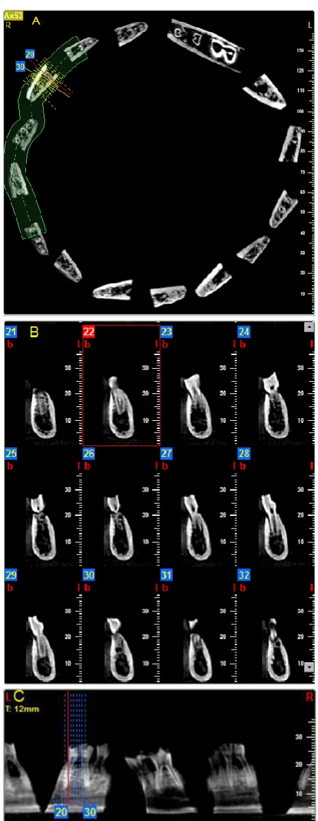
presentation included slides of PA digital radiographs and the other included sagittal, coronal and axial CBCT planes of each lesion (Fig. 2).
Four observers (two oral and maxillofacial
radiologists, one periodontist and one
endodontist), who were aware of the study design but blinded to the presence or absence of the lesions, separately observed the images on a 17-inch laptop monitor (Inspiron N65 KS, Dell,
Fujian, China) with 1366×768 pixels resolution.
Distance from the observers to the monitor was adjusted at 20-30 cm and no time limitation was set for observation of images. Also, the observers were allowed to change the magnification and brightness of images and used a 5-point scale to express their opinion regarding the presence or absence of PA lesions:
1. Definite absence of lesion
2. Probable absence of lesion
3. Unidentifiable
4. Probable presence of lesion
5. Definite presence of lesion
Images were randomly and separately evaluated and the observers recorded their opinion in datasheets. Observations were made in four sessions with one-week intervals as follows:
In the first session, digital radiographs were evaluated. In the second session, 10% of the digital radiographs were re-evaluated (to assess intra-observer agreement). In the third session, CBCT scans were evaluated. In the fourth session, 10% of the CBCT scans were re-evaluated (to assess intra-observer agreement). Diagnostic parameters including sensitivity, specificity, positive predictive value (PPV) and negative predictive value (NPV) were separately calculated for each imaging modality and the data were analyzed using one-way ANOVA and Tamhane’s post hoc test. Kappa and weighted kappa statistics were used to assess the intra- and inter-observer agreements.
RESULTS
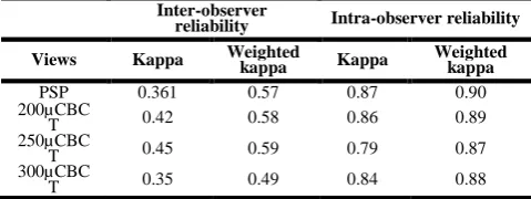
In this study, bone blocks, each with two posterior teeth (15 without PA lesions and 45 with PA lesions 1mm in diameter), were evaluated. The PSP digital radiographs and CBCT scans taken with 200, 250 and 300μ voxel sizes were evaluated. Expressing the parameters as absolute values indicated definite presence of lesion and was equal to the dichotomous yes/no scale in other studies. Expressing the parameters as complete values included possible options as well and increased the accuracy of the study. The inter-observer agreement was calculated using kappa and weighted kappa statistics and the results are shown in Table 1. The highest sensitivity belonged to CBCT scans taken with 200 and 250μ voxel sizes (P<0.001). The sensitivity of digital radiography was almost equal to that of CBCT scans with 300μ voxel size.
Table 1: Calculation of kappa and weighted kappa statistics for assessment of inter-observer and intra-observer reliability
Inter-observer
reliability Intra-observer reliability Views Kappa Weighted kappa Kappa Weighted kappa
PSP 0.361 0.57 0.87 0.90
200µCBC
T 0.42 0.58 0.86 0.89
250µCBC
T 0.45 0.59 0.79 0.87
300µCBC
T 0.35 0.49 0.84 0.88
PSP: Photostimulable phosphor plate, CBCT: Cone beam computed tomography
The specificity of CBCT with 200 and 250μ voxel sizes was higher than that of digital radiography and CBCT with 300μ voxel size. Kappa values are related to absolute and weighted kappa values are related to complete diagnostic parameters. The Cochrane Q test revealed no significant difference in the kappa values of the four images (P=0.913). The Cochrane Q test showed no significant difference in the weighted kappa values of the four images either (P=0.675). The inter-observer reliability was also assessed using kappa and weighted kappa statistics (Table 1).
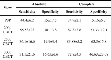
Complete sensitivity of the three CBCT voxel sizes ranged from 73% to 88% while complete specificity ranged from 46% to 73%. Complete sensitivity and complete specificity of PSP digital radiography were 75% and 51%, respectively, which were lower than the corresponding values for CBCT. However, among the four imaging groups, 200μ voxel size provided the highest sensitivity, specificity, NPV and PPV. The mean sensitivity, specificity, PPV and NPV of the four types of images are shown in Tables 2 and 3. Significant differences were found in complete sensitivity and complete NPV between 200 and 250μ voxel sizes with 300μ voxel size and PSP digital radiography (P<0.001). No significant differences were noted in other parameters between PSP digital radiography and CBCT or different voxel sizes.
DISCUSSION
Table 2: The mean complete and absolute sensitivity and specificity for detection of periapical bone defects on the four types of images
View
Absolute Complete
Sensitivity Specificity Sensitivity Specificity
PSP 44.4±6.2 15±17.5 74.9±2.1 51.6±6.3
200µ
CBCT 55.58±23 30±13.8 87.8±3.8 73.33±12.1
250µ
CBCT 36.1±16.6 19.9±9.4 83.88±5.2 63.3±15.8
300µ
CBCT 31.1±21.6 16.65±6.6 72.8±4.5 46.63±23.08
PSP: Photostimulable phosphor plate, CBCT: Cone beam computed tomography
Stavropoulos and Wenzel in their study reported a sensitivity rate lower than that obtained in the current study [7]. In their study, no difference was noted in detection of various size defects (1 and 2mm); but, the diagnostic values were lower than those in the current study. Moreover, voxel size was not reported in their study and assessments were only made in a specific FOV. Although in their study CBCT showed greater efficacy for detection of lesions, the CBCT unit used in our study was newer with more features and thus, provided greater accuracy. This may explain the higher diagnostic values obtained in our study. Sensitivity and specificity of CBCT scans in a study by Patel et al, on human
mandible were reported to be 100%[12]. In their
study, sample size was smaller than that in most similar studies and relatively large size of defects enhanced the diagnoses. This is among the most important reasons explaining the high diagnostic accuracy of CBCT reported in their study. Considering the differences in CBCT systems, selection of FOV and voxel size affect the quality and clarity of images and consequently yield different diagnostic values. Moreover, our study was conducted on sheep mandible, which is different from the human mandible. Sheep bone has more lacunae, which may result in higher frequency of false positive responses. A gold standard is necessarily required for in vivo studies.
Table 3: The mean complete and absolute NPV and PPV for detection of periapical bone defects on the four types of images
View Absolute Complete
Sensitivity Specificity Sensitivity Specificity
PSP 44.4±6.2 15±17.5 74.9±2.1 51.6±6.3
200µ
CBCT 55.58±23 30±13.8 87.8±3.8 73.33±12.1
250µ
CBCT 36.1±16.6 19.9±9.4 83.88±5.2 63.3±15.8
300µ
CBCT 31.1±21.6 16.65±6.6 72.8±4.5 46.63±23.08
PSP: Photostimulable phosphor plate, CBCT: Cone beam computed tomography
sensitivity and specificity values obtained in our study. However, in our study, the difference between some resolutions was significant. In the current study, we chose observers with different dental specialties. Among the four observers, absolute sensitivity (detection of lesion) of the radiologist was higher than that of others; whereas, complete specificity (diagnosis of absence of lesion) of the endodontist was higher than that of others; however, this difference was not significant (P=0.403). The results of the current study confirmed that CBCT had higher
diagnostic sensitivity than PSP digital
radiography. Also, our findings confirmed that the smaller the voxel size in CBCT, the higher the diagnostic accuracy. Based on recent studies, implant periapical lesion (IPAL; infection at the implant peri-apex) is among the major causes of implant failure [27-29]. Several etiologies have been recognized for IPAL such as the presence of bone defects in tooth socket [30]. Implant placement at the extraction socket of a tooth with endodontic lesion is another cause of IPAL [27]. Although the prevalence of IPAL is relatively low, accurate evaluation of tooth socket before implant placement is necessary even if sufficient time has passed and the extraction socket has healed [27]. This can only be done by three-dimensional imaging techniques such as CBCT, and development of IPAL can be prevented as such. Moreover, when it comes to anatomical structures such as the sinus floor or alveolar canal, lesions as small as 1-2mm are important; this indicates the significance of the current
study. For lesions adjacent to critical
structures/landmarks, where over-drilling even for a few millimeters would be contraindicated, use of more accurate imaging techniques prior to implant placement seems necessary, and this study points to the indication of CBCT for such cases.
CONCLUSION
The results of this study showed that
high-resolution CBCT images had higher accuracy than intraoral PSP digital radiographs for detection of artificially created PA lesions; CBCT can be used prior to implant placement in the extraction sockets of teeth suspected for endodontic pathologies to prevent IPAL or in case of proximity to critical structures, where over-drilling would be contraindicated. The results of this ex vivo animal study cannot be ideally generalized to the clinical setting; but, if CBCT is used for assessment of the presence of PA lesions, FOV size must be taken into account to minimize the patient radiation dose.
ACKNOWLEDGEMENT
The authors would like to thank Dr. Mojdeh Kalantar Motamedi for help in translating this manuscript.
REFERENCES
1- Gusiyska A. Cone beam computed tomography in the diagnosis of chronic apical periodontitis and clinical decisions: A review. Int J Sci Res (Raipur) 2015 Feb;2(4):1655-59.
2- Lemagner F, Maret D, Peters OA, Arias A, Coudrais E, Georgelin-Gurgel M. Prevalence of apical bone defects and evaluation of associated factors detected with cone-beam computed tomographic images. J Endod. 2015 Jul;41(7):1043-7.
3- Nair PN. Pathogenesis of apical periodontitis and the cause of endodontic failures. Crit Rev Oral Biol Med. 2004 Nov 1;15(6):348-81.
4- Orstavik D, Ford TP. Radiology of apical periodontitis. Essential Endodontology. 2nd ed., Oxford, Blackwell Munksgaar, 2008:202.
5- Santos O, Pinhiero LR, Umetsubo OS, Cavalcanti MG. CBCT-based evaluation of integrity of cortical sinus close to periapical lesions. Braz Oral Res. 2015;29.
6- Van Steenberghe D, Naert I. The first two stage dental implant system and its clinical application. Periodontol 2000. 1998 Jun;17:89-95.
beam dental CT, intra oral digital and conventional film radiography for the detection of periapical lesions An ex vivo study in pig Jaws. Clin Oral Investig. 2007 Mar;11(1):101-6.
8- López-Martínez F, Gómez Moreno G, Olivares-Ponce P, Eduardo Jaramillo D, Eduardo Maté Sánchez de Val J, Calvo-Guirado JL. Implants failures related to endodontic treatment. An observational retrospective study. Clin Oral Implants Res. 2015 Sep;26(9):992-5.
9- Alamri HM, Sadrameli M, Alshalhoob MA, Sadrameli M, Alshehri MA. Application of CBCT in dental practice. Gen Dent. 2012 Sep-Oct;60(5):390-400.
10- Pecoraro M, Azadivatan-le N, Janal M, Khocht A. Comparison of observer reliability in assessing alveolar bone height on direct digital and conventional radiographs. Dentomaxillofac Radiol. 2005 Sep;34(5):279-84.
11- Deman P, Atwal P, Duzenli C, Thakur Y, Ford NL. Dose measurements for dental cone-beam CT: a comparison with MSCT and panoramic imaging Phys Med Biol. 2014 Jun 21;59(12):3201-22.
12- Patel S, Dawood A, Mannocci F, Wilson R, Pitt Ford T. Detection of periapical bone defects in human Jaws using cone beam computed tomography and intraoral radiograph. Int Endod J. 2009 Jun;42(6): 507-15.
13- Tanimoto H, AraiY. The effect of voxel size on image reconstruction in cone-beam computed tomography. Oral Radiol.2009 Dec;25(2):149-53. 14- Sogur E, Baksi BG, Grondahl HG, Lomcali G, Sen BH. Detectability of chemically induced periapical lesions by limited cone beam computed tomography, intra-oral digital and conventional film radiography. Dentomaxillofac Radiol. 2009 Oct;38 (7):458-64.
15- Durack C, Patel S, Davies J, Wilson R, Mannocci F. Diagnostic accuracy of small volume cone beam computed tomography and intraoral periapical radiography for the detection of simulated external inflammatory root resorption. Int Endod J. 2011 Feb;44(2):136-47.
16- Tsai P, Torabinejad M, Rice D, Azevedo B.
Accuracy of cone-beam computed tomography and periapical radiography in detecting small periapical lesions. J Endod. 2012 Jul;38(7):965-70.
17- Estrela C, Bueno MR, Leles CR, Azevedo B, Azevedo JR. Accuracy of cone Beam computed tomography and panoramic and periapical radiography for detection of Apical periodontitis. J Endod. 2008 Mar;34(3):273-9.
18- Estrela C, Bueno MR, Azevedo BC, Azevedo JR, Pécora JD. A new periapical index based on cone beam computed tomography. J Endod. 2008 Nov;34 (11):1325-31.
19- Schropp L, Alyass NS, Wenzel A, Stavropoulos A. Validity of wax and acrylic as soft-tissue simulation materials used in in vitro radiographic studies. Dentomaxillofac Radiol. 2012 Dec;41(8): 686-90.
20- Worthington P, Rubenstien J, Hatcher DC. The role of cone-beam computed tomography in the planning and placement of implants. J Am Dent Assoc. 2010 Oct;141 Suppl 3:19S-24S.
21- Moura MS, Guedes OA, De Alencar AH, Azevedo BC, Esterela C. Influence of length of root canal obturation on apical periodontitis detected by periapical radiography and cone beam computed tomography. J Endod. 2009 Jun;35(6):805-9.
22- De Paula-Silva FW, Wu M, Leonardo MR, da Silva LA, Wesselink PR. Accuracy of apical radiography and Cone beam computed tomography scans in diagnosing apical periodontitis using histopathological findings as gold standard. J Endod. 2009 Jul;35(7):1009-12.
23- Liang YH, Jiang L, Gao XJ, Shemesh H, Wesselink PR, Wu MK. Detection and measurement of artificial periapical lesions by cone-beam computed tomography. Int Endod J. 2014 Apr;47(4): 332-8.
24- Kamburoğlu K1, Kursun S. Comparison of diagnostic accuracy of CBCT images of different voxel resolutions to detect simulated small internal resorption cavaties. Int Endod J. 2010 Sep;43(9):798-807.
tomography scans compared with intraoral image modalities for detection of caries lesions. Dentomaxillofac Radiol. 2014 Feb;37(1):18-22. 26- Hedesiu M, Baciut M, Baciut G, Nackaerts O, Jacobs R; SEDENTEXCT Consortium. Comparison of cone beam CT device and field of view for the detection of simulated periapical bone lesions. Dentomaxillofac Radiol. 2012 Oct;41(7):548-52. 27- Romanos GE, Froum S, Costa-Martins S, Meitner S, Tarnow DP. Implant periapical lesions: etiology and treatment options. J Oral Implantol. 2011;37(1): 53-63.
28- Penarrocha-Diago M, Maestre-Ferrin L,
Cervera-Ballester J, Penarrocha-Oltra D. Implant periapical lesion: diagnosis and treatment. Med Oral Patol Oral Cir Bucal. 2012 Nov 1;17(6):e1023-7.
29- Lefever D, Van Assche N, Temmerman A, Teughles W, Quirynen M. Etiology, microbiology and therapy of periapical lesions around oral implants: a retrospective analysis. J Clin Periodontol. 2013 Mar;40(3):296-302.