R E S E A R C H
Open Access
Rapid-Onset Obesity with Hypothalamic
Dysfunction, Hypoventilation, and
Autonomic Dysregulation (ROHHAD):
exome sequencing of trios, monozygotic
twins and tumours
Sarah F. Barclay
1, Casey M. Rand
2, Lauren A. Borch
1, Lisa Nguyen
1, Paul A. Gray
3, William T. Gibson
4,
Richard J. A. Wilson
5, Paul M. K. Gordon
1, Zaw Aung
1, Elizabeth M. Berry-Kravis
6, Diego Ize-Ludlow
7,
Debra E. Weese-Mayer
2,8†and N. Torben Bech-Hansen
1*†Abstract
Background:Rapid-onset Obesity with Hypothalamic Dysfunction, Hypoventilation, and Autonomic Dysregulation (ROHHAD) is thought to be a genetic disease caused byde novomutations, though causative mutations have yet to be identified. We searched forde novocoding mutations among a carefully-diagnosed and clinically
homogeneous cohort of 35 ROHHAD patients.
Methods:We sequenced the exomes of seven ROHHAD trios, plus tumours from four of these patients and the unaffected monozygotic (MZ) twin of one (discovery cohort), to identify constitutional and somaticde novosequence variants. We further analyzed this exome data to search for candidate genes under autosomal dominant and recessive models, and to identify structural variations. Candidate genes were tested by exome or Sanger sequencing in a replication cohort of 28 ROHHAD singletons.
Results:The analysis of the trio-based exomes found 13de novovariants. However, no two patients hadde novo variants in the same gene, and additional patient exomes and mutation analysis in the replication cohort did not provide strong genetic evidence to implicate any of these sequence variants in ROHHAD. Somatic comparisons revealed no coding differences between any blood and tumour samples, or between the two discordant MZ twins. Neither autosomal dominant nor recessive analysis yielded candidate genes for ROHHAD, and we did not identify any potentially causative structural variations.
Conclusions:Clinical exome sequencing is highly unlikely to be a useful diagnostic test in patients with true ROHHAD. As ROHHAD has a high risk for fatality if not properly managed, it remains imperative to expand the search for non-exomic genetic risk factors, as well as to investigate other possible mechanisms of disease. In so doing, we will be able to confirm objectively the ROHHAD diagnosis and to contribute to our understanding of obesity, respiratory control, hypothalamic function, and autonomic regulation.
Keywords:ROHHAD, Obesity, Hypothalamic dysfunction, Autonomic dysregulation, Hypoventilation, Genomics, Genetics, Exome sequencing, Next-generation sequencing
* Correspondence:[email protected] †Equal contributors
1Department of Medical Genetics, Cumming School of Medicine, Alberta
Children’s Hospital Research Institute, University of Calgary, Calgary, AB, Canada
Full list of author information is available at the end of the article
Background
Rapid-onset Obesity with Hypothalamic Dysfunction, Hypoventilation, and Autonomic Dysregulation (ROH-HAD) is a complex and devastating disease whose etiology is poorly understood, despite its initial description 50 years ago [1]. The condition, previously termed“late-onset cen-tral hypoventilation syndrome with hypothalamic dysfunc-tion”is extremely rare, with fewer than 100 cases reported in the literature, and occurs sporadically, with no clear-cut family history for inheritance of the phenotype [1–7]. In addition to the hypothalamic, respiratory and autonomic manifestations that are hallmarks of the disease, about 40 % of ROHHAD patients develop benign tumours of neural crest origin [2–4]. Because of the heralding feature of the rapid-onset obesity (20–30 lb over a 3–6 month period in, typically, a 2–7 year old, otherwise healthy, child), affected children should come to the attention of their pediatricians early in the clinical course. However, due to the variable timing and onset of other features, coupled with the presumption of exogenous obesity, a ROHHAD diagnosis is often delayed or missed, potentially leading to fatal central hypoventilation, cardiorespiratory arrest, and impaired neurocognitive development. Con-versely, many children with marked and potentially rapid weight gain, who do not meet the additional clinical cri-teria for ROHHAD, are inappropriately labeled as having ROHHAD. While diagnostic criteria and knowledge of the disease course have improved since the introduction of the ROHHAD acronym by Ize-Ludlow and coworkers in 2007 [2], these issues remain a challenge. Thus, unam-biguous diagnosis of ROHHAD is difficult but essential– not only for scientific inquiry but also for appropriate patient care. The identification of a diagnostic marker (genetic or otherwise) has the potential to decrease mor-bidity and mortality of patients with ROHHAD, and to guide future intervention and research on this disease.
As many ROHHAD features are reminiscent of other neurocristopathies of genetic origin, such as Congenital Central Hypoventilation Syndrome (CCHS) [7], a genetic basis to ROHHAD has been hypothesized. The sporadic appearance of a syndrome with tumour predisposition and an apparently primary disturbance of body growth is consistent with somatic mosaicism or constitutional inheritance of a de novodominant mutation (as seen in Sotos syndrome [8], Proteus syndrome [9], CLOVES syn-drome [10], and Weaver synsyn-drome [11]), though epigen-etic mechanisms could also be postulated, as have been shown to occur in Beckwith-Wiedemann syndrome [12]. However, previous candidate gene studies in ROHHAD using Sanger sequencing have not revealed a clear candi-date gene that can account for the full ROHHAD pheno-type [2, 4, 13].
To date, most known disease-causing mutations have been identified within the coding portion of the genome.
Whole exome sequencing (WES) among small, carefully-phenotyped cohorts has proven to be a very effective means of identifying the cause of rare pediatric diseases, a fact well demonstrated in a large national exome sequen-cing effort (FORGE Canada Consortium) that found disease-causing variants for 146 rare pediatric diseases, in-cluding variants in 67 genes not previously linked to any human disease [14]. Thus, WES is a highly cost-effective and efficient method for solving the genetic basis of rare diseases. To search for both constitutional and somaticde novosequence variants that could be the cause of this se-vere pediatric disease, we used WES to analyze seven ROHHAD trios, the unaffected monozygotic (MZ) twin of one of these seven patients, and tumours isolated from four of these seven patients as a discovery cohort. We also assembled a replication cohort of 28 singleton ROHHAD cases in which to analyze candidate genes identified through WES.
Methods
Criteria for preliminary diagnosis
The basic criteria for consideration of the diagnosis of ROHHAD were published in Ize-Ludlow et al. (2007) [2]. Briefly, features included: 1) onset of rapid and extreme weight gain after age 1.5 years (typically 2–7 years) in a pre-viously non-obese and seemingly normal child, 2) evidence of hypothalamic dysfunction, 3) alveolar hypoventilation, and 4) features of autonomic dysregulation.
Patient selection
in thediscovery cohortfor trio analysis. The other 28 par-ticipants formed thereplication cohort, of which exome sequence data were obtained for 9 and the remaining 19 were analyzed by Sanger sequencing, only in genes of interest.
Ethics, consent and permissions
This study was approved by the Ann & Robert H. Lurie Children’s Hospital of Chicago IRB (study ID: 2009– 13904) and the University of Calgary Conjoint Health Research Ethics Board (study ID: REB13-0164_REN2). All participants provided informed consent to partici-pate, including consent to publish the data herein.
Sample collection and DNA extraction
Genomic DNA was isolated from blood and tumour sam-ples, using a Puregene reagent kit (Qiagen). Tumours in-cluded both ganglioneuroma and ganglioneuroblastoma samples (Table 1).
Whole exome sequencing and analysis
Exome captures were completed using the Agilent Sure-Select V5 + UTRs capture kit. Massively parallel sequen-cing was performed on a SOLiD platform, at the Alberta Children’s Hospital Research Institute (ACHRI), and se-quences were aligned to the human reference genome (GRCh37) using Lifescope Genomic Analysis Software 2.5 (Life Technologies), (see Table 2). Variants were called using DeNovoGear [15] for the trio, twin, and tumour analyses; and the Genome Analysis Toolkit (GATK) Haplotype Caller (Version 3.3) [16] for the re-cessive and dominant analyses; and annotated for filtra-tion and prioritizafiltra-tion using ANNOVAR [17]. For the trio analysis, candidate variants were identified as novel or rare (MAF < 0.005, according to 1000 Genomes Pro-ject [18], the Exome Variant Server [EVS; [19]] and the Exome Aggregation Consortium [ExAC; [20]]) exonic, UTR, or splice site (within 2 bp of an exon) variants not within a segmental duplication. Candidate variants were further filtered to exclude UTR variants for the recessive analysis, and both UTR and synonymous variants for the dominant analysis. Candidate variants were assessed
using Combined Annotation Dependent Depletion (CADD) [21] and PolyPhen-2 [22] (for non-synonymous variants) to predict deleteriousness. CADD scores are phred-scaled, so a score of 10 indicates the pathogenicity score of a variant is in the top 10 % of the scores for all possible human variants; a score of 20 indicates it is in the top 1 % etc. PolyPhen-2 scores represent the prob-ability that the variant is deleterious.
Structural variation analysis
Bellerophon v1.03 [23] was used with the default settings to search for chromosomal translocation and inter-chromosomal insertions located inside exons in all 16 ROHHAD proband exomes (the 7 discovery cohort trio probands and the 9 replication cohort exomes) and 4 tumour samples. FishingCNV v2.1 [24] was used to search for genomic copy number variants (CNVs) in the seven trio ROHHAD probands within the discovery co-hort. Exomes from 22 healthy individuals, sequenced with the same enrichment kit (Agilent SureSelect cap-ture kit V5 + UTRs), were used as controls. We used Benjamini-Hochberg multiple testing correction to de-tect CNVs with a p-value < 0.05.
Variant validation and mutation analysis by sanger sequencing
The NCBI Primer Blast tool [25] was used to design site-specific primers to amplify genomic regions of interest. The amplicons were purified using the E.Z.N.A. Cycle Pure spin purification protocol (Omega Biotek) and then sequenced using the fluorescent dideoxy terminator method (Sanger method). Amplicon sequences were com-pared to the reference genome (GRCh37) using Mutation Surveyor (SoftGenetics) in order to identify variant posi-tions within the exons or UTRs. Allde novocandidate var-iants identified by DeNovoGear [15] with a posterior probability of 0.9 or greater were validated in this way, as were select candidate variants identified in the singleton (autosomal dominant and autosomal recessive) analyses. Selected candidate genes identified in the discovery cohort of 7 ROHHAD probands underwent mutation analysis by Sanger sequencing in 19 additional ROHHAD patients.
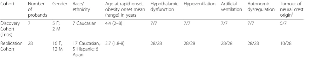
Table 1Phenotype of probands with Rapid-onset Obesity with Hypothalamic Dysfunction, Hypoventilation, and Autonomic Dysregulation (ROHHAD) in discovery and replication cohorts
Cohort Number of probands
Gender Race/ ethnicity
Age at rapid-onset obesity onset mean (range) in years
Hypothalamic dysfunction
Hypoventilation Artificial ventilation
Autonomic dysregulation
Tumour of neural crest origina
Discovery Cohort (Trios)
7 5 F;
2 M
7 Caucasian 4.4 (2–8) 7/7 7/7 7/7 7/7 5/7
Replication Cohort
28 16 F;
12 M
17 Caucasian; 5 Hispanic; 6 Asian
3.7 (1.8-8) 28/28 28/28 28/28 28/28 10/28
a
Table 2Details of exome sequencing
Cohort Sequencing sitea(Capture Kit, Sequencing platform, Aligner)
Participant ID
Description Mean
depth of coverage
Mean depth of coverage, cohort average (SD)
% of target region covered at least 20×
% of target region covered at least 20×, cohort average (SD)
Discovery cohort
ACHRI (Agilent SureSelect V5 + UTRs, Life Technologies SOLiD 5500xl, LifeScope 2.5)
18 Proband 141.4 132.7 (14.1) 91.88 931.79 (0.81)
31 Mother of 18 143.9 91.62
22 Father of 18 136 90.93
37 Monozygotic twin of 18 133.5 90.97
25 Proband 125.4 92.01
51 Mother of 25 149.6 92.81
50 Father of 25 151.2 92.84
27 Proband 139.2 91.87
32 Mother of 27 128 91.11
33 Father of 27 126.3 90.94
41 Proband 124.5 90.92
47 Mother of 41 142.1 92.97
46 Father of 41 139.4 92.65
42 Proband 133.4 92.04
43 Mother of 42 127.2 91.55
44 Father of 42 119.1 90.91
45 Proband 122.2 91.73
48 Mother of 45 136.3 92.43
49 Father of 45 165.1 93.58
57 Proband 124.7 91.45
59 Mother of 57 113.4 90.55
58 Father of 57 98.5 91.57
18 Tumour 257 249.8 (40) 94.74 95.11 (0.54)
27 Tumour 290.2 95.15
41 Tumour 258.3 95.85
57 Tumour 193.9 94.69
Replication Cohort
WASH U (Illumina All Exon 65 MB, Illumina HiSeq 2000, NovoAlign 2.07.13)
5 Proband 86.9 86.9 82.70 82.70
BGI (Agilent SureSelect V4, Illumina HiSeq 2000, BWA 0.5.9)
20 Proband 27.6 28.1 (1.7) 50.41 50.95 (1.79)
21 Proband 31.7 54.78
23 Proband 27.8 49.96
24 Proband 27.2 50.70
26 Proband 27.8 51.12
28 Proband 28 50.53
39 Proband 26.4 49.18
Perkin Elmer Corp (Agilent SureSelect Human All Exon 38 MB, Illumina HiSeq 2000, Bowtie 0.12.7)
A032 Proband 76.6 76.6 65.96 65.96
a
Mutation analysis by exome sequencing
WES was obtained for nine members of the ROHHAD replication cohort, and was used to search for candidate variants in the genes identified as potential candidates through the analysis of the trio exomes (i.e., genes carrying de novoor compound heterozygous variants in one of the seven trios; or heterozygous, rare, protein-altering variants in three of the seven trio probands). Exome captures were completed using the Agilent SureSelect capture kit, 38 MB or V4, or the Illumina All Exon 65 MB kit. Mas-sively parallel sequencing was performed on either a SOLiD or Illumina platform, at one of three institutions, and sequences were aligned to the human reference gen-ome (GRCh37) using BWA 0.5.9 [26], Lifescope Genomic Analysis Software 2.5 (Life Technologies), NovoAlign 2.07.13 (Novocroft), or Bowtie 0.12.7 [27] (see Table 2). Variants were called using the GATK Haplotype Caller (Version 3.3) [16] and annotated for filtration and prioritization using ANNOVAR [17]. The coverage of these nine exomes was not sufficient for a robust whole exome analysis (Table 2), but provided a reasonable data set in which to perform an exploratory mutation analysis (Additional file 1 shows the low-coverage proportions for each candidate gene that underwent mutation analysis in these replication exomes).
Results
Cohort characteristics
A total of 35 ROHHAD patients in whom the clinical diagnosis was confirmed were included in this cohort for genetic investigation (Table 1). Specifically, 7 of these pa-tients were included in the initial, trio-based, exome se-quencing analysis (discovery cohort), while 28 other ROHHAD patients were included in the secondary ex-ome and Sanger sequencing analysis (replication cohort). In addition, neural crest tumours from four ROHHAD patients (all of whom were trio probands) were analyzed. Blood DNA from 14 parents of the probands (7 trios) and from the monozygotic twin of one ROHHAD pro-band (discordant for the ROHHAD phenotype) was also included in our analysis. Our cohort represents a highly homogeneous group of ROHHAD patients, and all of the 35 patients included in the study showed rapid-onset obesity, hypothalamic dysfunction, hypoventilation, and autonomic dysregulation; all required artificial ventila-tion; and 15 (43 %) developed tumours of neural crest origin. Copy number variation (CNV) analysis by array comparative genomic hybridization (CGH), using the Nimblegen 720 k platform, in 26 of the 35 ROHHAD patients did not identify any ROHHAD-specific CNVs. Table 1 reveals a slight gender bias within our cohort (21 females, 14 males), however in our extensive experience studying ROHHAD we have not identified any specific gender prevalence, and careful review of
ROHHAD referrals subsequent to this study identified equal distribution of females and males in our broader ROHHAD cohort.
Exome sequencing
WES was completed on a total of 31 individuals includ-ing 16 ROHHAD patients (7 as part of trios in the dis-covery cohort and 9 in the replication cohort), 15 unaffected relatives (parents and one monozygotic twin), and tumour samples from four of the ROHHAD pro-bands (Table 2).
De novo inheritance model - trio analysis
To identifyde novovariants, we sequenced the exomes of seven ROHHAD trios. In this set of exomes, we achieved 130-fold mean coverage (after mapping and removal of PCR duplicate reads) with an average of 91.8 % +/−0.81 % (SD) of bases covered at least 20-fold (Table 2). Using DeNovoGear [15] to analyze the trios, 13 candidate de novovariants were identified and validated by Sanger ana-lysis (Table 3). Each patient carried between zero and three exonic or UTR de novo variants (average 1.86 per exome; 0.71 amino acid altering per exome), a value that is consistent with previous findings [28] and the expected mutation rate [29]. However, no two of these seven ROH-HAD patients hadde novovariants in the same gene.
Somatic mutation model (tumour and MZ twin analysis) It is possible thatde novomutations giving rise to ROH-HAD may be so severe that they are normally incompat-ible with life. Such a mechanism has been hypothesized for mutations in AKT1, which cause the Proteus syn-drome, but have only been found in a mosaic state and never in all somatic cells [9]. Similarly, surviving patients with ROHHAD may be mosaic for the disease-causing mutation, carried in only a subset of their cells and tissues. If this model were applicable, the subset of ROHHAD pa-tients with neural crest tumours would be the most likely to have an identifiable mutation, and the tumours them-selves would be the most likely tissue to carry the causa-tive mutation. To test this somatic mutation hypothesis, we sequenced the exomes of tumours from four of the seven patients that were included in the trio analysis.
We assumed that the tumour samples would contain a variable amount of normal non-tumourous tissue. Because this mosaic state would reduce our ability to de-tect rare mutations, we increased the sequencing cover-age to approximately double that obtained from the trios. This achieved a mean coverage of 260-fold (after mapping and removal of PCR duplicate reads) with an average of 95.1 % +/−0.54 % (SD) of targeted bases cov-ered at least 20-fold (Table 2). Since each analyzed tumour sample came from a ROHHAD case in a trio,
DeNovoGear [15] was used to compare the tumour sam-ples to the parental samsam-ples. Anyde novovariants iden-tified in a tumour were then compared to the de novo variants identified in that patient’s genomic DNA to sin-gle out any de novo variants unique to the tumour. All 13 of the de novo variants identified in the trio analysis were also present in the corresponding tumours; how-ever, no additional tumour-only variants were detected.
Our cohort also contains a pair of monozygotic (MZ) twins discordant for the ROHHAD phenotype [30]. To evaluate the possibility of somatic mosaicism for a causa-tive mutation in the affected twin, we used DeNovoGear to compare the variant profile between the twins, but did not identify any sequence variants in the affected twin’s exome that were not also present in the unaffected twin.
Autosomal recessive model
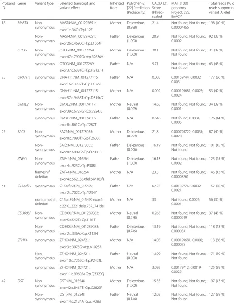
To identify potential recessive candidates, the trios were used to identify genes containing compound heterozygous inherited rare coding variants (or homozygous, as long as each parent was carrying the variant). We identified be-tween one and four compound heterozygous candidate genes in each patient. These are genes that contained two rare variants inherited in trans (i.e., one from each parent) (Table 5). No individual compound heterozygous candidate was seen in more than one individual, none of the nine
Table 3De novovariants observed in the exomes of seven ROHHAD cases (discovery cohort)
Proband ID Gene Selected transcript and variant effect
Variant type CADD [21] (Phred scaled)
PolyPhen-2 [22] prediction (probability)
Genomic position (GRCh37)
Patient 18 CD5 NM_014207:c.1406A > G:p.E469G
Non-synonymous
21.1 Deleterious (1.000) chr11:60893229
Patient 25 CD36 NM_001127444:c.1399A > G:p.R467G
Non-synonymous
19.84 Neutral (0.006) chr7:80303443
C17orf53 NM_024032:c.1046 T > C:p.I349T
Non-synonymous
5.791 Neutral (0.085) chr17:42226217
NEK7 NM_133494:c.*2404A > G 3′UTR 12.83 N/A chr1:198291053
Patient 27 MAPKAPK5 NM_139078:c.664 T > A:p.C222S
Non-synonymous
22.6 Deleterious (1.000) chr12:112321388
PPP1R16B NM_015568:c.477C > A:p.D159E
Non-synonymous
29.7 Deleterious (0.999) chr20:37529233
TAF1 NM_004606:c.4356C > T:p.R1452=
Synonymous 14.37 N/A chrX:70627913
Patient 41 PDE11A NM_001077358:c.783C > T:p.D261=
Synonymous 11.94 N/A chr2:178592832
FAM155B NM_015686:c.*652G > A 3'UTR 1.311 N/A chrX:68750448
Patient 42 PLCXD3 NM_001005473:c.*9191 T > C 3'UTR 4.216 N/A chr5:41311356
Patient 45 CCT4 NM_006430:c.*613G > A 3'UTR 4.916 N/A chr2:62095797
WDFY4 NM_020945:c.8577G > T:p.T2859=
Synonymous 1.393 N/A chr10:50174711
FAM199X NM_207318:c.828C > T:p.S276=
Synonymous 15.08 N/A chrX:103432819
All variants are heterozygous
replication exomes were found to contain two sequence variants in any of these genes, and none of these candidates have a known function that would strongly suggest an as-sociation with ROHHAD. Thus, we have not pursued any of them further at this time. We acknowledge that, in light of the fact that ROHHAD may be highly genetically het-erogeneous, one or more of these genes may indeed be disease-causing in an individual proband – but further studies would be required in order to gain additional evi-dence in support of any particular gene.
Autosomal dominant model
Though a familial dominant inheritance model is not consistent with the sporadic occurrence of this disease, we did search the 7 ROHHAD patient exomes for any genes that contained rare protein-altering (i.e., missense, nonsense, splice-site and indel) variants in all or a large
subset of the patients after censoring the de novo or inherited status. After further filtering by visual inspec-tion in the Integrative Genomics Viewer (IGV; [31, 32]), multiple variant calls were removed as probable false positives (e.g. eliminating rare variants on reads that hosted multiple other rare variants – we assumed that these reads were poorly mapped). At this stage, we iden-tified no genes with candidate variants in the same gene among 4 or more of the 7 patients. We did identify four genes (FRAS1, RELN, RIF1, POLE) that each had different rare, protein-altering variants among 3 of the 7 patients (Table 6). We searched the 9 exomes from the replica-tion cohort for rare protein-altering variants in these four genes and identified only one additional patient with a candidate variant in RIF1 (Table 6). All four of theRIF1 variants were validated by Sanger sequencing. All four RIF1 variants as well as all variants in the three
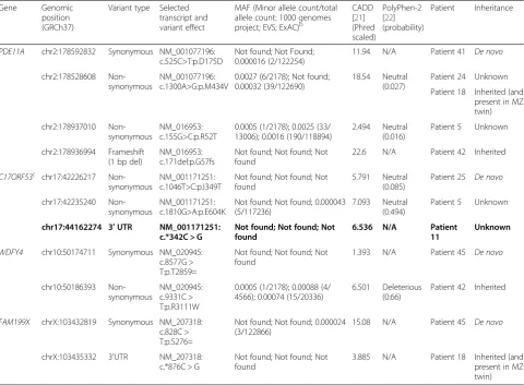
Table 4Results of extendedde novoanalysisaandC17ORF53mutation analysis
Gene Genomic
position (GRCh37)
Variant type Selected transcript and variant effect
MAF (Minor allele count/total allele count: 1000 genomes project; EVS; ExAC)b
CADD [21] (Phred scaled)
PolyPhen-2 [22] (probability)
Patient Inheritance
PDE11A chr2:178592832 Synonymous NM_001077196: c.525C>T:p.D175D
Not found; Not Found; 0.000016 (2/122254)
11.94 N/A Patient 41 De novo
chr2:178528608 Non-synonymous
NM_001077196: c.1300A>G:p.M434V
0.0027 (6/2178); Not found; 0.00032 (39/122690)
18.54 Neutral (0.027)
Patient 24 Unknown
Patient 18 Inherited (and present in MZ twin)
chr2:178937010 Non-synonymous
NM_016953: c.155G>C:p.R52T
0.0005 (1/2178); 0.0025 (33/ 13006); 0.0016 (190/118894)
2.494 Neutral (0.016)
Patient 5 Unknown
chr2:178936994 Frameshift (1 bp del)
NM_016953: c.171del:p.G57fs
Not found; Not found; Not found
22.6 N/A Patient 42 Inherited
C17ORF53c chr17:42226217 Non-synonymous
NM_001171251: c.1046T>C:p.I349T
Not found; Not found; Not found
5.791 Neutral (0.085)
Patient 25 De novo
chr17:42235240 Non-synonymous
NM_001171251: c.1810G>A:p.E604K
Not found; Not found; 0.000043 (5/117236)
7.093 Neutral (0.494)
Patient 5 Unknown
chr17:44162274 3′UTR NM_001171251:
c.*342C > G
Not found; Not found; Not found
6.536 N/A Patient
11
Unknown
WDFY4 chr10:50174711 Synonymous NM_020945: c.8577G > T:p.T2859=
Not found; Not found; Not found
1.393 N/A Patient 45 De novo
chr10:50186393 Non-synonymous
NM_020945: c.9331C > T:p.R3111W
0.0005 (1/2178); 0.00088 (4/ 4566); 0.00074 (15/20336)
6.501 Deleterious (0.66)
Patient 42 Inherited
FAM199X chrX:103432819 Synonymous NM_207318: c.828C > T:p.S276=
Not found; Not found; 0.000024 (3/122866)
15.08 N/A Patient 45 De novo
chrX:103435332 3'UTR NM_207318: c.*876C > G
Not found; Not found; Not found
3.885 N/A Patient 18 Inherited (and present in MZ twin)
a
All 16 ROHHAD exomes were searched for candidate variants (as described in methods: novel or rare (MAF < 0.005) exonic, UTR, or splice site (within 2 bp of an exon) variants not within a segmental duplication) within the 13 genes identified as containingde novovariants in one ROHHAD proband
b
1000 Genomes Project (http://www.1000genomes.org); EVS = Exome Variant Server (http://evs.gs.washington.edu/EVS/); ExAC = Exome Aggregation Consortium (http://exac.broadinstitute.org)
c
Table 5Compound heterozygous variants observed in exomes of seven ROHHAD cases (discovery cohort)
Proband ID
Gene Variant type Selected transcript and variant effect
Inherited from
Polyphen-2 [22] Prediction (Probability)
CADD [21] Score (Phred-scaled
MAF (1000 genomes Project; EVS; ExAC)a
Total reads (% of reads supporting variant Allele)
18 MAST4 Non-synonymous
MAST4:NM_001297651: Mother Deleterious (0.998)
21.4 Not found; Not found; 0.00004466
198 (40 %)
exon1:c.34C>T:p.L12F
Non-synonymous
MAST4:NM_001297651: Father Deleterious (1.000)
20.9 Not found; Not found; 0.0002
92 (35 %)
exon26:c.4690C>T:p.L1564F
OTOG Non-synonymous
OTOG:NM_001277269: Mother Deleterious (1.000)
20.1 Not found; Not found; Not found
31 (32 %)
exon47:c.7907G>A:p.R2636H
synonymous OTOG:NM_001277269: Father N/A 9.71 Not found; Not found; Not found
63 (48 %)
exon37:c.6381C>T:p.H2127H
25 DNAH11 synonymous DNAH11:NM_001277115: Father N/A 0.005 0.00159744; 0.0032; 0.003
177 (36 %)
exon16:c.3237T>C:p.L1079L
synonymous DNAH11:NM_001277115: Mother N/A 0.002 0.000199681; 0.0027; 0.0024
53 (49 %)
exon57:c.9468T>C:p.D3156D
DMXL2 Non-synonymous
DMXL2:NM_001174117: Mother Neutral (0.029)
14.65 Not found; Not found; 0.0001
34 (32 %)
exon39:c.6727G>C:p.V2243L
synonymous DMXL2:NM_001174116: Father N/A 0.646 Not found; 0.0004; 0.0005
126 (44 %)
exon8:c.861C>T:p.T287T
27 SACS
Non-synonymous
SACS:NM_001278055: Mother Deleterious (0.999)
21.8 0.000798722; 0.0035; 0.0028
87 (40 %)
exon8:c.7898T>G:p.F2633C
Non-synonymous
SACS:NM_001278055: Father Deleterious (0.996)
16.19 Not found; Not found; Not found
101 (45 %)
exon8:c.6009G>T:p.Q2003H
ZNF44 Non-synonymous
ZNF44:NM_016264: Father Deleterious (1.000)
16.13 Not found; Not found; 0.0002
123 (45 %)
exon4:c.923C>T:p.P308L
frameshift deletion
ZNF44:NM_016264: Mother N/A 23.3 Not found; Not found;
0.00008261
145 (43 %)
exon4:c.562_563del:p.M188fs
41 C15orf39 synonymous C15orf39:NM_015492: Father N/A 6.427 0.00139776; 0.0032; 0.0021
157 (38 %)
exon2:c.702C>T:p.Y234Y
nonframeshift deletion
C15orf39:NM_015492:exon2: Mother N/A 33 Not found; 0.0026; 0.0001
56 (30 %)
c.2210_2221del:p.737_741del
CD300LF Non-synonymous
CD300LF:NM_001289083: Mother Neutral (0.278)
0.265 Not found; Not found; 0.0000249
37 (43 %)
exon5:c.542T>C:p.I181T
Non-synonymous
CD300LF:NM_001289083: Father Deleterious (0.746)
13.19 Not found; Not found; 0.000033
118 (43 %)
exon2:c.336A>C:p.K112N
ZFHX4 synonymous ZFHX4:NM_024721: Mother N/A 14.05 0.000199681; 0.0002; 0.000075
113 (36 %)
exon3:c.3075G>A:p.A1025A
Non-synonymous
ZFHX4:NM_024721: Father Neutral (0.000)
1.699 Not found; Not found; Not found
171 (39 %)
exon10:c.7262C>T:p.P2421L
synonymous ZFHX4:NM_024721: Mother N/A 3.092 0.00179712; 0.0019; 0.0025
125 (39 %)
exon11:c.9960A>G:p.Q3320Q
42 DST
Non-synonymous
DST:NM_015548: Mother Deleterious (1.000)
15.35 Not found; Not found; Not found
197 (43 %)
exon42:c.8467T>C:p.C2823R
Non-synonymous
DST:NM_015548: Father Neutral
(0.144)
12.02 Not found; Not found; Not found
127 (39 %)
genes in which three of 16 probands have candidate variants (FRAS1, RELN, POLE), were inherited from unaffected parents, with both maternal and paternal transmission (Table 6).
Additionally, 187 genes were identified with poten-tial variants in 2 of the 7 discovery cohort patients (Additional file 2), but these have not been inspected in IGV, and a portion of them are likely false positive calls. While it remains possible that sequence variants in one or more of these genes are causative of ROH-HAD, with such a large number of candidate genes, and without inheritance information to support any particular one, it presently remains difficult to obtain enough genetic evidence to identify one as a causative gene. In one attempt to narrow down this list, we looked only at loss-of-function (LOF) variants, but did not identify any gene with LOF variants in more than one proband.
Structural variation analysis
In addition to identifying SNVs and small indels (as de-scribed in the previous sections), we also analyzed the ROHHAD exome data (including the tumour samples) for larger variations such as chromosomal transloca-tions and inter chromosomal insertransloca-tions and CNVs. No CNVs and no exonic translocations or insertions were detected in any of the samples. However, with exome data alone, we cannot rule out the possibility of fusion events in intronic or intergenic regions.
Mutation analysis
We performed mutation analysis (by Sanger sequencing) of two candidate genes (C17ORF53andMAPKAPK5) in 19 additional ROHHAD patients.
C17ORF53
In total, we identified three novel or rare (MAF < 0.005) variants in a search of the exons and UTRs of the C17ORF53gene among 35 ROHHAD patients (16 by ex-ome sequencing and 19 by Sanger sequencing) (Table 4). Two were non-synonymous variants, and one was in the 3’UTR. We do not have genetic samples from the parents of two of the patients carrying sequence variants in this gene, and so are unable to determine whether or not they are de novovariants. Since very little is known about the protein encoded by this open reading frame, we do not have information to model the effect these sequence vari-ants would have. Further investigation into the protein (when and where it is expressed, and what its function is) and the effect of these variants would be required to fur-ther implicate them in ROHHAD.
MAPKAPK5
Improvement in a subset of phenotypic features in Pa-tient 27 was observed shortly after a regimen of caffeine treatment (100 mg BID) was initiated [33]. A study found that transcript levels ofMAPKAPK5(in which Pa-tient 27 is carrying a de novomissense variant; Table 3) were transiently increased in response to forskolin
Table 5Compound heterozygous variants observed in exomes of seven ROHHAD cases (discovery cohort)(Continued)
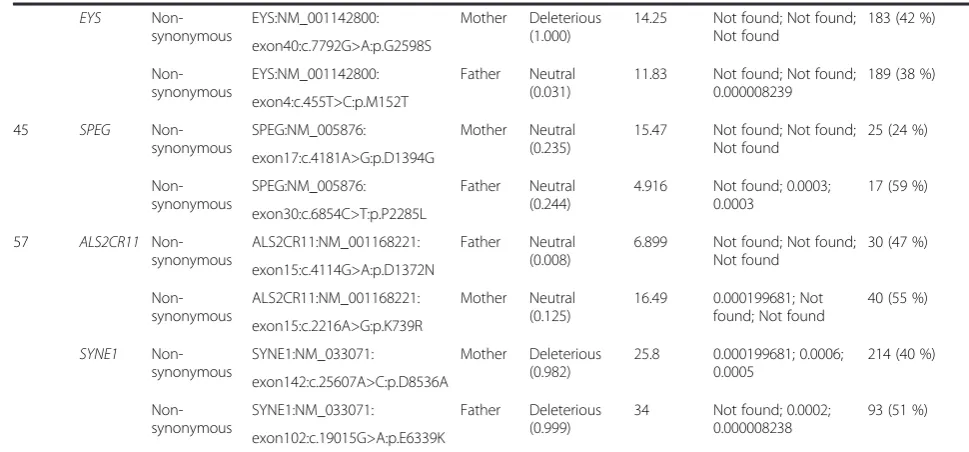
EYS Non-synonymous
EYS:NM_001142800: Mother Deleterious (1.000)
14.25 Not found; Not found; Not found
183 (42 %)
exon40:c.7792G>A:p.G2598S
Non-synonymous
EYS:NM_001142800: Father Neutral (0.031)
11.83 Not found; Not found; 0.000008239
189 (38 %)
exon4:c.455T>C:p.M152T
45 SPEG
Non-synonymous
SPEG:NM_005876: Mother Neutral (0.235)
15.47 Not found; Not found; Not found
25 (24 %)
exon17:c.4181A>G:p.D1394G
Non-synonymous
SPEG:NM_005876: Father Neutral (0.244)
4.916 Not found; 0.0003; 0.0003
17 (59 %)
exon30:c.6854C>T:p.P2285L
57 ALS2CR11 Non-synonymous
ALS2CR11:NM_001168221: Father Neutral (0.008)
6.899 Not found; Not found; Not found
30 (47 %)
exon15:c.4114G>A:p.D1372N
Non-synonymous
ALS2CR11:NM_001168221: Mother Neutral (0.125)
16.49 0.000199681; Not found; Not found
40 (55 %)
exon15:c.2216A>G:p.K739R
SYNE1 Non-synonymous
SYNE1:NM_033071: Mother Deleterious (0.982)
25.8 0.000199681; 0.0006; 0.0005
214 (40 %)
exon142:c.25607A>C:p.D8536A
Non-synonymous
SYNE1:NM_033071: Father Deleterious (0.999)
34 Not found; 0.0002; 0.000008238
93 (51 %)
exon102:c.19015G>A:p.E6339K
a
treatment (which, like caffeine, elevates cAMP) [34], leading us to hypothesize thatMAPKAPK5might be in-volved in this patient’s ROHHAD phenotype. However, mutation analysis in our ROHHAD cohort did not iden-tify any additional novel or rare exonic, splice site, or UTR sequence variants inMAPKAPK5.
Discussion
Our hypothesis that ROHHAD might be caused by germlinede novomutations or by mosaicism for somatic mutation(s) in one or more genes was tested using seven trios, including neural crest tumours of four of these pa-tients and the unaffected MZ twin of one (Table 2), as well as an additional 28 singleton ROHHAD patients used for mutation analysis of candidate genes. Despite the homogeneity of this ROHHAD cohort, and the depth
of coverage established in our exome sequencing, we did not identify any two patients who hadde novovariants in the same gene, anyde novocoding variants unique to the tumours, or any variants discordant between the affected and unaffected MZ twins. We also did not identify any structural variations in the exomes. While the sporadic oc-currence of ROHHAD suggests a de novo inheritance model, we were also able to use the exome sequencing data to assess autosomal recessive and autosomal domin-ant inheritance models. Neither model revealed a major gene that would explain ROHHAD in our cohort. One or more of the genes containing a)de novovariants (Table 3, Table 4), b) inherited compound heterozygous variants (Table 5), or c) inherited heterozygous variants in 3 or 4 patients (Table 6), may be causative of ROHHAD in some cases, but further genetic and/or functional evidence is
Table 6Genes with candidate variants observed in three of seven ROHHAD cases
Gene Proband ID
Variant type Selected transcript and variant effect
Polyphen-2 [22] Prediction (Probability)
CADD [21] score (Phred-scaled) MAF (1000 genomes project; EVS; ExAC)a Inherited from
No. of reads supporting reference call, No. of reads supporting variant call
FRAS1 41 Non-synonymous
NM_001166133:
exon27:c.3500G>A:p.R1167H
Neutral (0) 0.01 Not Found; 0.000083; 0.000058 Mother 64,50 42 Non-synonymous NM_001166133: exon29:c.3963G>C:p.K1321N Neutral (0.076)
9.266 Not Found; 0.000082; 0.0000516 Mother 96,57 57 Non-synonymous NM_001166133: exon38:c.5003A>G:p.N1668S Deleterious (0.996)
15.68 Not Found; Not Found; Not Found
Father 86,72
RELN 57
Non-synonymous NM_005045: exon54:c.8798C>T:p.T2933I Neutral (0.124) 16.6 0.000599042; 0.0002; 0.0002 Father 110,78 27 Non-synonymous NM_005045: exon51:c.8254G>A:p.G2752S Neutral (0.405)
23.3 Not Found; Not Found; Not Found Mother 12,14 45 Non-synonymous NM_005045: exon26:c.3651C>G:p.I1217M Deleterious (0.812) 19.32 0.000998403; 0.0034; 0.0027 Father 45,18
RIF1 18
Non-synonymous
NM_018151:
exon30:c.6314T>C:p.M2105T Neutral (0.002)
9.267 Not Found; Not Found; 0.0000165
Mother 59,50
41 Father 37,41
42 Splicing NM_018151:
exon32:c.6825 + 2 T > C
N/A 23.9 Not Found;
Not Found; Not Found Father 48,34 23 Non-synonymous NM_001177665: exon4:c.346C>T:p.R116C Neutral (0.09)
14.47 Not Found;
0.0004; 0.0003
Unknown 84,90
POLE 27
Non-synonymous
NM_006231:
exon43:c.5965G>A:p.A1989T
Neutral (0.043)
15.8 Not Found; Not Found; Not Found Mother 37,20 41 Non-synonymous NM_006231: exon41:c.5659G>A:p.V1887M Neutral (0.305)
10.07 Not Found; 0.0006; 0.0005 Mother 43,36 42 Non-synonymous NM_006231: exon13:c.1288G>A:p.A430T Deleterious (0.977) 24.5 0.000998403; 0.000077; 0.0007 Father 23,21
Bolded row represents variant identified in replication exomes
a
required before these variants could be considered truly pathogenic for the ROHHAD phenotype. In particular, it is difficult to determine the significance of theRIF1 find-ing (that 4 of 16 patients have inherited a rare protein-altering variant in this gene). The gene is large (41 exons encoding 2472 amino acids), and harbours a substantial number of variants (2492 variants in the ExAC database [20], 937 of which are rare protein-altering variants, and 20 of which are loss-of-function (LOF) variants [i.e., nonsense, frameshift, or canonical splice site mutations]). Thus, additional evidence - genetic (i.e., identifying add-itional ROHHAD patients withRIF1mutations) or func-tional (i.e., discovering aRIF1 function that could explain the ROHHHAD phenotype)–would be required to prove that this is more than a random chance occurrence. Anin silico search using Ingenuity Pathway Analysis (Qiagen) for functional connections among these 31 genes did not identify any potential links between different candidate genes from more than two patients.
Previous studies postulated eight genes (NDN,ASCL1, PHOX2B,NTRK2, BDNF, HTR1A, OTP, andADCYAP1) as candidate ROHHAD genes, based on their roles in the development or function of the systems (hypothal-amic, autonomic, and neuroendocrine) known to be deficient in ROHHAD, but failed to identify any disease-associated variants in these genes among their cohorts [2, 4, 13]. Likewise, we did not identify any candidate vari-ants in any of these eight genes in our 7 ROHHAD patient exomes (for each gene, at least 96 % of the coding bases were covered at least 20-fold with the exception of OTP, for which one of three exons was not effectively captured), confirming that none of these eight genes is a major ROHHAD gene. A recent study described a novel non-sense mutation in the Smith-Magenis gene,RAI1,in a pa-tient previously diagnosed with ROHHAD (though the authors note that deeper evaluation revealed his pheno-type was not consistent with ROHHAD or Smith-Magenis syndrome) [35]. We did not identify any candidate vari-ants inRAI1in our 7 ROHHAD patient exomes, despite the fact that >96 % of coding bases were covered at least 20-fold, suggesting that mutations in this gene are not characteristic of ROHHAD.
Although the present analysis did not uncover a causa-tive gene for ROHHAD, it does not completely rule out the possibility that ROHHAD is caused byde novocoding mutations. There are several aspects of the disease that may lead our analysis, as currently designed, to miss ade novocoding answer, even if one exists. For one, the possi-bility that somatic mosaicism represents the major mech-anism for ROHHAD complicates our analysis in that we may not have sampled the major tissue(s) carrying the ROHHAD-causing mutations, such as the brainstem or hypothalamic nuclei. Our use of the patients’ neuroendo-crine tumours represents one approach to addressing this
challenge, because these tissues would be expected to con-tain the causative ROHHAD genetic lesion if one exists. The fact that we did not identify any coding mutations distinct to the tumours suggests that the genetic lesion(s) we are looking for may be rare and highly specific (such as regulatory mutations or fusion genes) or are located out-side the exonic region. Secondly, there is the possibility that ROHHAD is characterized by a high level of genetic heterogeneity, whereby a discovery set of seven probands is not sufficiently large to contain more than one patient with a mutation in any single ROHHAD gene. However, we think that this is unlikely, as we did extend our exome analysis to include a total of 16 ROHHAD patients, and our entire study cohort included a total of 35 ROHHAD patients, suggesting that exome sequencing of additional trios or individuals is not likely to lead to an answer for the underlying genetics of ROHHAD. Finally, some limita-tions remain as to what can be discovered, even within the coding region, by WES. The mutations causing ROHHAD may be of a nature that is difficult to detect by WES, such as triplet repeat expansion. Alternatively, causative mutations might have occurred in parts of genes poorly enriched by our capture kit, and thus not covered by our sequencing. A list of those genes reveals several genes with known functions that could potentially be linked to ROHHAD; those will require a more extensive follow up sequencing project.
Given the many recent successes of WES in identifying disease-causing mutations, our study represents a major effort toward discovering the cause of ROHHAD. Though no coding pathogenic mutations for this disease were identified, our experimental design was sufficiently robust for us to conclude that exome sequencing of trios and tu-mours is not likely to be an efficient means of identifying the cause of ROHHAD. A reasonable next step would be whole genome sequencing to look for de novomutations beyond the coding exome, that may disrupt promoter re-gions, transcription factor binding sites, microRNA genes or binding sites, or other important non-coding regions.
that all the study subjects actually had ROHHAD, or that the intervention modified the phenotype. No technology yet available can rule out an autoimmune etiology for a particular disease, though autoimmune-mediated inflam-mation cannot explain a predisposition to neoplasia.
Conclusion
Using a comprehensive, well-designed and carefully exe-cuted exome sequencing study in an expertly-phenotyped cohort of 35 ROHHAD patients, we were not able to identifyde novopathogenic mutations causing ROHHAD. Our findings suggest that clinical exome sequencing is unlikely to be a useful diagnostic test in patients with true ROHHAD. However, because ROHHAD is a dev-astating disease that is characterized by a high inci-dence of cardiorespiratory arrest in childhood and by the need for artificial ventilation for life-support, it re-mains imperative to refine the search for genetic risk factors for ROHHAD, as well as to investigate other possible mechanisms of disease.
Additional files
Additional file 1: Low-coverage proportions of candidate genes in replication exomes.(XLSX 43 kb)
Additional file 2: Genes carrying putative candidate mutations in 2 of 7 ROHHAD probands in the discovery cohort.(XLSX 44 kb)
Competing interests
The authors declare that they have no competing interests.
Authors’contributions
SFB designed the study, planned and carried out the bioinformatic data analysis, designed and performed variant validations, designed mutation analysis, led data interpretation, and drafted and revised the paper. CMR designed the study, assisted with case designation, collected and extracted DNA from tissue samples, assisted with data interpretation, and drafted and revised the paper. LAB and LN designed and performed variant validations and mutation analyses, and revised the draft paper. PAG conceived the initial study, and contributed to the design of the overall study, assisted with data interpretation, and revised the draft paper. RJAW designed the study, assisted with data interpretation, and revised the draft paper. WTG designed the study, assisted with clinical assessments/case designation, assisted with data interpretation, and revised the draft paper. PMKG performed exome sequence alignments, assisted with planning bioinformatics analyses, designed and performed structural variation analyses, and revised the draft paper. ZA wrote scripts for bioinformatic data analysis and revised the draft paper. EMB-K and DI-L assisted with clinical assessments/case designation, assisted with data interpretation, and revised the draft paper. DEW-M initiated the international collaboration and continues to serve as its leader, conceived and designed the study, clinically assessed all patients, led the interpretation of clinical data/case designation, assisted with genetics data interpretation, and drafted and revised the paper. NTB-H designed and planned the study, led and supervised genetic analyses, assisted with data interpretation and drafted and revised the paper. All authors read and approved the final manuscript.
Acknowledgments
Funding for the exome and Sanger sequencing at the ACHRI Genomic and Bioinformatic Platform and UCDNA Services, University of Calgary was generously provided by ROHHAD Fight, Inc. SFB, NTBH, RJAW, CMR and DEW-M were also funded, in part, by ROHHAD Fight, Inc. SFB is also funded by the ACHRI-CIHR Training Program. NTBH is also funded by CIHR. PAG is
funded by NIH HL089742. RJAW is funded by AIHS and CIHR. WTG is funded by the CFRI and the CIHR. Sincere thanks to Richard Pon for his assistance in designing and planning the exome sequencing experiments. The authors would like to thank the Exome Aggregation Consortium and the groups that provided exome variant data for comparison. A full list of contributing groups can be found at http://exac.broadinstitute.org/about.
Author details
1Department of Medical Genetics, Cumming School of Medicine, Alberta
Children’s Hospital Research Institute, University of Calgary, Calgary, AB, Canada.2Center for Autonomic Medicine in Pediatrics (CAMP) in Department of Pediatrics, Ann & Robert H. Lurie Children’s Hospital of Chicago and Stanley Manne Children’s Research Institute, Chicago, IL, USA.3Department of Anatomy and Neurobiology, Washington University of Medicine, St Louis, MO, USA.4Department of Medical Genetics, University of British Columbia and Child & Family Research Institute, Vancouver, B.C., Canada.5Department of Physiology and Pharmacology, Alberta Children’s Hospital Research Institute and Hotchkiss Brain Institute, Cumming School of Medicine, University of Calgary, Calgary, AB, Canada.6Departments of Pediatrics, Neurological Sciences, and Biochemistry, Rush University Medical Center, Chicago, IL, USA.7Department of Pediatrics, Division of Pediatric Endocrinology, University of Illinois at Chicago, Chicago, IL, USA. 8Northwestern University Feinberg School of Medicine, Chicago, IL, USA.
Received: 3 July 2015 Accepted: 29 July 2015
References
1. Fishman LS, Samson JH, Sperling DR. Primary alveolar hypoventilation syndrome (Ondine’s Curse). Am J Dis Child. 1965;110:155–61. 2. Ize-Ludlow D et al. Rapid-onset obesity with hypothalamic dysfunction,
hypoventilation, and autonomic dysregulation presenting in childhood. Pediatrics. 2007;120(1):E179–88.
3. Bougneres P et al. Endocrine manifestations of the rapid-onset obesity with hypoventilation, hypothalamic, autonomic dysregulation, and neural tumor syndrome in childhood. J Clin Endocrinol Metab. 2008;93(10):3971–80. 4. De Pontual L et al. Delineation of late onset hypoventilation associated with
hypothalamic dysfunction syndrome. Pediatr Res. 2008;64(6):689–94. 5. Onal H, Ersen A. A case of late-onset central hypoventilation syndrome with
hypothalamic dysfunction: through a new phenotype. Turk J Pediatr. 2010;52(2):198–202.
6. Paz-Priel I, Cooke DW, Chen AR. Cyclophosphamide for rapid-onset obesity, hypothalamic dysfunction, hypoventilation, and autonomic dysregulation syndrome. J Pediatr. 2011;158(2):337–9.
7. Weese-Mayer DE et al. An official Ats clinical policy statement: congenital central hypoventilation syndrome: genetic basis, diagnosis, and management. Am J Respir Crit Care Med. 2010;181(6):626–44.
8. Douglas J et al. Nsd1 mutations are the major cause of sotos syndrome and occur in some cases of weaver syndrome but are rare in other overgrowth phenotypes. Am J Hum Genet. 2003;72(1):132–43.
9. Lindhurst MJ et al. A mosaic activating mutation in Akt1 associated with the Proteus syndrome. N Engl J Med. 2011;365(7):611–9.
10. Kurek KC et al. Somatic mosaic activating mutations in Pik3ca Cause Cloves syndrome. Am J Hum Genet. 2012;90(6):1108–15.
11. Gibson WT et al. Mutations in Ezh2 cause Weaver syndrome. Am J Hum Genet. 2012;90(1):110–8.
12. Weksberg R et al. Beckwith-Wiedemann syndrome demonstrates a role for epigenetic control of normal development. Hum Mol Genet. 2003;12 Spec No 1:R61–8.
13. Rand CM et al. Rapid-onset obesity with hypothalamic dysfunction, hypoventilation, and autonomic dysregulation: analysis of hypothalamic and autonomic candidate genes. Pediatr Res. 2011;70(4):375–8.
14. Beaulieu CL et al. Forge Canada consortium: outcomes of A 2-year national rare-disease gene-discovery project. Am J Hum Genet. 2014;94(6):809–17. 15. Ramu A et al. Denovogear: De Novo Indel and point mutation discovery
and phasing. Nat Methods. 2013;10(10):985–7.
17. Wang K, Li M, Hakonarson H. Annovar: functional annotation of genetic variants from high-throughput sequencing data. Nucleic Acids Res. 2010;38(16):E164.
18. Abecasis GR et al. An integrated map of genetic variation from 1,092 human genomes. Nature. 2012;491(7422):56–65.
19. Exome Variant Server, Nhlbi Go Exome Sequencing Project (Esp), Seattle, Wa. December, 2014. Available From: Http://Evs.Gs.Washington.Edu/Evs/. 20. Exome Aggregation Consortium (Exac), Cambridge, Ma. Available From:
http://exac.broadinstitute.org.
21. Kircher M et al. A general framework for estimating the relative pathogenicity of human genetic variants. Nat Genet. 2014;46(3):310–5. 22. Adzhubei IA et al. A method and server for predicting damaging missense
mutations. Nat Methods. 2010;7(4):248–9.
23. Hayes M, Li J. Bellerophon: a hybrid method for detecting interchromosomal rearrangements at base pair resolution using next-generation sequencing data. BMC Bioinformatics. 2013;14 Suppl 5:S6. 24. Shi Y, Majewski J. Fishingcnv: a graphical software package for detecting
rare copy number variations in exome-sequencing data. Bioinformatics. 2013;29(11):1461–2.
25. Ye J et al. Primer-blast: a tool to design target-specific primers for polymerase chain reaction. BMC Bioinformatics. 2012;13:134. 26. Li H, Durbin R. Fast and accurate short read alignment with
burrows-wheeler transform. Bioinformatics. 2009;25(14):1754–60. 27. Langmead B et al. Ultrafast and memory-efficient alignment of short Dna
sequences to the human genome. Genome Biol. 2009;10(3):R25. 28. O’roak BJ et al. Exome sequencing in sporadic autism spectrum disorders
identifies severe de novo mutations. Nat Genet. 2011;43(6):585–9. 29. Lynch M. Rate, molecular spectrum, and consequences of human mutation.
Proc Natl Acad Sci U S A. 2010;107(3):961–8.
30. Patwari PP et al. Monozygotic twins discordant for Rohhad phenotype. Pediatrics. 2011;128(3):E711–5.
31. Robinson JT et al. Integrative genomics viewer. Nat Biotechnol. 2011;29(1):24–6.
32. Thorvaldsdottir H, Robinson JT, Mesirov JP. Integrative Genomics Viewer (Igv): high-performance genomics data visualization and exploration. Brief Bioinform. 2013;14(2):178–92.
33. Gordon, SC, et al. The evolving phenotype in a patient with Rapid-Onset Obesity With Hypothalamic Dysfunction, Hypoventilation, And Autonomic Dysregulation (Rohhad) and response to caffeine treatment. Am J Respir Crit Care. 2015. Med(191): P. A5923, 2015 - Presented At American Thoracic Society International Conference, Denver, Co, May 2015.
34. Gerits N et al. The transcriptional regulation and cell-specific expression of the Mapk-activated protein kinase Mk5. Cell Mol Biol Lett. 2009;14(4):548–74. 35. Thaker VV et al. Whole exome sequencing identifies rai1 mutation in a
morbidly obese child diagnosed with rohhad syndrome. J Clin Endocrinol Metab. 2015;100(5):1723–30.
36. Ouvrier R et al. Idiopathic hypothalamic dysfunction: a paraneoplastic syndrome? Lancet. 1995;346(8985):1298.
37. Sirvent N et al. Hypothalamic dysfunction associated with neuroblastoma: evidence for a new paraneoplastic syndrome? Med Pediatr Oncol. 2003;40(5):326–8.
38. Sartori S et al. Intrathecal synthesis of oligoclonal bands in rapid-onset obesity with hypothalamic dysfunction, hypoventilation, and autonomic dysregulation syndrome: new evidence supporting immunological pathogenesis. J Child Neurol. 2014;29(3):421–5.
39. Chow C et al. Rapid-Onset Obesity with Hypothalamic Dysfunction, Hypoventilation, and Autonomic Dysregulation (Rohhad) syndrome may have a hypothalamus-periaqueductal gray localization. Pediatr Neurol.
2015;52(5):521–5. Submit your next manuscript to BioMed Central
and take full advantage of:
• Convenient online submission
• Thorough peer review
• No space constraints or color figure charges
• Immediate publication on acceptance
• Inclusion in PubMed, CAS, Scopus and Google Scholar
• Research which is freely available for redistribution