INTERNATIONAL JOURNAL OF PHARMACEUTICAL, CHEMICAL AND BIOLOGICAL SCIENCES
Available online atwww.ijpcbs.com
PHARMACOGNOSTICAL AND PHYTOCHEMICAL INVESTIGATION OF
ETHANOLIC EXTRACT ON LEAVES OF OCIMUM BASILICUM LINN
Deivam Sundarraju1*, J. Anbu2, R. Reeta3, KL. Senthilkumar4 and Ashwini Anjana5
1Department of Pharmacognosy, Faculty of Pharmacy, Padmavathi College of Pharmacy, Dharmapuri, Tamil Nadu, India.
2M.S. Ramaiah College of Pharmacy, MSR Nagar, Mathikere, Bangalore-560 054, Karnataka, India.
3Omsakthi College of Nursing, Dharmapuri, Tamil Nadu, India. 4Padmavathi College of Pharmacy, Dharmapuri, Tamil Nadu, India. 5Department of Pharmacology, VELS University, Chennai, Tamil Nadu, India.
INTRODUCTION
Pharmacognostical study is the preliminary step in the standardization of crude drugs. The detailed pharmacognostical evaluation gives valuable information regarding the morphology, microscopical and physical characteristics of the crude drugs. Pharmacognostic studies have been done on many important drugs and the resulting observations have been incorporated in various pharmacopoeias.1 Despite the modern
techniques, identification of plant drugs by pharmacognostic studies is more reliable. According to the World Health Organization, the macroscopic and microscopic description of a medicinal plant is the first step towards establishing the identity and the degree of purity of such materials and should be carried out before any tests are undertaken.2 Ocimum basilicum (Linn.) belongs to the Labiatae family.
It is popularly known as “Kali Tulsi” in Hindi, is a widely grown plant of Hindus. Different parts of the plant have been claimed to be valuable in
wide spectrum of diseases. It is a small perennial, tropically growing shrub of Asian origin. Medicinal plants from Labiatae family having anti-oxidants property. It is closely related with the prevention of degenerative illness, such as cardiovascular, neurological diseases, cancer and oxidative stress dysfunctions. A plant flavanoid has been found to be effective against ulcer in experimental animals. In our earlier studies, it has antipyretic, antiemetic, diuretic and cardiotonic properties 3-5.
MATERIALS AND METHODS
Collection and authentication of plant material
The plant Ocimum basilicum Linn. Leaves were collected in the month of September 2010 from the surrounding area of Chennai, Tamilnadu, India. The plant material was taxonomically identified by Botanist Dr.P.Jayaraman, Director, Plant Anatomy Research Centre, Chennai,
Research Article
ABSTRACT
Ocimum basilicum (Linn.) belongs to the Labiatae family. It is popularly known as “Kali Tulsi” in Hindi and is reported to have good medicinal values in Indian system of medicines. The present study was aimed at pharmacognostic and preliminary phytochemical screening of ethanolic extract on leaves of Ocimum basilicum. The pharmacognostic investigations were carried out in terms of organoleptic, macroscopy, microscopic and physico-chemical parameters including determination of ash values and extractive values. The preliminary phytochemical screening of ethanolic extract on leaves was revealed the presence of various phytoconstituents like alkaloids, flavonoids, phenols, coumarin and tannins, triterpenoids, phytosterols and saponins. The results of the plant Ocimum basilicum which may furnish a basis of judging the authenticity of the plant and also to differentiate the drug from its allied species and detect adulterants.
Tamil Nadu.
Macroscopical studies Organoleptic Characters
In organoleptic evaluation, appropriate parameters like taste, odour, size, shape and colour of the leaves and leaf powder were studied6,7.
Morphological Characters
Morphological investigations of the plant leaves were studied.
Microscopical studies 8-18
Preparation of specimen
The healthy leaves were selected carefully for the study of microscopical characters.
Sectioning of specimen
The paraffin embedded specimens were sectioned with the help of Rotary Microtome. The thickness of the sections was 10-12μm. De -waxing of the section was done by customary. The sections were stained with toluidine blue (0.25 % having a pH of 4.7) as per the specified method. Since Toluidine blue is a polychromatic stain, the staining results were remarkably good and some cytochemical reactions were also observed. The dye rendered pink colour to the cellulose walls, blue to the lignified cells, dark green to suberin, violet to the mucilage and blue to the protein bodies. Wherever necessary, sections were also stained with safranin, fast-green and N/10 Iodine to identify the presence of lignified cells and starch grains.
To study the histology of stomata, venation pattern and trichome distribution, paradermal sections (parallel to the surface of leaf) were taken. The clearing of leaf was done with 5% sodium hydroxide solution or epidermal peeling by partial maceration, employing Jeffrey’s maceration fluid. The mounting of macerated/cleared materials was performed with glycerine. Powdered materials of different parts were cleared with sodium hydroxide solution and mounted in glycerine medium after staining. Different cell components were studied and measured.
Photomicrographs
Microscopic descriptions of tissues can be described with micrographs wherever necessary. Photographs of different magnifications were taken. For normal observations bright field was used. For the study of crystals, starch grains and lignified cells, polarized light was employed. Since, these structures have birefringent property under polarized light; they appear bright against dark
background. Magnifications of the figures are indicated by the scale-bars. Descriptive terms of the anatomical features are as given in the standard anatomy books.
Physico-chemical studies Ash Values
The ash values are useful to determine the quality and purity of the crude drug. Ash contains inorganic radicals like phosphate, carbonates and silicates of sodium, potassium, magnesium, calcium, etc. Such variable are then removed by treating with acid. Different ash values like total ash, water soluble ash, acid insoluble ash and sulphated ash were determined as per standard procedure.
Extractive Values
Extractive values are useful for evaluation of crude drugs. It gives an idea about the nature of the chemical constituents present in the crude drug. Different extractive values like water soluble extractive and alcohol soluble extractive value were determined as per standard procedure.
Foreign Organic Matter
Foreign organic matter was determined from the weight of the drug taken in terms of percent w/w as per standard procedure.
Reaction of Powdered Drug with Different Reagents19
Powder of Ocimum basilicum react with Different Chemical Reagents such as Con.Sulphuric acid, Con.Nitric acid, Con.Hcl, 5% Iodine solution, Glacial acetic acid and 80% Sulphuric acid.
Preparation of extract
The Ocimum basilicum (L.) leaves were dried under shade and then powdered with a mechanical grinder. The powder was passed through sieve and stored in an airtight container for further use.
Procedure (Ethanolic Extract)
The coarse powder material (100 g) was extracted in continuous hot extraction in Soxhlet apparatus using Ethanol. The solvent was removed by distillation under reduced pressure, which produced a greenish sticky residue (yield 10%w/w with respect to dried plant material). The concentrated crude extract was stored and used for the further study.
Preliminary Phytochemical Screening 20, 21
constituents as detailed below and results have been tabulated in the table-3.
RESULTS AND DISCUSSION Macroscopic evaluation
Organoleptic characters was carried out on the leaves of Ocimum basilicum
Colour : Light green Odour : Aromatic
Taste : Sweet
Shape : Elliptical oblong Size : 2.5-5cm
Fig. 1: The Leaves of Ocimum basilicum
Morphological Characters
Ocimum basilicum is an erect aromatic herb,
70-90 cm in height, light green leaves with opposite arrangements and average leaf length 4.5cm. Elliptical oblong obuse, entire, petiole 1.3-2.5cm long, woody green stems, flowers are violet in simple or much branched racemes, odour- aromatic, pleasant, taste- sweet, characteristic.
Microscopic evaluation
Microscopic features of O.basilicum Leaf: The leaf is dorsiventral and the midrib is prominent (Fig.2.1, 2.2).It consists of short wide adaxidal hump and wide and fairly thick abaxial part (Fig.2.2).The midrib is 720µm thick; the adaxial hump is 300µm wide; the abaxial part is 800 µm wide. The epidermal layer of the midrib is single-layered; the cells being small elliptical and thin walled. The ground tissue of the midrib is parenchymatous, the cells being angular, thin walled and compact. The vascular strand is single, horizontally widened and collateral it includes several thin parallel lines of xylem elements, alternating with thin walled parenchyma cells. The phloem elements are situated at the basal part of the vascular bundle; the elements are in small groups of 4 or 5 cells.
The lateral vein is similar to the midrib in general structure (Fig.2.3).The lateral vein has prominent adaxial hump, wide abaxial part and parenchymatous ground tissue. The vascular bundle is single, collateral with limited number of xylem strands.
Fig. 2.1: T.S. of leaf
Fig. 2.2: T.S of leaf through midrib
Fig. 2.3: T.S of leaf through lateral vein
(AdE-Adaxial Epidermis, AdH-Adaxial Hump, GT-Ground Tissue, La-Lamina, LM-Leaf Margin, LV-Lateral Vein, MR-midrib, Ph-Phloem, PM-Palisade mesophyll, SM-Spongy mesophyll, VS-Vascular strand, X-Xylem).
Lamina (Fig .3.1)
lower sides).The lamina is 170µm thick. The adaxial epidermis is slightly thicker and the cells are narrow and cyclindrical.The abaxial epidermis is thin; the cells are narrow and oblong. The mesophyll tissue consists of a single adaxial band of cylindrical, less compact palisade cells. Below the palisade zone is the spongy parenchyma where the cells are small and form thin filaments around the air-chambers.
Petiole (Fig .3.2)
The petiole is pot shaped in sectional view. The adaxial side flat and the abaxial side semicircular. It is 900µm in vertical plane and 1.5mm in horizontal plane. It is continuous layer of thin, elliptical epidermal cells.
The ground tissue is parenchymatous; the cells are angular, thin walled and compact. Two or three layers of cells inner to the epidermis, especially in the adaxial region and smaller, collenchymatous. The vascular system includes a large crescent shaped main bundle, a pair of small, circular marginal bundles and a another pair of circular wing bundles(Fig.3.2). The arc-shaped main bundle consists of several short straight lines of small thin walled xylem elements and small clusters of sieve elements situated at the abaxial part of the vascular bundle. The marginal and wing bundles are circular with a cluster of xylem and a small rest of phloem elements.
Fig. 3.1: T.S of lamina
Fig.3.2: T.S of Petiole
Fig. 3.3: T.S of Petiole-enlarged
(AdE-Adaxial Epidermis, AdS-Adaxial Side,Col-Collenchyma, Ep-Epidermis, GT-Ground Tissue,MB-Median Bundle, Pa-Parenchyma,Ph-Phloem, PM-Palisade mesophyll, SM-Spongy mesophyll, VS-Vascular strand, W-Wing,WB-Wing Bundle, X-Xylem)
Epidermal Cells and Stomata Type
The structure of the epidermis and stomata were studied with paradermal section of the lamina as well as epidermal peelings prepared by partial maceration of the leaf. In the paradermal sections, the epidermal cells and the stomata appears in surface view (Fig.4.1,4.2).Both adaxial and abaxial epidermal cells have highly wavy, thick anticlinal walls and the cells are amoeboid in outline. The abaxial epidermal cells (Fig.4.1) are slightly smaller than the adaxial epidermal cells (Fig.4.2).The stomata are exclusively diacytic type; the stoma has two subsidiary cells, located at the polar region of the guard cells. The common wall of the subsidiary cells will at right angles with guard cell (Fig.5.1, 5.2). The stomata are 15x20 µm in size.
Fig. 4.1: Paradermal section of the lamina showing abaxial amoeboid epidermal cells
Fig.4.2: Adaxial epidermal cells and stomata
(AW-Adaxial Wall, GC-Guard Cell, EC-Epidermal cell, Sc-Subsidiary cell, St-Stomata)
Fig. 5.1:. Epidermal peeling showing stomata and wavy anticlinal walls
Fig. 5.2: A single diacytic stoma
(GC-Guard Cell, EC-Epidermal cell, SC-Subsidiary cell, ST-Stomata)
Venation Pattern of the Lamina (Fig.6.1,6.2)
The venation of the lamina is reticulate type. The lateral veins and veinlets become gradually thin and the ultimate branches of the veinlets well defined vein-islets. The islets are wide, polygonal in outline; the vein boundaries are straight or slightly wavy. Most of the vein-islets have vein-terminations. The vein-terminations are thin, long, either simply or branched once or twice (Fig.6.2).
Fig. 6.1: Reticulate venation of the lamina
Fig. 6.2: Vein islet & vein terminations
(VI-Vein Islet, VT-Vein Termination)
Epidermal Trichomes
Glandular types of epidermal trichomes are abundant on the lamina. They are aroma secreting glands. The glandular trichomes are two types; some of the glands are smaller in size and are capitate type; others are larger and peltate type.
Peltate Glands (Fig.7.1, 7.2; 8.1, 8.2; 9.2)
Peltate glands have two-celled stalk and a circular, horizontal plate of secretary body. The stalk of the gland arises from epidermal cell which is surrounded by many radiating rosette of cells (Fig.8.1, 8.2).The secretary body is 90 µm in diameter.
Capitate Glands
Fig. 7.1: Paradermal section of the lamina showing glandular trichomes
Fig. 7.2: Paradermal section of the lamina showing glandular trichomes
(Ec- Epidermal cell, GTr-Glandular Trichome, ST- Stomata)
Fig. 8.1: Glandular trichomes in surface view
Fig. 8.2: Larger and smaller type of glandular trichomes (Bc-Body cell, Ec- Epidermal cell, GTr-Glandular Trichome, Stc-Stalk cell)
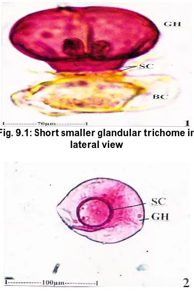
Fig. 9.1: Short smaller glandular trichome in lateral view
Fig. 9.2: Larger glandular trichome in surface view
(Bc-Body cell, GH-Glandular Head, Stc-Stalk cell)
Determination of Physico-Chemical Studies. The results were shown in Table no.1
Table 1: Determination of physical constants: (Ash values, Extractive values and foreign
organic matter)
Evaluation parameters Yield (% w/w)
Total ash 9.40
Acid insoluble ash 6.30
Alcohol soluble extractive 5.10
Water soluble extractive 8.13
Foreign organic matter 0.22
Reaction of Powdered Drug with Different Reagents
The behaviour of powder with various reagents under ordinary light by naked eye is given in Table. 2
Table 2: Behaviour of the Powder of Ocimum basilicum with Different Chemical Reagents
Treatment Colour Powder with different chemical
reagents
Observation
Powder as such Pale green
Powder + Conc. sulphuric acid Greenish Black Powder + Conc. nitric acid Brownish yellow Powder + Conc. Hydrochloric acid Pale yellow
Powder + 5% Iodine Brownish Yellow
Powder + glacial Acetic acid Pale green
Preliminary Phytochemical Screening
The preliminary phytochemical screening with the various qualitative chemical tests revealed the presence of various phytoconstituents like Alkaloids, glycosides, Acids, triterpenoids, tannins, phytosterols, flavonoids and saponins. The results were shown in Table 3.
Table 3: Results of Phytochemical screening
Constituents Ethanolic Extract of
Ocimum basilicum
Alkaloids +
Acids +
Phenolic compounds -
Flavanoids +
Glycosides +
Coumarin -
Tannins +
Phytosterols +
Saponins +
Triterpenoids +
( + ) = indicates presence (-) = indicates absence
CONCLUSION
The present study is related to pharmacognostical, physical constants and preliminary phytochemical screening of Ocimum
basilicum Linn. leaves provided useful information about its correct identity and evaluation. It helps to differentiate from the closely related other species of Ocimum
basilicum Linn. Phytochemical study is also
useful to isolate the pharmacologically active principles present in the drug. The other parameters observed are also useful for the future identification of the plant and serves as a standard monograph for identification and evaluation of plant.
ACKNOWLEDGEMENT
The authors wish to thank to Dr. Ishari. K. Ganesh, Chancellor, Vels University for providing the facilities necessary to carry out the research work. Authors wish to thank to Botanist Dr. P. Jayaraman, Director, Plant Anatomy Research Centre, Chennai, Tamil Nadu for providing valuable information about the plant and its identification.
REFERENCES
1. Sharma SK. Recent approach to herbal formulation development and standardization. http//pharmainfo.net. 2004
2. World Health Organization. Quality control methods for medicinal plant material. WHO Library. 1998 p 110-115. 3. Singh S. Evaluation of Gastric anti ulcer
activity of fixed oil of Ocimum basilicum
and its possible mechanism of action. Indian J Experi Biol. 1998;36:253-257. 4. Muralidharan A and Dhananjayan R.
Cardiac Stimulant activity of Ocimum Basilicum Extracts. Indian J Pharmacol. 2004;36:163-166.
5. Szollosi R and Szollosi LV. Total antioxidant power in some species of Labiatae. J Acta Biologica. 2002;46(3-4):125-127.
6. Orient Longman. Indian Medicinal Plants. 1995;4:160-161.
7. Kirtikar KR and Basu BD. Indian medicinal plants. 2003;3:1961-1964. 8. Easu K. Plant Anatomy Wiley and sons.
New York, 1964;767.
9. Esau K. Anatomy of seed plants. John Wiley and sons. New York, 1979;550. 10. Gamble JS. Flora of the Presidency of
Madras. Vol. I, II, & III. Botanical survey of India: Calcutta India. 1935.
11. Henry AN, Kumari GR and Chitra V. Flora of Tamilnadu Botanical Survey of India: Southern Circle, Coimbatore India. 1987;(3):258.
12. Johansen DA. Plant Microtechnique. Mc Graw Hill Book Co. New York. 523. 13. Metcalfe CR and Chalk L. Anatomy of the
Dicotyledons. Vol. I & II. Clarendon Press Oxford. 1950.
14. Metcalfe CR and Chalk L. Anatomy of the Dicotyledons. Vol. I & II. Clarendon Press Oxford. 1979; 276.
15. O’Brien TP, Feder N and Mc Cull ME. Polychromatic Staining of Plant Cell walls by toluidine blue-O. Protoplasma, 1964;(59):364-373.
16. Sass JE. Elements of Botanical Microtechnique. Mcgraw Hill Book Co New York. 1940;222.
17. Wallis TE. Text book of Pharmacognosy, CBS Publishers and Distibutors Shahdara Delhi India. 1985.
18. Yoga Narasimhan SN. Medicinal Plants of India. Vol. II. Tamilnadu Regional Research institute (Ay.) Bangalore India. 2000;715.
19. Ansari MM, Ahmad J, Ahmad A and Ansari SH. Pharmacognostic characterization and standardization of Morus alba stem bark. J Medic Aromatic Plant Sci. 2006;28:31-36.
20. Harborne JB. Phytochemical methods. Chapman and Hall London. 1999;60-66. 21. Kokate CK, Purohit AP and Gokhale SB.
Text Book of Pharmacognosy.1st edition