Published online November 20, 2014 (http://www.sciencepublishinggroup.com/j/js) doi: 10.11648/j.js.20140206.11
ISSN: 2330-0914 (Print); ISSN: 2330-0930 (Online)
European society of gastrointestinal endoscopy on bile
duct injuries
Syed Adnan Kabir
1, Syed Irfan Kabir
2, Roma Patel
3, Thomas Kallachil
1, Imran Inam
11
Surgical Department, Victoria Infirmary Hospital, Glasgow, G429TY 2
Heatherwood and Wexham Park hospital, Slough, Berkshire SL22AL 3
Surgical Department, Lincoln County Hospital, Lincoln, LN25QY
Email address:
Adnankabir58@hotmail.com (S. A. Kabir)
To cite this article:
Syed Adnan Kabir, Syed Irfan Kabir, Roma Patel, Thomas Kallachil, Imran Inam. European Society of Gastrointestinal Endoscopy on Bile Duct Injuries. Journal of Surgery. Vol. 2, No. 6, 2014, pp. 82-87. doi: 10.11648/j.js.20140206.11
Abstract:
The aim of this article is to describe the pathophysiology, diagnosis, classification, and management of BDI based on the relevant available literature, in particular the recent recommendations from the European Society of Gastrointestinal Endoscopy (ESGE). It is a known fact that bile duct injuries (BDI) are associated with a high morbidity and mortality, posing impaired quality of life along with substantial financial burdens to patients and the society in general. Depending on the type of duct injury, successful management is based upon the time of recognition of injury, patient condition, presence of complications and availability of professional expertise (radiologists, endoscopists and hepato-biliary surgeons). Appropriate management may include endoscopic, per-cutaneous and surgical interventions with imaging playing a significant role in initial diagnosis, assessment and treatment of such injuries.Keywords:
Injury, Leak, Bile Duct, Cholecystectomy, Drainage, Endoscopic Management, Stent1. Introduction
Bile duct injury (BDI) may lead to serious life threatening complications such as bile leakage, bilioma formation, intra-abdominal abscess or collection, cholangitis and biliary cirrhosis secondary to chronic strictures, and are notoriously associated with high morbidity and mortality with impaired quality of life, causing substantial financial burdens to patients and the society in general.
It can commonly occur in two situations i.e. iatrogenic (hepato-biliary surgery) and in trauma (rarely), both needing specific diagnostic and therapeutic management plans achieved with a multidisciplinary team (MDT) approach. [1]
Though, successful outcomes of BDI commonly depend on factors such as timely recognition of injury, patient condition, presence of complications and availability of professional experts i.e. radiologists, endoscopists and hepato-biliary surgeons, appropriate actual treatment options for such injuries are complex decisions and depends on the type and site of the duct injury identified, and may include endoscopic, per-cutaneous and surgical interventions with imaging playing a significant and important role in its initial diagnosis, assessment and treatment of such injuries. [2]
Therefore, the aim of this article is to describe the pathophysiology, diagnosis, classification, and management of BDI based on the available literature, in particular the recent recommendations of the European Society of Gastrointestinal Endoscopy (ESGE).[3]
2. Pathophysiology Hepato-Biliary
Surgery (Cholecystectomy)
Epidemiology
Today, laparoscopic cholecystectomy (LC) has become the gold standard for symptomatic gallstones. However, it is associated with a higher incidence of BDI when compared to an open cholecystectomy (OC). Although very rare, it has been reported by various authors that the incidence of iatrogenic BDI has risen from 0.1% to 0.2% in the era of OC to 0.4% to 0.7% in the era of LC.[4][5]
anatomical details.[7][8]
Never the less, risk factors for iatrogenic BDI depend on the conditions of surgery undertaken, intense inflammation is an independent risk factor and some surgeons recommending conversion to OC when encountered with a difficult situation.[9][10]
Other risk factors reported in the literature include incorrect interpretation of atypical anatomy (accounting for 66% to 97% of BDI causes). 11
The risk is even higher in a thin patient with a narrow main bile duct, which can be misrecognized as the cystic duct. However, many authors report that the most frequent type of injury encountered by the operating surgeon is the intra- or post- operative loss of substance of the cystic duct or the common bile duct (CBD). Furthermore, in 70% to 80% cases the surgeon operating is unaware of having incurred the injury.[11][12],
Delayed leaks are commonly due to thermal or vascular injury during dissection, 13 and, rarely, retained stones in the bile duct can result in suture breakdown by obstructing bile flow. This emphasizes the importance of identifying CBD stones before or during surgery by echo-endoscopy, MRCP or intra-operative cholangiogram.[13]
It is also Important to maintain good visibility within the surgical field as adhesions and insufficient knowledge of anatomical relationships increase the risk of BDI; other risk factors include haste and failure to consider the possibility of atypical routes of vessels and bile ducts (Luschka’s ducts) which have a prevalence of approximately 0.5%. If unrecognized during surgery, this has been reported as the second commonest cause of post-operative bile leaks. [14]
3. Liver Transplantation
Biliary complications are seen in 5% to 30% of cases following liver transplant surgery; common being anastomotic strictures (42% of the cases), followed by BDI (representing 27% of these complications).[15]
While BDI usually presents early and has been reported to correlate with whether the biliary anastomosis was a Roux-en-Y hepatico-jejunostomy or a hepatico-hepaticostomy.15, [16]
Strictures on the other hand are chronic complications and develop over a period of 3 months following liver transplantation. In addition to the previous, Riediger et al[17] in their systematic review and meta-analysis have concluded that anastomotic stenting has not been shown to prevent anastomotic strictures. Moreover, stent removal may itself result in bile leakage, therefore insertion of a T-tube is not recommended routinely.
4. Trauma
It is a rare cause of BDI reportedly identified in approximately 0.1% of the patients with multiple traumas. [18] It commonly presents at three different levels i.e. at the origin of the left branch of the bile duct, the hilum and the
point where the bile duct penetrates into the duodeno-pancreatic block.
Different mechanisms for such pattern of injury have been suggested. The most common being compression of the bile duct against the vertebral column, resulting in avulsion of the main bile duct between the extra- and fixed intra-pancreatic portions.[19][20]
5. Diagnosis Iatrogenic Bile Duct Injury
Intraoperative
It has been estimated that 25% to 32% of bile duct injuries are identified intra-operatively during laparoscopic cholecystectomies, and may be repaired without delay if a surgeon with experience in bile duct repair is available.[21]
Unfortunately, most injuries are not recognized at the index surgery hence patients may present days, weeks, months or even years later. In many circumstances when there is suspicion of a bile leak an intra-operative cholangiogram can help in the diagnosis.[22] However, the value of intra-operative cholangiograms in the diagnosis of intra-operative BDI is widely debated amongst hepato-biliary specialists. There is no consensus on the value of intra-operative cholangiography to detect or prevent BDI.39
While few are of the view that intra-operative cholangiography can have false positive results (in 1-3% of cases)[23] that can result in unnecessary exploration of biliary tract. It can also create a risk of tear at the confluence of the cystic duct and CBD, thus increasing the overall costs of the procedure. Where as, others are in favour of its routine use because of the medico-economic and medico-legal value of cholangiography, as well as the advantage of early treatment of BDI. [24][25]
6. Post-Operative
It is important to note that in the past, it was recommended to place drains in the gallbladder bed for the early detection of postoperative bile leaks. However, intra-abdominal drains are no longer recommended routinely as they increase length of hospital stay and are a high risk for infection.[26][27]
BDI’s that are not identified during the intraoperative period may present days, weeks, months or even years later. Patients may present with signs and symptoms associated with bile leakage, bile duct transection or ligation, such as biliary peritonitis, cholangitis, and jaundice. However, the greater frequency of nonspecific initial symptoms such as abdominal pain, malaise, nausea and anorexia may account for the common delays in the early diagnosis. Later presentation may include recurrent cholangitis and secondary biliary cirrhosis due to stricture formation.[28] [29][30]
be normal or only mildly elevated due to absorption of bile from the peritoneal cavity.[31]
Nevertheless, it is important that patients who have undergone hepato-biliary surgery and have failed to recover as expected undergo evaluation for the possibility of BDI with the above mentioned signs raising alarms for urgent imaging in order to identify the presence of bile within the abdomen.39
In the early post-operative phase, ultrasonography (USS) and computed tomography (CT) scan play an important role in the initial assessment of the situation, revealing intra-abdominal collections and ductal dilatation (biliary tree may not be dilated if there is bile leakage). Other imaging such as hepato-biliary scintigraphy does confirm a leak but unfortunately lacks the anatomical detail to identify the specific leak site. In such situations, cholangiograms could be of paramount importance to visualize the exact location and extent of injury.33
MRCP is a non-invasive diagnostic investigation that provides an excellent delineation of the biliary anatomy. However, it can miss minor bile leakages. ERCP has the added advantage of not only being able to identify the biliary anatomy and at the same time evaluate the injury, but also used therapeutically (if indicated) to bypass a leak or stricture by placement of stents.33
In order to define the anatomy of the biliary tree proximal to the injury, PTC has been found to be more useful (notably in those patients with complete ductal ligation or transection). In addition, PTC can be followed by placement of a percutaneous trans-hepatic biliary catheter, which is useful in decompressing the proximal biliary system in biliary obstruction, or to control bile leaks. PTC and drainage also allow decompression of the biliary tree in the early postoperative period after definitive repair of the bile duct injury, as well as providing access for postoperative cholangiography. [32]
7. Diagnosis in Trauma
In trauma cases, the diagnostic modalities depend on various factors i.e. how stable the patient is at onset of management and other vital organs injured requiring urgent management. Hence the diagnosis can classically be made at three different points; immediately at emergency laparotomy (performed for other intra-abdominal injuries) in an unstable patient, early in the stable patient presenting with non-specific symptoms, or late after the initial trauma as a result of complications.[33]
Non-specific symptoms after initial trauma such as nausea, vomiting, abdominal discomfort and low-grade pyrexia are all indications for repeat imaging (USS or CT scan) to look for intra-abdominal fluid or bile duct dilatation. In case of a suspected BDI, MRCP is recommended to be the most accurate investigation before therapy (whether endoscopic or surgical) is undertaken to fully define complex or multi focal injuries. Late diagnosis usually leads to very complex, and often fatal situations. Therefore it is important that BDI is diagnosed as early as possible.[34]
8. Classifications of Bile Duct injuries
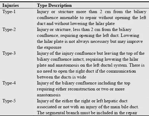
The first classification of BDI was described by Bismuth (Fig-1). This was before the era of LC. It was meant to be a guide for surgical repair and has been fairly well correlated with outcomes after treatment. [35]
Since then, many classifications of BDI have been proposed. Important to note is that of McMahon et al,[36] which identifies minor injuries (requiring a simple repair or insertion of a T-tube) and major injuries (possibly requiring surgical repair such as hepatico-jejunostomy). However, the most widely used classification today is that of Strasberg et al [37] (Fig 2). This is basically a simplification of the Bismuth classification with inclusion of injury induced by LC.
In conclusion, these classifications have limited value when it comes to clinical practice, mainly because the important factor intervening in therapy is the existence of a complete circumferential trans-section (needing surgical alternatives) or incomplete trans-section (needing endoscopic alternatives) of the main bile duct.36
Fig 1. Bismuth Classification System for Bile Duct
Injuries Type Description
Type-1 Injury or stricture more than 2 cm from the biliary confluence amenable to repair without opening the left duct and without lowering the hilar plate
Type-2 Injury or stricture, less than 2 cm from the biliary confluence, requiring opening the left duct. Lowering the hilar plate is not always necessary but may improve the exposure
Type-3 Injury of the injury confluence but leaving the top of the biliary confluence intact, requiring lowering the hilar plate and anastomosis on the left ductal system. There is no need to open the right duct if the communication between the ducts is wide
Type-4 Injury of the biliary confluence including the top requiring either reconstruction or two or more anastomosis
Type-5 Injury of the either the right or left hepatic duct associated or not with an injury of the main bile duct. The segmental branch must be included in the repair
Fig 2. Strasberg et al.’s classification of bile duct injury
Type A Bile leak from a minor duct still in continuity with the common bile duct
Type B Occlusion of part of biliary tree
Type C Bile leak from duct not in communication with common bile duct
Type D Lateral injury to extra hepatic bile ducts
9. Management of BDI Based on the
Recommendations of the European
Society of Gastrointestinal Endoscopy
(ESGE) and Literature
intra-operatively, characterization of the bile ducts can be obtained through intra-operative cholangiography. However, when the injury is recognized post-operatively, MRCP with 3-D reconstruction is recommended as it not only provides accurate mapping but also detects associated stones.[38] Characterization helps guide the best therapeutic plan i.e. endoscopic or surgical repair.
According to the recommendations made by the ESGE (2012),[39] endoscopic treatment is effective in more than 90% of patients who present with incomplete circumferential injury (iatrogenic or trauma) of the bile duct. The main purpose of such treatment is to reduce the pressure gradient of bile flow in the bile duct, lowering the pressure gradient between the bile duct and at the level of the sphincter of Oddi. This increases bile flow into the duodenum hence enhances the healing process. This can be achieved with various options i.e. either by sphincterotomy alone, or with the insertion of a plastic stent, or a naso-biliary drain. The latter two strategies generally need repeated interventions.
Literature has shown that that sphincterotomy with eventual stone extraction and insertion of a plastic stent allows bile duct healing in more than 90% of the cases. In addition to the above, plastic stents in particular improve healing when compared to sphincterotomy alone. On the contrary, no difference has been found between stent placements with sphincterotomy versus stent without sphincterotomy. Moreover, sphincterotomy with stent placement carries a short-term risk of pancreatitis and long-term risk of stricture formation (especially in young patients) thus confirming avoidance of sphincterotomy for this indication. [40]
There may be a case where sphincterotomy alone is performed to allow temporary drainage. Literature has shown that if stents are used for temporary drainage, biliary abnormalities such as stones, sludge or residual leak may become evident upon stent removal. Sandha et al42 proposed a therapeutic algorithm in which it recommends sphincterotomy alone in case of minimal leaks (< 200 mL/24 h). However, in severe leaks, strictures, contraindications to sphincterotomy or poor post-sphincterotomy drainage, it recommends insertion of a plastic stent for 4 to 6 weeks.[41] This approach has claimed to provide satisfactory results in more than 90% of the cases out of a series of 207 patients. In support to the above, two prospective studies with a total of 56 patients has also shown that in the absence of associated strictures, sphincterotomy alone improves the leak in more than 90% of the cases (within an average delay of 11 days).[42][43] A randomized controlled study has shown that insertion of a plastic stent enhanced healing significantly when compare to sphincterotomy alone.[44]
A large retrospective study has reported bile duct anomalies (sludge, stones or bile leaks) in 25% of cases at the time of stent removal. As a result, the ESGE recommends passage of a balloon catheter at the time of stent removal (when this strategy is chosen) in order to obtain an accurate cholangiogram.[45]
In case of biloma formation as a result of bile leak, combined endoscopic stent placement with radiological percutaneous drainage of the bile collection is deemed necessary. However, in a case of complete trans-section of the bile duct (identified either intra-operatively or early post-operatively) due to biloma formation, treatment is most often surgical (hepatico-jejunostomy).[46]
In conclusion, the ESGE recommends that all the advantages and disadvantages of each strategy need to be discussed with the patient, explaining that several procedures under general anesthesia may be necessary when stent insertion is chosen.
In all cases, a precise and meticulous workup of the bile leak must be made during the initial ERCP in order to characterize the leak and look for associated anomalies (stones, stricture), which may influence the therapeutic strategy. In their absence, the ESGE recommends insertion of a 7F plastic stent without initial sphincterotomy with removal between 4 to 8 weeks. Sphincterotomy alone can be envisioned if a new operation needs to be avoided (elderly patient with multiple co-morbidities). At the time of stent removal, new cholangiograms should be obtained to ensure that all debris has been removed. This cholangiography is best performed with a balloon catheter occluding the lower portion of the bile duct to ensure a high quality-mapping images.39
10. In Conclusion
Iatrogenic (mainly during laparoscopic cholecystectomy) is the most frequent cause of BDI. These are complex situations requiring a multi-disciplinary-radio-medico-surgical collaboration to achieve better management outcome. BDI can be recognized either intra- or post-operatively, this influences the type of diagnostic strategy and therapeutic management needed to deal with such a complication. Depending on the situation characterization of the biliary tree is a must and should be obtained by intra-operative cholangiogram or MRCP to help plan ideal therapeutic management plan. Endoscopy is recommended in the absence of complete trans-section of the main bile duct with insertion of a stent for 4 to 8 weeks as the first-line therapy; it is effective in more than 90% of patients. Sphincterotomy is not recommended as it increases early and late morbidity by 15%.
There are no recommendations in case of stent failure after 4 to 8 weeks, and the choice between a repeat, longer endoscopic stent or surgery is not well defined. In case of complete circumferential trans-section of the main bile duct, hepatico-jejunostomy is essential for optimal healing.
References
[2] Lau WY, Lai EC, Lau SH. Management of bile duct injury after laparoscopic cholecystectomy: a review. ANZ J Surg 2010; 80(1-2):75–81.
[3] Dumonceau JM, Tringali A, Blero D, et al. Biliary stenting: indications, choice of stents and results: European Society of Gastrointestinal Endoscopy (ESGE) clinical guideline. Endoscopy 2012; 44:277—98.
[4] Adamsen S, Hansen OH, Funch-Jensen P, Schulze S, Stage JG, Wara P. Bile duct injury during laparoscopic cholecystectomy: a prospective nationwide series. J. Am. Coll. Surg. 1997; 184: 571–8.
[5] Lai EC, Lau WY. Mirizzi syndrome: history, present and future development. ANZ J Surg 2006; 76: 251–7.
[6] Joseph M, Phillips MR, Farrell TM, Rupp CC. Single incision laparoscopic cholecystectomy is associated with a higher bile duct injury rate: a review and a word of caution. Ann Surg2012; 256(1):1—6.
[7] Parmeggiani D, Cimmino G, Cerbone D, et al. Biliary tract injuries during laparoscopic cholecystectomy: three casereports and literature review. G Chir 2010; 31:16—9.
[8] Southern surgeons group. A prospective analysis of 1518 laparoscopic cholecystectomies. The Southern Surgeons Club. N Engl J Med 1991; 324:1073—8.
[9] Georgiades CP, Mavromatis TN, Kourlaba GC, et al. Isinflammation a significant predictor of bile duct injuryduring laparoscopic cholecystectomy? Surg Endosc 2008; 22:1959—64.
[10] Nuzzo G, Giuliante F, Persiani R. The risk of biliary ductal injury during laparoscopic cholecystectomy. J Chir (Paris) 2004; 141:343—53.
[11] Karvonen J, Gullichsen R, Laine S, Salminen P, Grönroos JM. Bileduct injuries during laparoscopic cholecystectomy: primaryand long-term results from a single institution. Surg Endosc2007; 21:1069-73.
[12] Davidoff AM, Pappas TN, Murray EA, et al. Mechanisms of major biliary injury during laparoscopic cholecystectomy. Ann Surg 1992; 215:196—202.
[13] Richardson MC, Bell G, Fullarton GM, West of Scotland Laparo- scopic Cholecystectomy Audit Group. Incidence and nature of bile duct injuries following laparoscopic cholecystectomy: an audit of 5913 cases. Br J Surg 1996; 83:1356—60.
[14] Balija M, Huis M, Szerda F, Bubnjar J, Stulhofer M. Laparoscopic cholecystectomy–accessory bile ducts. Acta Med Croatica 2003; 57:105—9.
[15] Greif F, Bronsther OL, Van Thiel DH, et al. The incidence, timing, and management of biliary tract complications after ortho topic liver transplantation. Ann Surg 1994; 219:40—5.
[16] O’Connor TP, Lewis WD, Jenkins RL. Biliary tract complications after liver transplantation. Arch Surg 1995; 130:312—7.
[17] Riediger C, Müller MW, Michalski CW, et al. tube or no T-tube in the reconstruction of the biliary tract during orthotopic liver transplantation: systematic review and meta-analysis. Liver Transpl 2010; 16:705—17.
[18] Sawaya Jr DE, Johnson LW, Sittig K, McDonald JC, Zibari
GB. Iatrogenic and non-iatrogenic extra-hepatic biliary tract injuries: a multi-institutional review. Am Surg 2001; 67:473—7.
[19] D’Amata G, Rahili A, Habre J, Karimd jee B, Sanchez Bueno F, Bourgeon A. Traumatic avulsion of the intrapancreatic common bile duct: case report. G Chir 2006; 27:27—30.
[20] Maier WP, Lightfoot WP, Rosemond GP. Extra hepatic biliary ductal injury in closed trauma. Am J Surg 1968; 116:103—8.
[21] Lau WY, Lai EC, Lau SH. Management of bile duct injury after laparoscopic cholecystectomy: a review. ANZ J Surg 2010; 80(1-2):75–81.
[22] Flum DR, Dellinger EP, Cheadle A, Chan L, Koepsell T. Intra operative cholangiography and risk of common bile duct injury during cholecystectomy. JAMA 2003;289:1639—44.
[23] Iorga C, Cirimbei S, Strambu V, Popa F.Intraoperative cholangiography still a current investigation. Journal of Medicine and Life Vol. 6, Issue 4, October-December 2013, pp.399-402.
[24] Flum DR, Dellinger EP, Cheadle A, Chan L, Koepsell T. Intra operative cholangiography and risk of common bile duct injury during cholecystectomy. JAMA 2003; 289:1639—44.
[25] Ausania F, Holmes LR, Ausania F, Iype S, Ricci P, White SA. Intraoperative cholangiography in the laparoscopic cholecystectomy era: why are we still debating? Surg Endosc 2012; 26:1193—200.
[26] El-labban G, Hokkam E, El-labban M, Saber A, Heissam K, El-Kammash S. Laparoscopic elective cholecystectomy with and without drain: a controlled randomised trial. J Minim Access Surg 2012; 8:90—2.
[27] Tzovaras G, Liakou P, Fafoulakis F, Baloyiannis I, ZacharoulisD, Hatzitheofilou C. Is there a role for drain use in electivelaparoscopic cholecystectomy? A controlled randomized trial.Am J Surg 2009; 197:759—63.
[28] Connor S, Garden OJ. Bile duct injury in the era of laparoscopic cholecystectomy. Br J Surg 2006; 93(2):158–168.
[29] Lee CM, Stewart L, Way LW. Post cholecystectomy abdominal bile collections. Arch Surg 2000; 135(5):538–542.
[30] Bauer TW, Morris JB, Lowenstein A, Wolferth C, Rosato FE, Rosato EF. The consequences of a major bile duct injury during laparoscopic cholecystectomy. J Gastro intest Surg 1998; 2(1):61–66.
[31] Wan Yee Lau, Eric C. H. Lai and Stephanie H. Y. Lau. Management of bile duct injury after laparoscopic cholecystectomy: a review. ANZ J Surg 80 (2010) 75–81
[32] Colin M. Thompson, Nael E. Saad, Robin R. Quazi, Michael D. Darcy, Daniel D. Picus, Christine O. Menias, Management of Iatrogenic Bile Duct Injuries: Role of the Interventional Radiologist. RadioGraphics 2013; 33:117–134
[33] Parks RW, Diamond T. Non-surgical trauma to the extra hepatic biliary tract. Br J Surg 1995; 82:1303—10.
[34] Ivatury RR, Rohman M, Nallathambi M, Rao PM, Gunduz Y, Stahl WM. The morbidity of injuries of the extra-hepatic biliary sys-tem. J Trauma 1985; 25:967—73.
[36] McMahon AJ, Fullarton G, Baxter JN, O’Dwyer PJ. Bile duct injury and bile leakage in laparoscopic cholecystectomy. Br JSurg 1995; 82:307—13.
[37] Strasberg SM, Hertl M, Soper NJ. An analysis of the problem of biliary injury during laparoscopic cholecystectomy. J Am CollSurg 1995; 180:101—25.
[38] M. Pioche, T. Ponchonom. REVIEW; Management of bile duct leaks. Journal of Visceral Surgery (2013) 150S, S33— S38
[39] Dumonceau JM, Tringali A, Blero D, et al. Biliary stenting: indications, choice of stents and results: European Society of Gastrointestinal Endoscopy (ESGE) clinical guideline. Endoscopy 2012; 44:277-98.
[40] Sugiyama M, Atomi Y. Risk factors predictive of late complications after endoscopic sphincterotomy for bile duct stones: long-term (more than 10 years) follow-up study. Am JGastroenterol 2002; 97:2763-7.
[41] Sandha GS, Bourke MJ, Haber GB, Kortan PP. Endoscopic
therapy for bile leak based on a new classification: results in207 patients. Gastrointest Endosc 2004; 60:567-74.
[42] Aksoz K, Unsal B, Yoruk G, et al. Endoscopic sphincterotomyalone in the management of low-grade biliary leaks due tocholecystectomy. Dig Endosc 2009; 21:158—61.
[43] Llach J, Bordas JM, Elizalde JI, et al. Sphincterotomy in the treatment of biliary leakage. Hepatogastroenterology2002; 49:1496—8.
[44] Marks JM, Ponsky JL, Shillingstad RB, Singh J. Biliary stenting ismore effective than sphincterotomy in the resolution of biliaryleaks. Surg Endosc 1998; 12:327—30.
[45] Coté GA, Ansstas M, Shah S, et al. Findings at endoscopic retrograde cholangio-pancreato-graphy after endoscopic treatment of post cholecystectomy bile leaks. Surg Endosc2010; 24:1752—6.