STUDIES OF TOLERANCE INDUCTION IN B LYMPHOCYTES
Sarah L. PARRY B.Sc. (Hons).
A thesis submitted for the degree of Ph.D. in the University of London. October, 1993
National Institute for Medical Research, Department of Cellular Immunology,
The Ridgeway, Mill Hill,
ProQuest Number: 10018666
All rights reserved
INFORMATION TO ALL USERS
The quality of this reproduction is dependent upon the quality of the copy submitted.
In the unlikely event that the author did not send a complete manuscript and there are missing pages, these will be noted. Also, if material had to be removed,
a note will indicate the deletion.
uest.
ProQuest 10018666
Published by ProQuest LLC(2016). Copyright of the Dissertation is held by the Author.
All rights reserved.
This work is protected against unauthorized copying under Title 17, United States Code. Microform Edition © ProQuest LLC.
ProQuest LLC
789 East Eisenhower Parkway P.O. Box 1346
ABSTRACT
The silencing and/or elimination of autoreactive clones in the B cell repertoire has been shown to occur at both immature and mature stages of B cell development. A number of factors which influence tolerance induction in B cells, such as the degree of antigen receptor crosslinking and the presence or absence of co-stimulatory signals have been identified. The main focus of the work presented in this thesis concerns an investigation of two in vitro models of B cell tolerance. In the first, the capacity of sIgM and sIgD receptors to generate negative signals was studied using slgM^ immature B lymphomas and their slgD^ transfectants. The data suggest that ligation of both sig isotypes by anti-Ig antibodies induced tolerizing signals leading to growth arrest in these cells. Extensive crosslinking of sIg was a key factor in inducing the growth inhibition. Multimerization of sIgM, but not sIgD, resulted in apoptotic cell death. In addition, extensive crosslinking of the transmembrane phosphatase CD45 with certain monoclonal antibodies caused growth inhibition, and induced apoptosis in a fraction of the immature B lymphoma cells. Crosslinked CD45 enhanced the growth inhibition induced by both anti-IgM and anti-IgD antibodies, suggesting CD45 may play a part in modulating tolerogenic signals generated via the antigen receptors in immature B cells.
The second model employed mature murine splenic B lymphocytes. The data shows that extensive crosslinking of either sIgM or sIgD by anti-Ig antibodies induced unresponsiveness in these cells, leading to massive apoptotic cell death. Apoptotic nuclei were detected within 4 hours after anti-Ig stimulation, but cells which survived for 12 -1 6 hours were abortively activated, as evidenced by increased levels of MHC Class II antigens. Both IL-4 and ligation of CD40 partially reversed the induction of
ACKNOWLEDGEMENTS
I would like to express my thanks to Dr. Gerry Klaus for his supervision and help throughout this project.
Many thanks also to my friends and colleagues in the Divisions of Cellular and Molecular Immunology for all their help and encouragement. I would like to make a special mention of the members and ex-members of Gerry’s lab. - Maggie, Mary, Rob, Michael, Has, Caroline, Jillian and Jessica - who have been a pleasure to work with, and have made the last three years an enjoyable experience.
A special thank-you to Colin, who has always encouraged and supported me.
SECTION Page No
ABSTRACT
ACKNOWLEDGEMENTS
TABLE OF CONTENTS 4
LIST OF FIGURES 8
LIST OF TABLES 12
ABBREVIATIONS 13
CHAPTER 1: INTRODUCTION. 15
1.0. General introduction 15
1.1. B cell differentiation 19
1.2. Structure of the Ig-receptor complex 22
1.3. Signal transduction via sig receptors 23
1.4. The role of CD45 in regulation of signal transduction
by antigen receptors 26
1.5. Mechanisms of B cell activation 30
1.6. B cell tolerance 35
1.6.1. Immature B cell tolerance 35
1.6.2. In vitro models for studying tolerance induction in
immature B cells 38
1.6.3. Tolerance in mature B cells 39
1.6.4. Parameters which influence B cell tolerance 44
1.7. Objectives of the current studies 48
CHAPTER 2: MATERIALS AND METHODS. 49
2.1. Cell and antibody preparations 49
2.1.1. B cell lymphomas 49
2.1.2. T cell lines and supernatants 50
2.1.3. Storage of cells in liquid nitrogen 51
2.1.4. Antibodies 51
2.1.5. Lymphokine preparations 52
2.1.6. Experimental animals 53
2.1.7. Preparation of small dense B cells 53
2.2. General materials and methods 54
2.2.1. Reagents 54
2.2.2. Isotopes 55
2.2.3. Percoll gradients 55
2.2.5. Preparation of fixed activated T cells (FAT cells) 56
2.2.6. Soluble CD40 ligand (CD40L) : preparation of
CD40L-CD8a chimeric protein 56
2.2.7. Membrane-bound CD40L : preparation of CD40L-transfected murine erythroleukaemia
cells (CD40L+MEL cells) 57
2.2.8. Preparation of antibody-coated tissue culture plastic
(Immobilised antibody) 57
2.2.9. Preparation of anti-phosphotyrosine sepharose beads 58
2.2.10. Preparation of non-specific protein A sepharose beads 58
2.3. Buffers and tissue culture reagents 59
2.3.1. Phosphate buffered saline (PBS) 59
2.3.2. RPMI 1640 tissue culture medium (TC medium) 59
2.3.3. Eagles modified complete medium (MEM) 60
2.3.4. NP40 lysis buffer 60
2.3.5. Hypotonic fluorochrome solution 60
2.3.6. TBE buffer 61
2.3.7. TE buffer 61
2.3.8. TAE buffer 61
2.3.9. PBA buffer 62
2.3.10. SDS-PAGE non-reducing sample buffer 62
2.3.11. Running buffer 62
2.3.12. SDS-PAGE gels (10%) 63
2.4. Bioassays 63
2.4.1. Estimation of DNA or RNA synthesis 63
2.4.2. Apoptosis analysis by immunofluorescence, using flow
cytometry 65
2.4.3. Apoptosis analysis by immunofluorescence, using
fluorescence microscopy 66
2.4.4. Apoptosis analysis by DNA extraction and gel
electrophoresis 66
2.4.5. Fluorescent antibody staining and analysis 67
2.5. Biochemical analyses 68
2.5.1. Anti-phosphotyrosine immunoprécipitation 68
2.5.2. Western blotting and ECL detection 69
CHAPTER 3: THE ROLE OF sIgM AND sT^D IN IMMATURE B
CELL LYMPHOMAS 70
3.1. Introduction 70
3.2. Surface staining and FACS analysis of the B cell
lymphomas 70
3.3. Growth inhibition of B cell lymphomas by soluble or
immobilised anti-sig mAb 71
3.4. Anti-IgD antibodies enhance growth inhibition induced
by anti-IgM antibodies 73
growth inhibition in immature B cell lymphomas 73
3.6. Reversal of sig-mediated inhibition by LPS 74
3.7. Anti-/x induces apoptosis in the B cell lymphomas 75
3.8. Reversibility of growth inhibition 76
3.9. Effects of lymphokines on sIg-mediated growth
inhibition 76
3.10. Effects of preactivated T cells 77
3.11. Ligation of CD40 does not reverse anti-Ig mediated
growth inhibition 78
3.12. Cyclosporin A does not reverse IgM-mediated growth
inhibition in B cell lymphomas 79
3.13.1. Discussion part I: The role of sIgM and sIgD intolerance
induction 102
3.13.2. Discussion part II: Modulation of B cell tolerance by T
cell-derived influences 109
3.13.3. Discussion part III: Biochemical signals involved in
Ig-mediated growth inhibition 114
CHAPTER 4: HYPERCROSSLINKING SURFACE IgM OR IgD RECEPTORS ON INDUCES UNRESPONSIVENESS AND
APOPTOSIS. 119
4.1. Introduction 119
4.2. Unresponsiveness in mature B cells induced by
immobilised anti-Ig antibodies 119
4.3. Crosslinking of biotinylated anti-Ig antibodies with avidin abrogates their capacity to induce DNA synthesis
in B cells 121
4.4. Hypercrosslinking of biotinylated anti-Ig antibody
inhibits proliferation by heterologous anti-Ig antibody 122 4.5. Hypercrosslinking of biotinylated anti-Ig Abs induces
progressive unresponsiveness to LPS 123
4.6. Crosslinking of bio-anti-Ig mAb with streptavidin
induces concentration dependent effects 124 4.7. IL-4 partially reverses inhibition induced by crosslinked
anti-Ig antibodies 124
4.8. Ligation of CD40 partially reverses inhibition induced
by crosslinked biotinylated anti-Ig 125
4.9. Hypercrosslinking biotinylated anti-IgM or IgD
antibodies induces apoptosis in mature B cells 126 4.10. Immobilised anti-IgM or anti-IgD antibodies induce
apoptosis in mature B cells 129
4.11. Hypercrosslinking sig induces abortive B cell activation 130 4.12. Induction of apoptosis by hypercrosslinked anti-Ig
antibodies is abrogated to varying degrees by IL-4,
CD40L and a monoclonal antibody to CD40 131 4.13.1. Discussion part I: The role of sIgM and sIgD in mature
4.13.2. Discussion part II: The role of T cell-derived help 163 4.13.3. Discussion part III: Biochemical signalling pathways
involved in mature B cell tolerance 164
CHAPTER 5: CD45 MONOCLONAL ANTIBODIES INDUCE
GROWTH INHIBITION ON IMMATURE B CELL LYMPHOMAS 167
5.1. Introduction 167
5.2. FACS analyses of CD45 expression on B cell
lymphomas 168
5.3. Hypercrosslinking of CD45 induces growth inhibition of
the immature B cell lymphomas 168
5.4. Hypercrosslinked CD45 and anti-Ig antibodies act
additively to induce growth inhibition 169
5.5. LPS has no effect on growth inhibition induced by
hypercrosslinked CD45RB antibodies 170
5.6. Effects of T cell derived influences on CD45-mediated
growth inhibition 171
5.7. Induction of apoptosis in B lymphomas by
hypercrosslinked CD45RB antibody 171
5.8. Investigation of tyrosine phosphorylation following
crosslinking of IgM and/or CD45 173
5.9.1. Discussion part I: The role of CD45 in the immature
model of B cell tolerance 188
5.9.2. Discussion part II: Signal transduction via CD45 in the
B cell lymphomas 193
CHAPTER 6: GENERAL DISCUSSION 198
LIST OF FIGURES FIGURE
3.1. FACS analyses of sIgM and sIgD expression on B cell
lymphomas 81
3.2. Growth inhibition of B cell lymphomas by soluble anti“|i
antibody 82
3.3. Titration of soluble anti-^ on the CH33 lymphoma 83 3.4. Growth inhibition of WEHI-ôM cells by soluble or
immobilised anti-Ig antibodies 84
3.5. Anti-IgD antibodies enhance growth inhibition induced by anti-IgM antibodies in the WEHI-ôM B cell
lymphoma 85
3.6. Hypercrosslinking sig induces marked growth inhibition
in the ECH408-1 B cell lymphoma 86
3.7. LPS partially reversed growth inhibition induced by
soluble anti-IgM mAb in the B cell lymphomas 87 3.8. LPS does not reverse growth inhibition induced by
hypercrosslinked anti-Ig Ab in ECH408-1 B cell
lymphoma 88
3.9. Hypercrosslinked anti-;* antibody induces apoptosis in
CH33 cells 89
3.10. Soluble anti-IgM mAb induces growth inhibition which
is reversible in WEHI-231 B cell lymphoma 90 3.11. Recombinant IL-4 does not protect against growth
inhibition induced by soluble or immobilised anti-IgM
antibody 91
3.12. Lymphokines secreted by Th2 cells do not protect against sIgM-mediated growth inhibition in WEHI-231 cells 92 3.13. Partial reversal of sIgM-mediated growth inhibition in
CH31 but not WEHI-231 by preactivated T cells 93 3.14. Preactivated T cells reverse sIgM-mediated growth
inhibition in CH33 94
3.15. Inhibition of CH33 cells following culture with
3.16. Proliferation of CH33 cells following culture with
activated DIO.D cells or DlO.d crude supernatant 96 3.17. CD40L does not reverse growth inhibition induced by
anti-IgM or anti-IgD antibodies in WEHI-ôM cells 97 3.18. Cyclosporin A does not reverse IgM-mediated growth
inhibition in the B cell lymphomas 98
4.1. Induction of unresponsiveness in mature B cells by pretreatment with immobilised anti-Ig monoclonal
antibodies 133
4.2. Effects of immobilised anti-Ig on RNA synthesis in B
cells 134
4.3. Induction of non-responsiveness in B cells is dependent on the period of culture with immobilised anti-Ig
monoclonal antibody 135
4.4. Effects of crosslinking biotinylated anti-/x or anti-0 mAb with avidin on their capacity to induce DNA synthesis in
B cells 136
4.5. Effects of crosslinking sIgM or sIgD receptors with biotinylated mAb plus avidin on the responses of B cells
to heterologous stimulation 137
4.6. Effects of crosslinking sig receptors with biotinylated F(ab’)2 fragments plus avidin on the response of B cells
to heterologous stimulation 138
4.7. Effects of pulsing B cells with crosslinked bio-anti-ô or
bio-anti-^ on subsequent responses 139
4.8. Hypercrosslinldng of biotinylated anti-Ig mAb induces
delayed unresponsiveness to LPS 140
4.9. Hypercrosslinldng of biotinylated anti-Ig antibodies with
streptavidin induces concentration-dependent effects 141 4.10. IL-4 partially reverses inhibition induced by immobilised
anti-Ig 142
4.11. Effects of IL-4 on responses to bio-anti-/* in mature B
cells 143
4.12. Ligation of CD40 and/or IL-4 partially reverses inhibition induced by hypercrosslinked biotinylated
4.13. DNA staining profiles of nuclei from B cells cultured
with crosslinked anti-Ig antibodies 145
4.14. Effects of crosslinked anti-Ig on scatter profiles of B cell
nuclei 146
4.15. Detection of apoptosis in mature B cells by gel
electrophoresis 147
4.16. Hypercrosslinldng sig with immobilised antibody induced
apoptosis in mature B cells 148
4.17. B cells do not recover from the unresponsive state
following removal of the immobilised anti-Ig antibodies 149 4.18. Effects of co-stimulation with IL-4 on the induction of
apoptosis in B cells 150
4.19. Effects of co-stimulation with anti-CD40 and/or IL-4 on
the induction of apoptosis in B cells 151 5.1. FACS analyses of CD45 expression on WEHI-231 175 5.2. Immobilised CD45 mAb-induced growth inhibition in
CH33 cells 176
5.3. Growth inhibition of WEHI-231 by immobilised
CD45RB antibodies 177
5.4. Soluble, crosslinked CD45RB antibodies induce growth
inhibition in WEHI-231 178
5.5. Soluble anti-^ and immobilised CD45RB antibodies act
synergistically to induce growth inhibition 179 5.6. Soluble anti-Ig and crosslinked bio-CD45RB antibodies
act synergistically to induce growth inhibition 180 5.7. CD45-induced inhibition is not reversed by bacterial
lipopolysaccharide (LPS) 181
5.8. Ligation of CD40 and/or IL-4 do not reverse growth
inhibition induced by crosslinked CD45RB 182 5.9. Effects of immobilised or soluble anti-/x, and
immobilised CD45RB antibodies on CH33 cells 183 5.10. Hypercrosslinked CD45RB antibody induces apoptosis
5.11. Tyrosine phosphoproteins in anti-phosphotyrosine
immunoprécipitations from anti-^ Ab treated WEHI-231
cells 185
5.12. Tyrosine phosphoproteins in anti-phosphotyrosine immunoprécipitations from WEHI-231 cells treated with
anti-fi and/or CD45RB antibodies 186
LIST OF TABLES TABLE
3.1. Summary of growth inhibition in the four cell lines by
anti-/x or anti-6 mAb 99
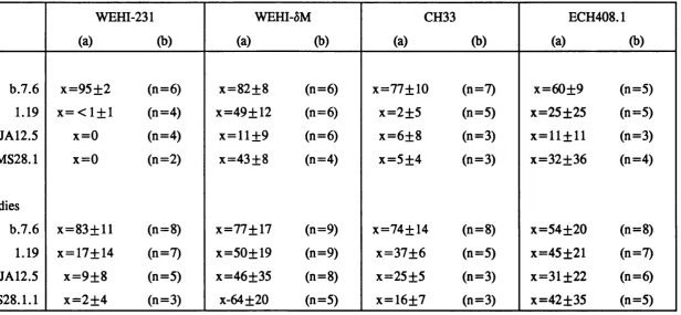
3.2. Induction of apoptosis in the B cell lymphomas by
soluble anti-^A or anti-6 monoclonal antibodies 100
3.3. Summary of apoptosis and cell cycle arrest by anti-/A
antibody in CH33 cells 101
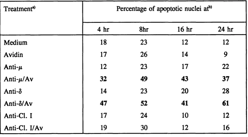
4.1. Time course of the appearance of apoptotic B cells after
culture with crosslinked anti-Ig antibodies 152 4.2. Effects of crosslinking sig with immobilised antibody
for 24 h 153
4.3. Effects of hypercrosslinldng sig receptors with
bio-anti-Ig plus avidin 154
5.1. Summary of apoptosis in CH33 by anti-^ or crosslinked
ABBREVIATIONS
Ab Antibody
anti-ptyr Anti-phosphotyrosine antibody
anti-/* Anti-IgM antibody
anti-6 Anti-IgD antibody
ATCC American type culture collection
Av Avidin
bio-Ab Biotinylated antibody
Bq Becquerel
BSA Bovine serum albumin
ca. Circa
Ca^+ Calcium
[Ca^^i Intracellular calcium concentration
CD40L CD40 ligand
Ci Curie
Con. A Concanavalin A
cpm Counts per minute
CR2 Complement receptor 2
CsA Cyclosporin A
DAG Diacylglycerol
dHgO Distilled water
DMSG Dimethylsulphoxide
ECL Enhanced chemiluminescence
EDTA Ethylene-diamine-tetraacetic acid
P (a b ’)2 Divalent antigen-binding fragment
FACS Fluorescein activated cell sorter
PCS Foetal calf serum
PITC Fluorescein isothiocyanate
PSC Forward scatter
h Hour
H-chain Heavy chain of Immunoglobulin
HEL Hen egg lysozyme
HrIL-2 Human recombinant IL-2
HRP Horse radish peroxidase
pH]TdR Tritiated thymidine
pH]Urd Tritiated uridine
i-Ab Immobilised antibody
ICAM Intercellular adhesion molecule
Ig Immunoglobulin
IL Interleukin
IP3 Inositol 1,4,5 triphosphate
kDa Kilodalton (molecular mass)
L-chain Light chain of Immunoglobulin
LCA Leucocyte common antigen
LPA Lymphocyte function-associated antigen
LPS Lipopolysaccharide
KH2PO4 Potassium dihydrogen phosphate
mAb Monoclonal antibody
MEL cells Murine erythroleukaemia cells
MEM Eagles modified complete medium
MPI Mean fluorescent intensity
MHC Major histocompatibility complex
Mr Molecular weight
NaCl Sodium chloride
Na^HPO^ Disodium hydrogen phosphate
NaOAc Sodium acetate
PAGE Polyacrylamide gel electrophoresis
PBS Phosphate-buffered saline
PGR Polymerase chain reaction
PI Propidium iodide
PIP2 Phosphatidyl inositol 4,5 bisphosphate
PLC Phospholipase C
PKC Protein kinase C
PTK Protein tyrosine kinase
PTPase Protein tyrosine phosphatase
rIL-4 Recombinant Interleukin 4
s-Ab Soluble antibody
sig Surface Immunoglobulin
SDS Sodium dodecyl sulphate
SF medium Serum-free medium
SSC Side scatter
SN Supernatant
TC medium Tissue culture medium
TCR T cell receptor
TD antigen T cell-dependent antigen T1 antigen T cell-independent antigen
Thcell T helper cell
TNF Tumour necrosis factor
U Units
CHAPTER 1
INTRODUCTION.
1.0. General introduction.
Immunological tolerance is defined as a state of specific, antigen-induced unresponsiveness to self. The immune system has the capacity to mount a response to virtually any foreign organism, tissue or macromolecule, yet in normal function avoids responding to self. Autoimmune disease results when the distinction between self and non-self breaks down. The mechanisms of tolerance to self antigen in either T or B lymphocytes are not yet clearly understood. This thesis presents data concerning an investigation of two in vitro models of B cell tolerance acting at different stages of B cell development. The molecules on the B cell surface responsible for antigen recognition are the surface immunoglobulin (sig) receptors. It is clear that the same cell surface Ig antigen receptors can deliver both activating and tolerizing signals to B cells, therefore it is impossible to consider B cell tolerance without also considering B cell activation. A wealth of information exists on the biochemical signalling pathways underlying B cell activation, and on the structure of the sig receptors, and these are discussed in the following sections. In addition, it is also necessary to consider T cell tolerance in any discussion of B cell tolerance, as T cells are known to be key regulators of the immune response. A brief description of T cell tolerance is given below and in section 1.6.3.
Historically, the word tolerance was first applied to the field by Owen (1945), who demonstrated that dizygotic cattle twins sharing a common placenta became
tolerant to each others tissue antigens if they had exchanged embryonic blood. These studies led Burnet and Fenner (1949) to predict that antigen introduced into the body during embryonic life, before the immune system had developed, would be mistaken for self and would not evoke an antibody response then, or if re-encountered later in life. Experimental induction of tolerance was first shown by Billingham et al. (1953), who injected spleen cells of adult mice into new bom mice of a different strain. Upon maturation, the recipient mice were tolerant to skin grafts of the donor strain, but rejected grafts of unrelated strains. These data, together with a number of subsequent studies (reviewed in Nossal, 1983), led to the generally accepted conclusion that antigens introduced in the perinatal period preferentially cause tolerance.
The development of Burnet’s clonal selection theory (1957) greatly increased the understanding of tolerance, as if immune recognition is encoded in a population of lymphocytes with one specificity per cell, then deletion of self-reactive clones would elegantly account for self tolerance. This idea was further elaborated by Lederberg (1959), who postulated that lymphocytes passed through an obligatory paralyzable state early in their ontogeny, during which antigenic encounter would silence or delete them (described as clonal abortion). If this early stage were passed without antigenic encounter, the cell would become inducible, i.e. susceptible to activation by clonal selection.
would be unable to produce antibodies.
CD4^, a/jS T cell receptor (TCR) bearing T cells (described as T helper cells) are thought to be orchestrators of the immune response, driving production of antibodies, secretion of lymphokines and involved in regulation of cytotoxic T cell clonal expansion. It is therefore particularly important that these CD4^ T cells discriminate between non-self and self, responding well to the former and being consistently unresponsive to the latter. Clonal deletion of immature T cells (thymocytes) has been demonstrated using mice transgenic for autoreactive antigen receptors (Kisielow et al., 1988), and has also been shown to occur in a non- transgenic mouse system using superantigens which engage a large and easily observable percentage of all T cells (Kappler et al. , 1987, reviewed in Marrack et al. , 1993). The clonal deletion, or negative selection, of thymocytes occurs in tandem with development into mature T cells, or positive selection, in the thymus cortex. Both these processes of selection involve reaction between TCRs on immature thymocytes and self MHC molecules and peptides on presenting cells (reviewed in von Boehmer, 1992). There is no clear understanding of how the TCR/MHC and peptide interaction induces these opposing effects, death versus survival, but a number of factors are thought to play a part in determining whether a thymocyte is deleted or survives to develop into a mature T cell. These factors include the type of antigen presenting cell a thymocyte encounters, and the particular microenvironments within the thymus cortex (reviewed in Marrack et al. , 1993). An alternative proposal is that positive selection depends on low affinity reactions between TCRs and MHC on any cell encountered, whereas tolerance depends on moderate or higher affinity reactions between the same TCR and MHC presenting cells (Sprent et ah, 1988). A number of studies have also demonstrated mature T cell tolerance, using superantigen models
or transgenic mouse models to show that mature T cells may become nonresponsive or die when confronted with peripheral self antigens (reviewed in Marrack et al. , 1993). Again, the mechanisms responsible for tolerance are not understood, although the form of antigen presenting cell and the in vivo microenvironment in which mature T cells encounter antigen are thought to be important in determining whether mature T cells are tolerized or activated.
Marrack et al. (1993), have proposed that the default pathway of CD4^ T cells confronted with antigen is tolerance rather than activation, and that these cells only respond when they are presented with antigen in the simultaneous presence of some non-specific inflammatory stimulus. These stimuli might include bacterial cell walls components, a or interferon or adjuvant. The stringent conditions for T helper cell activation should ensure that autoreactive CD4^ T cell immune responses are minimal, and prevent the specific T cell dependent activation of autoreactive B cells. However, given that B cell precursors would not be tolerant of self, the possibility arises that autoimmunization might follow if a self molecule came into association with a foreign molecule that could act as a carrier, thus being capable of inaugurating T cell help. Therefore, in order to prevent autoimmunity, tolerance mechanisms must also act directly on B cells. Numerous studies have now demonstrated B cell tolerance mechanisms acting at both immature and mature stages of B cell development (reviewed in Nossal, 1983, Goodnow, 1992). The processes involve both clonal deletion and functional silencing, and investigation of B cell tolerance is the main interest of the present study.
believed to have two major functions, firstly to provide specific B cells with the ability to concentrate, process and present antigen to speciAc helper T lymphocytes and secondly, to generate transmembrane signals that regulate cellular activity. Surface Ig (sig) molecules on mature B cells act as receptors through which activation signals may be transmitted to the cell following binding of multivalent antigen or anti- Ig antibodies used as surrogate antigen (reviewed in Cambier and Ransom, 1987). However, generation of the primary V-region repertoire by Ig gene rearrangements results in receptors with random specificity, and there is no evidence for a bias against self specificity. Consequently, in order to avoid autoimmunity and maintain a state of self tolerance, the specificity of the emerging lymphocytes must be constantly surveyed, and autoreactive cells are functionally or physically eliminated. These negative tolerizing signals are also mediated by the sig molecules following binding of self antigen. As yet there is no clear understanding of the cellular and molecular events leading to elimination or activation of B cells, and determining the mechanisms of immunological tolerance (both B and T cell tolerance) to self antigens remains a fundamental problem.
1.1. B cell differentiation.
The stage of B cell development is believed to be involved in determining whether B cells are activated or tolerized following antigen binding (Nossal, 1983). The differentiation of haematopoietic stem cells into immunocompetent B cells, and later fully mature antibody secreting cells or memory cells can be conveniently divided into two distinct stages. The first stage of clonal development (precursor B cells to immunocompetent B cells) is antigen independent and involves a series of
differentiation steps which are controlled by the inductive microenvironment of primary lymphoid tissues, which in mammals are the fetal liver and bone marrow for B cells (and the thymus for T cells). The second stage is antigen dependent, and occurs within the secondary lymphoid organs. Thus clonal selection through antigen recognition results in expansion of specific clones which either terminally differentiate into effectors, or give rise to memory cells.
In B progenitor cells the pseudo light chain is thought to associate with a protein, pl45, which anchors the complex in the plasma membrane. Expression of the pl45 protein is lost in pre-B cells, and X5 becomes disulphide linked to the fi chain. As the B cells develop further, ^-X5-VpreB is replaced by classical sig molecules (reviewed in Jongstra and Misener, 1993). The /*-X5-VpreB has been shown to mediate Ca^^ signals (Jongstra and Misener, 1993), and is thought to be involved in regulation of Ig light chain gene rearrangements (Kitamura et û/., 1992). However, the exact consequences of Ig receptor ligation in B cells, particularly pre-B cells, are far from clear.
The pre-B cells develop into immature B cells which express surface IgM, together with CD 19, CD20, CD21 (CR2), and Class II proteins. These immature B cells are particularly susceptible to tolerance induction (reviewed in Nossal, 1983), and, unlike their mature counterparts, do not re-express sig following capping with anti-Ig antibodies (Raff et al. , 1975), which might correlate with their capacity to be tolerized. As they mature, B cells synthesize and express IgD in addition to IgM, and during this phase of differentiation whilst levels of sIgD increase, those of sIgM decrease. A small proportion may additionally undergo switch recombination to Cy, Ca or Ce gene segments, leading to sIgG, sIgA or sIgE expression, this latter switching process being influenced by T cell derived factors. Resting (Gq) immunocompetent B cells (IgM^IgD^) represent one of the major cellular components of the immune system. Antigen, usually in conjunction with signals from Th cells, induces resting B cells to enter cell cycle, to clonally expand and to differentiate into antibody secreting cells or memory cells.
1.2. Structure of the Ig-receptor complex.
All five classes of immunoglobulin, IgM, IgD, IgA, IgE and IgG can be expressed on the cell surface, although the majority of resting immunocompetent B cells express sIgM and sIgD. It is not yet clear how the sig receptors are coupled to their second messenger generating systems, as the short and conserved cytoplasmic tails of the and 6 heavy chains (lysine-valine-lysine) preclude a direct role in signalling. However, both isotypes are non-covalently associated in the cell membrane with disulphide-linked heterodimers of glycosylated transmembrane proteins. sIgM and sIgD are associated with the subunits IgMa (pp32) and IgDa (pp33) respectively, as well as the subunits pp34 (Ig-y) and pp37 (Ig-/3)(Hombach et al. , 1990, Campbell and Cambier, 1990, Wienands et a/., 1990), each heterodimer consisting of an a subunit disulphide-linked to a jS or y subunit. Sequence analyses have revealed that Ig-j3 and Ig-7 are encoded by the B29 gene (Friedrich et al., 1993), and Ig-a is encoded by the mb-1 gene, with apparent isotype-specific differences in molecular weight due to differential N-linked glycosylation (Campbell et al. y 1991, Wienands and Reth, 1991, Venkitaramen et a l , 1991).
The precise role of the Ig-associated proteins in Ig-mediated signal transduction is not understood. However, Kim et al. (1993), used chimeric molecules composed of the extracellular and transmembrane domains of CD8a and the cytoplasmic domains of Ig-a and Ig-jS to show the Ig associated proteins have a signalling function. Crosslinking of these chimeric molecules with anti-CD8 antibody induced tyrosine kinase activation and Ca^'*' mobilization in a B lymphoma line. In addition. Gold et al. (1991) have shown that Iga and j3 are tyrosine phosphorylated following receptor stimulation, and Clark et al. (1992) have suggested distinct roles in signalling for Ig-a and Ig-jS subunits, as Ig-a but not Ig-/3 was found to associate with src-family tyrosine kinases.
1.3. Signal transduction via sig receptors.
The biochemical signals which underlie B cell activation, or B cell tolerance, are not yet fully understood. However, numerous studies have indicated that sIgM and sIgD induce similar early activation events upon crosslinking. Ligation of sig results in the rapid hydrolysis of phosphatidylinositol 4,5 bisphosphate (PIP2) by a specific phospholipase C, yielding the second messengers inositol 1,4,5 triphosphate (IP3) and 1,2 diacylglycerol (DAG) (Bijsterbosch et al., 1985, Ransom et al., 1986, Klaus et al., 1987). IP3 causes the release of calcium sequestered in internal stores, and is also rapidly metabolized into other inositol polyphosphates such as inositol 1,3,4,5 tetraphosphate and inositol 1,3,4 triphosphate (Fahey et ah, 1987). These inositol phosphates are thought to regulate plasma membrane Ca^"*" channels, allowing Ca^^ ion influx and inducing a further sustained elevation of [Ca^^]; (Ransom et al., 1988). The serine threonine kinase Protein Kinase C (PKC), translocates rapidly from
the cytosolic to the membrane compartments following anti-Ig stimulation (Nel et al. , 1986, Chen et al., 1986), where it is activated by DAG (Hombeck and Paul, 1986). In a series of studies, Mond and co-workers have shown that B cell activation may occur by PIP2 dependent and also PIP2 independent pathways (for example, Brunswick et al. , 1989, Mond et al. , 1990, Brunswick et al. , 1991, Pecanha et al. ,
1991). These authors demonstrated B cell activation in the absence of detectable increases in [Ca^^]; or detectable amounts of PKC, and suggest that other signal transduction pathways, in addition to PIP2 hydrolysis, may be initiated by crosslinked sig, culminating in B cell activation.
The precise molecular coupling between sIg and PLC activation is unclear. There is evidence for both GTP-binding protein (G protein) involvement (Harnett and Klaus, 1988, Gold et al. , 1987, Monroe and Haider, 1989), and the activation of protein tyrosine kinases (Gold et al., 1990, Campbell and Se Aon, 1990, Brunswick et al., 1991). As yet neither the G protein(s) nor the operative G protein effectors are known, although one candidate is p21™, which has been shown to co-redistribute with ligated sig (Grazadei et al., 1990). Another possibility are the G^-like proteins G14
and Gi5, which activate PLC-j8. Wilkie et al. (1991), have shown that the a-subunit of Gi5 is preferentially expressed in myeloid and B cell lineages, and G a^ is expressed in bone marrow adherent (stromal) cells, certain early myeloid cells and progenitor B cells.
On the other hand, several studies have used protein tyrosine kinase inhibitors to show that tyrosine phosphorylation is important for PIP2 hydrolysis and PKC activation, and is also a very early event in sig-mediated signalling (Carter et al.,
shown to co-precipitate with the complex. These include the src family PTKs p53/56*^, p59‘^ , p56‘‘"*' and pSS**"^ (Yamanashi et a l , 1991, Burkhart et a l , 1991, Campbell and Sefton, 1992), the latter being the only src-tyrosine kinase which is expressed exclusively in B cells (Dymecki et a l, 1990). In addition, a 72 kDa protein kinase (PTK72), that may be the product of the syk gene (Taniguichi et a l , 1991), is also associated with the Ig receptor (Hutchcroft et a l , 1991). Some of the PTK substrates have been identified, and a number of them are molecules thought to be important in signal transduction, although as yet their relative importance in B cell signalling is not known. The substrates include the Ig-associated src PTKs themselves (Yamanashi et a l , 1991, Burkhardt et a l , 1991), PTK72 (Hutchcroft et a l , 1991), the vav proto-oncogene product (Bustello and Barbacid, 1992), the 42 kDa mitogen- activated protein kinases (MAP kinases)/extracellular signal regulated (ERK) kinases (Cassilas et a l , 1991, Gold et a l , 1992b), sig-associated proteins Ig-a and Ig-jS (Gold et a l , 1991), CD22 (Shulte et a l , 1992), PI3 kinase or an associated protein (Yamanashi et a l , 1992, Gold et a l , 1992), and the p21”* GTPase-activating protein (GAP) (Gold et a l , 1993). In addition, murine B cells express PLC72 and to a lesser extent PLC7I (Hempel and Defranco, 1991), and both of these related isoenzymes are phosphorylated on tyrosine following stimulation of B cells with anti-Ig antibodies (Padeh et a l , 1991, Carter et a l , 1991, Roifman and Wang, 1992), which is thought to activate the PLC isoenzymes.
Together these studies have led to the suggestion that ligation of sig can simultaneously activate PLC7 and PLC# isoforms via PTK and G-protein mediators, respectively (reviewed in Defranco, 1992). Alternatively, the data may reflect the combined control of a single PLC isotype. A precedent for the latter type of regulation comes from the stimulation of PLC by the epidermal growth factor
receptor in hepatocytes, which activates an inhibitory G protein (GJ, as well as tyrosine phosphorylation (Yang et a/., 1991).
Within one hour of stimulation of B cells with anti-Ig, mRNA levels of the immediate-early response genes egr-1, c-fos and c-myc are increased. In addition, levels of mRNA encoding MHC Class II antigens increase, resulting in elevated surface expression of Class II proteins detectable within six hours (reviewed in Cambier and Campbell, 1990). The downstream events by which PKC and presumably other kinases act to mediate changes in expression of the immediate early response genes remain largely undefined. Ligation of B cell antigen receptors leads to tyrosine phosphorylation of MAP serine kinases, which are implicated in the phosphorylative control of transcription regulators (Cassilas et al. , 1991, Gold et aï. ,
1992). There is evidence that mig may activate MAP kinases by both PKC-dependent and independent mechanisms (Gold et a l , 1992) via a cascade of several serine/threonine kinases including raf kinase. Further downstream, the ets-1 proto oncogene product, a DNA binding protein, is phosphorylated following ligation of sig, and it has been suggested that calcium/calmodulin kinase II is the enzyme responsible (Fisher et a/., 1991). These authors also suggest that phosphorylation of Ets-1 is associated with an increase in c-fos transcription, but how this regulation is brought about, or how ligation of sig activates this calcium/calmodulin kinase is still unclear.
1.4. The role of CD45 in regulation of signal transduction by antigen receptors.
event (reviewed in Cambier, 1993). Discovery of the importance of PTKs in signal transduction via the TCR or sig receptor complexes has led to considerable interest in the regulatory function of protein tyrosine phosphatases (PTPases), and their possible role in B cell activation or tolerance induction. One such FTPase is CD45, a transmembrane protein believed to be essential for the signal transduction function of B and T cell antigen receptors in mature lymphocytes (Pingel and Thomas, 1989, Justement et a l , 1991). CD45 (also known as T200, B220, Leucocyte Common Antigen or Ly-5 in the mouse) is expressed on all nucleated cells of haematopoietic origin (reviewed in Thomas and Lefrancois, 1988, Trowbridge, 1991), and defines a family of isoforms ranging in Mr from 180 - 220 kDa. These are encoded by a single gene with alternative splicing of three exons (designated 4, 5 and 6, which encode the regions A, B and C respectively), generating eight possible isoforms. The A, B and C exons are inserted close to the amino terminus, and contain multiple sites for O-linked glycosylation. Thus, higher Mr isoforms are generated both by modification of the polypeptide structure of the extracellular domain and by the addition of carbohydrate. In contrast, the large intracellular domain of 705 amino acids is highly conserved, and has intrinsic PTPase activity, independent of the external domain (Ostergaard et al. , 1989).
CD45 splicing appears to be regulated in both a cell type-specific and a developmental manner (Thomas and Lefrancois, 1988). Most mature B cells express the highest molecular weight isoform of CD45, B220 (exons A, B and C) (Johnson et a l , 1989), whilst mature T lymphocytes express multiple forms (reviewed in Trowbridge, 1991). Monoclonal antibodies to CD45 have been used in lieu of a known ligand to address the role of CD45 in signal transduction. The CD45 Abs can be divided into those which recognise epitopes common to all members of the family
(pan CD45), and those which recognise only particular isoforms, termed CD45R Abs. This second group can be further subdivided according to the exon on which their binding is dependent, e.g. CD45RB Abs recognise an epitope requiring expression of the B exon, but could potentially recognise B, AB, BC, or ABC containing isoforms (Trowbridge, 1991, Johnson et al. y 1989).
The structural variation in the extracellular domain of CD45, together with the strictly regulated expression of the different isoforms, is thought to be of functional significance, presumably allowing binding of specific ligands, and perhaps regulating PTPase activity. As yet ligands for CD45 have not been identified. CD22, a B cell transmembrane protein of unknown function, can bind CD45 (Stamenkovic et al. y
1991), but it is not clear whether CD22 is a physiological ligand of CD45.
Most studies on the function of CD45 have concentrated on its role in signal transduction in T cells. Evidence for the involvement of CD45 in regulating TCR signal transduction has come from analysis of TCR signalling in CD45-deficient mouse T cell clones, which do not proliferate or produce cytokines in response to antigen, or anti-TCR mAbs (Pingel and Thomas, 1989). Engagement of the TCR on two different CD45-deficient T cell lines failed to induce PTK activity, PIP2
(Justement et al., 1991). This suggests that, as for T cells, CD45 may regulate the non-receptor PTKs associated with the sig complex.
In a number of studies in which CD45 is crosslinked to the TCR, inhibition of cell proliferation and early TCR-mediated signal transduction events have been observed (Ledbetter et al. , 1988). It has recently been suggested that inhibition of T cell activation by CD45-TCR crosslinking is not specific for CD45, but is dependent on the expression level of the surface protein to which the TCR is crosslinked (Alexander et al. , 1992). Since CD45 is expressed on the T cell surface at a ratio of ca. 10 : 1 with respect to TCR, if anti-TCR and anti-CD45 antibodies are used at saturating concentrations, it is more probable that a TCR will be crosslinked to a CD45 molecule, preventing TCR aggregation, and hence inducing non-specific inhibition of TCR signal transduction. However, this model cannot account for a number of studies where certain mAbs have negative effects on TCR-induced signals even without mAh crosslinking, an effect dependent on CD45 PTPase activity (Goldman et a l , 1992). Similarly, a number of studies in both murine B cells (Yakura et al., 1983, Hasegawa et a l , 1990), and human B cells (Mittler et al., 1987, Gruber et al., 1989) have shown CD45-mediated inhibition of antigen or anti-Ig driven activation and differentiation in the absence of physical co-crosslinking of CD45 and sig molecules. As yet it is not possible to reconcile the CD45-mediated negative effects with the conclusion from studies on CD45-deficient lines that CD45 is essential for TCR and Ig signal transduction. Thus the precise role of CD45 in both B and T cells is not yet clear.
1.5. Mechanisms of B cell activation.
Historically, self tolerance has been thought to involve a mirror image of the processes involved in immunity (Goodnow, 1990). Therefore, an understanding of B cell activation may aid the understanding of B cell tolerance. The immune response, as defined by clonal selection theory (Burnet, 1957), is initiated by regulation of the proliferation and differentiation of individual clones of lymphocytes, each of which bears a unique antigen receptor produced by DNA recombination during lymphocyte development. The generation of antibody secreting cells following primary immunization requires the interaction of an immunogen with sIgM and/or sIgD which are co-expressed by mature resting B lymphocytes. This interaction facilitates antigen internalisation, processing and presentation, and also initiates transduction of information across the plasma membrane.
polysaccharides such as dextran, Ficoll or levan, and polypeptides and their multivalent hapten conjugates. Their effects on B cells are mimicked by polyclonal heterologous anti-Ig antibodies. Characteristically the TI-2 antigens persist in the body for a prolonged period, perhaps due to the absence of appropriate catabolic enzymes. The fact that such molecules have the capacity to crosslink sig receptors with relatively great efficiency is thought to underlie their ability to induce B cell proliferation and differentiation without the requirement for cognate T cell help. TD antigens, which include proteins and hapten-protein conjugates, are unable to stimulate B cells singly, and require additional T cell derived stimuli to induce B cell proliferation and differentiation. The TD antigens are usually paucivalent or univalent, and are relatively poor crosslinkers of sig. The majority of monoclonal anti-Ig antibodies mimic TD antigen effects (Cambier et al. , 1982, Rudddich et al. ,
1988).
Mond and co-workers (Brunswick et al. , 1988, Rehe et al. , 1991, Pecanha et al. , 1991), have developed a model system to study the effects of TI-2 antigen on B cells, using monoclonal anti-Ig antibody conjugated to high molecular weight dextran (anti-Ig-dex), which tightly crosslinks the sig receptors. They showed that anti-Ig-dex induced B cell proliferation and differentiation at very low concentrations (pico- or nanogram/ml anti-Ig), which did not modulate sig expression, or generate detectable levels of PKC activity or Ca^^ mobilisation. In contrast, the concentration of unconjugated monoclonal anti-Ig (i.e. TD antigen) required to stimulate B cells induced receptor downregulation, and marked PIP2 hydrolysis and PKC activation. Since anti-Ig-dex does not modulate sig, it is able to mediate prolonged and repetitive stimulation of the B cells. The authors suggest that this characteristic, of mediating prolonged sig signalling, enables the TI-2 antigens to activate B cells in the absence
of T cells, whereas TD antigens are unable to mediate repetitive sig signals and require the added stimulation conferred by cognate T cell help to induce B cell activation.
Activation of B lymphocytes from the resting Gq state into cell cycle by signals which crosslink their sig molecules is characterised by several sequential stages. Within minutes/seconds of sig crosslinking PIP; hydrolysis, phosphorylation of protein tyrosine and Ca^^ mobilisation can be detected, and later (within hours) increases in a number of mRNA species such as those for c-myc, c-fos and Class II MHC antigens. These events, together with cell enlargement and an increase in membrane expression of Class II antigens, are thought to characterise the first stage of B cell activation, a transitional excitation state between Go and G%, termed Gix (Klaus e ra /., 1985).
requirements and the induction of S phase (in the absence of T cell help) had high physiochemical binding requirements, fulfilled only by the extensive crosslinking of polyclonal anti-Ig or TI-2 antigens.
TD antigens cannot directly induce B cell proliferation, and require helper signals for their clonal expansion and further differentiation. This helper function is provided by CD4^ helper T cells (Th), and includes both contact-mediated signals and cytokines (reviewed in Parker, 1993). It has also been shown that T cell derived helper signals, although not requisite, enhance the immune responses to TI-2 antigens (Cambier et al., 1982). Interaction between Th cells and B cells can be conveniently considered in a number of distinct phases: firstly, a recognition phase that results in the formation of physical conjugates of Th and B cells. This phase involves recognition via the TCR of processed antigen-MHC Class II complexes on B cells. In addition, numerous other ligand-receptor pairs are thought to participate in the joint recognition process, including CD4/Class II MHC, LFA-l/ICAM-1, CD2/LFA-3, CD28/B7(BB1) and the heat stable antigen with its ligand. Some of these adhesion/recognition molecules may also act as co-stimulatory molecules, modifying the course of Th cell or B cell activation (reviewed in Parker, 1993). For example, Linsley et al. , (1991), using Ig fusion proteins of B7 and CD28, have shown that B7- Ig co-stimulates T cell proliferation via its interaction with CD28, and augments the expression of IL-2 mRNA. In addition, there is consistent evidence that the LFA- 1/ICAM-l interactions at least enhance the delivery of T cell help (Tohma and Lipsky, 1991). CD2 on the T cell has also been proposed to act in the delivery of help (Sen et al., 1992), although other groups have reported no effect of anti-CD2 antibodies on delivery on non-cognate help (Tohma and Lipsky, 1991).
The second phase of T cell-B cell interaction involves activated T cells
expressing cell surface molecule(s) which activate B cells in a Class II unrestricted, antigen non-specific manner (Noelle et a l , 1991). A number of studies indicate that CD40, a B cell membrane protein related to the nerve growth factor receptor, is the major receptor on B cells that is triggered by activated Th cells (for example Lane et al., 1993, Malisewsld et a l , 1993). The physiological ligand for CD40 (CD40L) was identified as a novel 39 kDa Th cell membrane protein whose expression is upregulated on activated Thl and Th2 cells (Armitage et a l , 1992). Recently evidence has indicated that CD40L gene defects are responsible for X-linked hyper- IgM syndrome (Aruffo et a l , 1993, Korthauer et a l , 1993, Guttler-Allen et a l ,
1993), showing the importance of this molecule in stimulating B cells in normal conditions. Signalling through CD40 has been shown to involve a receptor-associated tyrosine kinase resulting in the increased production of IP3 and the activation of a number of serine/threonine kinases (reviewed in Noelle and Snow, 1993). Subsequent to B cell activation, continued growth, differentiation and isotype switching are dependent on lymphokines, in particular lL-4 and lL-5 in the mouse. Numerous studies have shown that lL-4 is necessary and sufficient for Th cell dependent B cell growth, whilst B cell differentiation to Ig synthesis involves both lL-4 and lL-5 (for example Boom et a l , 1988, Noelle et a l , 1991, reviewed in Noelle and Snow,
1993).
(reviewed in Fearon, 1993), which appear linked to the intracellular enzyme PI3- kinase, and may also recruit the src kinase p59^. Antibodies to CD72, an integral B cell membrane protein homologous to CD23, are also mitogenic for murine and human B cells, alone or in combination with lymphokines (reviewed in Parker, 1993).The ligand for CD72 is CD5, which is expressed on all mature T cells and a subset of B cells, and crosslinked anti-CD5 antibody delivers activating signals to T cells (Van der Velde et a/., 1991). Whether the CD72/CD5 interaction is co stimulatory for T cells, B cell or both in T/B collaboration is not yet clear. Finally, mAbs to CD22, a marker of mature B cells and member of the Ig family, enhance responses to anti-Ig (Clark. 1993). It remains to be determined how these different transmembrane proteins interact with and modify sig-mediated signals in response to ligation with antigen or anti-Ig.
1.6. B cell tolerance.
The mechanisms by which B cells discriminate between self and non-self are far from clear. However, numerous studies suggest that several mechanisms are involved acting at both immature and mature stages of B cell development.
1.6.1. Immature B cell tolerance.
Following the development of Burnet’s theory of clonal selection (1957), the dominant explanation for self tolerance was the clonal abortion (or negative selection) of autoreactive "forbidden" clones. This was believed to occur during a unique stage in the development of each clone, immediately after the lymphocyte expresses its
antigen receptor, and before it becomes competent to respond positively to antigenic stimulation. The process of clonal abortion would filter out autoreactive clones from the randomly generated repertoire.
The discovery of T and B lymphocytes necessitated a reappraisal of this simple model for the prevention of autoimmunity (Miller and Mitchell, 1969). Autoreactive B cells can readily be recovered from apparently normal animals or activated in situ in experimental autoimmune disease (for example, Steele and Cunningham, 1976, Temynck and Aurameas, 1986, Conger et al., 1987, Okamoto et al., 1992). However, since antigen-specific B cells need to collaborate with antigen-specific T cells to mount efficient antibody responses to the majority of foreign antigens, the failure to produce antibodies to self antigens could merely reflect clonal deletion of self antigen-specific T cells, rather than any change in the B cells themselves. Nevertheless, a number of situations are possible where foreign antigen-specific T cells could interact with self reactive-B cells. For example, a foreign antigen could become non-covalently associated with self antigen, as in the interaction between a viral DNA binding protein and self DNA, or when a foreign antigen cross-reacts with a self antigen. The absence of high affinity autoantibodies to self antigens in these situations therefore indicates that during the development of the immune system self tolerance not only operates in T cells, but also in B cells.
Sidman and Unanue, 1975). More recent studies have dramatically demonstrated clonal deletion of B cells using transgenic mice in which genes of an anti-H-? antibody were introduced into mice of the H-2'' haplotype (Nemazee and Burki, 1989a and b). These mice showed a reduction in B cell number and absence of the transgenic idiotype in the remaining peripheral B cells. Similar deletion was observed in double transgenic mice expressing both sig specific for hen egg lysozyme (HEL) and a membrane bound form of HEL (Hartley et al. , 1991), and has been suggested to account for B cell tolerance in mice transgenic for anti-CDS Ig fi chain (Brombacher et al.^ 1991). There are also indications that autoreactive B cells are deleted in Ig transgenic mice expressing a high affinity antibody to double stranded DNA (Erikson et a l , 1991). Inactivation, or clonal anergy, was first observed by Nossal and co-workers (Nossal and Pike, 1980, Pike et al. , 1982), who demonstrated that low doses of antigen could render immature B cells specifically unresponsive to antigenic and mitogenic stimulation. This type of tolerance was also demonstrated using mice which are doubly transgenic for a secreted form of HEL and anti-HEL sig, whose B cells are unresponsive to antigenic or mitogenic stimulation (Goodnow et al., 1988). Anergy has also been inferred to account for the absence of secreted anti-DNA antibody in transgenic mice expressing anti-single stranded DNA antibody genes (Erikson et a l , 1991). In an interesting recent tolerance model, a variable degree of both clonal anergy and clonal deletion has been inferred in sig transgenic mice expressing an anti-erythrocyte autoantibody originally derived from the New Zealand Black mouse strain (Okamoto et al., 1992). Compared with control mice carrying either the heavy or light chain genes of this antibody, mice expressing heavy plus light Ig chains exhibit fewer transgenic idiotype bearing B cells, suggesting clonal deletion. The remaining B cells secrete antibody poorly in response to LPS,
suggesting that they are anergic. Tolerance appears to break down spontaneously in some of the mice, resulting in detectable levels of autoantibody bound to circulating erythrocytes, leading to erythrocyte destruction and anaemia.
Very recent work has described a third potential mechanism for the induction of B cell tolerance, whereby immature autoreactive B cells may attempt to alter their antigen receptor specificity through Ig light chain gene rearrangement (Tiegs et al. , 1993, Gay et al. , 1993). Such receptor editing may be a valuable means of salvaging autoreactive B cells, but the physiological importance of this mechanism has not yet been fully established.
1.6.2. In vitro models for studving tolerance induction in immature B cells.
immature or neonatal B cells, suggesting that these lymphomas may provide a model for studying B cell tolerance (Pennell and Scott, 1986).
The intracellular signals that control growth arrest and apoptosis in the immature B cells and their neoplastic counterparts are largely unknown. However, there is evidence that ligation of sIgM on immature B cells and immature B lymphomas triggers both the phosphoinositide pathway (Fahey and Defranco, 1987, Monroe et al. , 1989, Page et al. , 1991), and results in activation of tyrosine kinases (Gold et al. , 1990, Campbell and Sefton, 1990). It has also been shown that anti-IgM or anti-kappa antibodies cause WEHI-231 cells to arrest in the phase of the cell cycle (Scott et al. , 1986), and failure of cells to progress through cell cycle accounts for the growth arrest in the B lymphoma.
It has been proposed that when the maturing B cells express sIgD they are no longer capable of being tolerized (Cambier et al. , 1977, Scott et al: 1977, Vitteta et al., 1977). Both WEHI-231 and CH33 cell lines have been transfected with ô-chain genes, producing the stable daughter lines WEHI-ôM (Wienands et al. , 1990) and ECH408-1 (Alez-Martinez et al., 1988), respectively which express high levels of sIgD. These transfectant lines therefore provide systems to examine the function of sIgD in B cell physiology.
1.6.3. Tolerance in mature B cells.
Both B and T cells recognise antigen through evolutionarily related antigen receptors, the sig receptors and the TCR, respectively. The antigen binding site of Ig molecules, and probably of the TCR, is formed primarily by the juxtaposition of six hypervariable polypeptide loops, the complementarity determining regions
(CDRs), three of which are encoded within each variable (V) domain of paired heavy and light Ig chains or a and TCR chains. Individual CDRs are numbered (CDRl, CDR2 and CDR3) in the order in which they occur in the primary sequence of each V domain. The TCR recognises foreign or self peptides complexed to self MHC molecules. Davis and Bjorkman (1988), have proposed that CDRl and CDR2 of the TCR, which show moderate diversity between different T cells, contact the MHC protein which also displays only moderate diversity, whereas CDR3, which shows great sequence diversity contacts the peptide. This model implies that TCR sequence diversity can be generated by somatic rearrangements of variable (V), diversity (D) and joining (J) segments within the thymus, as CDRl and CDR2 are encoded within V a and VjS germline gene segments and CDR3 is encoded by the V ia or VDJj3 junction where sequence diversity approaches randomness (Davis and Bjorkman, 1988). Since the entire repertoire of TCR specificities is generated by gene rearrangements within the thymus, and V region specificity of a given T cell does not change once that cell has emigrated from the thymus, T cells reacting strongly with self antigens will arise only in this site. Therefore self tolerance in the T cell repertoire is imprinted predominantly by "central" mechanisms of clonal deletion or inactivation (negative selection) acting within the thymus (reviewed in Goodnow,
1990).
and it is therefore necessary for the antibody molecule repertoire to generate greater diversity in CDRl and CDR2 than is found in the TCR repertoire. In mammals, the Ig V region repertoire is generated in two phases. In the first a preimmune repertoire of B cell clones expressing predominantly lower affinity antibodies is generated by V-D-J recombination in the bone marrow and fetal liver, resulting in mature primary slgM^sIgD^ B cells. Secondly, after contact with foreign antigen and Th cells in peripheral lymphoid organs a poorly understood process of somatic hypermutation occurs, which introduces point mutations throughout the V» and regions, and thus generates variability in all three CDRs. Successive cycles of hypermutation and continuous selection by antigen of cells with high affinity sig receptors specific for that antigen are thought to account for the process of affinity maturation of the antibody response during the establishment of B cell memory, and result in an immune repertoire of secondary B cells programmed to produce high affinity antibodies (reviewed in Rajewsky et a l , 1987).
It has now been demonstrated that somatic hypermutation and affinity maturation of immunoglobulins are associated with the generation of memory B cells in germinal centres (reviewed in Rajewsky, 1992). Berek et a l (1991) have shown that somatic antibody mutants accumulate and are selected for high antigen binding affinity in purified populations of splenic germinal centre B cells in the course of an immune response. In addition, Jacob et a l (1991), demonstrated ongoing somatic hypermutation in B cell populations picked from individual germinal centres involved in a particular immune response, and identified in histological sections. The latter authors also showed that B cells proliferating in neighbouring periarteriola sheath- associated foci did not undergo hypermutation. While the mechanisms of somatic hypermutation are not yet understood, a model has been proposed by Maclennan and
colleagues to account for the generation and selection of mutant cells (reviewed in Maclennan and Grey, 1986). Cells recruited into germinal centres accumulate somatic mutations during proliferative expansion as centroblasts. These differentiate into centrocytes, resting cells destined to apoptotic death unless rescued by specific signals delivered by the microenvironment. One of these signals is given by antigen deposited on the surface of the germinal centre follicular dendritic cells (FDCs) through the sig receptors of the responding cells, and provides the basis for the selection of high affinity mutants. Recent evidence suggests that antibody selection is not only determined by affinity for the antigen, but also by the rate of antigen binding, favouring the selection of cells which bind to their target antigen rapidly (Foote and Milstein, 1991). Such rapidly binding cells may be particularly successful in the competition for FDC binding and hence escape apoptotic death.
The CD40 protein on the B cell surface appears to deliver a second signal required for the generation of long-lived memory B cells in germinal centres (Liu et al., 1989). Another set of signals are soluble CD23 and IL -la, which may drive the cells to plasmablast differentiation, rather than memory cell differentiation (Liu et al. , 1991a). The rescue of centrocytes from apoptosis by these signals is thought to involve the induction of bcl-2 protein expression (Liu et al. , 1991b), a gene product which is known to be involved in rapid programmed cell death in a number of lymphocyte models (Hockenbury et al., 1990, Ishida et al., 1992). Throughout the process of mutation generation, which coincides with germinal centre development (Weiss et al., 1992), there appears to be stringent selection of B cells that express functional sig with high affinity for antigen, and cells unable to express their sig receptors are rapidly eliminated from the system.
the T cell repertoire, is formed by V-D-J recombination within primary lymphoid organs has important implications for self tolerance (reviewed in Goodnow, 1990). Since most immature B cells arising by gene recombination in the preimmune repertoire express sig antigen receptors with relatively low affinity, self reactive cells at this stage of B cell development are unlikely to bind sufficient levels of self antigen to be rendered tolerant, with the exception of those self antigens that are present at very high concentration or in multivalent form. In addition, somatic hypermutation may generate antibodies with new specificities, not seen in the preimmune repertoire. For example, hypermutation could result in a non-self specific B cell acquiring a new autoreactive specificity (Diamond and Scharff, 1984), or convert a nontolerant B cell with low affinity into a high affinity, potentially pathogenic autoantibody producing B cell clone. Given the various possibilities for collaboration between autoreactive B cells and foreign antigen specific T cells, it is therefore essential for B cell tolerance to be induced not only by central mechanisms within the bone marrow acting on immature B cells, but also by peripheral mechanisms with the potential to silence mature autoreactive B cells following hypermutation in secondary lymphoid tissues.
It is thought that several mechanisms exist to limit the damaging potential of mature immunocompetent lymphocytes (both B and T) possessing antigen receptors specific for self. Firstly, many self antigens are sequestered from the immune system and are not normally presented at adequate levels in a form that can be recognised by autoreactive cells. Another mechanism that has been proposed is the specific suppression of autoreactive cells by a special regulatory immune response directed against either self antigens or the idiotypes of the autoreactive antigen receptors. However, active suppression has not been definitively demonstrated, and remains to be adequately explained (reviewed in Parker and Eynon, 1991). Thirdly, it has been
shown that mature autoreactive B cells are inactivated, or deleted in the peripheral lymphoid organs and tissues as they recirculate and encounter self antigen. Direct evidence for this latter proposal comes from studies using transgenic mouse models, which demonstrated the induction of both clonal anergy and clonal deletion in mature B cells (Goodnow et al.j 1989, Russell et al., 1991,). Ligation of both sIgM and sIgD produces seemingly identical tolerizing effects (Brink et a l , 1992).
1.6.4. Parameters which influence B cell tolerance.
Several factors are thought to be involved in determining whether self-reactive B cells are deleted, rendered anergic or activated, including i) the stage of B cell development, ii) the degree of receptor engagement, and iii) the presence of additional signals.
i) The state of maturity of B cells.