University of Pennsylvania
ScholarlyCommons
Publicly Accessible Penn Dissertations
Fall 12-22-2010
Structure-Based Design of Inhibitors Targeting
Influenza A Virus M2 Proton Channel (A/M2)
Jun WangUniversity of Pennsylvania, [email protected]
Follow this and additional works at:http://repository.upenn.edu/edissertations
Part of theBiochemistry Commons,Biophysics Commons,Medicinal-Pharmaceutical Chemistry Commons,Organic Chemistry Commons, and theStructural Biology Commons
This paper is posted at ScholarlyCommons.http://repository.upenn.edu/edissertations/1558
For more information, please [email protected].
Recommended Citation
Wang, Jun, "Structure-Based Design of Inhibitors Targeting Influenza A Virus M2 Proton Channel (A/M2)" (2010).Publicly Accessible Penn Dissertations. 1558.
Structure-Based Design of Inhibitors Targeting Influenza A Virus M2
Proton Channel (A/M2)
Abstract
Influenza A virus M2 (A/M2) forms a homotetrameric channel in viral membranes that is highly selective for protons. A/M2 has been extensively studied by electrophysiologists, biophysicists, structural biologists and biochemists in order to understand the mechanism and selectivity of proton conductance from the structural basis. Medicinal chemists have also studied A/M2 as therapeutic target for anti-flu drugs. However, research on A/M2 drug binding lead to entirely different binding sites of two very similar anti-flu drugs. In light of the urgency in developing novel antivirals against drug resistant A/M2 mutants, it is imperative to solve this discrepancy in order to guide the next generation of antiviral discovery. This highly contentious debate was settled in favor of pore blocking through collaborate efforts with Dr. Mei Hong in Iowa State University. We showed by solid state NMR that the single high affinity pharmacologically relevant drug binding site locates at the N-terminal lumen with amine pointing towards C-terminal using 13C-2H rotational echo double-resonance NMR distance measurement of 13C-labeled M2TM and deuterated amantadine in lipid bilayers. Guided by the high resolution structure of drug-complexed M2, rational drug design based on BL-1743 scaffold lead to a series of spiran amines, which are not only 10 fold more active than amantadine, but also has moderate activity against drug resistant mutants V27A and L26F. Subsequent optimization improved the potency with IC50s down to low micromolar range. By substituting the carbon quaternary center with silicon, silaspiran amines were designed and synthesized and show higher antiviral potency than their carbon analogs. Finally in searching for novel scaffolds, a library of inhibitors was selected based on the structure and activity relationship results from previous studies and screened against M2. It was found that A/M2 is able to
accommodate inhibitors with diverse scaffolds. In conclusion, this thesis study solved the A/M2 drug binding site controversy and developed a series of potent A/M2 inhibitors that are promising as drug candidates.
Degree Type Dissertation
Degree Name
Doctor of Philosophy (PhD)
Graduate Group Chemistry
First Advisor William F. DeGrado
Keywords
Influenza A virus, M2 proton channel, A/M2 inhibitors, drug binding site, drug design
Subject Categories
Biochemistry | Biophysics | Medicinal-Pharmaceutical Chemistry | Organic Chemistry | Structural Biology
STRUCTURE-BASED DESIGN OF INHIBITORS TARGETING INFLUENZA A VIRUS M2 PROTON CHANNEL (A/M2)
Jun Wang
A DISSERTATION
in
Chemistry
Presented to the Faculties of the University of Pennsylvania in Partial Fulfillment of the
Requirements for the Degree of Doctor of Philosophy
2010
______________________________ Professor William F. DeGrado Supervisor of Dissertation
______________________________ Professor Gary A. Molander
Graduate Group Chair
Committee Members:
E. James Petersson, Assistant Professor of Chemistry Virgil Percec, Professor of Chemistry
ii
DESIGN AND PHARMACOLOGICAL CHARACTERIZTION OF INHIBITORS
OF INFLUENZA A VIRUS M2 PROTON CHANNEL
COPYRIGHT
2010
iii
ACKNOWLEDGMENT
The results and findings presented in this thesis study would not have reached so deeply
without the collaborative efforts from several partner research labs. It has been a fruitful
and joyful journey by having opportunities to interact with individuals with different
expertise. My sincere gratitude first goes out to my supervisor Dr. DeGrado, thank you
for taking me into your group and providing me with guidance that ultimately lead me to
complete this thesis, including drug design, synthesis plan, NMR study, assay
development etc. Your vision, intellect, persistence and patience have influenced me in
every angle and help shape me into a dedicated scientist.
Members in the DeGrado lab created a friendly environment that I enjoyed working in.
Specially thanks to the M2 team members, Dr. Yibing Wu, Dr. Alexei Polishchuk, Dr.
Cinque Soto and Dr. Rudresh Acharya. Thank you for teaching me new techniques and
offering suggestions through many insightful discussions. Thanks Dr. Hyunil Jo and Dr.
Ivan Korendovych for exchanging ideas and thoughts in many aspects of this project.
This thesis study would not have been possible without contributions from our
collaborators; their contributions are sincerely acknowledged and listed below:
To Dr. Mei Hong in Iowa State University, thank you for the solid state NMR work
presented in Chapter 3 and 4 that were performed by Dr. Sarah Cady in your lab, which
leading to solve the drug binding site debate.
To Dr. Larry Pinto and Dr. Robert Lamb in Northwestern University, thank you for all
the electrophysiology assay data and plaque reduction assay results collected by Dr.
iv To Dr. Michael klein in Temple University, thank you for molecular dynamics simulation
of the spiran amines on A/M2 binding performed by Dr. Giocomo Fiorin and Dr.
Vincenzo Carnevale in your lab.
I would like to thanks members of my thesis committee, Dr. E. James Petersson, Dr.
Virgil Percec and Dr. Donna Huryn. Thank you for serving in my committee and your
suggestions from the past committee meetings.
Finally, to my wife Lijuan Huang, who sacrificed her career by relocating to
Philadelphia, and supported me along the way. I am also grateful of having my parents
v
ABSTRACT
DESIGN AND PHARMACOLOGICAL CHARACTERIZTION OF INHIBITORS
OF INFLUENZA A VIRUS M2 PROTON CHANNEL
Jun Wang
Supervisor: Professor William F. DeGrado
Influenza A virus M2 (A/M2) forms a homotetrameric channel in viral membranes that is
highly selective for protons. A/M2 has been extensively studied by electrophysiologists,
biophysicists, structural biologists and biochemists in order to understand the mechanism
and selectivity of proton conductance from the structural basis. Medicinal chemists have
also studied A/M2 as therapeutic target for anti-flu drugs. However, research on A/M2
drug binding lead to entirely different binding sites of two very similar anti-flu drugs. In
light of the urgency in developing novel antivirals against drug resistant A/M2 mutants, it
is imperative to solve this discrepancy in order to guide the next generation of antiviral
discovery. This highly contentious debate was settled in favor of pore blocking through
collaborate efforts with Dr. Mei Hong in Iowa State University. We showed by solid state
NMR that the single high affinity pharmacologically relevant drug binding site locates at
the N-terminal lumen with amine pointing towards C-terminal using 13C-2H
rotational-echo double-resonance NMR distance measurement of 13C-labeled M2TM and deuterated
vi M2, rational drug design based on BL-1743 scaffold lead to a series of spiran amines,
which are not only 10 fold more active than amantadine, but also has moderate activity
against drug resistant mutants V27A and L26F. Subsequent optimization improved the
potency with IC50s down to low micromolar range. By substituting the carbon quaternary
center with silicon, silaspiran amines were designed and synthesized and show higher
antiviral potency than their carbon analogs. Finally in searching for novel scaffolds, a
library of inhibitors was selected based on the structure and activity relationship results
from previous studies and screened against M2. It was found that A/M2 is able to
accommodate inhibitors with diverse scaffolds. In conclusion, this thesis study solved the
A/M2 drug binding site controversy and developed a series of potent A/M2 inhibitors that
vii
TABLE OF CONTENTS
Title Page ... i
Copyright Notice ... ii
Acknowledgment ... iii
Abstract ...v
Table of Contents ... vii
Chapter 1. Introduction of Influenza A Virus M2 Proton Channel and its Importance as Antiviral Drug Targets 1.1 Influenza A virus infection is a serious human health threat ...1
1.2 Influenza virus family and compositions ...2
1.3 Influenza A virus replication cycle and antiviral drug targets ...6
1.4 Influenza A virus chemotherapy ...8
1.5 A/M2 structure and drug binding site ...12
1.6 Literature review of A/M2 inhibitors ...14
1.7 Introduction of M2 drug screening assays ...17
1.7.1 Two electrode voltage patch clamp assay ...17
1.7.2 Plaque reduction assay ...18
1.7.3 Circular Dichroism binding assay ...20
1.8 Drug design principle of A/M2 inhibitors ...21
1.9 References ...26
Chapter 2. Aim, Focus and Perspective of Influenza A Virus M2 Channel Inhibitor Development 2.1 Aim and focus of A/M2 inhibitor development ...32
2.2 Two controversial A/M2 drug binding sites ...35
2.3 Identification of the pharmacologically relevant A/M2 drug binding site ...39
2.4 Synthesis of A/M2 fragments for structural studies ...43
2.5 Structural based design of A/M2 inhibitors ...46
2.6 Perspective ...49
2.7 References ...50
Chapter 3. Structure of the Amantadine Binding Site of Influenza M2 Proton Channels In Lipid Bilayers 3.1 Abstract ...54
3.2 Maintext ...55
3.3 Methods Summary ...67
3.4 Methods...68
3.5 References ...72
viii
Chapter 4. Specific Binding of Adamantane Drugs and Direction of Their Polar Amines in the Pore of Influenza M2 Transmembrane Domain in Lipid Bilayers and Dodecylphosphocholine Micells Determined by NMR spectroscopy
4.1 Abstract ...93
4.2 Introduction ...94
4.3 Materials and Methods ...99
4.4 Results and Discussion ...106
4.5 Conclusions ...123
4.6 References ...125
4.7 Supporting Information ...130
Chapter 5. Discovery of Spiro-piperidine Inhibitors and Their Modulation of the Dynamics of the M2 Proton Channel from Influenza A Virus 5.1 Abstract ...141
5.2 Introduction ...142
5.3 Results and Discussion ...147
5.3.1 Structure-activity relationship (SAR) study of BL-1743 ...147
5.3.2 Structural basis for activity ...157
5.3.3 Solid state NMR characterization of AM2-TM bound to spiro-piperidine 9 ...159
5.4 Conclusions ...165
5.5 Experimental details...166
5.6 References ...179
Chapter 6. Design and Pharmacological Characterization of Inhibitors of Amantadine-Resistant Mutants of the M2 Ion Channel of Influenza A Virus 6.1 Abstract ...183
6.2 Introduction ...184
6.3 Materials and Methods ...187
6.4 Results ...193
6.4.1 Structure-activity relationship (SAR) of 3-substituted spiro-[5,5]-undecanes...193
6.4.2 Inhibition effect of spiran amine compound 8 on wt and amantadine insensitive A/M2 channels ...195
6.4.3 Competition among inhibitors ...202
6.4.4 Voltage dependence of inhibition of A/M2 channel activity by amantadine BL-1743 and spiran amine ...208
6.5 Discussion ...209
6.6 References ...215
ix
Chapter 7. Design Inhibitors Targeting Drug Resistant Influenza A Virus M2 Proton Channel
7.1 Abstract ...237
7.2 Introduction ...238
7.3 Results ...241
7.3.1 Homology modeling of V27A mutant indicates V27A mutant creats a larger cavity at N-terminal luminal site ...241
7.3.2 Design and synthesis of inhibitors targeting V27A mutant based on shape complementary principle ...242
7.3.3 Spiran amines are active against A/M2-V27A mutant in recombinant Oocyte electrophysiology assay ...246
7.3.4 Further optimization yielded spiroadamantane as one of the most potent V27A inhibitor ...248
7.3.5 Spiran amines were shown to be active in plaque reduction assay...249
7.3.6 Solid state NMR (ssNMR) indicated binding of spiran amine in the central cavity of V27A ...250
7.3.7 Molecular Dynamics (MD) simulation of drug binding ...251
7.4 Discussion ...253
7.5 Methods...254
7.6 References ...257
7.7 Supporting Information ...260
Chapter 8. Exploring Silaspiran Amines as Potent Inhibitors of Influenza A Virus M2 channel 8.1 Abstract ...271
8.2 Introduction ...271
8.3 Results and discussion ...274
8.4 Conclusion ...278
8.5 Experimental details...278
8.6 References ...282
Chapter 9. Exploring the Requirements for the Hydrophobic Scaffold and Polar Amine in Inhibitors of M2 from Influenza A Virus 9.1 Abstract ...286
9.2 Introduction ...286
9.3 Results and discussion ...288
9.3.1 SAR of polar head group ...289
9.3.2 Diverse hydrophobic scaffolds ...295
9.4 Conclusion ...296
1
Chapter 1
Introduction of influenza A virus M2 proton channel and its importance
as antiviral drug targets
1.1 Influenza A virus infection is a serious human health threat
The influenza virus, most notably the H1N1and H5N1 strains, poses a serious threat to
human health. Each year in the US, the virus is directly responsible for the death of
36,000 people, 114,000 hospitalizations,1 70 million missed workdays and 38 million lost
school days in a regular flu season.2 The estimated loss to the economy is $3-15 billion.
During pandemics (1957 and 1968) approximately 70% of the US population was
infected and in 1918/19 the estimate of death associated with Spanish influenza ranges
from 20-40 million (1 in 100 people worldwide). Typical flu infections are mild and
generally lead to a full recovery, whereas highly pathogenic strains of the virus often
cause life-threatening illness even among healthy adults. The outbreak of highly
pathogenic H5N1 avian influenza virus in 1997 and 2004/5 caused the death of millions
of chickens and had a very high mortality rate among the limited number of infected
humans. During the H1N1 pandemic in 2009,3 the flu virus rapidly spread throughout the
world when no vaccine was available. This caused great concern about the transmission
of avian virus mutants among humans. Influenza viruses can mutate in two ways:
antigenic drift and antigenic shift.4 Antigenic drift due to point mutations of the viral
negative strand RNAs generates slightly modified antigens on the virus surface, while
2 populations and viruses that infect humans (Figure 1.1). The latter is a more serious
problem since it produces different antigenic determinants, for which there is little
pre-existing immunity. Thus there is a great need for anti-influenza therapeutics. Currently
there are two classes of licensed small-molecule drugs in use to treat influenza virus
infection: amantadine and rimantadine, which target the virus′ M2 proton channel, and
Tamiflu (oseltamivir), which targets the virus surface protein neuraminidase.5
Figure 1. 1 | Reassortment of influenza viruses from different species. Figure from (Carolyn Buxton Bridges, MD, Influenza Branch, Division of Viral and Rickettsial Diseases, Centers for Disease Control and Prevention, Atlanta, Georgia: "Human influenza viruses and the potential for inter-species transmission." )
1.2 Influenza virus family and compositions
There are three types of influenza viruses (A, B and C), all belong to orthomyxoviridae
3 with influenza A and do not cause pandemics. Influenza A viruses infects multiple
species, including human, swine, birds and ferrets. It is one of the most serious human
diseases. Symptoms of flu infection include high fever, headache, tiredness, dry cough
and body aches, and it is more deadly for people with health complications, like pregnant
women, young children, seniors older than 65, and people with chronic medical
conditions (such as asthma, diabetes, and heart disease).7 Flu viruses are transmitted
through the airway by sneeze or cough of infected people. Occasionally, people can also
get infected by indirect contact mediated by viruses contaminated surfaces, then touching
their eyes, mouth and nose.
Influenza virus contains a genome made up of 8 single strand RNA segments, each
encoding a different viral protein (Figure 1.2). The reassortment of RNA segments from
different species of influenza A viruses, called antigenic shift, allows the virus to avoid
detection by the human immune defense system. This is highly problematic from the
standpoint of vaccine development since it is impractical to predict when, where and how
reassortment may occur. Antigenic shifts can lead to viruses with novel immunological
signatures to which pre-existing immunity in the population is lacking. This has the
potential to cause deadly worldwide pandemic disease if the virus also has high
replicative fitness and human-human transmissibility. In comparison, antigenic drift
caused by point mutation of one particular virus species only leads to annual epidemic
4 Figure 1. 2 | Influenza virus membrane proteins and genetic compositions. Figure from 8
with permission from Elsevier.
There are three membrane associated proteins displayed on influenza virus’ surface,
hemagglutinin (HA), neuraminidase (NA), and M2 protein, each of which play an
essential role in the viral replication cycle and are targets of vaccines and antivirals.
Currently there are 16 hemagglutinin subtypes (H1-H16) and 9 neuraminidase subtypes
(N1-N9), and they are the antigenic determinants of influenza virus and can undergo all
possible combinations giving rise to different influenza A viruses subtypes, e.g. H1N1,
H5N1, H3N2 etc. The majority of viral envelop proteins are hemagglutinin (80%) and
neuraminidase (17%) at approximately a 4 to 1 ratio. M2 represents a small portion (3%)
5 Inside the virus are viral ribonucleoproteins (vRNPs) structures that contain eight
different segments of negative sense, single stranded RNA, encoding for the eleven viral
proteins10 (Figure 1.2). RNA segment 1 codes for polymerase basic protein 2 (PB2), 2 for
polymerase basic protein 1 (PB1), 3 for polymerase acidic protein (PA), 4 for
hemagglutinin (HA), 5 for nucleocapsid protein (NP), 6 for neuraminidase (NA), 7 for
matrix protein 1 (M1) and 2 (M2) and 8 for non-structural protein 1 (NS1) and 2 (NS2).
PB1, PB2 and PA are the three P(polymerase) proteins, which are present at 30-60 copies
per virion. NP is the predominant structural protein subunit of the nucleocapsid and also
plays an essential role in viral RNA replication. Viral matrix protein (M1) underlies the
lipid bilayer through association with the cytoplasmic tail of M2, and is the most
abundant virion protein. NS2 (also known as NEP) protein is a structural component of
the virion and appears to form an association with M1 protein and is involved in the
export RNP complex from the nucleus. NS1 protein, when expressed, is not packed
inside the virions and plays a role in suppressing host antiviral responses.
6 Figure 1.3 Nomenclature of influenza virus. Figure from11
1.3 Influenza A virus replication cycle and antiviral drug targets8, 12-15
Influenza virus enters the cell by first interacting with the cell surface receptor containing
sialic acid through hemagglutinin, then internalized through receptor mediated
endocytosis (Figure 1.4). Once in the endosome, the low pH inside the endosome
activates the M2 proton channel, which functions to equilibrate the pH of the virus
interior with that of the acidic endosome. The lowering of the pH within the virus has two
outcomes: first, it triggers the conformational change of hemaglutinin to the low-pH
form, leading to fusion of viral and cellular membrane. Second, the low pH in viral
interior leads to disruption of the interactions between viral ribonucleoprotein (vRNP)
complex and the M1 protein, an important step in viral uncoating.16 The structure of
hemagglutinin was solved by X-ray crystallography17 which lead to the design of viral
fusion inhibitors that either prevent or accelerate the low pH form HA formation.
Additionally, for some subtypes of influenza A virus, the M2 proton channel activity
helps maintain a neutral pH in the lumen of the trans-Golgi network to prevent premature
triggering of the hemagglutinin to the low-pH form.18-20 The adamantane class of
antivirals block this uncoating process.21 Once the viral RNA is transported to the
nucleus, the negative sense strands of RNA are transcribed to positive sense RNA
(mRNA) first before being replicated and translated into viral proteins. Viral RNA
replication and transcription are carried out by three polymerase subunits, PA, PB1, PB2
7 inhibitors,22 and there is a growing interest in developing viral polymerase inhibitors as
structural information becomes available.23 Next, the translation of mRNA into viral
proteins can be inhibited by small interfering RNAs (siRNAs),24 and its efficiency was
further demonstrated in an animal study.25 In the late stage of viral replication, the viral
proteins repack into progeny virions, but before they can be released into the circulation
system, neuraminidase has to cleave the sialic acid receptor. This process can be inhibited
by neuraminidase inhibitors (eg, Tamiflu and Zanamivir).26 Moreover,
immunomodulators are also in development to reduce the inflammation initiated by
8 Figure 1. 4 | Influenza virus replication cycle and checkpoints for antivirals inhibition.
Figure from 27 with permission from NPG.
1.4 Influenza A virus chemotherapy
While immunization offers the first line defense against influenza A virus infection, its
effectiveness is greatly diminished due to the extraordinary ability of the viruses to
change their genetic makeup and escape from the host immune system. Thus, novel
antivirals are in great need to fight against influenza virus infections during pandemics
and seasonal epidemics. In theory, small molecule inhibitors targeting any of the
checkpoints during influenza virus replication cycle can be developed into antiviral
drugs.28 Currently there are two classes of small-molecule drugs on the market to treat
influenza virus infection (Figure 1.5): amantadine and rimantadine, which target the
virus′ M2 proton channel, and Tamiflu (oseltamivir) and zanamavir, which targets the
virus surface protein neuraminidase. A few other anti-flu drugs targeting NS1, NP and
viral polymerases are in still in development.12
Figure 1. 5 | Chemical structures of four proven antivirals drugs for treatment of influenza virus infection.
Compared with neuraminidase, A/M2 protein is more conversed with only a few major
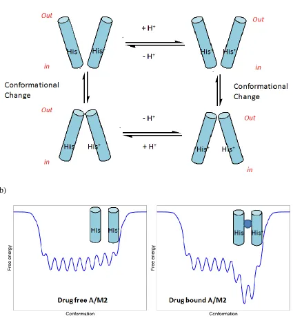
9 Part of the reason for the low mutation rate of A/M2 is due to the stringent sequence
requirement for A/M2 function. To conduct protons, A/M2 has to allow formation and
interchange of a number of functional conformers, each required for gating and
conducting protons (Figure 1.6). Any mutation which shifts these low energy barrier
interconvertable ensembles to one or more stable conformations is potentially disfavored
due to the loss of function. It has been shown in vitro through analytical
ultracentrifugation (AUC) experiments that mutation of pore lining residues and residues
at the helix-helix interface of A/M2 to alanine and phenylalanine result in a more stable
homotetramer compared with wild-type, except His37, which is required for selective
proton conductance.32, 33 Thus, the amino acid sequence of M2 is not optimized for
overall thermodynamic stability, but rather a compromise between stability and function.
From the drug development point of view, it would be ideal to target the most
conservative protein such that antivirals are effective across different subtypes of flu
viruses. In this regard, A/M2 is advantageous over neuraminidase as a drug target in
influenza virus inhibition; despite both of them are proven drug targets. Nevertheless, a
combination of antivirals drugs are desired in the case of highly virulent flu outbreaks. 12,
10 a)
b)
11 A large number of drug resistant mutations were selected in cell culture under
amantadine selection pressure, including L26F; V27 to A, S, G, or D; A30 to T, E, or P;
S31N 19, 35 (Figure 1.7), and a subset of these mutations were also found in infected
patients that were treated with amantadine.36 Not all of the mutant viruses maintain the
transmissibility of wt viruses and many of these mutations have the tendency to revert in
the absence of drugs.19, 37 Only three major mutations were identified from large scale
genome sequencing of transmissible viruses from 1918 to 2008,29, 38 among which S31N
is the predominant drug resistant mutation in H3N2 and H1N1 subtypes, followed by
V27A and L26F. The latter two mutations were found to be predominant in certain flu
seasons. A highly conductive channel would be toxic to the infected host cell and would
lead to cell death before the assembly and budding of the progeny virus. Another function
of A/M2 is to equilibrate the pH across the Golgi in order to prevent the premature
conformational change of the viral fusion protein, hemagglutinin (HA).18-20 A/M2 and
hemagglutinin have co-evolved and acted in concert with each other. A particularly
acid-labile hemagglutinin would require an A/M2 variant like D44N with higher proton
conductance capability to maintain neutrality in the late Golgi to prevent a premature
conformational change in the hemagglutinin before reaching the cell surface.39 Taken
together, only three major mutations in A/M2, namely S31N, V27A and L26F satisfy
both the functional and evolutionary requirements of influenza viruses, thus becoming
12 a)
b)
Figure 1. 7 | Amino acid sequence of influenza A virus strain A/Udron/72 and its mutations. a) Domain structure of influenza A virus M2 protein. b) A/M2 drug resistant mutations isolated from cell culture treated with amantadine.
1.5 A/M2 structure and drug binding site
AM2 is a homotetrameric, type III integral membrane protein containing: 1) a short
N-terminal region that is important for the protein’s incorporation into the virion;40 2) a
transmembrane (TM) helix that is required for tetramerization, drug binding and proton
channel formation;41-44 3) a C-terminal amphiphilic helix that is involved in membrane
localization, budding and scission;45, 46 and 4) a C-terminal tail that interacts with the
matrix protein M1.47 Although native A/M2 protein is palmitoylated (Cys50) and
phosphorylated (Ser64), these modifications have no effect on its channel-forming
properties in cell membrane.48, 49 The protein also has cysteine residues at position 17 and
19 on the viral N-terminal exterior, which form a mixture of covalent dimers and
tetramers.50 However, these disulfides are not essential for M2 assembly into functional
13 fully active in oocytes.48, 51 Structures of the drug complexed transmembrane domain of
A/M2 (A/M2TM) were solved using X-ray crystallography52 and solution NMR53 (Figure
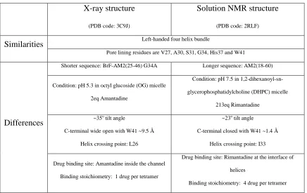
1.8). The overall structures were similar, showing left handed packing of a
homotetrameric bundle. The differences between these two structures are: (1) The helix
tilt in the X-ray structure is 30-40o, while the solution NMR structure is 23o. This
difference might result from several factors: (a) Solubilization environment - crystal was
grown in octylglucoside (OG) micelle at pH 5.5, which confers the open conformation of
M2. In contrast, the NMR structure was solved in 1,2-Dihexanoyl-sn
-glycerophosphocholine (DHPC) at pH 7.5, which confers the closed conformation of M2.
(b) The protein sequences used for X-ray and solution NMR studies were different. The
X-ray structure was solved with transmembrane segment (residues 25-46), solution NMR
study used a longer sequence with an extra C-terminal amphiphilic helix (residues
20-60). (2) The striking difference is where the drug binds. In the X-ray structure, the
electron density inside the cavity at the N-terminal lumen matches that of amantadine,
leading to a pore blocking mechanism of drug action. In solution NMR, four rimantadine
molecules were found on the C-terminal exterior side at the helices interface. The
transmembrane helices are in close contact such that there was not enough room to
accommodate rimantadine inside the channel. Based on this structure, an allosteric
inhibition mechanism was proposed where the drug would selectively stabilize the closed
14 From a drug discovery perspective, it is imperative to figure out which drug binding site
is the pharmacological relevant binding site. This is one of the goals of my thesis work.
Figure 1. 8 | X-ray and solution NMR structures of drug bound A/M2TM. Left: X-ray structure of A/M2TM (25-46)G34A with electron density inside the channel matching amantadine (PDB code: 3C9J). Right: Solution NMR structure of A/M2TMC (21-61) with four rimantadine binding to the peripheral of the channel (PDB code 2RLF). Figures from 52, 53 with permission from ACS and NPG.
1.6 Literature review of A/M2 inhibitors
Amantadine was first discovered by scientists at DuPont to have antiviral activity in 1964
21
and was approved by the U S Food and Drug Administration (FDA) in 1966 for the
treatment of influenza A virus infection in 1966. Subsequently, a close structural analog
of amantadine, rimantadine, received FDA approval in 1994 and has been shown to have
higher potency and reduced central neuron system (CNS) side effects. Unfortunately
most of the currently circulating strains of influenza virus have become resistant to the
adamantane class of drugs, causing the CDC to issue a recommendation to discontinue
15 drug on the market. Increasing number of oseltamivir resistant cases have been reported
in recent years.55, 56 Thus there is great need to develop novel antivirals with higher
potency and broad spectrum inhibition. Extensive medicinal chemistry efforts have been
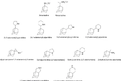
devoted to synthesize amantadine analogs, including 1-substituted adamantane,57, 58
2-substituted adamantane,59-61 2,2-disubstitued adamantane, 2,2-spiro adamantane57, 62, 63
and 1,2-annulated adamantine64 (Figure 1.9). Quite a few compounds have been
identified to be more potent than rimantadine.4, 5 But the cytopathic effect (CPE) assay65
that was used to test these compounds is not target specific, so it is difficult to correlate
the structures with M2 inhibition. Moreover, the influenza A virus strains that were tested
lack the S31N or V27A M2 mutation, so it is not clear whether any of these compounds
might work against virus carrying amantadine resistant mutations.
16 Aside from amantadine, only very few scaffolds other than adamantane have been
explored for antiviral activities against influenza A virus (Figure 1.10), including
1-norbornylamine,66 tricyclic ICI 130685,67 BL-1743,68 bicycle[2,2,2]octane69 and
isopinocampheylamine.70 There are also a few patented polycyclic compounds that claim
to have antiviral activity, e.g. pentacycloundecane amines,71 aminopolycyclodecanes,72
aminotricyclononanes,73 pentacycloalkane amines,74 but their detailed mechanism of
action have not been fully explored. In this regard, despite the importance of A/M2 as a
proven drug target and the urgency to develop potent A/M2 blockers, there is very
limited structure and activity relationship information about A/M2 inhibitors. Moreover,
a majority of the current circulating influenza A viruses carry drug resistant mutations
such as L26F, V27A, S31N and S30T, none of which have been shown to be druggable.
In this thesis study, I aim to identify novel scaffolds that may provide new avenues for
developing antagonists of A/M2. The spirene guanidine analogue,
2-[3-azaspiro(5,5)undecanol]-2- imidazoline (BL-1743) was choosen as an initial hit
compound for the structure and activity relationship study, as its antiviral activity has
been shown to be A/M2 specific, and it is also easier to synthesize and diversify from the
17 Figure 1. 10 | Structures of non-adamantane based anti-influenza A viruses agents.
1.7 Introduction of M2 drug screening assays
1.7.1 Two electrode voltage patch clamp assay
The gold standard of measuring compound activity on an ion channel is the patch clamp.
Even though it is low throughput, the high quality data generated from this assay offers
insights in guiding drug design. In the patch clamp assay, a constant electrical potential
(voltage clamp) is applied across an electrically isolated area (patch) of membrane
containing the protein of interest, and the current is recorded directly in real time under
different conditions, e.g. in the presence or absence of inhibitor.
The protein assayed can either be expressed in cell membrane or reconstituted in
artificially formed bilayer-like vesicles. Both single channel and whole cell current
recording are possible, but in the case of A/M2, due to the extremely small conductance
18 screening assay experiment, Oocyte expressing A/M2 channel is clamped at -20 mV.
Xenopus laevis (African clawed frog) oocytes are large cells with exceeding 1 mm in
diameter and are therefore easy to manipulate, and can be induced to express transport
proteins of interest by intracytoplasmic injection of mRNA encoding the desired protein.
Initially oocyte is bathing at pH 8.5 buffer, where A/M2 channel is closed; then A/M2
channel is activated by lowering the buffer solution to pH5.5. When the inward A/M2
current reaches maximum, the 100 μM inhibitor was applied for 2 min at pH5.5 buffer
solution. The remaining current after 2 min application was compared to the maximum
current before the application of the inhibitor. The potency of the inhibitors was
expressed as percentage inhibition of A/M2 current by 2 min application of 100 μM
inhibitors. Typical trace of the current change is shown in Figure 1.11.
Figure 1. 11| Current trace of A/M2 channel upon acidic activation and compound inhibition.
1.7.2 Plaque reduction assay
To test the in vivo activity of inhibitors, plaque reduction assay of recombinant influenza
A virus was performed. In the plaque reduction assay, the infectious ability of virus on
MDCK cells was monitored in the presence of different doses of inhibitors. If the
19 localized area of ‘plaque’, which can be monitored as areas of dead/destroyed cells
detected by general cellular stains or as areas of infected cells detected by
immuno-staining. On the other hand, if the virus replication is inhibited by potent inhibitors, no or
less plaque will form under the same condition. In a typical procedure, confluent
monolayers of MDCK cells were incubated with the wt Udorn virus [100 plaque-forming
units (pfu) per well] and V27A/L38F mutant virus (1000 and 100 pfu per well) in a
DMEM/1%bovine serum albumin mixture for 1 h at 37 oC. The inoculums were
removed, and the cells were washed with phosphate-buffered saline (PBS). The cells
were then overlaid with DMEM-containing 0.6% Avicel microcrystalline cellulose (FMC
BioPolymer, Philadelphia, PA) and NAT (2.0 μg/mL). To examine the effect of drugs on
plaque formation, monolayers were preincubated with DMEM supplemented with the
indicated concentrations of the drugs at 37 oC for 30min, and virus samples were
preincubated with a DMEM/1% BSA mixture with the indicated concentrations of the
drugs at 4 oC for 30 min before infection. Two to three days after infection, the
monolayers were fixed and stained with a naphthalene black dye solution (0.1%
naphthalene black, 6% glacial acetic acid, and 1.36% anhydrous sodium acetate).
20
1.7.3 Circular Dichroism binding assay
The structural changes resulted from drug binding to M2TM can be monitored by circular
dichroism (CD) spectroscopy. M2TM adopts a α-helix structure when reconstituted in
detergents, showing minimal at θ223 and θ209 in CD spectrum. Under condition of
intermediate to high peptide/detergent ratio (>1:400), binding of drug promotes a shift of
monomer/tetramer equilibrium to drug bound tetramer, which has significantly higher
intensity of ellipticity at 223nm (θ223). Drug binding constants are derived by plotting the
change at θ223 against drug concentration. In practice, the ellipticity ratio θ223/ θ209 instead
of the absolute value at θ223 is used for data fitting in order to minimize the small
concentration differences during titration. This assay was first used for amantadine
binding and later applied to other inhibitors. Binding constants derived from the CD
binding assay are in general agreement with electrophysiology assay data. Typical trace
21 Rimantadine
Amantadine
Figure 1. 13 | CD binding assay of amantadine and rimantadine to M2TM in DPC micelle. Figure from75 with permission from ACS.
1.8 Drug design principle of A/M2 inhibitors
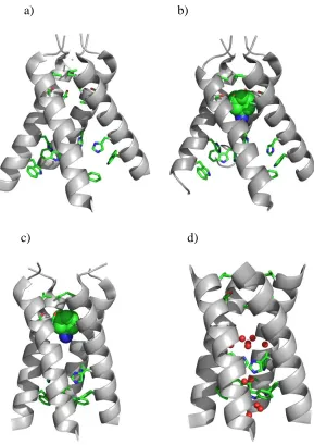
The design of A/M2 inhibitors started with our X-ray crystal structures of A/M2TM
(25-46) (PDB: 3C9J, 3BKD) solved in n-octylglucoside (OG) micelle76 (Figure 1.14), and
also the most recent SSNMR structure of amantadine complexed A/M2TM77 (PDB:
2KQT). These structures represent conformational states from higher (pH 7.5) to lower
pH (pH 5.5). In principle, any conformation of A/M2, either at high, medium or low pH
can be used for the design of A/M2 inhibitors, because amantadine binds at both high and
low pH, despite its higher potency at elevated pH.78 A high resolution (1.6 Å) X-ray
structure76 of drug free A/M2TM (PDB: 3LBW) that was recently solved reveals not only
22 water clusters in the channel. This water-His37 box like structure was suggested to play
an important role in A/M2 proton conductance mechanism by delocalizing protons over
the entire region.76 This high resolution structure offers a new starting point for the next
generation of anti-flu drug design.
a) b)
c) d)
23 All previous reported A/M2 inhibitors share the same structural similarity: a positively
charged amino or amino derivative (guanidine, etc) group attached to a large hydrophobic
aliphatic scaffold. A variety of amantadine analogues with more hydrophobic and bulkier
positively charged groups have several-fold greater potencies against most forms of the
virus that lack the S31N mutation.4, 5 The large variety of amines that can be
accommodated in the site argues against a highly specific interaction between the amine
and the channel.58 This conclusion is consistent with the amantadine-complexed
AM2-TM structure (PDB: 3C9J), in which the electron density map fits better with the
positively charged amantadine amine pointing down toward His37, but the distance is too
far (6.8 Å) for effective hydrogen bonding between the imidazoles and the amine (or
ammonium group). Overlaying the SSNMR structure of amantadine complexed
A/M2TM (PDB: 2KQT) with the high resolution X-ray crystal structure of drug free
A/M2TM (PDB: 3LBW) generates a model of wt M2 with the crystal waters in the entry
cluster (Figure 1.15). In this model, amantadine is within hydrogen bonding distance with
the crystal waters which suggests amantadine inhibiting M2 by not only physically
blocking the channel, but also altering the pKas of the His37 residue79 such that it can no
24 Figure 1.15 | Overlaying A/M2TM structures of SSNMR structure (PDB: 2KQT) and X-ray structure (PDB: 3LBW).
The pore lining residues of wt A/M2 in the drug binding site are V27, A30, S31 and G34
that forms a hydrophobic pore. In the SSNMR structure of amantadine complexed
A/M2TM (PDB: 2KQT), V27 constrains the drug binding site in the N-terminal with the
pore dimension ~4 Å, and the channel expands to the widest at G34 with an inner
diameter of ~8 Å (Figure 1.16). In the channel axis, the drug binding site consists of two
helix turns of A/M2 backbone with the distance of ~ 9 Å (the distance of one turn
α-helix is 5.4 Å, the tilt angle of the α-helix is 30o, thus the distance in the channel axis is 5.4
Å*2*cos30o = 9.4 Å). The drug binding site dimension in the channel axis can be further
extended to 11 Å if the water clusters above His37 are displaced. This suggests the A/M2
drug binding site has enough room to accomendate molecules larger than
25 Figure 1. 16 | A/M2 drug binding site dimension analysis
For the inhibitor to fit inside the channel, it has to meet two criteria: hydrophobic match
and Shape complementary. These are the guidelines for A/M2 inhibitor drug design.
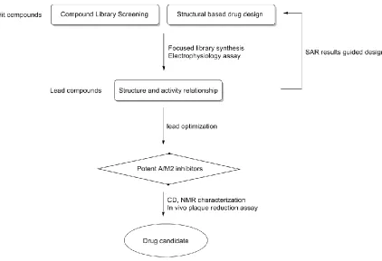
The general procedure of A/M2 inhibitor discovery (Figure 1.17) starts from a rational
drug design based on the hit compounds identified from high throughput screening or a
de novo structural based design. The inhibitors designed are next synthesized and assayed
in A/M2 specific electrophysiology assay. The structure and activity relationship (SAR)
correlation results from the assay are used as guidelines for the next round of structure
based rational design using the same scaffold, or selection of structurally diverse
compounds that meet the criteria of SAR results. The inhibitors designed or selected are
subjected to the next round of electrophysiology assays. The most potent inhibitors are
further characterized both in plaque reduction assay and in biophysical assays such as
26 change of A/M2. The ultimate goal is to identify drug candidates that can be further
pursued in animal test and clinical trials.
Figure 1. 17 | A/M2 inhibitor discovery scheme.
1.9 References
1. Fiore, A. E.; Shay, D. K.; Broder, K.; Iskander, J. K.; Uyeki, T. M.; Mootrey, G.; Bresee, J. S.; Cox, N. J., Prevention and control of influenza - Recommendations of the
Advisory Committee on Immunization Practices (ACIP), 2008. MMWR Morb Mortal
Wkly Rep 2008, 57, (RR7, Suppl. S), 1-60.
2. Keech, M.; Scott, A. J.; Ryan, P. J. J., The impact of influenza and influenza-like illness on productivity and healthcare resource utilization in a working population.
Occupational Medicine-Oxford 1998, 48, (2), 85-90.
3. Neumann, G.; Noda, T.; Kawaoka, Y., Emergence and pandemic potential of swine-origin H1N1 influenza virus. Nature 2009, 459, (7249), 931-939.
4. De Clercq, E., Antiviral agents active against influenza A viruses. Nat. Rev. Drug Discov. 2006, 5, (12), 1015-1025.
27 6. Palese, P., and M. L. Shaw. Orthomyxoviridae. The viruses and their replication. . In Fields Virology 5th Edition. Knipe DM, Howley PH (Eds). Lippincott Williams & Wilkins, PA, USA 1647-1689.
7. http://www.cdc.gov/flu/keyfacts.htm.
8. Krug, R. M.; Aramini, J. M., Emerging antiviral targets for influenza A virus.
Trends Pharmacol. Sci. 2009, 30, (6), 269-277.
9. Holsinger, L. J.; Nichani, D.; Pinto, L. H.; Lamb, R. A., Influenza A virus M2 ion channel protein: a structure-function analysis. J. Virol. 1994, 68, (3), 1551-1563.
10. Lamb, R. A.; Choppin, P. W., THE GENE STRUCTURE AND REPLICATION
OF INFLUENZA-VIRUS. Annu. Rev. Biochem. 1983, 52, 467-506.
11. http://en.wikipedia.org/wiki/Influenza.
12. Boltz, D. A.; Aldridge, J. R.; Webster, R. G.; Govorkova, E. A., Drugs in Development for Influenza. Drugs 2010, 70, (11), 1349-1362.
13. Klumpp, K., Recent advances in the discovery and development of anti-influenza
drugs. Expert Opin. Ther. Pat. 2004, 14, (8), 1153-1168.
14. Wang, G. T., Recent advances in the discovery and development of anti-influenza
drugs. Expert Opin. Ther. Pat. 2002, 12, (6), 845-861.
15. De Clercq, E.; Neyts, J., Avian influenza A (H5N1) infection: targets and strategies for chemotherapeutic intervention. Trends Pharmacol. Sci. 2007, 28, (6), 280-285.
16. Martin, K.; Heleniust, A., Nuclear transport of influenza virus ribonucleoproteins: The viral matrix protein (M1) promotes export and inhibits import. Cell 1991, 67, (1), 117-130.
17. Russell, R. J.; Kerry, P. S.; Stevens, D. J.; Steinhauer, D. A.; Martin, S. R.; Gamblin, S. J.; Skehel, J. J., Proc Natl Acad Sci U S A. . 2008, 105, (46), 17736-17741.
18. Sugrue, R. J.; Bahadur, G.; Zambon, M. C.; Hallsmith, M.; Douglas, A. R.; Hay,
A. J., Specific structural alteration of the influenza haemagglutinin by amantadine.
EMBO J. 1990, 9, (11), 3469-3476.
19. Grambas, S.; Hay, A. J., Maturation of influenza A virus hemagglutinin--estimates of the pH encountered during transport and its regulation by the M2 protein.
Virology 1992, 190, (1), 11-18.
20. Sakaguchi, T.; Leser, G. P.; Lamb, R. A., The ion channel activity of the influenza virus M(2) protein affects transport through the Golgi apparatus. J. Cell. Biol.
1996, 133, (4), 733-747.
21. Davies, W. L.; Hoffmann, C. E.; Paulshock, M.; Wood, T. R.; Haff, R. F.; Grunert, R. R.; Watts, J. C.; Hermann, E. C.; Neumayer, E. M.; McGahen, J. W., Antiviral Activity of 1-Adamantanamine (Amantadine). Science 1964, 144, (362), 862-&. 22. Öberg, B., Rational design of polymerase inhibitors as antiviral drugs. Antiviral Res. 2006, 71, (2-3), 90-95.
23. Yuan, P.; Bartlam, M.; Lou, Z.; Chen, S.; Zhou, J.; He, X.; Lv, Z.; Ge, R.; Li, X.; Deng, T.; Fodor, E.; Rao, Z.; Liu, Y., Crystal structure of an avian influenza polymerase PAN reveals an endonuclease active site. Nature 2009, 458, (7240), 909-913.
28 25. Ge, Q.; Filip, L.; Bai, A.; Nguyen, T.; Eisen, H. N.; Chen, J., Inhibition of influenza virus production in virus-infected mice by RNA interference. Proc. Natl. Acad. Sci. U. S. A. 2004, 101, (23), 8676-8681.
26. Moscona, A., Neuraminidase Inhibitors for Influenza. N. Engl. J. Med. 2005, 353, (13), 1363-1373.
27. von Itzstein, M., The war against influenza: discovery and development of sialidase inhibitors. Nat. Rev. Drug Discov. 2007, 6, (12), 967-974.
28. Das, K.; Aramini, J. M.; Ma, L. C.; Krug, R. M.; Arnold, E., Structures of influenza A proteins and insights into antiviral drug targets. Nat. Struct. Mol. Biol. 2010,
17, (5), 530-538.
29. Furuse, Y.; Suzuki, A.; Kamigaki, T.; Oshitani, H., Evolution of the M gene of the influenza A virus in different host species: large-scale sequence analysis. Virol J
2009, 6.
30. Deyde, V. M.; Xu, X. Y.; Bright, R. A.; Shaw, M.; Smith, C. B.; Zhang, Y.; Shu, Y. L.; Gubareva, L. V.; Cox, N. J.; Klimov, A. I., Surveillance of resistance to adamantanes among influenza A(H3N2) and A(H1N1) viruses isolated worldwide. J. Infect. Dis. 2007, 196, (2), 249-257.
31. Saito, R.; Sakai, T.; Sato, I.; Sano, Y.; Oshitani, H.; Sato, M.; Suzuki, H., Frequency of amantadine-resistant influenza A viruses during two seasons featuring cocirculation of H1N1 and H3N2. J. Clin. Microbiol. 2003, 41, (5), 2164-2165.
32. Stouffer, A. L.; Ma, C.; Cristian, L.; Ohigashi, Y.; Lamb, R. A.; Lear, J. D.; Pinto, L. H.; DeGrado, W. F., The Interplay of Functional Tuning, Drug Resistance, and Thermodynamic Stability in the Evolution of the M2 Proton Channel from the Influenza A Virus. Structure 2008, 16, (7), 1067-1076.
33. Stouffer, A. L.; Nanda, V.; Lear, J. D.; DeGrado, W. F., Sequence Determinants
of a Transmembrane Proton Channel: An Inverse Relationship between Stability and Function. J. Mol. Biol. 2005, 347, (1), 169-179.
34. Govorkova, E. A.; Webster, R. G., Combination Chemotherapy for Influenza.
Viruses 2010, 2, 1510-1529.
35. Hay, A. J.; Wolstenholme, A. J.; Skehel, J. J.; Smith, M. H., The molecular basis of the specific anti-influenza action of amantadine. EMBO J. 1985, 4, (11), 3021-3024.
36. Shiraishi, K.; Mitamura, K.; Sakai-Tagawa, Y.; Goto, H.; Sugaya, N.; Kawaoka,
Y., High frequency of resistant viruses harboring different mutations in amantadine-treated children with influenza. J. Infect. Dis. 2003, 188, (1), 57-61.
37. Suzuki, H.; Saito, R.; Masuda, H.; Oshitani, H.; Sato, M.; Sato, I., Emergence of amantadine-resistant influenza A viruses: Epidemiological study. J. Infect. Chemother.
2003, 9, (3), 195-200.
38. Furuse, Y.; Suzuki, A.; Oshitani, H., Large-Scale Sequence Analysis of M Gene
29 41. Ma, C.; Polishchuk, A. L.; Ohigashi, Y.; Stouffer, A. L.; Schon, A.; Magavern, E.; Jing, X.; Lear, J. D.; Freire, E.; Lamb, R. A.; DeGrado, W. F.; Pinto, L. H., Identification of the functional core of the influenza A virus A/M2 proton-selective ion channel. Proc Natl Acad Sci U S A 2009, 106, (30), 12283-8.
42. Kochendoerfer, G. G.; Salom, D.; Lear, J. D.; Wilk-Orescan, R.; Kent, S. B.; DeGrado, W. F., Total chemical synthesis of the integral membrane protein influenza A virus M2: role of its C-terminal domain in tetramer assembly. Biochemistry 1999, 38, (37), 11905-13.
43. Salom, D.; Hill, B. R.; Lear, J. D.; DeGrado, W. F., pH-dependent tetramerization and amantadine binding of the transmembrane helix of M2 from the influenza A virus.
Biochemistry 2000, 39, (46), 14160-70.
44. Cady, S. D.; Luo, W.; Hu, F.; Hong, M., Structure and function of the influenza A M2 proton channel. Biochemistry 2009, 48, (31), 7356-64.
45. Rossman, J. S.; Jing, X.; Leser, G. P.; Lamb, R. A., Influenza Virus M2 Protein Mediates ESCRT-Independent Membrane Scission. Cell 2010, 142, (6), 902-13.
46. Rossman, J. S.; Lamb, R. A., Autophagy, apoptosis, and the influenza virus M2 protein. Cell Host Microbe 2009, 6, (4), 299-300.
47. McCown, M. F.; Pekosz, A., Distinct domains of the influenza a virus M2 protein cytoplasmic tail mediate binding to the M1 protein and facilitate infectious virus production. J Virol 2006, 80, (16), 8178-89.
48. Holsinger, L. J.; Shaughnessy, M. A.; Micko, A.; Pinto, L. H.; Lamb, R. A., Analysis of the posttranslational modifications of the influenza virus M2 protein. J. Virol.
1995, 69, (2), 1219-1225.
49. Thomas, J. M.; Stevens, M. P.; Percy, N.; Barclay, W. S., Phosphorylation of the M2 Protein of Influenza A Virus Is Not Essential for Virus Viability. Virology 1998, 252, (1), 54-64.
50. Holsinger, L. J.; Lamb, R. A., Influenza Virus M2 Integral Membrane Protein Is a Homotetramer Stabilized by Formation of Disulfide Bonds. Virology 1991, 183, 32-43. 51. Sakaguchi, T.; Tu, Q. A.; Pinto, L. H.; Lamb, R. A., The active oligomeric state of the minimalistic influenza virus M-2 ion channel is a tetramer. Proc. Natl. Acad. Sci. U. S. A. 1997, 94, (10), 5000-5005.
52. Stouffer, A. L.; Acharya, R.; Salom, D.; Levine, A. S.; Di Costanzo, L.; Soto, C. S.; Tereshko, V.; Nanda, V.; Stayrook, S.; DeGrado, W. F., Structural basis for the function and inhibition of an influenza virus proton channel Nature 2008, 452, (7185), 380-380.
53. Schnell, J. R.; Chou, J. J., Structure and mechanism of the M2 proton channel of influenza A virus. Nature 2008, 451, (7178), 591-U12.
54. http://www2a.cdc.gov/HAN/ArchiveSys/ViewMsgV.asp?AlertNum=00240.
55. de Jong, M. D.; Thanh, T. T.; Khanh, T. H.; Hien, V. M.; Smith, G. J. D.; Chau, N. V.; Cam, B. V.; Qui, P. T.; Ha, D. Q.; Guan, Y.; Peiris, J. S. M.; Hien, T. T.; Farrar, J., Oseltamivir Resistance during Treatment of Influenza A (H5N1) Infection. N. Engl. J. Med. 2005, 353, (25), 2667-2672.
30 57. Kolocouris, N.; Kolocouris, A.; Foscolos, G. B.; Fytas, G.; Neyts, J.; Padalko, E.; Balzarini, J.; Snoeck, R.; Andrei, G.; De Clercq, E., Synthesis and Antiviral Activity Evaluation of Some New Aminoadamantane Derivatives. 2. J. Med. Chem. 1996, 39, (17), 3307-3318.
58. Aldrich, P. E.; Hermann, E. C.; Meier, W. E.; Paulshock, M.; Prichard, W. W.; Synder, J. A.; Watts, J. C., Antiviral agents. 2. Structure-activity relations of compounds related to 1-adamantanamine. J. Med. Chem. 1971, 14, (6), 535-543.
59. Setaki, D.; Tataridis, D.; Stamatiou, G.; Kolocouris, A.; Foscolos, G. B.; Fytas, G.; Kolocouris, N.; Padalko, E.; Neyts, J.; Clercq, E. D., Synthesis, conformational characteristics and anti-influenza virus A activity of some 2-adamantylsubstituted azacycles. Bioorg Chem. 2006, 34, (5), 248-273.
60. Kolocouris, A.; Tataridis, D.; Fytas, G.; Mavromoustakos, T.; Foscolos, G. B.; Kolocouris, N.; De Clercq, E., Synthesis of 2-(2-adamantyl)piperidines and structure anti-influenza virus a activity relationship study using a combination of NMR spectroscopy and molecular modeling. Bioorg. Med. Chem. Lett. 1999, 9, (24), 3465-3470.
61. Zoidis, G.; Kolocouris, N.; Foscolos, G. B.; Kolocouris, A.; Fytas, G.; Karayannis, P.; Padalko, E.; Neyts, J.; De Clercq, E., Are the 2-isomers of the drug rimantadine active anti-influenza A agents? Antivir. Chem. Chemother. 2003, 14, (3), 153-164.
62. Kolocouris, N.; Zoidis, G.; Foscolos, G. B.; Fytas, G.; Prathalingham, S. R.; Kelly, J. M.; Naesens, L.; De Clercq, E., Design and synthesis of bioactive adamantane spiro heterocycles. Bioorg. Med. Chem. Lett. 2007, 17, (15), 4358-4362.
63. Kolocouris, N.; Foscolos, G. B.; Kolocouris, A.; Marakos, P.; Pouli, N.; Fytas, G.; Ikeda, S.; De Clercq, E., Synthesis and Antiviral Activity Evaluation of Some Aminoadamantane Derivatives. J. Med. Chem. 1994, 37, (18), 2896-2902.
64. Zoidis, G.; Kolocouris, N.; Naesens, L.; Clercq, E. D., Design and synthesis of 1,2-annulated adamantane piperidines with anti-influenza virus activity. Bioorg. Med. Chem. 2009, 17, (4), 1534-1541.
65. Shih, S.-R.; Chu, T.-Y.; Reddy, G.; Tseng, S.-N.; Chen, H.-L.; Tang, W.-F.; Wu,
M.-s.; Yeh, J.-Y.; Chao, Y.-S.; Hsu, J.; Hsieh, H.-P.; Horng, J.-T., Pyrazole compound BPR1P0034 with potent and selective anti-influenza virus activity. J. Biomed. Sci. 2010,
17, (1), 13.
66. Garcia Martinez, A.; Teso Vilar, E.; Garcia Fraile, A.; De La Moya Cerero, S.; Rodriguez Herrero, M. E.; Martinez Ruiz, P.; Subramanian, L. R.; Garcia Gancedo, A., Synthesis of substituted 1-norbornylamines with antiviral activity. J. Med. Chem. 1995,
38, (22), 4474-4477.
67. Alnakib, W.; Higgins, P. G.; Willman, J.; Tyrrell, D. A. J.; Swallow, D. L.; Hurst, B. C.; Rushton, A., Prevention and treatment of experimental influenza A virus infection in volunteers with a new antiviral ICI 130,685. J. Antimicrob. Chemother. 1986, 18, (1), 119-129.
31 virus M2 protein in Saccharomyces cerevisiae: identification of a novel inhibitor of influenza virus. Antimicrob. Agents Chemother. 1995, 39, (10), 2204-2209.
69. Whitney, J. G.; Gregory, W. A.; Kauer, J. C.; Roland, J. R.; Snyder, J. A.; Benson, R. E.; Hermann, E. C., Antiviral agents. I. Bicyclo[2.2.2]octan- and -oct-2-enamines. J. Med. Chem. 1970, 13, (2), 254-260.
70. Zhao, X.; Li, C.; Zeng, S.; Hu, W., Discovery of highly potent agents against influenza A virus. Eur. J. Med. Chem. 2010, In Press, Accepted Manuscript.
71. Miller, L. S. Pentacyclo-undecane amines active against influenza viruses. US3449422-A, 1969.
72. Stedman, R. J. Amino-and aminomethyl-tetracyclononanes - antiviral agents. US3641148-A, 1972.
73. John, R. E.; Glenside, H. 3 and 9 amino or 3 and 9 aminomethyltricyclo (3 3 1 o37 - nonanes and 1 amino and 1 aminomethyl tricyclo (3 3. US3496228-A, 1970.
74. LAB, S. K. F. Pentacyclic amines of (I), (II) and (III) and pharmaceutically acceptable acid addn. salts. where 1 group A (in II) or B (in III) = (alk)nNRR' and the others ar. FR5107-A; GB1068655-A; US3418368-A; AU6602263-A; FR5107-M;
BE678377-A; CH469655-A; JP69005213-B; JP70003377-B; JP70007299-B;
US3542868-A; CA894267-A, 1966.
75. Balannik, V.; Wang, J.; Ohigashi, Y.; Jing, X. H.; Magavern, E.; Lamb, R. A.; DeGrado, W. F.; Pinto, L. H., Design and Pharmacological Characterization of Inhibitors of Amantadine-Resistant Mutants of the M2 Ion Channel of Influenza A Virus.
Biochemistry 2009, 48, (50), 11872-11882.
76. Acharya, R.; Carnevale, V.; Fiorin, G.; Levine, B. G.; Polishchuk, A. L.; Balannik, V.; Samish, I.; Lamb, R. A.; Pinto, L. H.; DeGrado, W. F.; Klein, M. L., Structure and mechanism of proton transport through the transmembrane tetrameric M2 protein bundle of the influenza A virus. Proc. Natl. Acad. Sci. U. S. A. 2010, 107, (34), 15075-15080.
77. Cady, S. D.; Schmidt-Rohr, K.; Wang, J.; Soto, C. S.; DeGrado, W. F.; Hong, M., Structure of the amantadine binding site of influenza M2 proton channels in lipid bilayers. Nature 2010, 463, (7281), 689-692.
78. Wang, C.; Takeuchi, K.; Pinto, L. H.; Lamb, R. A., Ion channel activity of influenza A virus M2 protein: characterization of the amantadine block. J. Virol. 1993,
67, (9), 5585-5594.
79. Hu, J.; Fu, R.; Cross, T. A., The Chemical and Dynamical Influence of the
Anti-Viral Drug Amantadine on the M2 Proton Channel Transmembrane Domain. Biophys J.
32
Chapter 2
Aim, focus and perspective of influenza virus M2 channel inhibitor
development
2.1 Aim and focus of A/M2 inhibitor development
Studying the mechanism of proton conductance through A/M2 and how drug binding
affects A/M2 dynamics is the first step towards structure-based design of M2 inhibitors.
The knowledge learned through structural and functional studies of A/M2 drug binding
can be used to guide rational design of potent A/M2 inhibitors targeting not only wild
type (wt), but more importantly pharmacologically relevant drug resistant mutants.
Structural studies of membrane protein are often challenging due to the difficulties
associated with production, assay, functional reconstitution, and structural
characterization of this class of proteins. Production of large quantities (tens to hundreds
of milligrams) of A/M2 protein and its fragments in high purity (>98%) are required for
structural and functional studies. This problem was addressed in two ways in this study:
(1) Bacterial expression with controlled protease digestion. This provided both
isotopically labeled A/M2 fragments (19-49 and 19-53) for NMR studies and natural
abundance full length A/M2 for liposome flux assay. (2) Solid phase synthesis. The
optimized protocol was applied for synthesis of selectively labeled (15N, 13C or 15N + 13N)
A/M2 peptides with different lengths for NMR or large scale natural abundance A/M2 for
33 reconstitution conditions for NMR studies of A/M2. A wild range of common detergents
and detergent/lipid combinations were screened using circular dichrorism (CD)
spectroscopy or 15N-1H HSQC NMR to identify conditions that stabilize M2TM in a
native like conformation in solution. It was found that n- dodecylphosphatidylcholine
(DPC), tetradecyl-N,N-dimethyl-3-ammonio-1-propanesulfonate (C14-betaine) and
n-dodecyl-α-D-maltoside (DM) gave high quality of 15N-1H HSQC spectra, highlighting
their promise for M2 structural determination by NMR.
Structural studies of drug-complexed A/M2 raised controversy about the mode of action,
specifically whether the drug inhibits the M2 function by directly blocking the channel or
allosterically modulating a conformational change of M2. From the drug design point of
view, it is imperative to solve this discrepancy so that medicinal chemists can focus on
the correct drug binding site. In collaboration with Dr. Mei Hong (Iowa State University),
we resolved the controversy using REDOR solid state NMR experiments using deuterium
labeled amantadine and M2TM with selective isotope labeled residues at both of the
above proposed drug binding sites.1 As shown in Chapter 3, this study showed that the
primary high affinity site was located at the N-terminal lumen, occupied by a single
amantadine; a secondary low affinity site at the C-terminal helices interface was only
detected with excess amantadine. Moreover, the pore binding model is further supported
34 Even though the N-terminal lumen was confirmed as a pharmacologically relevant drug
binding site, the orientation of the amine from drug inside the channel was not
determined. Extensive computational investigations have been carried out to define the
mechanism of amantadine/rimantadine action as well as orientation of the amine,
including molecular dynamics simulations,5-8 rigid-body docking,9, 10 and small molecule
probe mapping.11, 12 Since these studies used a variety of different M2 structures/models
as starting points with varying protonation states, it is not surprising that no consensus
has yet emerged. A recent molecular dynamic simulation study by Khurana et al5 found
that the orientation of the amine were not the same under different protonation states of
His37: the amine was almost equally distributed up and down at low protonation, while it
preferentially pointed towards His 37 when His37 was more solvated at greater
protonation. Clearly, there is a need to experimentally determine the precise location of
drug in the site. Depending on whether the amine points towards the N or C-terminus, the
mechanism by which drug binding inhibits this proton channel is different. These results
will facilitate the optimization of existing drugs. In Chapter 4, this question has been
addressed using both M2TM reconstituted in lipid bilayers as well as DPC micelles. 13
C-2H rotational-echo double-resonance (REDOR) NMR experiments of 13C-labeled M2TM
and methyl-deuterated rimantadine in lipid bilayers showed that the polar amine points to
the C-terminus of the channel, with the methyl group located close to Gly34. Specific 2
H-labeling of the drugs permitted assignment of drug-protein cross peaks in solution NMR
experiments of A/M2TM(22-46) in dodecylphosphatidylcholine (DPC) micelles,
35 bilayer-bound M2TM. These results strongly suggest that adamantane drugs inhibit
A/M2 function by direct physical occlusion of the pore and perturb the pKa of the proton
sensing His37 through water mediated hydrogen bonding.
A major effort in this dissertation is devoted to structure based rational design of A/M2
inhibitors presented in Chapter 5, 6, 7, 8 and 9. Guided by the X-ray crystal structures of
M2TM and pore binding model, a series of spiran amines have been designed,
synthesized and assayed. Highly potent inhibitors were identified that are not only active
against the wt A/M2, but also against the drug resistant mutants V27A and L26F.
2.2 Two controversial A/M2 drug binding sites
Two studies published in 2008 raised a controversy about the A/M2 drug binding site
among the public.13-15 From the drug discovery standpoint, it is imperative to validate
which site is the pharmacological relevant drug binding site. Before planning experiments
to solve the issue, it is necessary to closely examine the two structures to see whether
either one of the structures agree with existing functional assay data, models built based
on experimental data and biophysical assays. The top and side view of the two drug
36 a) b)
c) d)