Original Research Article.
International Journal of Medical Research ProfessionalsP-ISSN: 2454-6356; E-ISSN: 2454-6364 DOI: 10.21276/ijmrp
A Study of Infective Endocarditis in Valvular Heart Disease in
North East India
Tribeni Sharma
1, Debabrata Goswami
2, Rajarshi Chakraborty
3*1Associate Professor, 2Professor, Department of Medicine, Gauhati Medical College and Hospital, Guwahati, Assam, India.
3*Registrar, Department of Medicine, Fakhruddin Ali Ahmed Medical College, Joti Gaon, Assam, India. ABSTRACT
Introduction: Infective Endocarditis (IE) is disease caused by infection of endothelial lining of intra-cardiac structures and is invariably fatal, if untreated.
Objectives: To study the incidence, clinical profile, microbiological profile of Infective Endocarditis in Valvular Heart Failure.
Methods: We conducted a hospital-based Observational
Descriptive study comprising of 150 patients of Valvular Heart Disease from July 2015 - June 2016 who were diagnosed on basis of thorough history, physical examination, Echocardiography, Blood Culture testing, etc. by application of Modified Dukes Criteria. The patients were admitted in Gauhati Medical College and Hospital, Assam, India and fulfilled the Inclusion and Exclusion criteria of the study. Statistical analysis was performed by Fisher’s Exact or Chi-Square Test.
Results: In our study, 22 out of 150 patients (14.67%) were confirmed to have Infective Endocarditis and the further results and observation were made out of these Definitive cases. Majority of cases (40.91%) were in their fourth decade. 72.73% cases were male. The origin was mostly rheumatic (77.27%). Fever and Congestive Heart Failure were the predominant symptom and complication respectively. Vegetation’s were observed in 90.91% cases. 35 % of cases had >/= 10 mm vegetation size, and they had more complications. Mitral valve
(36.36%) wasthe predominantly involving valve. Blood culture positivity was 36.36%. Staphylococcus aureus (50%) was the most common organism isolated. All organisms were sensitive to Vancomycin and Linezolid. Mortality was 13.64%.
Discussion: IE is more common in Rheumatic heart disease.
In Indian scenario, more emphasis should be given on Echocardiography in diagnosing IE. The predominant organism is Staphylococcus aureus, sensitive to Vancomycin and Linezolid.
Key Words: Infective Endocarditis, Echo-Cardiography, Blood-Culture, Staphylcoccus aureus,
*Correspondence to:
Dr. Rajarshi Chakraborty,
Registrar, Department of Medicine, Fakhruddin Ali Ahmed Medical College, Joti Gaon, Assam, India.
Article History:
Received: 29-01-2018, Revised: 24-02-2018, Accepted: 28-03-2018
Access this article online
Website: www.ijmrp.com
Quick Response code
DOI:
10.21276/ijmrp.2018.4.2.043
INTRODUCTION
Infective Endocarditis (IE) is a disease caused by microbial infection of endothelial lining of intra-cardiac structures and is invariably fatal, if untreated.1 in recent years, the epidemiology
and microbiology of IE has changed. The incidence of IE is 1.7 – 7.2 cases per 100,000 person-years.2 The female to male ratio
has been stable over years to 1:2.3 In developed countries, IE is
now affecting older age group and patients with no previous known valve disease, unlike developing nations where rheumatic heart disease is still the main risk factor for development of IE. IE is a diagnostic and therapeutic challenge to clinicians. Today the Duke’s Criteria presented in 1994 and subsequent modifications are considered to be the most accurate in diagnosing IE. Classically, the predominant causative agent has been Streptococcus viridans. However, Staphylococcus aureus has now become the primary pathogen of Endocarditis.4
There have been various studies on Infective Endocarditis in India and abroad which have been discussed here with references. We aim to find the incidence, clinical profile and microbiology of IE in Valvular Heart Disease in North-East Indian tertiary care hospital.
MATERIALS AND METHODS
presenting with features of IE were started on antibiotic coverage, and thoroughly investigated. Patients with SLE, Non-Valvular cardiac defects, chemotherapeutics usage, age below 12 years were excluded from the study. A detailed history of present symptoms, past history of IE/ Valvular heart disease, a thorough clinical examination esp clinical stigmata of IE like splinter hemorrhage, Roth spot,Osler nodes, Janeway lesions, clubbing, splenomegaly were meticulously searched for.
Routine blood tests, urine examination, CRP, ESR, Chest Xray (P/A), ECG, 2D Trans-Thoracic Echocardiography ACUSON CV70 Siemens, and at least 3 sampling of Blood Culture testing using BACTEC for aerobic, facultative anaerobic and fungal organisms via Sabouraud’s Dextrose Agar media (first sampling done before first dose of IV antibiotic, second at least 12 hours apart and third done 12 hours of second sampling from different venipuncture sites) were performed in all cases.
All patients underwent a comprehensive Trans-thoracic 2D Echocardiography (TTE) analysis using two-dimensional, Doppler, M- mode imaging using ACUSON CV70 Siemens analyser with standardized view on same machine. The detailed Echocardiographic examination was done to evaluate the condition of the valvular apparatus, cardiac anatomy, presence of vegetations, abscess formation, perforation etc. Statistical
analysis of significance was performed wherever applicable using SPSS version 16.0, GraphPad Software, San Diego California USA (www.graphpad.com). The p- value was calculated by using Fisher’s Exact Test, or Chi- Square Test. All the statistical graphs were prepared using Microsoft Excel 2007 and Microsoft Word 2007.
RESULTS AND OBSERVATIONS
A total of 150 patients of Valvular heart disease were studied, of which 22 cases (14.67%) were confirmed to have Infective Endocarditis with the help of Modified Dukes’ Criteria and the further observations were discussed of these definite cases of IE. Most of the cases in this study were in the fourth decade of life (40.91%) with a median age of 39 years.
About 2/3rd of patients were male (72.73%) and 77.27% of the
total IE cases were rheumatic in origin. Fever was the predominant symptom observed in 100% cases of IE, followed by changing quality of murmur (77.27%) of cases. The most common complication was Congestive Heart Failure (59.09%). Vegetations were observed in 90.91% of cases diagnosed as Infective Endocarditis by Modified Dukes’ Criteria. There was 35% of cases having >/= 10mm size of vegetation; such vegetations had more complications.
Table 1: Age Distribution
Age group No. of patients Percentage
12 –20 years 1 4.54%
21 – 30 years 4 18.18%
31 – 40 years 9 40.91%
41 – 50 years 7 31.83%
51 – 60 years 1 4.54%
61 – 70 years 0 0 %
>70 years 0 0%
Table 2: Sex Distribution (Male: Female ratio = 2.7:1)
Sex No. of patients Percentage
Male 16 72.73%
Female 6 27.27%
Table 3: Types of Underlying Heart Disease:
ORIGIN
Male Female Total
No. % No. % No. %
Rheumatic Heart Disease 12 54.55% 5 22.72% 17 77.27%
Ischaemic Heart Disease 3 13.63 % 1 4.55 % 4 18.18%
Prosthetic Valve Disease 0 0 % 0 0 % 0 0%
Intravenous Drug Abusers 1 4.55 % 0 0 % 1 4.55%
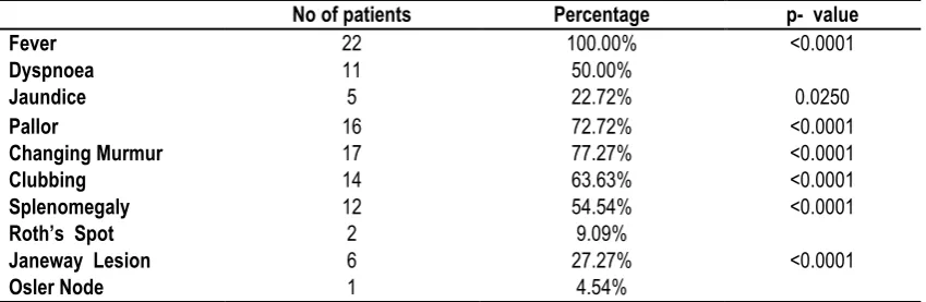
Table 4: Clinical Presentation of Infective Endocarditis
No of patients Percentage p- value
Fever 22 100.00% <0.0001
Dyspnoea 11 50.00%
Jaundice 5 22.72% 0.0250
Pallor 16 72.72% <0.0001
Changing Murmur 17 77.27% <0.0001
Clubbing 14 63.63% <0.0001
Splenomegaly 12 54.54% <0.0001
Roth’s Spot 2 9.09%
Janeway Lesion 6 27.27% <0.0001
Table 5: Complications in Infective Endocarditis
No. of patients Percentage p-value
Congestive Heart Failure (CCF) 13 59.09% 0.0056
Neurological (NEURO) 6 27.27% 0.0007
Peripheral Embolism (P E) 9 40.91% 0.0020
Acute Kidney Injury (AKI) 9 40.91% 0.0199
Table 6: Presence of Vegetations
No. of I E Cases Percentage p-value
Present 20 90.91%
<0.0001
Absent 2 9.09%
Total 22 100%
Table 7: Size of Vegetations >/= 10 Mm
No. of patients Percentage
More Than / Equal To 10 Mm 7 35.00%
Less Than 10 Mm 13 65.00%
Total 20 100%
Table 8: Valvular Involvement
Valve(S) No. Of Patients Percentage
Mitral 8 36.36%
Aortic 6 27.27%
Tricuspid 4 18.18%
Pulmonary 0 0%
Multivalvular (Aotic + Mitral)
2 9.09%
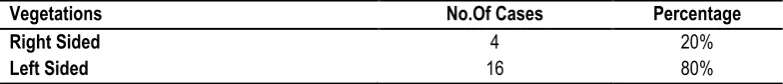
Table 9: Right Sided Vs Left Sided Events
Vegetations No.Of Cases Percentage
Right Sided 4 20%
Left Sided 16 80%
Table 10: Laboratorical Investigations
Tests No. of patients Percentage p-value
Anaemia 15 68.18% 0.0001
Leucocytosis 21 95.45% <0.0001
Increased Creatinine 9 40.91% 0.0020
Increased Bilirubin 6 27.27%
Increased CRP 22 100.00% <0.0001
Increased ESR 22 100.00% <0.0001
Table 11: Relation of Complications With Vegetations Sized >/= 10mm Size Of
Vegetation
AKI Neurological
Complication
CCF Peripheral
Embolism
+ - + - + - + -
>/= 10 Mm 6 1 5 2 7 0 6 1
< 10 Mm 3 10 1 12 6 7 1 12
P Value 0.0166 0.0072 0.0445 0.0012
Table 12: Blood Culture Testing
No. Of Cases Percentage P-Value
Positive 8 36.36%
<0.0001
Table 13: Organism Isolation
ORGANISM No. of patients Percentage
Staphylococcus aureus 4 50.00%
CONS 2 25.00%
Streptococcus 2 25.00%
Total 8 100%
Table 14: Prognosis
No. of patients Percentage p-value
Survived 19 86.36%
0.0101
Expired 3 13.64%
Total 22 100%
Mitral valve involvement was seen predominantly (36.36%), followed by Aortic valve 27.27 %.Left sided events were more common (80%).
Among the laboratory finding, increased ESR and CRP were seen in all cases, followed by Leucocytosis (95.45%), anemia (68.18%),increased serum creatinine (40.91%), hyperbilirubinemia (27.27%).
Blood culture positivity was seen in 36.36% of cases. The most common organism isolated was Staphylococcus aureus (50%), followed by 25% each of CONS and Streptococcus. All the organisms were sensitive to Vancomycin and Linezolid preparations. Mortality was seen in 13.64% of cases.
DISCUSSION
150 patients with different types of Valvular Heart Disease, admitted in Gauhati Medical College have been studied thoroughly for the presence of Infective Endocarditis in relation to the clinical features, detection of vegetations in 2D Echocardiography and specifically for Blood Culture and Sensitivity. The diagnosis of Infective Endocarditis has been established through Modified Duke’s Criteria. A total of 22 patients out of 150 patients have been diagnosed to have Infective Endocarditis in this study.
Age Distribution
Most of the cases in this study were in their fourth decade of life. With a maximum number, 9 patients (40.91%) were observed in the age group of 31-40 years with the median age of 39 years (Table 1). Most of the Indian studies noted a similar age distribution. Study conducted by Ghosh et al 20145 has found in
their study that the median age of their subjects was 31 years. However, most of the European studies have noted a higher age group distribution, 52 years in Netzer et al 20046, 44 +/- 20 years
in Castillo et al7, 59.5 +/- 17.2 in French Survey8, 56 +/- 17 in
European Heart Survey.9
Sex Distribution
In the present study out of 22 patients 16 patients (72.73%) were male and 6 patients (27.27%) were female, the Male – Female Ratio is 2.7: 1 (Table 2). This sex distribution of this study is found to be similar to the findings in most of the Indian studies. Among the different Indian studies, Garg et al10 study showed similar
result of greater preponderance among men (72.73%) with Male : Female ratio of 2.7:1; Khan et al 11study had Male : Female ratio
of 2.7 :1; Ghosh et al5 had ratio of 3.4:1,; Gupta et al 15had ratio of
3.3:1; Choudhury et al17 showed ratio of 2.5:1. The European
studies too showed similar results with male preponderance as in
study by Netzer et al6 3:1; French Survey8 2.4:1; EIRA-2
Investigators12 had ratio of 2.3:1.
Types of Underlying Heart Disease
In this study, all the patients having underyingvalvular heart disease have been studied. Among them, majority (77.27%) were having rheumatic heart disease (Table 3), followed by Ischaemic heart disease (18.18%) and intravenous drug abuse related valvulitis (4.55%). This type of result is in accord with study by Ghosh et al 20145 and Garg et al10. However, Western studies
have observed an increase in Prosthetic valve Endocarditis and Intravenous drug abuse related Endocarditis as evident in Netzer et al6 study, Castillo et al study7, French survey8, EIRA-2
Investigators12 study.
Clinical Spectrum
In this present study, fever is the most prevalent complaint (100.00%) as evidenced (in Table 4). In other studies, both Indian and Western, fever is also the most predominant feature; Ghosh et al 20145 showed 91 %, Fournier et al13 showed 93 %. Clinically,
there is change in murmur character present in 77.27 % which is in accordance with study of Fournier et al13 (79.3%). Pallor has
been seen in 72.72% in this study; similar result is found in Ghosh et al5. Dyspnoea is found in 50 % cases similar to Ghosh et al5
study. Clubbing is observed in 63.63 % cases, 59 % in Ghosh et al5 study. Splenomegaly is evident in 54.54 % cases here, 55% in
Ghosh et al study5. Roth’s spot is seen in 9.09% cases here, 4.5%
in Ghosh et al5 study.Janeway lesion is seen in 27.27 % cases,
23% in Ghosh et al5 study and Osler Nodes are found similarly in
both these studies – 4.5%.
Complications
The most common complication is Congestive Heart Failure which has been seen in 13 out of 22 patients (59.09%) in this study (Table 5); this is near to the observation in Ghosh et al5 (50 %),
Gupta et al15 (47%)and Heiro et al14 study (55%), (50 %) in Hoen
et al and Alla et al.8 The next most common complications
observed are Acute kidney Injury (40.91%) and Peripheral Embolism (40.91%); Acute kidney injury is seen in 36 % in Ghosh et al5, 33% in Gupta et al15, 39% in Netzer et al6. Peripheral
embolism is seen in 31 % in Heiro et al.14 Neurological
complications have been observed in 6 out of 22 patients (27.27%) which is similar to Heiro et al14 (31%) and 27 % in
Ghosh et al.5
Presence of Vegetations
al10 89.9 %, French survey8 91 %, Castillo et al7 86 %, Ghosh et
al5 100 %.
Large Vegetations
In the present study, large vegetations (>/= 10 mm) have been seen in 35 % of cases, 7 out of 22 cases (Table 7). In other studies, similar data have been seen, 27 % in Ghosh et al5, 29 %
in Gupta et al15 It is also observed that the presence of large
vegetations is associated with an increased number of all complications like CCF, neurological, AKI, Peripheral embolism as evident from the statistically significant p value (Table 11).This is also observed in Ghosh et al.5
Valvular Involvement
In this study, the predominant valve involvement seen is Mitral valve in 36.36% cases (8 in 22 cases) (Table 8); followed by Aortic valve 27.27% (6 in 22 cases) Tricuspid valve 18.18 % (4 in 22 cases) and Multivalvular in 9.09 % (2 in 22 cases). Similar observation is seen in Khan et al11 Mitral 43 %, Aortic 23%.; Garg
et al10– Mitral 36%, Aortic 35%; Gupta et al15 Mitral 70%, Aortic
52%. However, Western studies have a higher involvement of
Aortic Valves as evident in Netzer et al6– Mitral 35%, Aortic 53%;
French survey8– Mitral 43%, Aortic 49%; EIRA -2 Investigators12
study – Mitral 37%, Aortic 42%.
Left Sided Vs Right Sided Events
Most of the vegetations were left sided in origin, 16 out of 20 cases,i.e., 80 % This is similar in Ghosh et al5 study (Table 9).
Laboratory Test Evaluation
Anaemia is observed in 68.18 % cases ,i.e, 15 in 22 cases; anaemia is observed in 50% in Fournier et al13.Leucocytosis is
observed in 95.45 % cases, increased creatinine in 40.91% cases (31% in Fournier et al13), hyperbilirubinaemia is observed in
27.27% cases. Increased CRP and ESR have been observed in 100 % cases (Table 10).
Blood Culture Positivity
Out of 22 patients of Infective Endocarditis, using the Modified Duke’s Criteria, Blood Culture test showed positive result in 8 cases, i.e, 36.36 % (Table 12). This is in accordance with observation in Math et al16 41%, Choudhury et al17 46%, Tariq et
al18 46% and Fournier et al13 31%. However, in other Western
countries, this incidence is much higher; 91% Netzer et al6,
Castillo et al7 88%, 91% in French survey8, 86% in European
Heart Survey9, 89 % in EIRA-212.
Organism Isolation with Culture Sensitivity
In this study, Staphylococcus aureus has been isolated in 4 cases, i.e, 50 % of Blood Culture positive cases (Table 13); Sensitive to Vancomycin, Linezolid, Tigecycline, Gentamycin and Resistant to Benzyl Penicillin, Cotrimoxazole, Ofloxacin, Cefoxitin. Coagulase Negative Staphylococcus has been isolated in 2 cases, i.e, and 25 % of Blood Culture positive cases, sensitive to Penicillin, Ceftriaxone, Vancomycin, Linezolid, Azithromycin and Resistant to Amikacin. Streptococcus has been isolated in 2 cases, i.e, and 25 % of Blood Culture positive cases, Sensitive to Ofloxacin, Vancomycin, Linezolid, Ceftriaxone, Cotrimoxazole and Resistant to Amoxycillin, Ampicillin, and Azithromycin.
Some of the Continental and Western studies need to be discussed here. Staphlococcusis predominantly found in studies like Choudhury et al17 36% cases, Castillo et al7 34%, Heiro et al14
40%, Ferrari et al19 48%, Khan et al11 52 %. Streptococcus has
been predominant in other studies like Netzer et al6 42%, French
Survey8 48%, EIRA-2 Investigators study.12
Mortality
In this study, 3 cases expired out of the 22 cases of Infective Endocarditis, i.e, 13.64% of cases (Table 14). In other studies, there is are several values like maximum of 25% in Choudhury et al17, to a minimum of 4.5% in Ghosh et al5; in between are Gupta
et al15 6.5%, EHS9 12.6%,Netzer et al6 15%, French Survey8 16%,
Castillo et al7 21%, Garg et al10 21%, Tariq et al18 23%, 24% in
EIRA-2 Investigators.12
CONCLUSION
Infective Endocarditis is an important serious complication of Valvular Heart Disease, but is oftenly missed in clinical evaluation. With proper understanding of varied clinical profile and application of investigations, one can diagnose this grave condition early and prevent or delay the morbidity/mortality consequences effectively. Age is a fluctuating variable in terms of geographical terrain. In developing nation, the presence of Infective Endocarditis is more in Valvular Heart Disease of rheumatic origin. There is a need for a strong suspicion for Infective Endocarditis clinically in every case of fever in Valvular Heart Disease. The presence of other clinical variables like Changing murmur, pallor, splenomegaly, clubbing, Janeway lesions etc increases the chances of suspecting Infective Endocarditis. In patients with Infective Endocarditis, more emphasis should be given to 2D Echocardiography for detection of vegetations. In Indian conditions, Blood culture positivity is much less as compared to Western situations, this is probably due to several reasons like partially treated Over-the-Counter cases, fastidious organisms, financial constraints for repeated blood culture testing, quality of blood culture procurement and testing, etc. The presence of vegetation of size >/= 10 mm has been found to have more complications. However, the predominant organism has been found to be Staphylococcus aureus, which is sensitive to Vancomycin and Linezolid preparation.
BIBLIOGRAPHY
1. Saptarsi MH, Patrick TO. Infective Endocarditis. Hurst's the Heart. 2011; 13th Edition; ch 86:1940-66.
2. Mylonakis E, Calderwood SB. Infective endocarditis in adults. New England Journal of Medicine. 2001 Nov 1; 345(18):1318-30.
3.Mouly S, Ruimy R, Launay O, Arnoult F, Brochet E, Trouillet JL, Leport C, Wolff M. The changing clinical aspects of infective endocarditis: descriptive review of 90 episodes in a French teaching hospital and risk factors for death. 2002 Nov 30; 45(4).
4.Fowler VG, Miro JM, Hoen B, Cabell CH, Abrutyn E, Rubinstein E, Corey GR, Spelman D, Bradley SF, Barsic B, Pappas PA. Staphylococcus aureus endocarditis: a consequence of medical progress. Jama. 2005 Jun 22; 293(24):3012-21.
5.Ghosh S, Sahoo R, Nath RK, Duggal N, Gadpayle AK. A Study of Clinical, Microbiological, and Echocardiographic Profile of Patients of Infective Endocarditis. International Scholarly Research Notices. 2014 Nov 4; 2014.
7. Castillo JC, Anguita MP, Ramirez A, Siles JR, Torres F, Mesa D, Franco M, Munoz I, Concha M, Valles F. Long term outcome of infective endocarditis in patients who were not drug addicts: a 10 year study. Heart. 2000 May 1; 83(5):525-30. 8. Hoen B, Alla F, Selton-Suty C, Béguinot I, Bouvet A, Briançon S, Casalta JP, Danchin N, Delahaye F, Etienne J, Le Moing V. Changing profile of infective endocarditis: results of a 1-year survey in France. Jama. 2002 Jul 3; 288(1):75-81. 9. Tornos P, Iung B, Permanyer-Miralda G, Baron G, Delahaye F, Gohlke-Bärwolf C, Butchart EG, Ravaud P, Vahanian A. Infective endocarditis in Europe: lessons from the Euro heart survey. Heart. 2005 May 1; 91(5):571-5.
10.Garg N, Kandpal B, Garg N, Tewari S, Kapoor A, Goel P, Sinha N. Characteristics of infective endocarditis in a developing country-clinical profile and outcome in 192 Indian patients, 1992–2001. International journal of cardiology. 2005 Feb 15; 98(2):253-60.
11. Khan NU, Farman MT, Sial JA, Achakzai AS, Saghir T, Ishaq M. Changing trends of infective endocarditis. J Pak Med Assoc. 2010 Jan 1; 60(1):24-7
12. Epidemiologic, clinical, and microbiologic profile of infective endocarditis in Argentina: A national survey. The Endocarditis Infecciosa en la República Argentina–2 (EIRA-2) Study. American heart journal. 2006 Feb 28; 151(2):545-52.
13. Werner M, Fournier PE, Andersson R, Hogevik H, Raoult D. Bartonella and Coxiella antibodies in 334 prospectively studied episodes of infective endocarditis in Sweden. Scandinavian journal of infectious diseases. 2003 Oct 1; 35(10):724-7.
14. Heiro M, Helenius H, Mäkilä S, Hohenthal U, Savunen T, Engblom E, Nikoskelainen J, Kotilainen P. Infective endocarditis in a Finnish teaching hospital: a study on 326 episodes treated during 1980–2004. Heart. 2006 Oct 1; 92(10):1457-62.
15.Gupta A, Gupta A, Kaul U, Varma A. Infective endocarditis in an Indian setup: Are we entering the'modern'era? Indian Journal of Critical Care Medicine. 2013 May 1; 17(3):140..
16. Math RS, Sharma G, Kothari SS, Kalaivani M, Saxena A, Kumar AS, Bahl VK. Prospective study of infective endocarditis from a developing country. American heart journal. 2011 Oct 31; 162(4):633-8.
17. Choudhury R, Grover A, Varma J, Khattri HN, Anand IS, Bidwai PS, Wahl PL, Sapru RP. Active infective endocarditis observed in an Indian hospital 1981–1991. The American journal of cardiology. 1992 Dec 1; 70(18):1453-8
18. Tariq M, Siddiqui BK, Jadoon A, Alam M, Khan SA, Atiq M, Smego RA. Clinical profile and outcome of infective endocarditis at the Aga Khan University Hospital. International Journal of Collaborative Research on Internal Medicine & Public Health. 2009.
19. Nunes MC, Gelape CL, Ferrari TC. Profile of infective endocarditis at a tertiary care center in Brazil during a seven-year period: prognostic factors and in-hospital outcome. International Journal of Infectious Diseases. 2010 May 31; 14(5):e394-8.
Source of Support: Nil.
Conflict of Interest: None Declared.
Copyright: © the author(s) and publisher. IJMRP is an official publication of Ibn Sina Academy of Medieval Medicine & Sciences, registered in 2001 under Indian Trusts Act, 1882. This is an open access article distributed under the terms of the Creative Commons Attribution Non-commercial License, which permits unrestricted non-commercial use, distribution, and reproduction in any medium, provided the original work is properly cited.