Original Research Article.
Profile of Hysterectomy Specimens: Prospective Clinco-Pathological Study
Ajaz Amin Zargar
1, Reyaz Ahmad Tasleem
2, Farooq Sidieq Dar
1, Ruby Reshi
2, Nusrat Ali
1,
Sheikh Imran Sayeed
3, Hilal Ahmad Wani
4*1Senior Resident, 2Professor, Department of Pathology, Government Medical College, Srinagar, J & K, India. 3Associate Professor, Department of Physiology, Government Medical College, Srinagar, J & K, India. 4*Research Scientist-II, Multidisciplinary Research Unit, Government Medical College, Srinagar, J & K, India. ABSTRACT
Background: Hysterectomy is the most common operation performed by gynaecologists. Few studies have been done describing the pathological findings in hysterectomy specimens and while examining the relationship between the preoperative clinical indications and pathological diagnosis.
Objectives: This study was undertaken to identify the most common pathologies in hysterectomy specimens and to correlate the findings with the clinical indications.
Methodology: A total of 378 cases were studied over a period of two years. Specimens were formalin fixed and tissue was adequately processed from them. The sections were stained routinely with hematoxylin and eosin stain.
Results: Menorrhagia (35.22%), fibroid uterus (26.63%) and uterovaginal prolapsed (13.05%) were the most common clinical indications for hysterectomy. The most common findings identified by histopathology were proliferative endometrium (39.41%) in endometrium, leiomyoma (31.48%) in myometrium, chronic cervicitis (50.26%) in cervix, ovarian cysts in ovaries and salpingitis in fallopian tubes.
Conclusion: The histopathological examination confirmed the clinical diagnosis in majority of cases. However, it is mandatory that every hysterectomy specimen should be subjected to detailed histopathological examination to find missed pathologies to ensure better postoperative management.
Keywords: Hysterectomy, Histopathology, Menorrhagia.
*Correspondence to:
Dr. Hilal Ahmad Wani, Research Scientist-II,
Multidisciplinary Research Unit,
Government Medical College, Srinagar, J & K, India. Article History:
Received: 31-01-2018, Revised: 22-02-2018, Accepted: 30-03-2018
Access this article online
Website:
www.ijmrp.com
Quick Response code
DOI:
10.21276/ijmrp.2018.4.2.069
INTRODUCTION
The uterus is an organ of conception stimulated continually by hormones, denuded monthly of its endometrial mucosa and subjected to a gamut of disorders. Varied changes that occur in endometrium reflect its responsiveness to either hormonal stimulation of circulating estrogen and progesterone levels or lack of it. The lesions that afflict the uterine corpus and can cause serious clinical complaints like abnormal uterine bleeding and chronic pelvic pain, constitute bulk of gynaecological pathologies like hyperplasias, polyps, adenomyosis, leiomyomas, endometriosis, inflammatory lesions like PID and neoplastic proliferation.1
Hysterectomy a common surgical procedure performed on women in the peri and postmenopausal period, considered a lifesaving procedure in women with certain types of cancer and in acute uterine haemorrhage, usually performed to relieve symptoms such as abnormal vaginal bleeding and pelvic pain and often also performed as a definite management for gynaecological diseases such as fibroids, endometriosis, adenomyosis, atypical endometrial hyperplasia and uterovaginal prolapse. Hysterectomy may constitute either a total hysterectomy, radical hysterectomy or
a vaginal hysterectomy. Total hysterectomy applies to the removal of the uterus and cervix.
When bilateral adnexae are removed it’s called as hysterectomy
with bilateral salpingo-oophorectomy. Radical hysterectomy a more extensive procedure done for cancer of uterus or cervix; includes removal of the uterus, cervix, surrounding tissue, upper vagina and the pelvic lymph nodes. Vaginal hysterectomy is performed predominantly for uterine prolapse whereas abdominal hysterectomy with or without salpingo-oophorectomy for fibroids and menstrual problems.2-6
MATERIALS AND METHODS
This was an observational study conducted in the Department of Pathology, Government Medical College, Srinagar and was prospective in nature.
The study material comprised of hysterectomies received in the department for a period of one year and ten months from 1st January 2015 to 31st October 2016.
The clinical information and the relevant investigation of the patients who underwent hysterectomy during this period was obtained from the histopathological requisition forms and clinical case sheets.
The hysterectomy specimens received were properly labeled, numbered and fixed in 10% buffered formalin. After a detailed gross examination of the specimens, multiple slices were taken from representative sites and processed. The paraffin blocks were sectioned and stained routinely with hematoxylin and eosin. A detailed microscopic examination of the stained sections was carried out and lesions were categorized as lesions of the uterine corpus which included the lesions of the endometrium and the myometrium, lesions of the cervix, lesions of the ovary and lesions of the fallopian tube.
RESULTS
The present observational study pertains to all the hysterectomy specimens received in the Department of Pathology, Government Medical College, Srinagar over the specified period.
A total of 378 cases were studied and the observations were made. The distribution of types of hysterectomies indicated maximum number of cases i,e 314 (83.06) were of total abdominal hysterectomy followed by vaginal hysterectomy comprising 64 cases (27.65%) as is shown in Table 1.
Break up of hysterectomy cases in relation to salpingo-oorphorectomy is shown in Table 2. Out, of 378 cases of hysterectomy, 268 cases (70.89%) had undergone bilateral salpingo-oophorectomy whereas 21 cases (5.5%) had only unilateral salpingo-oophorectomy. In 89 cases (23.54%) no oophorectomy had been done.
The age distribution of hysterectomy cases show maximum number of patients were in the age group of 41-50 years i.e. 169 cases (44.70%) followed next by the age group of 31- 40 years i.e. 113 cases (29.89%).
63 cases (16.66%) were seen in 51-60 years age group, 20 cases (5.29%) in 61-70 years age group and 9 cases (2.38%) were seen in 20-30 years age group. Least number of cases i.e., 4 (1.05%) were seen in > 70 year age group. Amongst them youngest patient was 25 years old and the oldest was 80 years old (Table 3).
Total number of cases did not come to 378 because many patients presented with more than one symptom, majority of the patients undergoing hysterectomy i.e. 205 cases (35.22%) presented with menorrhagia followed by fibroid uterus with 155 cases (26.63%), uterovaginal prolapse with 76 cases (13.05%), ovarian cyst formed the presenting feature in 35 cases (6.01%), endometrial hyperplasia in 29 cases (4.98%), postmenopausal bleeding in 20 cases(3.43%), uterine polyp 15 cases (2.57%). Other less common presenting feature were serous cystadenoma in 5 cases (0.85%), carcinoma cervix and cervical polyp 4 cases each (0.68%), malignant ovarian tumour in 3 cases (0.51%). The clinical indications of hysterectomy are shown in Table 4.
Table 1: Distribution of types of hysterectomies
Type cases (n=378) (%)
Abdominal 314 83.06
Vaginal 64 16.93
Table 2: Hysterectomy specimens in relation to Salpingo-oophorectomy
Type cases
(n=378)
(%)
Total abdominal hysterectomy with bilateral Salpingo-oophorectomy
268 70.89
Total abdominal hysterectomy with unilateral Salpingo-oophorectomy
21 5.55
Hysterectomy without Salpingo-oophorectomy
89 23.54
Table 3: Age distribution of Hysterectomy specimens
Age (years) cases (n=378) (%)
20-30 9 2.38
31-40 113 29.89
41-50 169 44.70
51-60 63 16.66
61-70 20 5.29
> 70 4 1.05
Table 4: Clinical indications of Hysterectomy
Indication cases (n = 582) (%)
Menorrhagia 205 35.22
Fibroid 155 26.63
Uterovaginal prolapse 76 13.05
Ovarian cyst 35 6.01
Endometrial hyperplasia 29 4.98
Pelvic inflammatory disease 27 4.63
Postmenopausal bleeding 20 3.43
Uterine polyp 15 2.57
Serous cystadenoma 5 0.85
Carcinoma cervix 4 0.68
Cervical polyp 4 0.68
Malignant ovarian tumour 3 0.51
Gross Examination of Hysterectomy Specimens
The gross examination of cervix as depicted in Table 7 shows majority of the cases i.e. 309 (81.74%), were unremarkable, cervical hypertrophy in 64 (16.93%) cases, polyp projecting into the cervical canal in 3 (0.79%) cases and a growth eroding the cervical lips in 2 (0.52%) cases.
The gross examination of ovaries as depicted in Table 8 shows right ovary unremarkable in majority of the specimens i.e., in 201 (69.31%) cases, cysts were observed in 82 (28.27%) cases, a solid mass in 7 (2.41%) cases. Left ovary was unremarkable in 200 (72.46%) cases, cysts were identified in 72 (26.08%) cases and a solid mass in 4 (1.44%) cases.
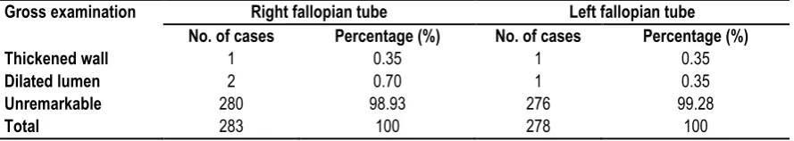
The gross examination of fallopian tubes as depicted in Table 9 shows right fallopian tube unremarkable in majority of the cases i.e. 280 (98.93%), lumen was dilated in 2 cases (0.70%) and the wall of the fallopian tube was thickened in 1 (0.35%) cases. Left fallopian tube unremarkable in 276 (99.58%) cases. The wall was thickened in one case and only one case showed dilated lumen. The histopathological examination of endometrium shows Proliferative phase endometrium in majority of cases i.e., 149 (39.41%), basal endometrium in 96 cases (25.39%) followed by Atrophic endometrium with 38 cases (10.05%), Secretory endometrium in 36 cases (9.52%), simple cystic hyperplasia in 18 cases (4.76%), endometrial polyps in 16 cases(4.23%).
Histologically these showed morphologies varying from functional, hyperplastic to atrophic, pseudodecidual changes were observed in 8 cases (2.11%), complex hyperplasia in 5 cases (1.32%) out of which one showed mild atypia and endometritis in 4 cases (1.05%). Malignant tumours observed were endometrial carcinoma in 6 cases (1.58%) and endometrial stromal sarcoma in one case (0.26). Aria stella reaction was seen in one case (0.26) (Table 10).
The histopathological examination of myometrial lesions as depicted in Table 11 shows majority of the cases i.e. 179 (47.35%) showed normal histology, the next major group was formed by Leiomyoma with 119 (31.48%) cases followed by adenomyosis with 45 (11.90%) cases and leiomyoma with adenomyosis in 35 (9.25%) cases.
The histopathological examination of cervical lesions shows majority of the cases i.e., 190 (50.26%) comprised of chronic cervicitis. 36 of these had hyperplastic lining ectocervix, 24 had squamous metaplasia of endocervix, 52 showed features of papillaryendocervicitis (13.75%), endocervical polyps were observed in 4 (1.05%) cases, squamous cell carcinoma was found in 3 (0.79%) and adenocarcinoma, cervical intraepithelial neoplasia in 1 (0.26%) case each. The cervix was histologically normal in 67 (17.72%) cases (Table 12).
Table 5: Gross examination of endometrium
Gross examination No. of cases (n = 378) Percentage (%)
Fleshy endometrium 19 5.02
Polypoid growth 12 3.17
Unremarkable 347 91.79
Table 6: Gross examination of myometrium
Gross examination No. of cases (n = 378) Percentage (%)
Leiomyomata 142 37.56
Thickened myometrium 9 2.38
Unremarkable 227 60.05
Table 7: Gross examination of cervix
Gross examination No. of cases (n = 378) Percentage (%)
Hypertrophied cervix 64 16.93
Polyp 3 0.79
Growth eroding the cervical lips 2 0.52
Unremarkable 309 81.74
Table 8: Gross examination of the ovaries
Gross examination Right Ovary Left ovary
No. of cases Percentage (%) No. of cases Percentage (%)
Cysts 82 28.27 72 26.08
Solid mass 7 2.41 4 1.44
Unremarkable 201 69.31 200 72.46
Total 290 100 276 100
Table 9: Gross examination of fallopian tubes
Gross examination Right fallopian tube Left fallopian tube
No. of cases Percentage (%) No. of cases Percentage (%)
Thickened wall 1 0.35 1 0.35
Dilated lumen 2 0.70 1 0.35
Unremarkable 280 98.93 276 99.28
Table 10: Histopathological examination of endometrium
Histopathological Diagnosis No. of cases Percentage (%)
Proliferative Endometrium 149 39.41
Basal Endometrium 96 25.39
Atrophic Endometrium 38 10.05
Secretory Endometrium 36 9.52
Simple cystic hyperplasia 18 4.76
Endometrial Polyp 16 4.23
Pseudodecidual change 8 2.11
Complex hyperplasia 5 1.32
Endometritis 4 1.05
Endometrial Carcinoma 6 1.58
Endometrial stromal sarcoma 1 0.26
Aria stella reaction 1 0.26
Table 11: Histopathology of myometrium
Histopathological diagnosis No. of cases Percentage (%)
Leiomyoma 119 31.48
Adenomyosis 45 11.90
Leiomyoma with adenomyosis 35 9.25
Normal histology 179 47.35
Table 12: Histopathological examination of cervix
Histopathological diagnosis No. of cases (n=378) Percentage
Chronic cervicitis 190 50.26
Papillary endocervicitis 52 13.75
Chronic cervicitis with hyperplastic lining ectocervix 36 9.52
Chronic cervicitis with squamous metaplasia of endocervix 24 6.32
Endocervical polyp 4 1.05
Squamous cell carcinoma 3 0.79
Cervical intraepithelial neoplasia 1 0.26
Adenocarcinoma 1 0.26
Normal histology 67 17.72
Table 13: Histopathological examination of ovaries
Histopathological diagnosis Right Ovary Left ovary
No. of cases Percentage (%) No. of Cases Percentage (%)
Follicular cyst 28 9.89 30 10.79
Cystic follicle 11 3.88 16 5.75
Corpus luteal cyst 48 16.96 34 12.23
Serous cyst 2 0.70 3 1.07
Endometriosis 5 1.76 4 1.43
Serous cystadenoma 12 4.24 3 1.07
Mucinous cystadenoma 3 1.06 4 1.43
Benign mature teratoma 2 0.70 1 0.35
Serous cystadenocarcinoma 1 0.35 3 1.07
Mucinous cystadenocarcinoma 1 0.35 3 1.07
Granulosa cell tumor 1 0.35 3 1.07
Brenner tumour 2 0.70 1 0.35
Adenocarcinoma 3 1.06 3 1.07
Transitional cell carcinoma 1 0.35 1 0.35
Clear cell adenocarcinoma 1 0.35 1 0.35
Normal histology 162 57.24 174 62.58
Table 14: Histopathological examination of fallopian tube
Histopathological diagnosis Right fallopian tube Left fallopian tube
No. of cases Percentage (%) No. of cases Percentage (%)
Hydrosalpinx 1 0.35 1 0.35
Saplingitis 2 0.70 1 0.35
Endometriosis 1 0.35 1 0.35
Normal histology 279 98.58 276 99.28
Total 283 100 278 100
Table 15: Correlation of clinical diagnosis with histopathological diagnosis
Preoperative diagnosis Confirmed by histopathology
No. of cases Percentage (%)
Fibroid (n = 155) 142 91.61
Uterovaginal prolapsed (n = 76) 76 100
Ovarian cyst (n = 35) 29 82.8
Endometrial hyperplasia (n = 29) 15 51.7
Pelvic inflammatory disease (n = 27) 17 62.9
Uterine polyps (n = 15) 13 86.6
Cervical polyps (n = 5) 3 60
Serous cystadenoma (n = 5) 5 100
Carcinoma cervix (n = 4) 4 100
Malignant ovarian tumor, (n = 3) 3 100
Carcinoma endometrium (n = 2) 2 100
Dermoid cyst (n = 1) 1 100
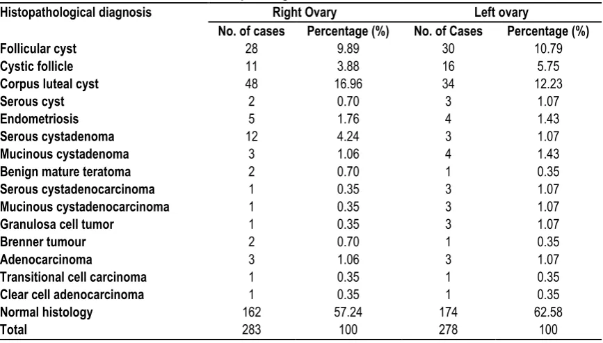
The histopathological examination of ovaries shows right ovary was normal in majority of the cases i.e. 162 (57.24%) while as Corpus luteal cyst were observed in 48 cases (16.96), follicular cysts in 28 (9.89%) cases, serous cystadenoma in 12 cases (4.24%), cystic follicle in 11 cases (3.88%) and endometriosis in 5 cases (1.76%). Adenocarcinoma and mucinous cystadenoma were seen in 3 cases each (1.06%). Serous cyst, benign mature teratoma, brenner tumour was seen in 2 cases (0.70%). One case (0.35%) each of serous cystadenocarcinoma, mucinous cystadenocarcinoma, granulosa cell tumour and transitional cell carcinoma were also observed. Left ovary was histologically unremarkable in majority of the cases i.e. 174 (62.58%), corpus luteal cyst were observed in 34 cases (12.23%), follicular cysts in 30 (10.79%) cases and cystic follicle in 16 cases (5.75%). Endometriosis and mucinous cystadenoma were seen in 4(1.43%) cases each, serous cyst, serous cystadenoma and serous cystadenocarcinoma in 3 (1.07%) cases each. while one (0.35%) case each of inclusion cyst benign mature teratoma, brenner tumour and transitional cell carcinoma were observed (Table 13). The histopathological examination of fallopian tube) shows right fallopian tube normal in majority of these cases i.e. 279 (98.58%), two (0.35%) cases of salpingitis were observed, one of chronic salpingitis and the other of tubercular salpingitis where as one case each of endometriosis and hydrosalpinx were also observed. Left fallopian tube normal in majority of the cases i.e. 276 (99.28%), one case each of hydrosalpinx and chronic salpingitis were observed (Table 14).
Pre-operative clinical diagnosis was available in 357 cases. Clinical diagnosis of fibroid uterus was given in 155 cases out of which 142 (91.61%) were confirmed by histopathology, pelvic inflammatory disease was confirmed histopathologically in 17 (62.96%) out of 27 cases, , ovarian cyst in 29 (82.8%) out of 35 cases, Endometrial hyperplasia in 15(51.7%) out of 29 cases,
uterine polyps in 13 (86.6%) out of 15 cases, Cervical polyp in 3(60%) out of 5 cases and carcinoma cervix, Uterovaginal proplapse, Adenomyosis, Dermoid cyst, Serous cystadenoma, malignant ovarian tumor and Carcinoma endometrium were observed in 100% of cases each (Table 15).
DISCUSSION
Hysterectomy is the most commonly performed major gynaecological surgery. It is a successful operation in terms of symptom relief, patient satisfaction and provides definitive cure to many diseases involving uterus as well as adnexae.7 This study
was conducted to analyse the pattern of lesions in hysterectomy specimens in our institution and to compare our findings with those of other workers.
The commonest estimated age range of hysterectomy in our study was 41-50 years. In present study the preferred approach in majority of cases is abdominal (83.06%) followed by vaginal route (16.93%) which is in accordance with the findings reported in other studies.8,9 Majority of cases (70.89%) had undergone
bilateral salpingo-oophorectomy along with hysterectomy. Mackenzie et al., 2004 have also reported bilateral salpingo-oopherectomy in 50% of their cases.10
In our study the most common clinical indication for hysterectomy is menorrhagia (35.22%) followed by fibroid uterus (26.63%). Menorrhagia was also reported as the most common clinical indication in other studies.11-14 The commonest endometrial
pathology observed in our study is atrophic endometrium (10.05%) whereas that reported by kleebkaow et al., (2008)15 was
3.8%, however Mehboob and Ahmad (2002)16 reported a higher
In India carcinoma and other malignancies of the body of uterus are not as frequently encountered as other gynaecological malignancies.18 Endometriod carcinoma being the commonest
form of endometrial carcinoma accounting for more than three fourth of all cases.19
In our present study, malignant tumours comprised of six cases of endometriod type and one case of endometrial stromal sarcoma. The endometrial carcinoma reported by Gousia 201014 was of
papillary adenocarcinoma type whereas both the tumours reported by Gazozai et al 200417 were endometrial stromal sarcoma.
Uterine leiomyomata is the most common tumour of the female genitalia, estimated to occur in 20-40% of women in their reproductive years20, the likelihood that leiomyomata will cause
symptoms is undoubtedly related to their number, size and location although it seems equally plausible that myomata may frequently represent an incidental rather than causal finding.21
Leiomyoma was the most common myometrial lesion observed bin present study, the same was true of other studies.14,22,23
22.72% cases of leiomyoma in present study showed foci of adenomyosis as well. Similar findings were observed by other studies.13,14,24
In the present study 43.75% cases of adenomyosis revealed the presence of leiomyoma as well. Similar findings were reported by Shaikh and Khan 199025, Ali 200524, Purandare and
Jhalam 199326, Bukhari and Sadiq 200722 and Praveen and
Tayyab 2008.13
Chronic cervicitis is an extremely common condition in adult females at least at microscopic level.27 Chronic cervicitis was
detected in 89.39% cases in present study. 9.6% cases of chronic cervicitis also had squamous metaplasia of endocervical lining, squamous metaplasia of cervix were reported by other studies.16,24
14.4% cases of chronic cervicitis in the present study depicted hyperplasia lining ectocervix and 13.75%of cases showed the features of papillary endocervicitis, these findings have not been reported in the earlier studies. The cervical carcinoma reported in present study included three cases of squamous cell carcinoma and one case of adenocarcinoma, their incidence in present study is close to that reported by Mehboob and Ahmad 2002.16
Non-neoplastic cysts are most common ovarian lesions observed in present study, similar findings were reported in other studies.11,13,28 The incidence of ovarian tumours in present study is
close to that reported in other studies.13,28,29 Fallopian tubes are
complex structures that represent more than conduits from ovary to endometrial cavity.30 In the present study, only significant
lesions were two cases of salpingitis.one case of hydrosalpinx and one case of endometriosis. Similar findings were observed in other study.30
CONCLUSION
Menorrhagia (35.22%) and fibroid uterus (26.63%) were most common clinical indication for hysterectomy. The most common finding identified by histopathology were proliferative phase (39.41%) in endometrium, leiomyoma (31.48%) in myometrium, chronic cervicitis(50.26%) in cervix, benign cysts in ovaries. It is mandatory that every hysterectomy specimen, even if it appears grossly normal should be subjected to detailed histopathological examination so as to find missed pathologies especially missed malignancies and establish definitive cause in number of cases designated abnormal uterine bleeding to ensure better
postoperative management. The present study provides a fair insight into the histopathological patterns of lesions in hysterectomy specimens in our institution and the results can be safely considered as reflection of disease pattern in Kashmir.
ACKNOWLEDGEMENT
The authors acknowledge the support provided by the Department of Pathology Govt. Medical College Srinagar. Indirect role played patients whose samples have been included in the study is highly acknowledged.
REFERENCES
1. Jamie Prat, Female reproductive system. Ivan Damjanov,
Anderson’s Pathology 10th edition, Mosby-Year Book, Inc.1996;
vol 2: p2261-2275.
2. Ranabhat SK, Shrestha R, Tiwari M, Sinha DP, Subedee LR. A retrospective histopathological study of hysterectomy with or without Salpingo-Ophorectomy specimens.JCMC2010;1(1):26-29. 3. G. Gupta, D.S. Kotasthane, V.D. Kotasthane, Hysterectomy: A clinico-pathological Correlation of 500 Cases. The Internet Journal of Gynaecology and Obstetrics. 2010; Vol. 14: No .1.
4. Conley G. Lacey, About Hysterectomy- surgical removal of the uterus, or womb. When you need an operative. American college of surgeons. p.1-11.
5. Sobande AA, Eskander M, Archibong EL. Elective hysterectomy: A clinicopathological review from Abha catchment area of Saudi Arabia. West Afr J Med 2005; 24: 31-5.
6. Mangala Gowri, Geetha Mala, Srinivasa Murthy, Vedavathy Nayak. Clinicopathological study of uterine leiomyomas in hysterectomy specimens. Journal of Evolution of Medical and Dental Sciences 2013; 2(46), Page 9002-9009.
7. Jaleel R, Khan A, Soomro N. Clinicopathological study of abdominal hysterectomies. Pak J Med Sci 2009;25(4).
8. Aksu F, Gezer A, Oral E. Seventeen-year review of hysterectomy procedures in a university clinic in Istanbul (1985-2001). Arch Gynecol Obstet 2004; 270 (4): 217-222.
9. Abdullah LS. Hysterectomy: A Clinicopathologic correlation, Bahrain Medical Bulletin 2006 (June); 28 (2).
10. Mackenzie IZ, Naish C, Rees M, Manek S. 1170 consecutive hysterectomies: indications and pathology.J. J Br Menopause Soc 2004;10 (3): 108-112.
11. Jamal S, Baqai S. A clinicohistopathological analysis of 260 Hysterectomies. Pak J Pathol 2001; 12 (2): 11-14.
12. Sobande AA, Eskander M, Archibong EI, Damole 10. Elective hysterectomy: a clinicopathological review from Abha Catchment Area of Saudi Arabia. West 4.11. .1 Med 2005; 24 (1): 31-35. 13. Parveen S, Tayyab S. A clinicopathological review of elective abdominal hysterectomy. J Surg Pak 2008; 13 (1).
14. Gousia Rahim Rather, Yudhvir Gupta, Subash Bardhwaj Patterns of lesions in Hysterectomy specimens: A prospective study Vol. 15 No. 2, April - June 2013.
15. Kleebkaow P, Maneetab S et al. Preoperative and postoperative agreement of histopathological findings in cases of endometrial hyperplasia.Asian Pacific J Cancer Prev2008;9:89-91. 16. Mehboob R, Ahmad N. Unexpected pathology at vaginal hysterectomy for genital prolapse. Pak J Med Res 2002; 41(4): 142-44.
18. Ronnet BM, Kurman RJ. Precussor lesions of endomjal carcinoma. In: Biaustein's Pathology of the Female Genital Tract (Kurman kJ, editor), 5th edition, Springer-Verlag, New York-Berlin-Heidelberg 2002: 467. 500.
19. Cherian A, Surin C, Jacob S, Prabhakar BR: Primary malignancies of the corpus uteri – retrospective five year analysis. Indian J Pathol Microbiol 1995; 38: 63-72.
20. Buttram VC, Reiter RC. Uterine leiomyomata—etiology, symptomatology and management. Feral Steril 1981; 36: 433445. 21. Reiter R C, Wagner PL, Gambone JC. Routine hysterectomy for large asymptomatic uterine leiomyomata: a reappraisal. Obstet Gynecol 1992;79(4):481-484.
22. Bukhari U, Sadiq S. Analysis of the underlying pathological lesions in hysterectomy specimens. Pak J Pathol 2007; 18 (4): 110-112.
23. Miller NF, Mich AA: Hysterectomy. Therapeutic Necessity or Surgical Racket? Am J Obstet Gynecol 1945; 51: 804-810. 24. Ali A. Incidence of adenomyosis in hysterectomies. Pak J Med Res 2005; 44 (1).
25. Shaikh H, Khan KS. Adenomyosis in Pakistani women: four year experience at the Aga Khan University Medical Centre, Karachi. J Clin Pathol 1990; 43: 817-819.
26. Purandare S,Jhalam L.Pathological picture in hysterectomy done for abnormal uterine bleeding.J Obstet G ynecol India 1993;43(1-5):418-421.
27. Rosai J. Female reproductive system. In: &nal. and Ackerman's Surgical pathology, 9th edition. Mosby, An Imprint of Elsevier, Missouri 2004; 2: 1523-1648.
28. Jha R, Pant AD, Jha A, Adhikari RC, Sayami G. Histopathological analysis of hysterectomy specimens. Nepal Med Assoc 2006; 45 (163): 283-290.
29. Samalia Modupeola OA, Adesiyun AG, Agunbiade OA, Mohammad-Duro A. Clinicopathological assessment of hysterectomies in Zaria. Eur J Gen Med 2009; 6 (3) 150-153. 30. Bagwan IN, Harke AB, Malpani MR, Deshmukh SD. Histopathological Study of Spectrum of Lesions Encountered in the Fallopian Tube. Obstet Gynecol 2004; 54 (4): 379-382.
[
Source of Support: Nil.
Conflict of Interest: None Declared.
Copyright: © the author(s) and publisher. IJMRP is an official publication of Ibn Sina Academy of Medieval Medicine & Sciences, registered in 2001 under Indian Trusts Act, 1882. This is an open access article distributed under the terms of the Creative Commons Attribution Non-commercial License, which permits unrestricted non-commercial use, distribution, and reproduction in any medium, provided the original work is properly cited.