International Journal of Current
Medical and Pharmaceutical
Research
Available Online at http://www.journalcmpr.com
DOI: http://dx.doi.org/10.24327/23956429.ijcmpr20170266
RESEARCH ARTICLE
COMPARATIVE STUDY OF OPERATIVE AND NON OPERATIVE MANAGEMENT OF INTRA-
ARTICULAR CALCANEAL FRACTURES – A PROSPECTIVE STUDY
*Arun G.Behin., Senthilnathan A., Prabhakar R and Harri Vishnu M
Department of Orthopaedics, Rajah Muthiah Medical College and Hospital, Annamalai University,
Chidambaram, Tamilnadu, India-608 002
ARTICLE INFO ABSTRACT
Introduction: Calcaneum is the most frequently fractured tarsal bone. These fractures result in long term disability with potential economic impact on the patient. Historically treatment of intra articular calcaneum fractures has varied from non-operative management with or without closed reduction to open reduction with internal fixation by various surgical approaches to primary Arthrodesis. In the past two decades with advent of CT scan, better implants and improved methods of fixation Operative treatment has now gained much popularity.
Aim of the Study: The aim is “To compare the results of operative and nonoperative treatment of intra -articular calcaneal fractures”.
Materials and Methods: The study included 40 patients of intra articular calcaneal fractures admitted in RMMCH from June 2015 to September 2017.Twenty patients with intra articular calcaneum fractures treated by non surgical method and Twenty patients treated surgically by percutaneous K- wire fixation, percutaneous cannulated cancellous screw fixation or by open reduction and internal fixation using locking plates with follow up of 6 months. The fractures were classified on the basis of X–Ray findings tongue type or joint depression type and by Computed Tomography (CT) findings as Sanders type I to IV. The patients were evaluated by Modified Maryland Foot Score, with excellent defined as 90 - 100 points, good as 75 – 89 points, fair as 50 – 74 points and poor as <50 points.
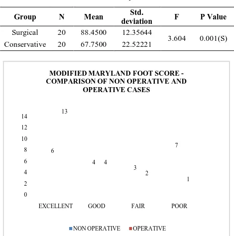
Results: In Non surgical management Excellent Modified Maryland Foot Score was achieved in 6 patients, Good in 4, Fair in 3 and poor in 7 patients. In Surgically treated patients Excellent Modified Maryland Foot score was achieved in 13 patients, good in 4, fair in 2 and poor in 1 patient.
Conclusion: Surgical treatment of intra-articular calcaneal fractures using various modalities like K-Wires, cannulated cancellous screws or by locking compression plate has better functional outcome than non surgical cases. Good timing of surgery is of paramount importance to avoid early complications. Reconstruction of subtalar anatomy prevents subtalar arthritis & maintains the foot biomechanics.
Copyright © 2017 Arun G.Behin et al. This is an open access article distributed under the Creative Commons Attribution License, which permits unrestricted use, distribution, and reproduction in any medium, provided the original work is properly cited.
INTRODUCTION
Calcaneal fractures is one of the most disabling fractures with frequent occurrence in the wage earning period of life. Calcaneal fractures constitutes approximately 1-2% of all fractures and 60% of all Tarsal fractures. 65-75% of calcaneum fractures are intra-articular and involve one or more of the three sub talar articulating facets. 80-90% occur in men between 21 - 45 years of age. Bilateral calcaneum fractures in 5-10% of cases. Fall from height is the most common cause of injury followed by Road traffic accident.
The therapeutic modality for displaced intra articular calcaneum fractures can be divided into conservative(7) and surgical management(8,9). Intra articular calcaneum fracture with comminution remains a challenge to the treating
orthopaedic surgeon, because of serious potential complications and controversies persisted regarding the best treatment.
Historically intra articular fractures have been treated non-operatively. Non-operative treatment led to increased morbidity due to incongruency of articular surface resulting in subtalar arthritis and post traumatic stiffness with loss of subtalar motion, widening of heel.
However in patients with associated diabetes mellitus, smokers, prolonged edema and skin blisters surgery cannot be done and has to be managed non-operatively. More over performing surgery in these patients can lead to wound complications(10) and the possibility of osteomyelitis.
Article History:
Received 17th July, 2017 Received in revised form 25th August, 2017
Accepted 5th September, 2017
Published online 28th October, 2017
Key words:
Calcaneum, Bohler’s Angle, Gissane Angle, Sub talar joint.
With the advent of CT scan with 3D reconstructions, appropriate timing of surgery, better implants – locking plates, improved methods of fixation and rehabilitation, operative treatment of intra articular calcaneum fracture has now become a standard method.
Operative goal includes - Restoration of congruity of subtalar articulation, restoration of Bohler’s(6) and Gissane angles(5), restoration of normal width and height of calcaneus, neutralization of varus deformity of fracture.
Surgical treatment includes closed reduction and percutaneous K-wire or cancellous screw fixation, open reduction and internal fixation using locking plates with or without bone grafting.
MATERIALS AND METHODS
40 patients (33 male and 7 female) with intra articular calcaneal fractures admitted in Rajah Muthiah Medical College Hospital from June 2015 to September 2017 were included in the study.
Patients were evaluated by X ray–AP, lateral and Harris axial view. The fractures were classified on the basis of computed tomography (CT) findings as Sanders type I to IV(3).
Inclusion Criteria
Patient aged between 18 to 70 years
Displaced Intra articular calcaneal fractures – Sander’s Type II, III & IV.
Bohler’s Angle < 20°
Gissane Angle > 135°
Exclusion Criteria
Extra articular fractures
Undisplaced Intra articular calcaneal fractures – Sander’s Type 1
Paediatric fractures
Bohler’s Angle 20-40°
Gissane Angle 105-135°
Compound fractures
Head injury and poly trauma patients with GCS less than 8.
Indications for Operative Management
1. Anterior process fractures with >25% involvement of calcaneo- cuboid articulation.
2. Displaced fractures of the calcaneal tuberosity
3. Irreducible body fractures with >30° varus or 40° valgus deformity.
4. Lateral or medial process fractures with >1.5 cm displacement.
5. Fracture-dislocations of the calcaneus. 6. Patients who are younger and active groups. 7. Patients who wants quick return to work
Indications for Nonoperative Management
1. Diabetes.
2. Smoking.
3. Prolonged edema for > 3 weeks. 4. Skin blistering.
5. Patients not willing for operative treatment after being explained about the nature of the fracture and necessity for intervention.
6. Fractures in patients with other medical comorbidities prohibiting surgery.
20 patients (15 male and 5 female) were treated non operatively and 20 patients (18 male and 2 females) were treated surgically.
Non Operative Management: Patients were given a below
knee slab and limb elevation. Cold fomentation and anti edema measures were given. After the edema subsides below knee cast is applied and the patient is advised non weight bearing for 8 weeks. After 8 weeks, X ray is taken for signs of union and depending on the uniting nature of the fracture allowed partial weight bearing at 10 to 12 weeks and full weight bearing at 13 to 15 weeks.
Surgical Management: The surgical methods used in our
study are as follows.
1. Closed Reduction and Internal Fixation with K-Wires. 2. Closed Reduction and Internal Fixation with Cannulated
Cancellous Screws.
3. Open Reduction and Internal Fixation with Lateral Calcaneal Locking plate.
The aim of surgery is to achieve anatomical reconstruction of all articular surfaces, restore Bohler’s and Gissane angles, to carry out primary stable fixation and begin early mobilization. Patients were given a below knee slab. Strict limb elevation and anti-edema measures were done and watched for the swelling to subside and appearance of “wrinkle sign”. Surgery was performed only after the edema had resolved. All patients were operated with an average of 7-15 days from the time of fracture if the soft tissue condition was good.
Surgical Techniques
Closed Reduction and Internal Fixation with K-Wires
The first step was to insert a 3-mm Steinmann pin superolateral to the Achilles tendon insertion, the pin was inserted into the tongue fragment with manipulation to restore the posterior facet as described by Essex-Lopresti(4). Then closed reduction of the fracture was done by introduction of 3 mm K-wire for traction in the dorso-cranial plane, the varus or valgus malaligment of the hind foot was corrected with the wire positioned orthogonally to the longitudinal axis of the calcaneus. The reduction was secured by multiple 2 mm K-wires, the number of which will vary according to the type of fracture and the degree of comminution. K-wires should secure
both the reduced articular surface and the overall alignment and should be arranged perpendicular to fracture lines as possible. The Steinmann pin was removed, after introducing of multiple K Wires, The wires were bent above the skin level.
Closed Reduction and Internal Fixation with Cannulated Cancellous Screws
Open Reduction and Internal Fixation with Lateral Calcaneal Locking plate
Surgery was performed in lateral position either in general or regional anaesthesia by extensile lateral approach in all patients. The land marks are lateral malleolus, calcaneo-cuboid joint and base of fifth metatarsal. Incision made in a right angled fashion with the vertical line starting 4cm above the lateral malleoli between fibula and tendoachilles and extended downward till the junction of dorsal and plantar skin. The horizontal line is extended distally up to the base of fifth metatarsal. The incision is carried straight down to the bone at its angle and then developed to allow a single, thick flap to be lifted from the periosteal surface. A “no touch” technique is employed by retracting the flap with K wires in the talus and cuboid. Reduction aided by periosteal elevator or osteotome. The posterior facet and the anterior facets were reduced. Reduction verified with C-arm. Low profile locking plates were contoured and positioned. The plate is secured by 3.5mm cancellous locking screws of various length. Subcutaneous tissue is closed using 2-0 vicryl and tension free skin closure done using 3-0 ethilon. Multiple small drain tubes inserted in between. Tight dressing and compression bandage applied. Below knee slab given.
Post-Operative Protocol and Follow Up
All patients were immobilized in posterior plaster splint and limb elevated. Drain is removed after 48 hours and first wound inspection done on 2nd day. Suture removal done after 13th day (13 to 18th day). After suture removal below knee cast applied. In patients treated with K-wires, K-wire removal done after 8 weeks. All patients were regularly followed up with X – rays and based on the signs of union, partial weight bearing allowed by 8 to 10 weeks and full weight bearing allowed at 12 to 14 weeks.
Functional outcomes were evaluated by Modified Maryland Foot Score, with excellent defined as 90 - 100 points, good as 75 – 89 points, fair as 50 – 74 points and poor as <50 points..
RESULTS
In our study of the 40 patients, 33 patients were male and 7 patients were female. 31 were due to fall from height and 9 were due to road traffic accidents. The mean age among operative group is 35 years and the mean age among non operative group is 34 years.
In the non operative patients, the mean Bohler’s angle was 10.05°, the mean Gissane angle was 149.05°, the mean height was 32.90 mm and the mean width was 56.70 mm.
In operative patients, the mean pre-operative Bohler’s angle was 9.9° and mean post-operative angle was 24.15° with P value <0.001 which is statistically significant.
In operative patients, the mean pre-operative Gissane angle was 149.6° and the mean post-operative angle was 122° with P value < 0.001 which is statistically significant.
In operative patients, the mean pre-operative height was 32.6 mm and the mean post-operative height was 44.0 mm with P value < 0.001 which is statistically significant.
In operative patients, the mean pre-operative width was 57.75 mm and the mean post-operative width was 51.85 mm with P value < 0.001 which is statistically significant.
In non operatively treated patients, Excellent Modified Maryland Foot Score Was achieved in 6 patients, Good in 4, Fair in 3 and poor in 7 patients. In Surgically treated patients Excellent modified Maryland Foot score were achieved in 13 patients, good in 4, fair in 2 and poor in 1 patient.
On comparison of Modified Maryland Foot Score of both Operative and Non operative patients the P Value is 0.001 which is statistically significant.
In Non operative patients: Complications encountered are heel pain (n=3), subtalar arthritis (n=3) and joint stiffness (n= 4).
In Operative patients: Complications encountered are heel pain (n=2), subtalar arthritis (n=1), joint stiffness (n=2), Wound dehiscence (n=1) and superficial infection (n=1) patient. Superficial infection and wound dehiscence patients were treated conservatively with daily dressing and broad spectrum antibiotics. Both cases settled well. There was no case of deep infection and bone infection.
Table 1 Sander’s Classification Distribution
Sander’s Classification
Group
Total
Operative Non operative
N % N % N %
Type II 4 20.0 5 25.0 9 22.5
Type III 14 70.0 13 65.0 27 67.5
Type IV 2 10.0 2 10.0 4 10.0
Total 20 100.0 20 100.0 40 100.0
Table 2 Operative Treatment-Measurements
Mean Std. Deviation
P value
Pre op Post op Pre op Post op
Bohler’s Angle (°) 9.90 24.1500 5.75738 6.78446 <0.001(S)
Gissane Angle (°) 149.60 122.00 5.56682 8.64505 <0.001(S) Height (mm) 32.6500 44.0000 1.66307 3.21182 <0.001(S) Width (mm) 57.7500 51.8500 1.58529 2.30046 <0.001(S)
Table 3 Modified Maryland Foot Score
Group N Mean Std.
deviation F P Value
Surgical 20 88.4500 12.35644
3.604 0.001(S)
Conservative 20 67.7500 22.52221
0 2 4 6 8 10 12 14
EXCELLENT GOOD FAIR POOR
6 4 3 7 13 4 2 1
MODIFIED MARYLAND FOOT SCORE -COMPARISON OF NON OPERATIVE AND
OPERATIVE CASES
Case I Operative Treatment
Pre op X-ray lateral Axial view C T scan
Post Op X-Ray lateral view Post op X-ray axial view Lateral view after union
Axial view after union Pre op gissane angle-143° Pre op bohler’s angle-15°
DISCUSSION
The optimal management of displaced intra articular calcaneal fractures remains a matter of debate despite advancements in diagnosis by means of imaging and in surgical techniques.
A lot of studies had been done on the management of displaced intra Articular calcaneal fractures with some studies showing no difference in the functional outcome of both non operative and operative cases and some studies showing better results in operative cases.
In 2013 Agren(2) conducted a Randomized controlled Multicentre Trial of 82 patients of 5 trauma centres with displaced intra articular calcaneal fractures. He concluded that Operative treatment was not superior to non operative treatment at one year of follow up but appear to have some benefits at eight to twelve years of follow up.
But there is reduced prevalence of post traumatic arthritis in operative cases but the need for sub-talar arthrodesis was not increased following non operative treatment.
David B. Thordarson(1) in 1996 done a randomized controlled study of 30 Patients of displaced intra articular calcaneal fractures and 15 patients were treated non operatively and 15 patients were operated with a follow up of 17 months. This study demonstrates Operative treatment with early mobilization is superior to Non operative treatment.
In our study all the patients were followed up for a minimum period of 6 months (6-12 months) and evaluated using Modified Maryland Foot Scoring System. Excellent and Good results were achieved in 85% (n=17) of operatively treated patients and only 50% (n=10) of non operatively treated patients. Fair and Poor results were achieved in 15% (n=3) of operatively treated patients and in 50% (n=10) of non
Case II Non Operative Treatment
X- ray lateral and axial view CT scan X-ray lateral after union- showing early
arthritic changes in sub talar joint
Gissane angle-149° Bohler’s angle -8°
operatively treated patients. On comparing the scores of both Operative and Non operative patients the P value is 0.001 which is statistically significant.
In operative treatment Bohler’s Angle, Gissane Angle, calcaneal height and width were restored in 85% of patients. Sub talar joint congruity is maintained so this delays the development of subtalar arthritis.
The incidence of complications in non operative patients : Sub talar arthritis – 15% (n=3), Joint stiffness – 20% (n=4) and Heel pain-15% (n=3) is higher compared to operatively treated patients : Sub talar arthritis- 5% (n=1), Joint stiffness- 10%(n=2) and Heel pain- 10%(n=2).
CONCLUSION
Operative treatment of intra-articular calcaneum fractures using closed reduction and internal fixation with K - wires, closed reduction and internal fixation with cannulated cancellous screws and open reduction and internal fixation with lateral calcaneal locking plate based on the type of fracture has better functional outcome than Non operative treatment.
Restoration of Bohler’s angle, Gissane angle, Calcaneal height and Calcaneal width are of paramount importance for better functional outcome after surgery.
Surgery should be done only after the edema subsides – WRINKLE SIGN to prevent soft tissue complications.
Reconstruction of subtalar anatomy prevents subtalar arthritis & maintains the foot biomechanics.
References
1. Thordarson DB, Krieger LE. Operative vs non operative treatment of intra-articular fractures of the calcaneus: a prospective randomized trial. Foot Ankle Int 1996 Jan;17(1):2-9.
2. Agren PH, Wretenberg P, Sayed-Noor AS. Operative versus nonoperative treatment of displaced intra-articular calcaneal fractures: a prospective, randomized, controlled multicentertrial. J Bone Joint Surg Am 2013 Aug 7;95(15):1351-1357.
3. Sanders R, Fortin P, DiPasquale T, Operative treatment in 120 displaced intraarticular calcaneal fractures. Results using a prognostic computed tomography scan classification. Clin Orthop Related Res 1993 May(290):87-95.
4. Essex-Lopresti P (1952), The mechanism, reduction technique, and results in fractures of the Os Calcis. Br J Surg 39:395-419.
5. Gissane W. Discussion on “Fracture of the oscalcis” (Proceedings of the British Orthopaedic Association). J
Bone Joints Surg 1947; 29:254-255.
6. Bohler, Lorenz. Diagnosis, pathology and treatment of fractures of the oscalcis. J Bone and Joint Surg, Jan 1931 - vol 13 issue 1 - ppg 75-89.
7. Pozo JL, Kirwan EO, Jackson AM. The long-term results of conservative management of severely displaced fractures of the calcaneus. J Bone Joint Surg Br 1984 May;66(3):386-390.
8. Hammesfahr JF. Surgical treatment of calcaneal fractures. Orthop Clin North America 1989;20(4):679-689.
9. Leung KS,Yuen KM, ChanWS. Operative treatment of displaced intra-articular fractures of the calcaneum.
Medium-term results. J Bone Joint Surg Br
1993;75(2):196-201.
10. Abidi NA, Dhawan S, Gruen GS, Wound-healing risk factors after open reduction and internal fixation of
calcaneal fractures. Foot Ankle Int 1998 Dec; 19(12):856-861.