*Corresponding author:Harkirat
Senior Resident, Department of Pathology, Govt. Medical College, Amritsar, India
INTERNATIONAL JOURNAL OF CURRENT MEDICAL AND
PHARMACEUTICAL RESEARCH
ISSN: 2395-6429, Impact Factor: SJIF: 4.656
Available Online at www.journalcmpr.com
Volume 4; Issue 1(B); January 2018; Page No. 2923-2927
DOI: http://dx.doi.org/10.24327/23956429.ijcmpr20180368
Research Article
PATTERN OF LYMPHADENOPATHY ON FNAC IN TERTIARY CARE HOSPITAL
- A CROSS SECTIONAL STUDY
Harkirat
1*., Amarjit Singh
2., Amar Jyoti
3., Mandeep Randhawa
4and N.S.Neki
51
Senior Resident, Dept. of Pathology, Govt. Medical College, Amritsar, India
2
Professor & Head, Dept. of Pathology, Govt. Medical College, Amritsar, India
3,4
Assistant Professor, Dept. of Pathology, Govt. Medical College, Amritsar, India
5
Professor & Head, Dept. of Medicine, Govt. Medical College, Amritsar, India
ARTICLE INFO ABSTRACT
Lymphadenopathy is one of the commonest clinical presentation of various neoplastic and non neoplastic diorders at all age groups. Fine needle aspiration cytology (FNAC) is a simple, quick, inexpensive and minimally invasive technique that helps in diagnosing multiple lesions avoiding the need for biopsy. The aim of the documented study is to observe the various cytomorphological patterns of lymphadenopathy through FNAC, and to assess their age and sex specific distribution. This study was conducted on 570 cases of lymphadenopathy over a period of 18 months from july 2016 to Dec. 2017 in the cytopathology section of department of pathology, Govt. Medical College, Amritsar. Cytological diagnosis was correlated with details of relevant clinical findings and investigations.
Out of total 570 cases studied, Tubercular lymphadenitis was found to be most common presentation of lymphadenopathy amounting to 249 cases (43.68%) followed by Reactive lymphadenitis (164 cases, 28.77%), Metastatic lymphadenitis (95 cases,16.67%), Lymphoproliferative disorders (40 cases,7.017%) and Suppurative lymphadenitis (22 cases,3.85%) respectively. Highest incidence of lymphadenopathy was found in patients of 21-40 yrs age group (221 cases, 38.77%) followed by age group of 0-20 yrs (189 cases,33.16%). Male to female ratio was 1:1.04 with slight male preponderance.
The present study concludes that Tubercular Lymphadenitis is arguably leading cause of lymphadenopathy at all ages with maximum incidence in children and adolescents. It also highlights the usefulness of FNAC in the early diagnosis of various causes of lymphadenopathy differentiating neoplastic from non-neoplastic causes, thus helping the clinician in arriving at an early diagnosis and timely management without any unnecessary surgical intervention.
Copyright © 2018 Harkirat et al. This is an open access article distributed under the Creative Commons Attribution License, which permits unrestricted use,
distribution, and reproduction in any medium, provided the original work is properly cited.
INTRODUCTION
Lymph nodes are most widely distributed and easily accessible component of lymphoid system.1 They are among the commonest aspirated organ for diagnostic purpose. Lymphadenopathy is the term used to describe the conditions in which lymph nodes become abnormal in size, number and consistency. 75 % of all lymphadenopathies are localized, with 50 % being seen in head and neck area. 25 % of lymphdadenopathies are generalized and are often a sign of significant systemic underlying disease.2 Cervical lymphdadenopathy is one of the commonest clinical presentation of patients attending the outdoor department.3 Based upon the different geographical areas, the etiology of lymphadenopathy varies eg. in tropical areas, tuberculosis is a main cause of lymphadenopathy in adults and children.4
However, its prevalence is only less than 5% among Indian children aged between 0-14 years.5 Nonetheless, in a large number of studies, the most common benign etiologies are non specific reactive changes in lymph nodes.6 Despite the low prevalence of malignancy among patients with lymphadenopathy, it remains to be the main concern of both patients and physicians. Studies have shown that its prevalence is low among patients with unexplained lymphadenopathy7. FNAC is a reliable and least expensive method for developing countries.8,9 It has become an acceptable and widely practiced minimally invasive technique as a first line investigation 10
MATERIALS AND METHODS
A descriptive cross- sectional study was conducted over a period of 18 months ie. from July 2016 to Dec.2017 in the
Article History:
Received 16th October, 2017 Received in revised form 25th November, 2017
Accepted 23rd December, 2017 Published online 28th January, 2018
Key words:
Fine needle aspiration cytology (FNAC), Lymphadenopathy, Tuberculous lymphadenitis, Reactive lymphadenitis, Metastatic
International Journal Of Current Medical And Pharmaceutical Research, Vol. 4, Issue, 1(B), pp.2923-2927, January, 2018
cytolopathology section of Department of Pathology, Govt. Medical College, Amritsar. Brief history including age, sex, size and site along with thorough clinical examination was carried out. The FNAC was done on cohort of 570 patients with clinically significant lymphadenopathy using 20-24 G needle without local anaesthesia. Aspirated material was
smeared onto slides, air dried and fixed in methanol for May- Grunwald Giemsa (MGG) stain and wet alcohol fixed for Hematoxylin and Eosin. Ziehl Neelson ( ZN ) stain for acid fast bacilli was also used wherever required. Review of all the cytological reports was done to explore the patterns of lymphadenopathy. Various diagnosis were noted and categorized into groups including Tubercular, Reactive, Suppurative, Metastatic and Lymphoproliferative disorders.
RESULTS
In the documented study, 570 patients were subjected to FNAC for lymphadenopathy. There were 291 males and 279 females with M:F ratio of 1:1.04, i.e. with a slight male preponderance. The age at presentation ranged from 1 month to 87 years. Maximum number of patients were in the age group of 21-40 years (221 cases, 38.77 %) followed by age group 0-20 years (189 cases, 33.16%), 41-60 years (118 cases, 20.70%), 61-80 years (41 cases, 7.19%) and >80 years (1 case, 0.17%) respectively.
Reactive lymphadenitis was noted in 164 cases (28.77%). Among these patients, 89 patients (54.26%) were in 0-20 age group and 55 patients (33.54%) were in 21-40 year age group followed by 16 cases (9.76%) in 41-60 year age group and 4 cases (2.44%) in 61-80 years age group.
Suppurative lymphadenitis was noted in 22 patients (3.85%) with majority (10 cases, 45.45%) of cases in 21-40 years age group. Tuberculous lymphadenitis accounted for a total of 249 cases (43.68%). This was the most common presentation of lymphadenopathy in the current study. Granulomatous inflammation with coexistent caseous necrosis was the dominant sub category in this group accounting for 231 cases
(92.77%) followed by 11 cases (4.41%) of granulomatous inflammation without caseation necrosis while suppurative granulomatous pathology was seen in only in 7 cases (2.81%). Tubercular lymphadenitis was seen maximum in the 21-40 year age group amounting to 129 cases (51.81%) followed by 0-20 age group i.e. 86 cases (34.54%). Of all the cases, 74 cases (29.7%) showed AFB positivity.
In the documented study, 23 cases (4.03%) were HIV positive and 1 case(0.17%) was HCV positive. Out of 23 cases of HIV, 12 cases (52.17%) showed reactive lymphoid hyperplasia, 7 cases (30.43%) showed tuberculous lymphadenitis, 2 cases of necrotizing lymphadenitis (8.69%) 1 case of NHL(4.35%) and 1 case(4.35%) of atypical lymphoid hyperplasia. Among the 23 cases of HIV positive patients, only 6 cases(26.08%) showed strong AFB positivity.
Metastatic carcinomatous deposits were observed in 95 cases (16.67%) predominantly above 40 years of age. Cervical lymph nodes were involved in all types of lymphadenopathies. Squamous cell carcinoma was the most common metastatic lesion in the documented study (61 cases, 64.21%) followed by adenocarcinoma (14 cases, 14.73%), and Non small cell undifferentiated carcinoma (11 cases, 11.58%). 4 cases (4.21%) showed metastatic deposits of small cell carcinoma, 3 cases (3.16%) showed metastatic deposits of Infilterating Table 2 Comparison of studies showing incidence of Lymphadenopathy
Sr.no. Studies lymphadenitis Tubercular lymphadenitis Reactive lymphadenitis Metastatic lymphadenitis Suppurative Lymphoma
1 Present 43.68% 28.77% 16.67% 3.85% 7.02%
2 Nirmal
Amit(2016) 44.60% 35.47% 10.79% 9.35% 1.08%
3 Kochhar A
K(2016) 38.52% 24.45% 30.37% 3.70% 2.22%
4 Chandran N(2014) 45.5% 27.2% 11.75% 9.5% 3.5%
5 Sharada
Mani(2017) 45% 27% 9% 2% -
6 Shrestha A K
(2009) 42% 23% 10% - 2%
7 Hira Chand(2009) 37.2% 41.5% 12.3% 3% 8.5%
8 Pardeep
Tandon(2016) 32% 45.92% 10.34% 7.68% 2.97%
9 Chandra
Lekha(2017) 37.5% 47.5% 13% - 2%
10 Khajuria (2006) 52.3% 37.2% 3.8% 1% 2%
Table 1 Distribution of various causes of Lymphadenopathy on FNAC
Age (years) Sex Reactive lymphadenitis Suppurative lymphadenitis Tuberculosis Metastasis Lymphoma Alh
HD NHL L
0-20 (189) M F 59 30 2 4 55 31 - - 1 2 3 2 2 - - 1
21-40 (221) M 21 2 52 9 3 3 1 3
F 34 8 77 3 1 7 - 1
41-60 (118) M 8 4 10 42 1 1 2 -
F 8 1 18 16 - 3 - -
61-80 (41) M 2 1 3 15 - 1 1 -
F 2 - 3 9 - - 1 -
>80 (1) M - - - 1 - - - -
Total (570) 164(28.77%) 22(3.85%) 249(43.68%) 95(16.67%) 8 20 7 5
40(7.017%)
ductal carcinoma of breast and in 2 cases (2.1%) metastasis in cervical lymph node were seen from papillary
thyroid. Metastatic lesions of lymph nodes were found to be more common in males (67 cases, 70.52%) as compared to females (28 cases, 29.47%).
Figure1 FNAC Smear showing Caseation Necrosis
Figure 2 FNAC Smear showing Epitheloid Cell
Figure 3 FNAC Smear showing Reactive Lymphadenitis
Figure 4 FNAC Smear showing Non Hodgkin Lymphoma
ductal carcinoma of breast and in 2 cases (2.1%) metastasis in cervical lymph node were seen from papillary carcinoma of thyroid. Metastatic lesions of lymph nodes were found to be more common in males (67 cases, 70.52%) as compared to
Documented study observed only 40 cases (7.017%) of lymphoproliferative disorders. 20 cases (50%) were of Non Hodgkin Lymphoma (NHL) while Hodgkin’s disease was seen only in 8 cases (20%).7 patients (17.5%) presented with Leukemic infiltration of lymph nodes (3 cases of ALL and 4 cases of SLL/CLL) and 5 cases (12.5%) showed atypical lymphoid hyperplasia. Lymphoproliferative disorders were seen in 21-40 year age group ( 15 cases
41-60 year age group (11 cases, 27.5%), 0
cases, 20%) and 61-80 years age group (6 cases, 15%). Lymphoproliferative disorders affected male patients (29 cases.72.5%) more than the females
DISCUSSION
Lymphadenopathy is defined as the enlargement of lymph nodes within contiguous anatomic regions. Viral,
mycobacterial infections are the most common causes of benign regional lymphadenopathy. Fine needle aspiration (FNAC) is inexpensive, compl
diagnosis of lymphadenopathy and it has reduced the need for surgical biopsy. In the documented study 570 cases of lymphadenopathy were subjected to FNAC
help the clinician in arriving at an early diagnosis i presenting with lymphadenopathy. Various patterns of lymphadenopathy were observed in our study namely Tuberculous lymphadenitis, Reactive lymphadenitis, Suppurative lymphadenitis, Metastatic lymphadenitis and Lymphoproliferative disorders includin
Non Hodgkin Lymphoma (NHL) and Atypical lymphoid hyperplasia. These patterns seen in our study were more or less same as reported in other studies in India and other developing countries.11,12
Most common cause of lymphadenopathy in t
was found to be Tuberculosis (249 cases, 43.68%). In India,tuberculous lymphadenitis is one of the most common
FNAC Smear showing Caseation Necrosis
FNAC Smear showing Epitheloid Cell Granuloma
FNAC Smear showing Reactive Lymphadenitis
FNAC Smear showing Non Hodgkin Lymphoma
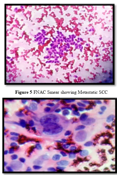
Figure 5 FNAC Smear showing Metastatic SCC
Figure 6 FNAC Smear of Hodgkin Lymphoma Showing RS Cell
Documented study observed only 40 cases (7.017%) of lymphoproliferative disorders. 20 cases (50%) were of Non Hodgkin Lymphoma (NHL) while Hodgkin’s disease was seen cases (20%).7 patients (17.5%) presented with Leukemic infiltration of lymph nodes (3 cases of ALL and 4 cases of SLL/CLL) and 5 cases (12.5%) showed atypical lymphoid hyperplasia. Lymphoproliferative disorders were 40 year age group ( 15 cases,37.5%) followed by 60 year age group (11 cases, 27.5%), 0-20 age group ( 8 80 years age group (6 cases, 15%). Lymphoproliferative disorders affected male patients (29 cases.72.5%) more than the females (11 cases,27.5%).
Lymphadenopathy is defined as the enlargement of lymph nodes within contiguous anatomic regions. Viral, bacterial or mycobacterial infections are the most common causes of benign regional lymphadenopathy. Fine needle aspiration (FNAC) is inexpensive, completely safe and quick method for diagnosis of lymphadenopathy and it has reduced the need for surgical biopsy. In the documented study 570 cases of enopathy were subjected to FNAC. Our aim was to help the clinician in arriving at an early diagnosis in cases presenting with lymphadenopathy. Various patterns of lymphadenopathy were observed in our study namely Tuberculous lymphadenitis, Reactive lymphadenitis, Suppurative lymphadenitis, Metastatic lymphadenitis and Lymphoproliferative disorders including Hodgkin’s disease, Non Hodgkin Lymphoma (NHL) and Atypical lymphoid hyperplasia. These patterns seen in our study were more or less same as reported in other studies in India and other developing
Most common cause of lymphadenopathy in the present study was found to be Tuberculosis (249 cases, 43.68%). In India,tuberculous lymphadenitis is one of the most common
FNAC Smear showing Metastatic SCC
International Journal Of Current Medical And Pharmaceutical Research, Vol. 4, Issue, 1(B), pp.2923-2927, January, 2018
type of lymphadenopathy encountered in clinical practice in contrast to very low frequency of 1.6% in developed countries.13 The highest incidence of tuberculous lymphadenitis was seen in 3rd and 4th decade of life i.e. in 21-40 year age group (129 cases, 51.81%). Our findings were almost similar to the studies done by Nirmala Chandran
11
,Nirmal Amit14 and Sharadamani 15 which showed incidence of tuberculosis as 45.5%,44.6% and 45% respectively while at variance to those of Kochhar3 (38.52%),Tandon16 (32%) and Khajuria12 (52.3%). Tariq Ahmed et al17 in 2008 in his study observed tuberculous lymphadenitis as the most common pathology of the cervical lymph node lesions. Studies of all these authors including the documented study suggest that Tuberculous lymphadenitis is the most common cause of lymphadenopathy. This is attributed to the low socio-economic status, poverty, overcrowding, malnutrition, incomplete treatment, resistance and increased incidence of HIV infected patients.18
Recently, according to WHO report 2017, an estimated 10.4 million new cases of tuberculosis have been detected worldwide, 10% of which were infected with HIV. An estimated 1.7 million people died from tuberculosis. Global efforts to combat tuberculosis have saved an estimated 53 million lives since 2000 and reduced the mortality rate by 37%. Despite these achievements, tuberculosis remains the top infectious killer.19
The second most common cause of lymphadenopathy in the documented study was Reactive lymphadenitis (28.77%). This was found to be common in the younger age group of <40yrs (144 cases, 87.80%). Since infections from the oral cavity, ears, nose and paranasal sinuses drain into the cervical lymph nodes; reactive lymphoid hyperplasia is a common finding.20 Similar findings were observed by Kochhar3 (24.45%) and Shreshta4 (23%) while Tandon16(45.5%) and Chandralekha21 (47.5%) observed high incidence of Reactive lymphoid hyperplasia than tuberculosis.
Metastatic lymphadenopathy was seen predominantly above 40 yrs of age (83 cases,87.37%) and was observed to be more common in males (67 cases,70.52%) . The superficial cervical lymph nodes are common sites of metastasis. FNAC has a documented higher sensitivity in diagnostic workup of metastatic malignancies which may be due to the fact that the metastatic carcinoma cells are usually abundant and their cytological features are dissimilar to that of the cells of normal or hyperplastic lymph nodes22. Present study documented 16.67% cases with metastatic lymphadenopathy. Similar findings were observed by Chandralekha21 (13%) and Hirachand23 (12.3%). Ruchi Khajuria12 observed only 3.8% cases of metastatic lymphadenopathy and this variation in results is due to difference in age groups of patients, as most of the metastatic lesions are common above 40 yrs of age. Metastatic Squamous cell carcinoma (SCC) was found to be in majority of cases (61, 64.21%) followed by Adenocarcinoma (14 cases, 14.73%). Similar high incidence of metastatic SCC was observed by Nirmal Amit14 (76.67%), Hirachand23 and Shilpa et al.24
The anatomical site of involved lymph node along with age , sex and cytomorphological patterns often give clue to the site of primary tumor. For example, upper cervical lymph nodes commonly harbours the metastatic deposits from aerodigestive tract and midcervical harbours metastasis from thyroid.3 Intranuclear cytoplasmic inclusions suggest metastasis from
papillary carcinoma of thyroid as observed in the documented study (2 cases,2.1%). FNAC is a useful prognostic tool in stage 3 cancers wherein metastasis to regional lymph nodes is usually found. It also aids in the diagnostic workup of a metastatic tumor of unknown origin14 as observed in the documented study (11 cases, 11.58%).
CONCLUSION
Tuberculosis stands out to be the most common cause of cervical lymphadenopathy in India and the top infectious killer. Present study also concludes Tuberculosis as the most common cause of Lymphadenopathy followed by Reactive lymphadenitis, metastatic lymphadenitiss (particularly squamous cell carcinoma), lymphoproliferative disorders and Suppurative Lymphadenitis. Documented study highlights the usefulness of FNAC in diagnosing the various patterns of lymphadenopathy thus helping the clinician in arriving at an early diagnosis and timely management of curable causes of lymphadenopathy like infections and tuberculosis. FNAC combined with clinical correlation can be used as a first line investigation in work up of lymph node lesions. It helps in early diagnosis and saving the patients from high mortality and morbidity.
References
1. Qasmi SA ,Kiani F,Malik AI,Salamatullah J,Farooq MO,Abassi MA. Cervical Lymphadenopathy: A Common Diagnostic Dilemma. Journal of International Surgery Pakistan. 2012 Apr-Jun;17(2):76-80
2. Richner S,Laifer G. Peripheral lymphadenopathy in immunocompetent Adults. Swiss Med Weekly.2010 Feb 20 ;140 (7-8):98-104.
3. Kochhar AK,Puri PL, Kochhar S. Role of fine needle aspiration cytology in assessment of cervical lymphadenopathy. International Journal of Medical Research and Review.2016 Jun ;4(6):876-880.
4. Shrestha AK, Chalise PR, Shrestha ML. Lymph node biopsies : a hospital based retrospective study. Journal of Nepal Medical Association. 2009 Oct – Dec ; 48 (176):306-9.
5. Narang P,Narang R,Mendiratta DK, Sharma SM,Tyagi NK. Prevalence of tuberculous lymphadenitis in children in Wardha district, Maharashtra state, India. International journal of Tuberculosis and Lung Disease. 2005;9:188-94.
6. Mohan A, Reddy MK, Phaneendra BV, Chandra A. Aetiology of peripheral lymphadenopathy in adults : Analysis of 1724 cases seen at a tertiary care teaching hospital in southern India. National Medical Journal of India. 2007 Mar- Apr; 20(2):78-80.
7. Bazemore AW, Smucker DR. Lymphadenopathy and malignancy. American Family Physician 2002 Dec1;66 (11):2103-10.
8. Rakhshan M, Rakhshan A. The diagnostic accuracy of fine needle aspiration cytology in neck lymphoid masses. Iranian journal of pathology. 2009 Sep 1;4 (4):147-50.
10. Ripunjaya M, Anne W. Utility of fine needle aspiration cytology of lymph nodes. IOSR Journal of Dental and Medical Sciences 2013 Jul-Aug;8 (5):13-18.
11. Chandran N,Dayananda SB,Radha. Causes of cervical lymphadenopathy- A Cytological Study. Journal of Evolution of Medical and Dental Sciences. 2014 Jan; 3(2):379-85.
12. Khajuria R, Goswami KC, Singh K, Dubey VK. Pattern of lymphadenopathy on fine needle aspiration cytology in Jammu. JK Science. 2006;8 (3):157-9.
13. Paul PC, Goswami BK, Chakrabarty S, Giri A,Pramanik R. Fine needle aspiration cytology of lymph nodes-an institutional study of 1448 cases over a five year period. Journal cytology, 2004; 21:187-90. 14. Nirmal Amit K, Kirti N, Jha J, Sudhir K. Role of fine
needle aspiration cytology in assessment of cervical lymphadenopathy in variable age groups: A retrospective study. Health Sciences, 2016;5 (11):306-10.
15. Sharadamani GS. Prevalence of various causes of lymphadenopathy in a rural setting in India. Indian journal of pathology and oncology. 2017 July-Sept;4(3):396-464.
16. Tandon P, Gautam W. Utility of FNAC in lymphadenopathy- A study of 638 cases in primary care setting. National journal of Laboratory Medicine. 2016 July;5(3): 11-15
17. Ahmed T, Naeem M, Ahmed S,Samad A. Fine needle aspiration cytology and neck swellings in surgical outpatient, Journal of Ayub Medical College Abottabad. 2008; 20(3):30-2.
18. Upadhyay GP, Thakker RM. Evaluation of fine needle aspiration cytology as the initial diagnostic test in cases of cervical lymphadenopathy. International journal of Research in Medical Sciences. 2016; 12:5103-7. 19. World Health Organisation: Global TB Report Oct
2017. WHO Report.
20. Baji SN, Anand V, Sharma R, Deore KS, Chokshi M. Analysis of FNAC of cervical lymph nodes: Experience over a two years period. International Journal of Medical Science and Public Health. 2014; 3 (5):607-9. 21. Janagam C, Atla BL. Role of FNAC in the diagnosis of cervical lymphadenopathy. International journal of Research and Medical Sciences.2017 Dec;5(12):5237-41.
22. Volmar KE, Singh HK, Gong JZ. The advantages and limitations of the role of core needle and fine needle aspiration biopsy of lymph nodes in the modern era. Hodgkin and Non- Hodgkin lymphomas and metastatic disease. Pathology case Reviews 2007;12(1):10-26. 23. Hirachand S, Lakhey M, Akhter J,Thapa B. Evaluation
of fine needle aspiration cytology of lymph nodes in Kathmandu Medical College teaching Hospital. Kathmandu University Medical Journal 2009;7(2):139-42.
24. Shilpa G, Nataraju G. Pattern of lymph node Diseases in a Tertiary Level Referral center: a cytological study of 943 cases. International journal of Biological and Medical Research. 2013;4(3):3448-52.
How to cite this article:
Harkirat et al (2018) 'Pattern of Lymphadenopathy on Fnac in Tertiary Care Hospital - A Cross Sectional Study', International Journal of Current Medical and Pharmaceutical Research, 4(1), pp. 2923-2927.