Original Research Article.
Anatomical Variations, Embryogenesis and Clinical Significance of
Profunda Femoris Artery: A Cadaveric Study
Aswinprakash Subramanian
1, Kosuri Kalyan Chakravarthi
2*, Jagadeesh Dhamodharan
3, R. Arulmoli
41Lecturer, Faculty of Medicine, Anatomy Unit,
Asian Institute of Medicine, Science and Technology (AIMST University), Bedong, Kedah, Malaysia. [Ex PG, Department of Anatomy, Chettinad Hospital and Research Institute, Tamil Nadu, India.] 2*Associate Professor Department of Anatomy,
Varun Arjun Medical College, Banthra, Shajahanpur, Uttar Pradesh, India. 3Senior Lecturer, Faculty of Medicine, Anatomy Unit,
Asian Institute of Medicine, Science and Technology (AIMST University), Bedong, Kedah, Malaysia. 4Professor, Faculty of Medicine, Anatomy Unit,
Asian Institute of Medicine, Science and Technology (AIMST University), Bedong, Kedah, Malaysia. ABSTRACT
Background: Accurate knowledge of variation pattern of origin and course of lower limb vessels particularly in the femoral region of thigh have received attention for anatomists and surgeons due to great clinical importance during angiographic imaging procedures as well as during surgeries that are performed in the femoral triangle.
Aim: Accordingly the study was designed to study the anatomical variations, embryogenesis and clinical significance of Profunda femoris artery in human Cadavers.
Methods: The study was conducted in the department of Anatomy, Chettinad Hospital and Research Institute. A total of 36 formalin fixed specimens irrespective of age and sex were utilized for the present study. The course of the profunda femoris artery was traced from its origin i.e. the femoral artery till its continuation as fourth perforator. The distance between the site of origin of profunda femoris artery and midpoint of the inguinal ligament was measured in millimetres by means of non-stretchable thread and scale. The mode of origin and length of origin of the lateral and medial circumflex femoral artery from the profunda femoris arteries was also studied.
Results: Variations in the branching pattern of profunda femoris artery was observed in 10 specimens (27.77%). The incidence percentage of origin of profunda femoris artery from posterolateral was 41.67% and from lateral, 41.67% were equally distributed. In 16.6% of cases the profunda femoris originated from posterior aspect. The distance of origin of profunda femoris from the midpoint of inguinal ligament on right side was commonly placed between 41 and 50mm, which are slightly higher than the average range; whereas on left side, it was of normal average between 31 and 40 mm. The distance
of lateral circumflex femoral artery from the profunda femoris artery was between 01-10mm on right side, and between 11-20 mm on the left side. The distance of origin of the medial circumflex femoral artery was between 01-40mm on both the sides, most of the times it originated between 11-20 mm on the left side.
Conclusions: The knowledge of anatomical variations of femoral artery and their branches in femoral triangle presented in this study importance during anterolateral thigh flap, aortopopliteal bypass, coronary artery bypass grafting, extracranial-intracranial bypass surgery. It also helps radiologists, orthopaedic, plastic and reconstructive surgeons, general and vascular surgeons to avoid unnecessary complication during surgical and therapeutic intervention.
Keywords: Arteriography, Cannulation, Profunda Femoris, Rete Femorale.
*Correspondence to:
Dr. Kosuri Kalyan Chakravarthi,
Associate Professor, Department of Anatomy, Varun Arjun Medical College,
Banthra, Shajahanpur, Uttar Pradesh, India.
Article History:
Received: 19-03-2018, Revised: 12-04-2018, Accepted: 15-05-2018 Access this article online
Website:
www.ijmrp.com
Quick Response code
DOI:
10.21276/ijmrp.2018.4.3.004
INTRODUCTION
The femoral region of thigh is used for various clinical procedures, particularly in respect to arterial cannulation, so the course and ramification of the vessels of the lower limbs have received attention for anatomists and surgeons. The profunda femoris
iliac arteries above and with the popliteal artery below and therefore maintain efficient collateral circulation for the lower limbs. The profunda femoris artery arises from the lateral aspect of femoral artery at about 3.5cm below the inguinal ligament and leaves the femoral triangle between pectineus and adductor longus; it descends between adductor longus and adductor brevis, then between adductor longus and adductor magnus finally it pierces the adductor magnus as fourth perforating artery and anastomoses with superior muscular branches of popliteal artery. In the femoral triangle, the profunda femoris artery gives off lateral and medial circumflex femoral arteries, during the rest of the course it provides muscular and three perforating branches. Accurate knowledge of anatomical variations regarding profunda femoris, medial and lateral circumflex femoral arteries are important for clinicians in the present era of various clinical procedures like arterial catheterization, arteriography, and femoral embolectomy, fracture reduction of acetabulum and head of femur. Accordingly the study was designed to study the
anatomical variations, embryogenesis and clinical significance of Profunda femoris artery in human cadavers.
MATERIALS AND METHODS
The study was conducted in the department of Anatomy, Chettinad Hospital and Research Institute, Tamil Nadu, India. A total of 36 formalin fixed human cadaveric specimens irrespective of age and sex were utilized for the present study. The femoral triangle was dissected according to the Cunningham’s manual.
After exposing the femoral artery and its major branches the origin of profunda femoris artery with its relation to femoral artery was studied. Photographs of variations of the profunda femoris arteries were taken for proper documentation. The distance between the site of origin of profunda femoris artery and midpoint of the inguinal ligament was measured in millimetres by means of non-stretchable thread and scale. The mode of origin and length of origin of the lateral and medial circumflex femoral artery from the profunda femoris arteries was also studied.
Figure 1: Unusual complete absence of medial circumflex artery and the descending branch originated in common with the profunda femoris artery.
Figure 2: Medial circumflex femoral artery directly taking origin from femoral artery.
Figure 3: Ascending branch of lateral circumflex femoral artery taking origin from femoral artery.
Figure 4: Lateral circumflex femoral artery originating from the femoral artery.
The medial circumflex femoral artery and profunda femoris artery arising as common stem from the femoral artery.
Table 1: Relative position of origin Vs distance of origin of profunda femoris artery from the femoral artery
Range (mm) Relative position of origin
Posterior Posterolateral Lateral
21-30 0 0 3
31-40 2 5 4
41-50 4 6 5
51-60 0 3 0
61-70 0 1 3
RESULTS
I. The percentage of incidence of origin of profunda femoris artery from posterolateral was 41.67% and from lateral, 41.67% were equally distributed. In 16.6% of cases the profunda femoris originated from posterior aspect (Table-1).
II. Variations in the branching pattern of profunda femoris artery were observed in 10 specimens (27.77%).
Variant 1: An unusual complete absence of medial circumflex artery and the descending branch originated in common with the
profunda femoris artery was observed in 1 specimen (Figure-1).
Variant 2: The medial circumflex femoral artery directly taking origin from femoral artery was noted in 2 specimens (Figure-2).
Variant 3: The lateral circumflex femoral artery and profunda femoris artery arising as common stem from the femoral artery was noted in 1 specimen (Figure-2).
Variant 5: The lateral circumflex femoral artery originating from the femoral artery was observed in 3 specimens (Figure-4).
Variant 6: The medial circumflex femoral artery and profunda femoris artery arising as common stem from the femoral artery was noted in 2 specimens (Figure-4).
III. The distance of origin of profunda femoris from the midpoint of inguinal ligament on right side was commonly placed between 41 and 50mm, which are slightly higher than the average range;
whereas on left side, it was of normal average between 31 and 40 mm. (Table-2).
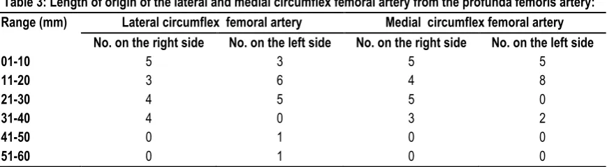
IV. The distance of lateral circumflex femoral artery from the profunda femoris artery was between 01-10mm on right side, and between 11-20 mm on the left side. The distance of origin of the medial circumflex femoral artery was between 01-40mm on both the sides, most of the times it originated between 11-20 mm on the left side (Table-3).
Table 2: Length (in mm) of origin of profunda femoris artery from the midpoint of inguinal ligament:
Range (mm) No. on right side No. on left side
21-30 1 2
31-40 4 7
41-50 9 6
51-60 2 1
61-70 2 2
Table 3: Length of origin of the lateral and medial circumflex femoral artery from the profunda femoris artery:
Range (mm) Lateral circumflex femoral artery Medial circumflex femoral artery
No. on the right side No. on the left side No. on the right side No. on the left side
01-10 5 3 5 5
11-20 3 6 4 8
21-30 4 5 5 0
31-40 4 0 3 2
41-50 0 1 0 0
51-60 0 1 0 0
DISCUSSION
The knowledge of the site of origin of profunda femoris artery is important while performing clinical procedures in the femoral region and hip joint replacement and also for avoiding iatrogenic arteriovenous fistula or severe secondary haemorrhage while performing femoral artery puncture.
Profunda Femoris Artery
Profunda femoris artery is useful for the ultrasound, arteriography, Doppler imaging, angiography and also magnetic resonance imaging.1 The most common site of origin of profunda femoris
artery mentioned in the text books are from the lateral aspect of femoral artery.2 LippertandPabst reported 48% originof profunda
femoris from posterolateral aspect of femoral artery.3Daksha Dixit
et al reported the origin of profunda femoris artery arising from posterior aspect of femoral artery in 39% of cases.4 M. B.
Samarawickrama et al reported the most frequent pattern of origin from posterior aspect in 46% of cases.5 In the present study, the
incidence of origin of profunda femoris artery from posterolateral was (15 specimens) 41.67% and from lateral, (15 specimens) 41.67% were equally distributed (Table-1). In (6 specimens) 16.6%, of cases the profunda femoris originated from posterior aspect (Table-1). Such knowledge of anatomical variations of side of origin of profunda femoris artery noted in our study may help the clinicians to avoid complications during femoral arterio-venous fistula while performing femoral artery puncture for surgical exposure of the common femoral and profunda femoris junction. During cannulation of femoral artery the puncture site is usually profunda femoris artery or femoral artery distal to the origin of profunda femoris artery, hence it’s necessary to know the level of
origin of profunda femoris artery. 6 The normal distance of origin of
the profunda femoris artery from midpoint of inguinal ligament is 35mm.7 Bannister et al and Snell found the average range of
origin from the mid-inguinal point to be 47.5mm.8,9 In the present
study the distance of origin of profunda femoris from the midpoint of inguinal ligament on right side was commonly placed between 41 and 50mm, which are slightly higher than the average range (Table 2); Whereas on left side, it was of normal average between 31 and 40 mm (Table 2). Marina Baptist et al 10 observed very
distal origin of profunda femoris artery to be 60-70mm, which is coinciding with the present study (Table 2). Nachicet Shankar et al observed the very proximal origin of profunda femoris artery to be less than 10mm distal to the inguinal ligament.11 This can be
compared with the present study with the proximal origin in the range of 21-30mm (Table 2). Such knowledge of variant anatomical data of proximal or distal origin of profunda femoris artery noted in this study useful in revascularization procedures done for non-healing ulcers and/or gangrene, to relieve the claudication pain and in preventing the necrosis of flap, when used in plastic and reconstructive surgery. 12, 13
Lateral Circumflex Femoral Artery
Uzel. M.et alreported the incidence of origin of lateral circumflex femoral artery from profunda femoral artery in 77.3% cases and from femoral artery inclusive of origin in common with femoral artery in 22.7%cases.14 In the studies conducted by Uzel et al14
from femoral artery including the common stem.15 Tanvaa Tansatit
et al found that lateral circumflex femoral artery was originating from the profunda femoral artery in 56.7% of cases and from the femoral artery in 43.3% of cases.16 Suthar K, Patil D et al,
dissected 50 lower limbs and found that the lateral circumflex femoral artery was arising from the profunda femoral artery in 80% of cases and from the femoral artery in 20% of cases.17 where as
in the present study, the most common origin of lateral circumflex femoral artery was arising from the profunda femoris artery in 86.26% of cases.
Lateral circumflex femoral artery is used for reconstruction of large defects in the face, anterolateral thigh flap and a perforator flap in the reconstruction of gunshot wounds. Its branches are used for aortopopliteal bypass, coronary artery bypass surgery.18 The
knowledge of the variations among the lateral circumflex artery and their branches is of importance when clinical procedures are undertaken in the femoral region and in hip replacement. Rui Fernandas and Jason Lee have reported the successful use of lateral circumflex femoral artery perforator flap as a reliable option for the immediate reconstruction of large defects secondary to gunshot wounds of face.19 It is the reliable source for small to
large soft tissue losses for reconstructing avulsed soft tissue losses in the head and neck. Yasuhiko Sugawara et al have reported the successful utilization of the lateral circumflex femoral artery as a midway outflow for aorto-lateral-circumflex-femoropopliteal artery sequential bypass on a patient with total occlusion of all major arteries in unilateral Iliofemoral region.20
Medial Circumflex Femoral Artery
Ravindranath et al 21 recorded a case of total absence of medial
circumflex femoral artery which is seldom reported. The absence of medial circumflex femoral artery rare variation observed in the present study draws the attention and raises the question regarding the preservation of arterial supply to the head of femur during surgical procedures of the thigh and hip joint. Knowledge regarding this type of arterial variations noted in this study may prevent iatrogenic injuries in taking pedicle flaps from the branches of these arteries. From the present study, the incidence of medial circumflex femoral artery (86.18%) arising from the profunda femoris artery can be compared with Lippshutz BB 22
(56%), 63% reported by Siddharth et al 23, 53% reported by Clarke
SM and Colborn GL 24 and 62.2% reported by Prakash et al.25 In
the study conducted by Marina Baptist et al,26 the average
distance between the origin of medial circumflex femoral artery from the origin of profunda femoris artery was between 11-20mm, this is in coincidence with the present study. 19.2% of medial circumflex femoral artery branching from femoral artery and also arising in common with profunda femoris recorded in the present study can be compared with Gautier et al of about 16.4%.27
Emmanuel Gautier, et al 27the medial circumflex femoral artery,
which exhibits different pattern, is an important artery in supplying blood to head and neck of the femur, adductor muscles and fatty tissue in acetabular fossa. Due to close association of the artery with the above mentioned structures, there is a high risk of the artery being severed during trauma or surgical procedures such as total hip arthroplasty. It has also a greater importance in plastic surgery operations with the vascular pedicle of grafts such as transverse upper gracilis flap and the medial circumflex perforator free flap. It is used in selective arteriography in idiopathic ischaemic necrosis.
EMBRYOLOGICAL BASIS FOR VARIATIONS
Variations in the vessels of lower limb are most often due to the artery developing from rete femorale in the ventral aspect of the thigh. It communicates with the external iliac artery above through rete pelvicum and sciatic artery. The primary sciatic artery grows out from the fifth lumbar intersegmental artery in the dorsal part of the thigh, when embryo is 10mm long and ends in plantar capillary plexus. As the development progresses anastomosis between the axis artery and rete femorale develops. It is generally accepted that increase in the blood flow in these capillaries determines the final mature arterial pattern. Therefore we can speculate that one possible reason for the observed variations could be increased blood flow in the rete femorale vessels. Thus, most of the appropriate channels enlarge while others contract and disappear, thereby establishing final arterial ramification.28
The anomalous pattern of lateral and medial circumflex femoral arteries may be due to
➢ Divergence in the mode and proximo-distal level of branching.
➢ Presence of unusual compound arterial segments.
➢ Aberrant vessels that connect with principal vessels, arcades or Plexuses.
➢ Unsuspected neural, mycological or osteo-ligamentous relationships.
CONCLUSION
We believe that the sound knowledge of anatomical variations of site of origin, course and branches of the profunda femoris artery presented in this study may helpful for various clinical procedures like arterial catheterization, arteriography, femoral embolectomy, fracture reduction of acetabulum and head of femur and helps in reducing the chances of intra-operative secondary haemorrhage and post-operative complications.
REFERENCES
1. M Uzel, E Tanyeli, M Yildirim. An anatomical study of the origins of the Lateral circumflex femoral artery in the Turkish population. Folia Morphol. 2008; 67: 4.
2. Standring S. (2004) Grays Anatomy, 40th ed., Philadelphia, Elsevier Churchill Livingstone, 1379-1380.
3. Lippert H, Pabst R. Arterial variation in man, classification and frequency. J F Bergmann-Verlag N Munche 1985; 416-20. 4. Daksha Dixit, Dharati M. Kubavat et al. A Study of Variations in The Origin Of Profunda Femoris Artery And Its Circumflex Branches. Int J Biol Med Res. 2011; 2(4): 1084 - 1089.
5. M B Samarawickrama, B G Nanayakkara. Branching pattern of the femoral artery at femoral triangle: a cadaveric study. Galle Medical Journal 2009; 14: 1.
6. Chleborad WP, Dawson DL. The profunda femoris artery: variation in clinical applications. Clin Anat. 1990; 3: 33–40.
7. Gray’s Anatomy. The Anatomical Basis of Clinical Practice.
Fortieth Edition. Elsevier Churchill Livingstone. 2008:1452. 8. Bannister L H, Berry M M, Collins P. Gray's Anatomy: Cardiovascular system, 38th Edition, Churchill Livingstone, Medical Division of Longman Group, UK Ltd. 1995: 1566-1568. 9. Snell R S. Clinical Anatomy of Medical Students. 4th Edn. Little Brown and Co. Boston. 1992: 607.
femoral artery; Professional Med J, 2007; 14 (3): 523-527. 11. Nachikat Shankar, Roopa R, Unusual bilateral origin of the Deep artery of thigh and associated variations. Int. Journal of Anat Variations 2009; 2: 99-101.
12. VuksanovicBozaric A, Stefanivic N, Pavlovic S, Duraskosvic R, Randelovic J. Analysis of deep femoral artery origin variances on fetal material. Facta Universities Medicine and Biology, 2007; 14 (3): 112-116.
13. Natale A, Belcastro M, Palleschi A, Baldi I. The mid-distal deep femoral artery: few important centimeters in vascular surgery. Ann Vasc Surg. 2007; 21:111-6.
14. Uzel M, Tanyeli E, Yildirim M. Anatomical study of the origin of lateral circumflex femoral artery in Turkish population. Folia Morphol (Warsz) 2008; 67(4): 226-230.
15. Fukuda H, Ashida M, Ishii R, Abe S, Ibukuro K. Anatomical variants of the lateral
femoral circumflex artery: an angiographic study. Surg Radiol Anat 2005; 27(3): 260-264.
16. Tansatit Tanvaa, Wanidchapholoi S, Sanguansit Pasinee.
“The anatomy of the lateral circumflex femoral artery in
anterolateral thigh flap.” J Med Assoc Thai. 2008; 91(9): 1404-09.
17. Suthar K, Patil D, Mehta C, Patel V et al. Cadaveric study: morphological study of branches of femoral artery in front of thigh. CIBTech Journal of Surgery. 2013; 2(2): 16-22.
18. Shridevi NS, Shiny Vinila BH , Kumarswamy R, Joanna Han YT. A study on origin of lateral circumflex femoral artery an original article. Anatomica Karnataka. 2012; 6(3):35-37.
19. Rui Fernandes, Jason Lee. The use of lateral circumflex femoral artery perforator flap in the reconstruction of gunshot wounds of the face. Journal oral and maxillofacial surgery, 2007; 11: 7.
20. Yasuhiko Sugawara et al Utilization of Lateral circumflex artery as Midway Outflow for Aortopopliteal grafting: Report of a case Surgery Today, 1998, 28 (9):967-970.
21.Ravindranath G, N Jayasree, N Ratnakar Rao, Ranadheer Reddy et.al., Medial circumflex femoral artery – A Rare Anamoly, Case Study. Journal of Anatomical Society of India, 2006; 55(1).
22.Lipchutz B B. Studies on blood vascular tree. A composite study of the femoral artery. Anat Rec, 1916; 10: 361-370. 23.Siddharth P, Smith N L, Mason R A, Giron F. Variational anatomy of the deep femoral artery. Anat Rec, 1985; 212 (2): 206–209.
24. Clark, S.M., and Colborn, G.L. (1993): The medial femoral artery: its clinical anatomy and nomenclature. Clinical Anatomy. 6: 94-105.
25. Prakash, Kumari J, Kumar bhardwaj A et.al., Variations in the origins of the profunda femoris, medial and lateral femoral circumflex arteries: a cadaver study in the Indian population. Rom J Morphol Embryol, 2010; 51 (1): 167-70.
26. Baptist M, Sultana F, Hussain T. Anatomical Variations; The origin of profunda femoris artery, its branches and diameter of femoral artery. Professional Med J. 2007; 14(3): 523-527. 27.Gautier E, Ganz K, Krügel N, Gill T, Ganz R, Anatomy of the medial femoral circumflex artery and its surgical implications. J Bone Joint Surg Br 2000; 82(5):679–683.
28. Bunyamin Sahin, Ahmet Uzun, Mehmet Emirzeoglu, Rengin Kosif, Sait Bilgic. A deep femoral artery passing in front of the femoral vein. Folia morphologica 2003; 62(2):143-6.
[
Source of Support: Nil. Conflict of Interest: None Declared.
Copyright: © the author(s) and publisher. IJMRP is an official publication of Ibn Sina Academy of Medieval Medicine & Sciences, registered in 2001 under Indian Trusts Act, 1882. This is an open access article distributed under the terms of the Creative Commons Attribution Non-commercial License, which permits unrestricted non-commercial use, distribution, and reproduction in any medium, provided the original work is properly cited.