Effect of Temperature towards Rice Husk Silica
Characterization with Different Preparation
Methods
Rosiah Osman
1, Nor Hapishah Abdullah
1, Khamirul Amin Matori
2, Mohd. Nizar Hamidon
1, Ismayadi Ismail
1,
Syazwan Mustaffa
11Institute of Advanced Technology (ITMA), University Putra Malaysia, Serdang, Selangor,Malaysia
2Dept.of Physics, Faculty of Science, University Putra Malaysia, Serdang, Selangor,Malaysia
[email protected], [email protected] , [email protected], [email protected], [email protected],
Abstract-- Rice Husk Ash (RHA) ceramic presented in this paper undergoes different preparation methods. This study reported a comparison in terms of product quality, yield, structure and processing time between acid leaching techniques (HCl) to the other non-treated acid leaching. In this research, the White Rice Husk Ash (WRHA) pellets were prepared by cleaning the Rice Husk (RH) with distilled water and HCl acid respectively. The raw material, RH was going through thermal treatment in order to produce white ash powder then milled into nano-sized powder via high-energy ball milling (HEBM). To subject the samples to a series of temperatures, the pressed pellets went through multi-sample sintering, where the multi-samples were sintered at 800ºC, 1000ºC, 1100ºC and 1200ºC for each batch. The percentage of density and porosity respectively increases and decreases with the increasing of sintering temperature. During the sintering process, the grain size increased, which caused less porosity and giving higher density in the sample. The comparative XRD plot of the sintered samples at various temperatures prepared by different cleaning route showed the difference between the amorphous and crystalline silica. The XRD spectra reveal that the main phase for crystallization was cristobalite and tridynamite. The FESEM images displayed the comparative morphological features of the samples. The grain sizes were observed to be smaller by acid leach treatment. Acid leaching of rice husks prior to combustion resulted in smaller particle size, larger surface area in comparison with ash from non-acid treated husks (NTR).
Index Term--Rice husk silica, cristobalite, acid-leach treatment, high energy ball milling.
I. INTRODUCTION
Rice Husk (RH) is an abundant waste material in all rice producing countries including Malaysia. Although it has been used as a composite in alloy and ceramic industry but still millions of tons were wasted worldwide. RH can be considered as suitable energy and silica resource in Asian countries. By heating at higher temperatures, the unburned carbon can be removed from the ashes [1] which lead to the crystallization of the ash from amorphous silica into cristobalite or tridymite. At lower temperature, the amorphous nature of rice husk ash silica will be formed [2]. The most ordinary form of crystalline silica found is quartz [3]. However, more research has been focused on the formation of cristobalite and tridymite [4]. The transformation has been reported that α-quartz can be form at below 573°C, ß-quartz at
573-870°C, ß-tridymite at 870-1470°C, and ß-cristobalite at 1470-1710°C [5]. Many methods have been developed to produce pure silica from rice husk ash (RHA) in low cost. It has been reported that purity of silica is highly affected from chemical treatment [6]-[9], than thermal treatment [10]. This process not only producing valuable silica powder but also has benefited reducing pollution problem by converting agriculture waste into useful product. The increasing interest to RH requires complete investigation on the effect of purification and heat treatment to their phase and microstructure.
There have been several reports on the formation of silica from rice husk [11–13]. Real et al. [14] found that a homogeneous size distribution of nanometric silica particles could be obtained by burning rice husk at 873–1073K in a pure oxygen atmosphere. Della et al. [15] observed that active silica with a high specific area could be produced from rice husk ash after heat-treating at 973K in air. Kalapathy et al.
[16] used an improved method to produce silica with lower
sodium content by adding silicate solution to pH 1.5 hydrochloric, citric, or oxalic acid solutions until pH 4.0 was reached. Kamath and Proctor [17] reported that the rice husk could be dissolved in sodium hydroxide solution, and then titrated with acid to obtain silica gel. These previous studies focused on the process of manufacturing silica from rice husk. However, the mechanism of thermal decomposition as well as the surface characteristic of product under nonisothermal condition has received much less attention.
In this paper, characterization of silica prepared by heating the rice husk in air is investigated. In addition, the rice husk is treated with different preparation methods, i) distilled water and ii) acid leaching technique to remove impurities from the samples. The presence of crystalline silica in RHA ceramics sintered at temperature range 800-1200 °C was examined by X-ray Diffraction (XRD). The microstructure of this crystalline silica is also observed by using scanning electron microscope.
II. METHODOLOGY
stage was consisted of washing RH in distilled water in order to remove clay and rock impurities and subsequently dried in an oven at 100°C for 24 h to remove water content. For acid leach treatment, HCl was used for acidic treatment with laboratory grade (Merck & Co.). The dried rice husk was refluxed with an acidic solution in a glass round-bottomed flask for 1 h. After the acidic solution was drained, the husk was washed repeatedly with distilled water until the filtrate was free from acid.
The acid-leached rice husk (ALRH) was then dried at room temperature for 24 h. The dried RH then was taken into crucible and placed it in electrical furnace for 6 h at 900°C to get a white ash. The ash was dry milled into nano-sized powder via high-energy ball milling (HEBM) in a hardened steel vial for 20 minutes using a SPEX8000D mill.The pellets with 13 mm diameter were formed and sintered at various temperatures at 800°C, 1000°C, 1100ºC and 1200°C for 6 h with heating rate and cooling rate of 4°C.
The crystal phases of the sintered RHA pellets were identified by XRD analysis. X-ray Diffractometer (PANAalytical (Philips) X’Pert Pro PW3050/60) with CuKα radiation (Bragg angle 2θ in angular range of 20 to 80°) equipped with a copper x-ray tube and scintillation detector was used. The microstructures and composition were analyzed by JEOL JSM 6400 Scanning Electron Microscope (SEM) with Oxford Inca Energy 200 EDX. The density of the samples was obtained by using the Archimedes principle.
III.RESULTSANDDISCUSSION
A. Particle Size-Surface Area
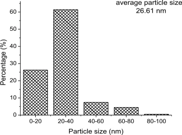
Fig. 1(a) and 2(a) show the micrograph of the RH and ALRH powder sample milled for 20 min which have been measured by using scanning transmission electron microscopy (STEM) for particle size confirmation. The particle size was measured by taking diameters of 200 particles, and it was found to have an average size around 68.59 nm and 26.61 nm for RH and ALRH respectively. The agglomerations of the particles, as can be observed in Fig. 2(a) occurred due to large surface area and subjected to repeated cold welding and fracturing of the HEBM process. The particle obtained from acid leach treatment has given smaller particle size compared to the common rice husk preparation.
0-40 40-80 80-120 120-160 160-200 0
5 10 15 20 25 30 35
40 average particle size: 68.59 nm
Particle size (nm)
P
e
rc
e
n
tag
e
(
%)
Fig. 1. (a) FeSEM image and (b) particle size distribution for RH
a)
b)
a)
0-20 20-40 40-60 60-80 80-100 0
10 20 30 40 50 60
average particle size: 26.61 nm
P
er
ce
ntag
e (
%)
Particle size (nm)
Fig. 2. (a) FeSEM image and (b) particle size distribution of ALRH
Fig. 3(a) shows the XRD pattern of RHA ceramics sintered at 800°C, 1000°C, 1100°C and 1200°C for 6 h revealed that the presence of crystalline phases. Cristobalite and tridymite were observed after sintering. A weak peak of tridymite at 20.7° accompanied with a strong peak of cristobalite at 21.8° could be seen in the figure. It was confirmed that the reaction sintering process can effectively transform phase to cristobalite. These results can be evidence that RHA ceramics start the transformation from amorphous silica to cristobalite accompanied with a small amount of tridymite. Shinohara and Kohyama in 2004 [18] found at higher temperature or after a long sintering time, the proportion of tridymite increased, whereas the proportion of cristobalite decreased in agreement with the result found in this study. The phase analysis for acid leach treatment as shown in Fig.3(b) reveals that the sintered samples from 800ºC to up to 1100ºC along with as milled sample were in amorphous phase. This result is consistent with the finding of Rohani et. al [16,19] which found that acid-leached silica shows amorphous state upon combustion at low temperature. The crystalline cristobalite phase only occurred at 1200 ºC sintering temperature. The amorphous phase convinced that the particle size observed in the acid leach treatment gave smaller values compared to non-acid leach treatment.
The present study revealed the phase analysis of RHA ceramics that formed by sintering. Two crystalline form of silica in RHA ceramics were cristobalite and tridymite. Silica of RHA is amorphous form at low temperature 700°C and 800°C [20] while crystalline silica occurred at temperature above 900°C. It is believed that the crystalline silica of ceramic RHA is correlated with their microstructure and also their composition.
Fig. 3. (a) XRD patterns of RH (non-treated acid)
Fig. 3. (b) XRD patterns of ALRH
Fig. 4. (a) FESEM images of RH sintered at different temperatures
0-200 200-300 300-400 400-500
0 10 20 30 40 50
Grain size (nm)
P
e
rc
e
n
ta
g
e
(
%
)
average grain size: 258 nm
100-200 200-300 300-400 400-500
0 10 20 30 40 50
average grain size: 291 nm
Grain size (nm)
P
er
ce
ntag
e (
%)
200-300 300-400 400-500 500-600 600-700 0
10 20 30 40 50
average grain size: 424 nm
Grain size (nm)
P
er
ce
nta
ge
(%
)
250-500 500-750 750-1000 1000-1250 1250-1500 0
10 20 30 40 50 60 70
average grain size: 456 nm
Grain size (nm)
Pe
rc
en
tag
e (
%)
Fig. 4. (b) Grain size distribution of RH sintered at different temperatures
Fig. 5(a) presented the surface morphology of acid treated ceramics sintered at 800°C, 1000°C, 1100°C and 1200°C respectively. Although the grain particle is not spherical and irregular, but it shown evidently the amount of pores decreased with increasing sintering temperature. The particle sizes observed in this series were observed to be smaller compared to non-acid treated as shown in Fig. 5 (b).
25-50 50-75 75-100 100-125 0 10 20 30 40 50 60
70 average particle size: 65 nm
Particle size (nm)
Pe rc en tag e ( %)
25-50 50-75 75-100 100-125
0 10 20 30 40 50
average particle size: 72 nm
Particle size (nm)
Pe rc en tag e ( %)
50-100 100-125 125-150 150-175
0 10 20 30 40 50
average grain size: 100 nm
Grain size (nm)
Pe rc en tag e ( %)
0-200 200-300 300-400 400-500
0 10 20 30 40 50 60 70
average grain size: 190 nm
Grain size (nm)
Pe rc en ta ge (% )
Fig. 5. (b) Grain size distribution of ALRH sintered at different temperatures
B. Density-Porosity
During the sintering process, shrinkage can occur and the samples produced an entirely dense material without having certain porosity. At higher sintering temperatures, the density is increased and the porosity is decreased. During the sintering process, the thermal energy generates a force that drives the grain boundaries to grow over pores, thereby the pore volume and hence the materials become
denser. However, the amorphous phase would be reduced through sintering at higher temperatures. As grain growth causes less grain boundaries to be present and less amorphous phase would be achieved due to the larger grain size. To give a better clarity, a graph of density and porosity percentage versus grain size was plotted as in Fig. 6(a) and Fig. 6(b).
800 900 1000 1100 1200
48 50 52 54 56 58 42 44 46 48 50 52 Po ro si ty (% )
sintering temperature (oC)
R el at ive D en si ty (% ) Relative density Porosity
ALRH800 ALRH1000 ALRH1100 ALRH1200 30
40 50
sintering temperature (o C) R el at ive D en si ty (% ) 54 56 58 60 62 64 66 68 70 Relative density Porosity Po ro si ty (% )
Fig. 6. Density and Porosity of (a) RH (b) ALRH
IV.CONCLUSION
In this research, ceramics produced from different preparation route of RH were well analyzed by XRD and FESEM. The XRD revealed the changes in crystal phase due to the effect of sintering temperature. RH was in crystalline phase at all sintering temperatures. ALRH ceramics start the transformation from amorphous silica at lower sintering temperature (800-1100ºC) and require a higher sintering temperature (1200oC) to form crystalline phase. FESEM is practical instrument to track structural changes that occur at the surface of ceramics. The morphology analysis of RHA ceramics showed that the microstructure of samples were
a)
related to the phase of crystal occurred. The microstructure of ALRH agreed with the XRD result in which the grain sizes were all appeared smaller compared to RH. On the other hand, EDX analysis confirm the composition of elements contain in RHA ceramics. By using HEBM, the process to obtain nanosize particles also could be performed with a very short milling time (10-20 mins).
ACKNOWLEDGEMENT
The researchers gratefully acknowledge the financial support for this study from Ministry of Science and Technology (MOSTI) Malaysia through Science Fund Grant (03-01-04-SF2201).
REFERENCES
[1] R.V. Krishnarao, J. Subrahmanyam and T. Jagadish Kumar, “Studies on the formation of black particles in rice husk silica ash,” Journal of the Europian Ceramic Society, vol. 21, no. 1, pp. 99-104, 2001. doi: 10.1016/S0955-2219(00)00170-9 [2] Adil Elhag Ahmed and Farook Adam, “Indium incorporated
silica from rice husk and its catalytic activity,” Microporous and Mesoporous Materials, vol. 103, no. 1-3, pp. 284-295, 2007. doi: 10.1016/j.micromeso.2007.01.055
[3] A.I. Zakharov, A.V. Belyakov and A.N. Trvigunov, “Forms of extraction of silicon compounds in rice husks,” Glass and Ceramics, vol. 50, no. 9-10, pp. 420-425, 1993. doi: 10.1007/BF00683590
[4] K. Kordatos, S. Gavela, A. Ntziouni, K.N. Pistiolas, A. Kyritsi, V. Kasselouri-Rigopoulou, “Synthesis of highly siliceous ZSM-5 zeolite using silica from rice husk ash,” Microporous and Mesoporous Materials, vol. 115, pp. 189–196, 2008. doi:10.1016/j.micromeso.2007.12.032
[5] A.R. West, Basic solid state chemistry. John Wiley & sons, UK, 1999.
[6] H.D. Banerjee, S. Sen and H.N. Acharya, “Investigations on the production of silicon from rice husks by the magnesium method,” Materials Science and Engineering, vol. 52, pp. 173-179, 1982. doi: 10.1016/0025-5416(82)90046-5
[7] P. Mishra, A. Chakraverty and H.D. Banerjee, “Production and purification of silicon by calcium reduction of rice-husk white ash,” Journal of Materials Science, vol. 20, pp. 4387-4391, 1985. doi: 10.1007/BF00559326
[8] A.A.M. Daifullah, N.S. Awwad, S.A. El-Reefy, “Purification of wet phosphoric acid from ferric ions using modified rice husk,”
Chemical Engineering and Processing, vol. 43, pp. 193-201, 2004. doi: 10.1016/S0255-2701(03)00014-X
[9] M. Patel, A. Karera and P. Prasanna, “Effect of thermal and chemical treatments on carbon and silica contents in rice husk,”
Journal of Materials Science, vol. 22, pp. 2457-2464, 1987. doi: 10.1007/BF01082130
[10] N. Ikram, M. Akhter, “X-ray diffraction analysis of silicon prepared from rice husk ash,” Journal of Materials Science, vol. 23, pp. 2379-2381, 1988. doi: 10.1007/BF01111891
[11] K. Kamiya, A. Oka, H. Nasu, J. Comparative Study of Structure of Silica Gels from Different Sources, Sol-Gel Sci. Technol. 19 (2000) 495.
[12] K.G. Mansaray, A.E. Ghaly, Determination of kinetic parameters of rice husks in oxygen using thermogravimetric analysis, Biomass Bioenerg. 17 (1999) 19.
[13] A. Chakraverty, P. Mishra, H.D. Banerjee Investigation of combustion of raw and acid-leached rice husk for production of pure amorphous white silica, J. Mater. Sci. 23 (1988) 21. [14] C. Real, M.D. Alcala, J.M. Criado, Preparation of Silica from
Rice Husks, J. Am. Ceram. Soc. 79 (1996) 2012.
[15] V.P. Della, I. Kuhn, D. Hotza, Rice husk ash as an alternate source for active silica production, Mater. Lett. 57 (2002) 818. [16] U. Kalapathy, A. Proctor, J. Shultz, An improved method for
production of silica from rice hull ash, Bioresour. Technol. 85 (2002) 285.
[17] S.R. Kamath, A. Proctor, Silica gel from rice hull ash: Preparation and characterization, Cereal Chem. 75 (1998) 484. [18] Y. Shinohara, N. Kohyama, “Quantitative analysis of tridymite
and cristobalite crystallized in rice husk ash by heating,”
Industrial Health, vol. 42, no. 2, pp. 277-285, 2004.
[19] Rohani Abu Bakar, Rosiyah Yahya andSeng Neon Gan, Production of High Purity Amorphous Silica from Rice Husk,
Procedia Chemistry 19 (2016) 189-195.
[20] M.G. Abd El Wahed, “Electrical behavior of blended cement made of rice husk ash,” Journal of Materials Science Letters,