International Journal of Medical Science and Current Research (IJMSCR)
Available online at: www.ijmscr.com
Volume2, Issue 1,Page No: 272-278
January-February 2019
Hydatid Cyst of the Heart
Dr Sajad Ahmad Bhat, Assistant Professor, Department of Biochemistry, West Kazakhastan Marat Ospanov State Medical University,Aktobe Kazakhstan
Dr Raisa Aringazina , Associate professor, Department of Internal Diseases, West Kazakhastan Marat Ospanov State Medical University,Aktobe Kazakhstan
Dr Rustem Isaev, Chief Doctor of Aktobe Medical Center Aktobe Kazakhstan Dr Ardak Esengulova, Director Hospital of Aktobe Medical Center Kazakhstan
Dr Anes Bekkuzhin, Associate рrofessor, Department of Biochemistry, West Kazakhastan Marat Ospanov State Medical University,Aktobe Kazakhstan
Gulsana Bekbergenova, Doctor of ultrasound diagnostics of Aktobe Medical Center Kazakhstan
Makhmutsultangali Otessin, Student of the faculty of General medicine, West Kazakhastan Marat Ospanov State Medical University,Aktobe Kazakhstan
*Corresponding Author:
Dr Raisa Aringazina
Associate Professor Department of Internal Diseases West Kazakhastan Marat Ospanov State Medical University
Aktobe Kazakhstan
Type of Publication: Case Report Conflicts of Interest: Nil
ABSTRACT
The article describes a clinical case of echinococcal heart cyst in a 37-year-old woman who was hospitalized in the Department of cardiology Aktobe medical center due to cardiac arrhythmia with a first stage heart failure clinically and was successfully operated in a cardiac surgery department. With the help of echocardiography (Echocardiography), the primary isolated echinococcal cyst of the left ventricle of the heart was diagnosed. Echinococcosis is a serious health problem in some endemic geographical regions of the world. Hydatid cyst of heart is uncommon. An important issue is early diagnosis and complex therapy. Surgical treatment of cardiac echinococcosis is better than conservative therapy. Therefore, surgical removal of the left ventricular echinococcal cyst was performed. Cyst extraction in the postoperative period in combination with chemotherapy prevented the risk of recurrence.
Keywords: Echinococcosis of the heart, hydatid cyst, left ventricle, parasites, diagnosis, treatment
INTRODUCTION
Echinococcosis is a parasitic invasion by the larval stage of the ribbon helminthes of Echinococcus, which occurs with the defeat of internal organs (liver, lungs, heart and brain) and the formation of echinococcal cysts in them. Pathology occurs in the territory of the Commonwealth of Independent States (CIS), where livestock - Kazakhstan, Kyrgyzstan, Uzbekistan, Russian Federation. In the world, the incidence of echinococcosis occurs with different
India, Pakistan, Iran, Iraq and some European countries [1,2,3,4,5,6,7,8,9]. Echinococcosis of the heart is extremely rare, only in 0.5-2% of patients [4, 9, 10]. In endemic regions, the incidence of the population can reach up to 5-10%. The primary isolated form of cardiac echinococcosis is about 0.45% [10]. According to WHO, infectious and parasitic diseases remain the leading causes of human
e
273
e
273
e273
e
273
e273
e
273
e273
e
273
e273
e
273
e
273
e
273
e
273
e273
e
273
e273
e
273
e273
e
273
e273
e
273
considered with livestock. But recently there has been a tendency of the disease growth among the urban population, which is not engaged in
agricultural work [18]. The relevance of
echinococcosis of the heart in comparison with its localization in other organs is associated with a rare occurrence. Echinococcus in left ventricle was observed for the first time in the Aktobe region of west Kazakhstan.
Case report
The 37-year-old patient was admitted to the cardiology Department of medical center aktobe with complaints of heart failure, breathlessness & weakness. From the anamnesis of the disease it was found that the periodic heartbeat became worried for a year, did not know the reason. However, two months before admission to the hospital patient complaint of dyspnea 13.06.16 g about 21.00 PM the
condition suddenly deteriorated, due to the
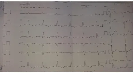
appearance of ventricular tachycardia on the electrocardiogram (ECG) (figure. 1), at the first aid stage 100mg lidocaine was used in/in bolus, then
4mg fractional. Paroxysm docked to sinus
tachycardia (Fig. 2).
Physical examination: When the patient was admitted in the hospital, patient was conscious, skin was clear.
tests were normal except erythrocyte sedimentation
rate (ESR 25-33 mm/h). Enzyme-linked
274
274
274
274
274
274
274
274
274
274
274
274
274
274
274
274
274
274
274
274
274
Figure 1.ECG (13.06.16) on stage ambulance paroxysmal ventricular tachycardia with heart rate 200 beats per minute.
Figure 2
e
275
e
275
e275
e
275
e275
e
275
e275
e
275
e275
e
275
e
275
e
275
e
275
e275
e
275
e275
e
275
e275
e
275
e275
e
275
Figure 3
ECG (14.06.16): Sinus rhythm 74 beats per minute, normal EOS position, violation of blood supply to the lower left ventricular wall.
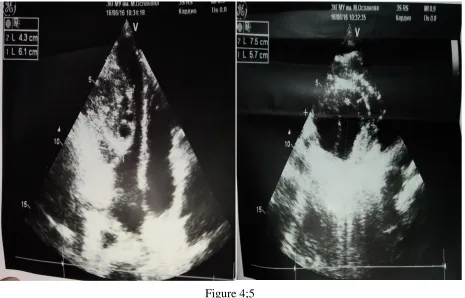
Figure 4;5
276
276
276
276
276
276
276
276
276
276
276
276
276
276
276
276
276
276
276
276
276
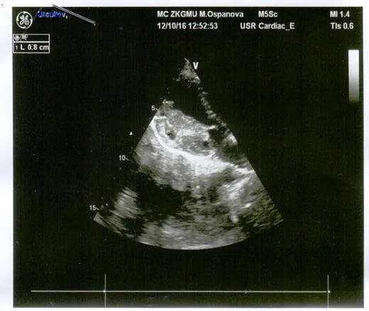
Figure 6
Daily ECG monitoring(14.06.16): basic sinus rhythm with heart rate 75 per minute, minimum heart rate 50 per minute, 01:41:39 h, maximum heart rate 126 per minute 18:56: 24 h (there was physical activity). Maximum pause-1.48 seconds, 01:41: 33h, due to sinus arrhythmia. Pauses more than 2 seconds no. During the period of ECG monitoring 247 polymorphic, single ventricular extrasystoles, 1B night hours, 3-supraventricular
extrasystoles were registered. There was no arrhythmia or conduction.
e
277
e
277
e277
e
277
e277
e
277
e277
e
277
e277
e
277
e277
e277
e277
e277
e
277
e277
e
277
e277
e
277
e277
e
277
occur from systemic or pulmonary circulation or as spread from adjacent organs [4, 11, 18]. The absence of a long-term clinical manifestation of ES is associated, echinococcal cysts grow slowly, on average, by 1-3 cm per year.
Analysis of the literature of observations shows [4, 11, 19, 20] that hydatid cysts of the heart are rare and often reaches a large size, rarely — plural. As for the localization of Echinococcus in the heart, it should be noted that there is not a single area of the heart that is not affected by echinococcosis. The most common cysts are located in the muscle of the left (LV) and right ventricles (RV), interventricular septum (MWP), rarely — in the atrium and pericardium. The most frequent localization of echinococcal cysts in the left ventricle and interventricular septum is associated with good blood supply [8]. Transthoracic
echocardiography, computed tomography and
magnetic resonance imaging are the most important tools for patient diagnosis and observation, and serological reactions provide good information about the disease [4,9,12,13,14].In the treatment of hydatid cysts of the heart must be surgical, with a combination of chemotherapy, in the case of isolated cardiac hydatid cyst before surgery is impossible – anthelmintic therapy because of the risk of development of the destruction of the wall of the cyst [4,15,16,17].
In the literature there is a description [11] of the history of the disease similar to the clinical symptom, the use of antiarrhythmic drug for relief of ventricular tachycardia with our observation, but the distinctive features are age (57 years), sex (male) localization in the right ventricle.If there are patients with myocardial and pericardial diseases of unknown cause for endemic regions, in differential diagnosis
recurrence of invasion.In addition, the result of surgical treatment is considered to be very important. Therefore, the treatment of ES should be combined surgical treatment with chemotherapy in the postoperative period in order to prevent relapses.
Echocardiography and serological test for
Echinococcus are the main methods in both diagnosis and control of ES recurrence.
The conclusion of our article is that in cystic lesions
of organs, regardless of their localization,
echinococcosis should be taken into account in the differential diagnosis.
Reference
1. Ibragimov A. Ultrasonic diagnosis of right
ventricle hydatid cysts: report of a rare case / Matkasimova Z., Raeva J., Parmankulova B.Journal of clinical medicine of Kazakhstan 2014:1 31: 48-51.
2. Polaykov N. In. Single (hydatid)
Echinococcus / Romich V. V., Sapharov R. V., Polaykov V. E. Research and practice in medicine. 2015: 2 .27-35.
3. Bahadir Sarli, Mehmet Ugurlu, Ahmet Oguz
Baktir, Ali Ihsan Tekin, Ahmet Tok, and Bayram Yagmur. Lone, Mobile. Left Atrial Hydatid Cyst. Tex Heart Inst J. 2016; 43(3): 261–263.
4. T.N.C. Padmanabhan K.V.K. Kumar
Mohammed Sadiq Azam Anil Kumar Bilolikar.
5. Primary echinococcus infection of the heart: a
rare type of cystic echinococcosis .European Heart Journal. 2017; 38.29-2255.
6. Vafae Eslahi, Aida; Kia, Eshrat Beigom;
278
278
278
278
278
278
278
278
278
278
278
278
278
278
278
278
278
278
278
278
278
Iranian journal of parasitology. Том: 12. : 2: 230-235
7. Muhaidi, M. J. Determination of the infective
strain of hydatid cyst in Iraqi cattle by using CO1 gene.Iraqi journal of agricultural sciences. Том: 48 Выпуск: 2 Стр: 644-649
8. Gurzu S, Beleaua MA, Egyed-Zsigmond
E, Jung I. Unusual Location of Hydatid Cysts: Report of Two Cases in the Heart and Hip Joint of Romanian Patients // Korean J Parasitol. 2017 Aug; 55(4):429-431. doi: 10.3347/kjp.2017.55.4.429. Epub 2017: 31.
9. Cardiac Echinococcosis Involving Left
Ventricular Myocardium in an 18-Year-Old Patient Zaprin G. Vazhev, Hristo A. Stoev
Department of Cardiovascular Surgery,
Medical University of Plovdiv, Plovdiv,
Bulagaria Folia Med (Plovdiv)
2018;60(2):308-13.
10.Moghul, Dunya; Hamidi, Hidayatullah.
Incidental finding of cardiac hydatid cysts, report of two cases //Moghul and Hamidi BMC Medical Imaging: 2018: 18:22.
11.Kahlfuß S, Flieger RR, Roepke TK, Yilmaz
K. Diagnosis and treatment of cardiac
echinococcosis Heart. Epub 2016:
1;102(17):1348-53.
12.Sushil Kumar Singh, Vikas Singh, Sarvesh
Kumar. Right ventricular hydatid cyst
presented as tachyarrhythmia. Journals.sager pub.com: 2018. 28
13.Uygur, Begum; Ustabasioglu, Fethi Emre;
Karakurt, Huseyin; с соавторами. An unusual cause of chest pain: An isolated huge cardiac hydatid cyst //Journal of clinical ultrasound. Том: 46 Выпуск: 4 Стр: 2018; 262-264.
14.Rostami, Alireza; Keykhali, Naser; Broujerdi,
Gholamreza Nouri. Heart and Lung Hydatid Cyst in a Pregnant Woman: A Case Report //Annals of Medical and health science research. Том: 7, Выпуск: 6, Стр: 2017:373-376
15.Noaman, H Hydatid Cyst of the Heart
/(Noaman, Haitham), Rawaf, S (Rawaf,
Salman), Majeed, A (Majeed, Azeem), Salmasi, AM (Salmasi, Abdul-Majeed) //Angiology: 2017. 68: 765-768.
16.Tanyeli, Omer; Dereli, Yuksel; Mercan, Ilker;
et al. New World's old disease: cardiac hydatid disease and surgical principles.
//Cardiovascular journal of Africa:
2017: 28: 304-308.
17.Gurzu, Simona; Beleaua, Marius Alexandru;
Egyed-Zsigmond, Emeric et al.
18.Unusual Location of Hydatid Cysts: Report of
Two Cases in the Heart and Hip Joint of
Romanian Patients//Korean journal of
parasitology. Tом: 55 Выпуск: 4 Стр. 2017 :429-431,
19.Wadhawa, Vivek; Shah, Jigar; Doshi, Chirag;
с соавторами. Surgical overview of cardiac echinococcosis: a rare entity//Interactive cardiovascular and thoracic surgery. Том: 27 Выпуск: 2. Стр: 2018 191-197.
20.Malikova M. S. Intracardiac echinococcosis /
Phrolova Y. V., Raskin V. V.,
Dzemeshkevich S. A., Dombrovskaya A.V.,
Mershina E. A., Sinitsyn V. E.,
Dzemeshkevich S. L. Russian journal of cardiology: 2015, 9 (125): 7-11
21.Rymer, Jennifer A.; Warraich,
Haider; Goldstein, Sarah; et al./A Rare
Presentation of Cardiac Cystic
Echinococcosis of the Ventricular
Septum.Conference: Scientific Sessions of the American-Heart-Association / Resuscitation
Science Symposium Location: Anaheim,
CAD. 2017.11-15
Nunes, Maria Carmo P.; Guimaraes Junior,
Milton Henriques; Diamantino, Adriana