International Journal of Medical Science and Current Research (IJMSCR) Available online at: www.ijmscr.com
Volume2, Issue 3,Page No: 132-141 May-June 2019
132
Medicine ID-101739732
A Comparative Study of Accuracy in Bracket Positioning With Modified Height Bracket
Positioning Gauge and Boonegauge: In Vivo Study
Dr. Vasundhara Bhide*, Dr.(Mrs).P.V.Hazarey, Dr.Harish Atram, Dr.Gayatri Kale, Dr.Sneha Waghmare, Dr. Mohini Dharmadhikari
1
Post Graduate student, 2 Professor and HOD, 3 Reader, 4,5,6 Post Graduate student Department of Orthodontics and Dentofacial Orthopaedics
Swargiya Dadasaheb Kalmegh Smruti Dental College and Hospital Wanadongri, Nagpur, Maharashtra, India
*Corresponding Author:
Dr. Vasundhara Bhide, BDS
Post Graduate student, Department of Orthodontics and Dentofacial Orthopaedics Swargiya Dadasaheb Kalmegh Smruti Dental College and Hospital
Wanadongri Nagpur, Maharashtra, India
Type of Publication: Original Research Paper Conflicts of Interest: Nil
ABSTRACT
Background: The aim of our clinical trial is to compare the accuracy in vertical and mesio-distal aspect of bracket positioning with modified height bracket positioning gauge and Boone gauge.
Materials & Methods: A Randomized Controlled Trial with a sample size of 20 patients who were required to undergo fixed orthodontic treatment. Existing height bracket positioning gauge was modified.The study was divided into two groups: Group A and B. In Group A (n=10) upper arch bracket positioning was to be done with Boone gauge and in lower arch with Modified height bracket positioning gauge and vice a versa in Group B (n=10). Digitization of pre-bonding and post-bonding casts was done with 3D scanning machine. Accuracy in bracket positioning between two gauges was assessed and compared by superimposition of pre and post bonding digital models for all 20 cases. Discrepancy was assessed in mesio-distal and vertical bracket position and statistical analysis was done.
Results: For mesio-distal bracket positioning, tooth numbers 12,14, 24 and 33 showed statistically significant difference whereas for vertical bracket positioning, tooth numbers 11,15,21,25 35 and 45 showed statistically significant difference.
Conclusion: Relatively less error was found in mesio-distal bracket postioning with the use of MHBPG as compared to Boone gauge as vertical arm in the modified gauge facilitated proper visualization by coinciding to the long axis of the tooth leading to increased accuracy. MHBPG was found to be more accurate in vertical bracket positioning as compared to Boone gauge.
Keywords: Bracket, Gauge, Bracket positioning, Digital scanning.
INTRODUCTION
Orthodontic treatment efficiency is highly increased with the introduction of newer techniques and modifications done to Preadjusted Edgewise Appliance. Andrews1 introduced the straight-wire
Pag
e
133
Pag
e
133
Pag
e
133
Pag
e
133
Pag
e
133
Pag
e
133
Pag
e
133
Pag
e
133
Pag
e
133
Pag
e
133
Pag
e
133
Pag
e
133
Pag
e
133
Pag
e
133
Pag
e
133
Pag
e
133
Pag
e
133
Pag
e
133
Pag
e
133
Pag
e
133
Pag
e
133
Andrews plane).1 FA point was selected because it was felt that, it was readily and consistently located.12 Ricketts3 (1976), and later Kalange4 (1999), recommended the use of marginal ridges as a guidance for the vertical positioning of brackets and bands. McLaughlin and Bennett5 advocated the positioning of brackets at a measured distance from the incisal edge, with different vertical positions recommended for different sized teeth.
In Orthodontics, bonding of brackets to enamel surfaces is the most crucial part of treatment mechanics. A gauge is used to measure and determine the bracket distance from the incisor or occlusal edge of the teeth. Proper use of gauge adds to the accuracy of the bracket position and in turn affects satisfactory initial alignment thus minimizes need for archwire bends and bracket repositioning.
Therefore, there is importance of bracket positioning devices for positioning of brackets at recommended vertical heights. Various attempts have been made in improving efficiency in bracket positioning which led to the emergence of various designs of bracket positioners.678
However several authors have conducted in vitro studies, i.e. on either stone models or typodonts comparing the accuracy of bracket placement using different gauges, conclusions of which will not be applicable to in vivo or clinical conditions. These studies also demonstrated that errors occurred most frequently in angular and vertical positioning.9101112 Several such studies used photographic method for analysis which in turn may have manual errors.
Therefore in present study attempt was made to modify existing height bracket positioning gauge. Vertical arm was added in the gauge for proper visualization of long axis of tooth. It was thought that if the vertical arm in the modified gauge correlates with the long axis of the tooth to be bonded then it will help the clinician to improve bracket positioning.
Therefore the present study was conducted in vivo with an objective to compare the accuracy of orthodontic bracket positioning with two different bracket positioning instruments i.e modified height bracket positioning gauge and Boone gauge. Assessment of accuracy in bracket positioning was done with the help of digital scanning of dental casts taken before and after bonding by superimposition of
digital images of pre and post bonding casts for both the gauges. The present study was carried out with following aim and objectives.
Aim
To compare the accuracy in vertical and mesio-distal aspect of bracket positioning with modified height bracket positioning gauge and Boone gauge.
Objectives
1. To evaluate and compare accuracy of bracket positioning in vertical aspect with modified height bracket positioning gauge and Boone gauge.
2. To evaluate and compare accuracy of bracket positioning in mesio-distal aspect with modified height bracket positioning gauge and Boone gauge.
Material and Methods
This was a Randomized Controlled Trial with a sample size of 20 patients. All these twenty individuals were required to undergo fixed orthodontic treatment.
Ethical approval Ethical and consent study
approvals were obtained from the Institutional Ethics Committee. Participation of each participant was voluntary and informed consent was obtained before commencement of the study. Individuals reporting to the departmental OPD of Orthodontics and Dentofacial Orthopaedics were clinically screened and those satisfying the following inclusion criteria were included in the study.
Inclusion criteria:
Individuals with presence of fully erupted teeth till 1st molar having normal tooth size for proper bracket positioning with mild to moderate crowding or spacing were selected for the study.
Exclusion criteria:
Individuals with severe crowding and rotations and missing teeth were excluded. Those individuals with tooth wear including attrition, abrasion, abfraction, fractured incisal edges and cusp tips and developmental dental anomalies were also excluded.
Methodology:
Pag
e
134
Pag
e
134
Pag
e
134
Pag
e
134
Pag
e
134
Pag
e
134
Pag
e
134
Pag
e
134
Pag
e
134
Pag
e
134
Pag
e
134
Pag
e
134
Pag
e
134
Pag
e
134
Pag
e
134
Pag
e
134
Pag
e
134
Pag
e
134
Pag
e
134
Pag
e
134
Pag
e
134
made and dental casts were prepared. Cervico-incisal and mesiodistal tooth size measurements were made on the dental casts using vernier calliper. From amongst 35 cases, 20 cases having their tooth size in normal range as given by Dr. Woelfel 13 were selected for the study. All these twenty individuals were required to undergo fixed orthodontic treatment.
Patients were divided into 2 groups, Group A and Group B. In Group A (10 patients) upper arch bracket positioning was to be done with Boone gauge with lead marking and in lower arch bracket positioning
was to be done with Modified height bracket positioning gauge. In Group B (10 patients) upper arch bracket positioning was to be done with Modified height bracket positioning gauge and lower arch with Boone gauge. These groups were made just for allocation of gauges in upper and lower arch, so that in each patient both the gauges are being used to avoid sample bias and better comparison of accuracy in bracket positioning between two gauges (BG and MHBPG).
Modified Height Bracket positioning gauge Bracket placement using modified gauge
Digital scanned image with estimated bracket positioning in Upper arch
Digital scanned image- Upper arch Superimposition of prebonding and post bonding image
Informed consent was taken from each individual. The pre-treatment records for this study were taken in the form of study models and Orthopantomogram (OPG) for each case. OPG was taken in Sirona Orthophos machine for all patients. It was used to determine the desired slot angulation of each bracket by evaluating the position of the roots. Estimation of bracket positioning was done as given by Mclaughlin Bennet and Trevisi14. individualized bracket positioning chart was prepared and followed in every
patient during bonding procedure. This served as a vertical reference guide for bracket placement.
Pag
e
135
Pag
e
135
Pag
e
135
Pag
e
135
Pag
e
135
Pag
e
135
Pag
e
135
Pag
e
135
Pag
e
135
Pag
e
135
Pag
e
135
Pag
e
135
Pag
e
135
Pag
e
135
Pag
e
135
Pag
e
135
Pag
e
135
Pag
e
135
Pag
e
135
Pag
e
135
Pag
e
135
decided. Individualized bracket positioning chart included vertical as well as horizontal positions of bracket placement
1st set of casts i.e pre-bonding cast along with individualized bracket positioning chart of every patient was sent to the CAD centre for 3D scanning of study models prior to bonding. Scanning was performed using Geomagic Control X software. With the help of individualized bracket positioning chart for each patient, digital analyzer marked the estimated bracket position as given in chart on the digital casts by using Geomagic Design X software. Digital analyzer was a person with the technical knowledge of Geomagic Design X software who did the markings on the digital casts. This scanning of pre-bonding casts was done to facilitate superimposition with post-bonding casts for comparing accuracy in bracket positioning.
For the selected individuals, direct bonding procedure was followed. 0.022 slot MBT brackets were used for each patient. Previously prepared individualized bracket positioning chart and OPG which was taken before initial bracket placement was referred during bracket positioning. From the OPG, by evaluating root position, desired slot angulation was determined and if the root alignment needs correction, appropriate adjustment was done in the slot angulation during initial bracket placement. Bracket positioning was done in group A and B using 2 different gauges according the respective gauge group.
Modification of the gauge:
Original height bracket postioning gauge (Denticon) was modified. It had an extension to correlate with the long axis of tooth for increased accuracy during bracket positioning. L shaped extension was made using 0.017x0.025 inch stainless steel archwire. L shaped extension was soldered onto the ends of the original height bracket positioning gauge.
After bonding, another set of study models were taken and dental casts were sent for 3D scanning and analyzation. Accuracy in bracket positioning with Boone gauge and modified HBPG was assessed and compared by superimposition of pre-bonding and post bonding digital models for all 20 cases. Discrepancy in bracket positioning was assessed in
mesio-distal and vertical bracket position and statistical analysis was done.
STATISTICAL ANALYSIS
All the samples were subjected to statistical analysis. Comparison of the two groups was done Wilcoxon rank sum test.
RESULTS
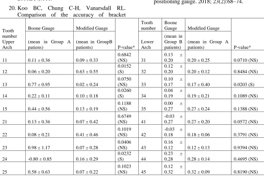
Table 1 shows the comparison between two gauges in mesio-distal bracket positioning where tooth number 12,14 and 24 showed the difference to be statistically significant. Tooth number 33 showed the difference to be statistically significant.
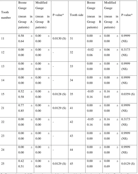
Table 2 shows the comparison between two gauges in vertical bracket positioning where tooth number 11,15,21 and 25 showed the difference to be statistically significant. Tooth number 35 and 45 showed the difference to be statistically significant.
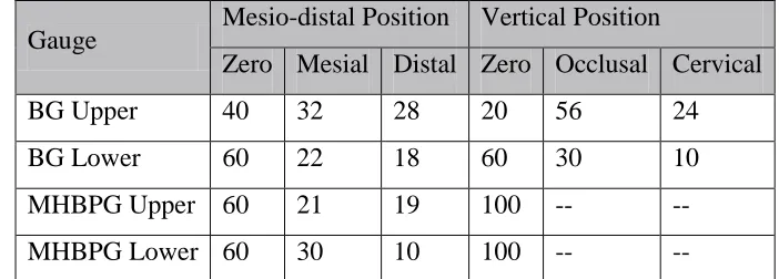
Table 3 provides the comparison of deviations in bracket positioning in mesio-distal and vertical position, evaluated using Pearson’s Chi-square test. In mesio-distal position, in the upper arch, the difference in the distribution of deviations of bracket positioning between BG and MHBPG was statistically significant with a p-value of 0.0183. However, in the lower arch, the difference in the distribution between the two techniques was statistically insignificant with p-value of 0.1723. In vertical position, the distribution of number of deviations across zero, Occlusal and cervical positions between BG and MHBPG was statistically highly significant with p-value < 0.0001.
DISCUSSION
Pag
e
136
Pag
e
136
Pag
e
136
Pag
e
136
Pag
e
136
Pag
e
136
Pag
e
136
Pag
e
136
Pag
e
136
Pag
e
136
Pag
e
136
Pag
e
136
Pag
e
136
Pag
e
136
Pag
e
136
Pag
e
136
Pag
e
136
Pag
e
136
Pag
e
136
Pag
e
136
Pag
e
136
According to McLaughlin et al.5, accuracy of bracket positioning is essential so that the built in features of the bracket system can be expressed fully and efficiently. In original edgewise appliance, bracket placement was normally carried out using gauges and irrespective of tooth size, standard millimeter measurements were taken from the incisal edge of each tooth. This caused incisal bracket placement for large incisors, which in turn led to variations in the amount of torque and in-out produced by the brackets. It is necessary to visualize the vertical long axis of the clinical crown of each tooth to achieve accuracy because errors my cause incorrect tip position of the teeth. McLaughlin et al proposed that the bracket wings need to be parallel to the long axis. But obtaining axial accuracy is the most difficult aspect. Many instruments are introduced for making the bracket positioning convenient, but very few give the easiest way to obtain axial accuracy.1617
Therefore in this study, existing height bracket positioning gauge was modified. To correlate with the long axis of tooth, modified guage had L shaped extension made from 0.017x0.025 inch stainless steel archwire for increased accuracy during bracket positioning. L shaped extension was soldered onto the ends of the original height bracket positioning gauge. Similar long axis extension was added by Saklecha and his co-workers18 where they initially took two inch band material of 0.180” × 0.006” and folded on to each other and then welded. Then a hole of 3 mm in diameter is drilled into the center of the band and over that a small piece of straight 0.016 × 0.022” SS wire bent into “L” shape is welded on one end of the band at the center. Their design was similar to the modification done in the present study but the procedure for its construction was more tedious and time consuming as compared to that done in present study. In addition to the tedious construction procedure, the wire for assessing the long axis of the tooth was on the opposite side of the end of the measuring side of the gauge which may account for visual errors in assessing long axis. Vertical arm in the present study was welded at the same side of the measuring side which assisted in minimizing both vertical and horizontal bracket placement errors.
Various researchers have modified the existing bracket positioning gauges but literature lacks studies regarding comparison of the accuracy between
modified gauge and existing available gauges. Therefore in present study, in addition to modification of existing height bracket positioning gauge, comparison in accuracy of bracket positioning with modified HBPG and boone gauge was done.
Bracket positioning might remain at risk of inaccuracy not only between different clinicians but also between different areas bonded by the same operator. Development of customized orthodontic philosophy and the incorporation of digital advances in orthodontics have given the ray of hope to eradicate this problem. Studies by Lahcen Ousehal and Laila Lazrak and his associate19 and Amir
Mohammadi and Seyed Moslemzadeh20 have
assessed the bracket positioning accuracy with the help of photographs. But results regarding efficiency of HBPG and BG were found to be contradictory like Lahcen Ousehal and Laila Lazrak19 concluded that BG provides better placement of the vertical bracket whereas Amir Mohammadi and Seyed Moslemzadeh20 found that HBPG has more vertical accuracy in bracket positioning. Reasons for these contradictory results may be attributed to the fact that both these authors have used photographic method for assessment of bracket positioning. This might have resulted in errors due to standardization of photographic technique and methods used.
Therefore to minimize the difficulty in standardization of photographic method and reduce photographic error as well as to increase the accuracy, 3D digital scanning of the dental casts was done before and after bonding of brackets. Dental casts along with individualized bracket positioning chart of every patient were sent to CAD centre for 3D scanning of study models prior to bonding. Bonding was then performed in individual patient with respective gauges as assigned to that particular patient. Post bonding study models were made and sent to the same CAD center for digital scanning purpose. Deviations or error in bracket positioning were evaluated by superimposing the pre-bonding and post-bonding scanned images. In this manner, digital scanning of dental casts excluded the shortcomings of photographic methods used by previous authors 46,47 as well as manual errors in evaluation were also excluded.
Pag
e
137
Pag
e
137
Pag
e
137
Pag
e
137
Pag
e
137
Pag
e
137
Pag
e
137
Pag
e
137
Pag
e
137
Pag
e
137
Pag
e
137
Pag
e
137
Pag
e
137
Pag
e
137
Pag
e
137
Pag
e
137
Pag
e
137
Pag
e
137
Pag
e
137
Pag
e
137
Pag
e
137
which they mainly studied bracket positioning on study models that too of one single patient. Whereas there is limited literature regarding in vivo studies comparing bracket positioning accuracy between two different gauges wherein bracket positioning is carried out in different patients. Therefore present study was conducted in vivo wherein bracket positioning was assessed in 20 individuals.
Table 1 shows the comparison of difference in mesio-distal bracket positioning between BG and MHBPG in upper and lower arch respectively. For upper arch, tooth numbers 12, 14 and 24 showed statistically significant differences between BG and MHBPG whereas for lower arch tooth number 33 showed statistically significant difference. For upper arch, mean mesio-distal error with the use of MHBPG and BG was 0.17 and 0.26 mm respectively, whereas for lower arch it was 0.18 and 0.08 for MHBPG and BG respectively. Overall mesio-distal error with MHBPG and BG was 0.175 and 0.17 respectively with no statistically significant difference. This finding was in agreement to a study by Amir Mohammadi and Seyed Moslemzadeh20 who conducted a study only in upper arch and observed that overall mean mesio-distal error with the use of HBPG and BG was 0.28 and 0.29 respectively, with no statistically significant differences (p=0.982). Results of present study indicate that MHBPG can be successfully used in upper arch where less error was observed as compared to lower arch. Possible explanation for this can be related to more difficulty experienced for bracket positioning in lower arch in visualizing mesio-distal bracket position and problems in maintaining strict isolation. However in lower arch BG showed improved accuracy mainly for canine. Possible reason for this may be attributed to variability in canine morphology which makes it difficult to determine proper mesio-distal position for bracket placement with MHBPG as it simultaneously determined both vertical as well as mesio-distal bracket position.
Table 2 shows comparison of difference in vertical bracket positioning between BG and MHBPG in upper and lower arch respectively. Tooth numbers 11,15,21,25,35 and 45 showed statistically significant difference with the use of two gauges ( BG and MHBPG). For upper arch mean error in vertical bracket placement with BG and MHBPG was 0.23 and 0 respectively. For lower arch mean error in
vertical bracket placement with BG and MHBPG was -0.01 and -0.13 respectively. From these observations it can be concluded that more accuracy was observed with MHBPG in upper as well as lower arch. This finding was similar to the studies done by Amir
Mohammadi and Seyed Moslemzadeh20 and Bon
Chan Koo and co-workers21 who also found
increased vertical accuracy with HBPG than BG.
Bon Chan Koo and co-workers21 compared bracket
placement accuracy between direct and indirect bonding procedures. Boone gauge was used for bracket positioning in direct bonding group wherein discrepancies were observed in vertical aspect which can be attributed to the fact that Boone gauge can get tilted and yield error in vertical position of brackets.
Taylor and his associate11 checked the reliability of positioning preadjusted brackets in an in vitro study model. They investigated bracket positioning accuracy by 12 operators and found that the accuracy in bracket positioning was more in vertical reference plane, which is in agreement to the findings of present study.
Armstrong et al,22 did the similar study comparing the vertical accuracy of bracket placement between experienced orthodontists and final dental students. He concluded that the accurate direct placement of orthodontic brackets to teeth does not appear to be related to clinical experience or specialist training. These results might seem to be inconsistent with those by Sergio Luiz Mota Junior et al23 who concluded that operators are prone to fail in the placement of orthodontic attachments when using the Boone gauge, despite their clinical experience in orthodontics. The choice of single operator in the present study reduces the errors related to difference in the competency level of different clinicians.
Literature lacks in vivo studies on comparison of bracket positioning accuracy with two different gauges. Therefore this study was carried out with an objective to compare bracket positioning in patients where two gauges i.e. one existing gauge and another modified gauge are compared.
Pag
e
138
Pag
e
138
Pag
e
138
Pag
e
138
Pag
e
138
Pag
e
138
Pag
e
138
Pag
e
138
Pag
e
138
Pag
e
138
Pag
e
138
Pag
e
138
Pag
e
138
Pag
e
138
Pag
e
138
Pag
e
138
Pag
e
138
Pag
e
138
Pag
e
138
Pag
e
138
Pag
e
138
more deviations in occlusal aspect. Possible reason for this might be attributed to the factor that Boone gauge can get tilted leading to more occlusal bracket placement. MHBPG showed improved accuracy with insignificant errors in vertical position.
Based on the findings of this study, it can be concluded that an error occurs in vertical as well as mesiodistal aspects with the use of both the gauges. However, there is a significant difference in terms of vertical error between the two gauges because of the modification of HBPG in present study leading to improved accuracy in bracket positioning. Precision in the present study was also enhanced with the use of digital scanning of dental casts.
CONCLUSION
1. Relatively less error was found in mesio-distal bracket postioning with the use of MHBPG as compared to Boone gauge as vertical arm in the modified gauge facilitated proper visualization by coinciding to the long axis of the tooth leading to increased accuracy.
2. MHBPG was found to be more accurate in vertical bracket positioning as compared to Boone gauge as no teeth showed significant error in vertical bracket positioning with the use of MHBPG.
3. Deviation in bracket positioning in mesial as well as distal aspect is seen with the use of both the gauges in both the arches.
4. Boone gauge showed more deviations in bracket positioning in vertical aspect occlusally so care should be taken not to allow tilting of BG while bracket positioning.
5. Use of digital scanning in the present study facilitated accuracy in evaluation of bracket positioning.
REFERENCES
1. Andrews LF. Straight-wire appliance origin, contoroversy, commentary. J Clin Orthod. 1976;10:99–114.
2. Andrews LF. The straight-wire appliance. Br J Orthod. 1979;6(3):125–43.
3. Ricketts RM. Bioprogressive therapy. USA Rocky ountion. 1979;
4. Kalange JT. Ideal appliance placement with APC brackets and indirect bonding. J Clin Orthod. 1999;9:516–26.
5. McLaughlin RP. Finishing and detailing with a preadjusted appliance system. JCO. 1991;4:251–64.
6. Kumar KK, Singh I, Goel S. A new bracket-positioning gauge. J Clin Orthod JCO. 2001;35(3):154.
7. Stockstill JW, Levy-Bercowski D, DeLeon E. A new bracket-placement device. J Clin Orthod JCO. 2008;42(7):412–4.
8. Aggarwal A, Nayak US. A new tool for orthodontic bracket placement. J Clin Orthod JCO. 2009;43(4):275.
9. Fowler P V. Variations in the perception of ideal bracket location and its implications for the pre-adjusted edgewise appliance. Br J Orthod. 1990;17(4):305–10.
10.Berman M. Maurice Berman--straight wire myths. Interview by Robert Kirschen. Br J Orthod. 1988;15(1):57.
11.Geoffrey Taylor N, Cook PA. The reliability of positioning pre-adjusted brackets: an in vitro study. Br J Orthod. 1992;19(1):25–34.
12.Balut N, Klapper L, Sandrik J, Bowman D. Variations in bracket placement in the preadjusted orthodontic appliance. Am J Orthod Dentofac Orthop. 1992;102(1):62–7.
13.Scheid RC. Woelfel’s dental anatomy. Lippincott Williams & Wilkins; 2012.
14.McLaughlin RP, Bennett JC, Trevisi HJ. Systemized orthodontic treatment mechanics. Elsevier Health Sciences; 2001.
15.Angle EH. The latest and best in orthodontic mechanism. Dent Cosm. 1929;71:409–21.
16.Smaha CN, Voth ED. A positioning device for direct bracket attachment. Am J Orthod. 1972;62(4):394–9.
Pag
e
139
Pag
e
139
Pag
e
139
Pag
e
139
Pag
e
139
Pag
e
139
Pag
e
139
Pag
e
139
Pag
e
139
Pag
e
139
Pag
e
139
Pag
e
139
Pag
e
139
Pag
e
139
Pag
e
139
Pag
e
139
Pag
e
139
Pag
e
139
Pag
e
139
Pag
e
139
Pag
e
139
18.Saklecha B. A new two bracket positioning gauge. :55–7.
19.Ousehal L, Lazrak L. The accuracy of brackets placement in direct bonding technique: a comparison between the pole-like bracket positioning gauge and the star-like bracket positioning gauge. Mohammadi A, Moslemzadeh SH. Comparison of the accuracy of bracket placement with height bracket positioning gauge and boone gauge. J Dent Res Dent Clin Dent Prospects. 2011;5(4):111.
20.Koo BC, Chung C-H, Vanarsdall RL. Comparison of the accuracy of bracket
placement between direct and indirect bonding techniques. Am J Orthod Dentofac Orthop. 1999;116(3):346–51.
21.Armstrong D, Shen G, Petocz P, Darendeliler MA. A comparison of accuracy in bracket positioning between two techniques— localizing the centre of the clinical crown and measuring the distance from the incisal edge. Eur J Orthod. 2007;29(5):430–6.
22.Luiz S, Júnior M, José M, Schmitberger CA. Evaluation of the prototype of a new bracket-positioning gauge. 2018; 23(2):68–74.
Tooth number Upper Arch
Boone Gauge Modified Gauge
P-value*
Tooth number
Boone
Gauge Modified Gauge
P-value* (mean in Group A
patiens)
(mean in GroupB patients)
Lower Arch
(mean in Group B patients)
(mean in Group A patients)
11 0.11 ± 0.36 0.09 ± 0.33
0.6842
(NS) 31
0.13 ±
0.20 0.20 ± 0.25 0.0710 (NS)
12 0.06 ± 0.20 0.63 ± 0.55
0.0152
(S) 32
0.12 ±
0.20 0.20 ± 0.12 0.8484 (NS)
13 0.77 ± 0.95 0.02 ± 0.24
0.0750
(NS) 33
0.10 ±
0.17 0.17 ± 0.40 0.0203 (S)
14 0.22 ± 0.11 0.10 ± 0.18
0.0260
(S) 34
0.04 ±
0.19 0.19 ± 0.21 0.1089 (NS)
15 0.44 ± 0.56 0.13 ± 0.19
0.1188
(NS) 35
0.00 ±
0.27 0.27 ± 0.24 0.1388 (NS)
21 0.13 ± 0.36 0.07 ± 0.42
0.6749
(NS) 41
-0.03 ±
0.27 0.27 ± 0.20 0.0572 (NS)
22 0.08 ± 0.21 0.41 ± 0.46
0.1019
(NS) 42
-0.03 ±
0.18 0.18 ± 0.06 0.3791 (NS)
23 0.98 ± 1.17 0.07 ± 0.28
0.0406
(NS) 43
0.16 ±
0.12 0.12 ± 0.13 0.9394 (NS)
24 -0.80 ± 0.85 0.16 ± 0.29
0.0232
(S) 44
0.23 ±
0.28 0.28 ± 0.14 0.4695 (NS)
25 0.58 ± 0.63 0.07 ± 0.22
0.1023
(NS) 45
0.12 ±
0.32 0.32 ± 0.09 0.8190 (NS)
Table 1 Comparison of the difference in mesio-distal bracket positioning (ideal estimated position-Post bonding position) between BG (Group A patients, n=10) and MHBPG (Group B patients, n=10) in upper arch; Comparison of the difference in mesio-distal bracket positioning (estimated position-Post bonding position) between BG (Group B patients, n=10) and MHBPG (Group A patients, n=10) in lower arch
Pag
e
140
Pag
e
140
Pag
e
140
Pag
e
140
Pag
e
140
Pag
e
140
Pag
e
140
Pag
e
140
Pag
e
140
Pag
e
140
Pag
e
140
Pag
e
140
Pag
e
140
Pag
e
140
Pag
e
140
Pag
e
140
Pag
e
140
Pag
e
140
Pag
e
140
Pag
e
140
Pag
e
140
Tooth
number
Boone
Gauge
(mean in
Group A
patients)
Modified
Gauge
(mean in
Group B
patients)
P-value* Tooth side
Boone
Gauge
(mean in
Group B
patients)
Modified
Gauge
(mean in
Group A
patients)
P-value*
11 0.58 ±
0.64
0.00 ±
0.00 0.0130 (S) 31
0.00 ±
0.00
0.00 ±
0.00
0.9999
(NS)
12 0.00 ±
0.00
0.00 ±
0.00 - 32
-0.02 ±
0.06
0.06 ±
0.00
0.3173
(NS)
13 0.00 ±
0.00
0.00 ±
0.00 - 33
0.00 ±
0.00
0.00 ±
0.00
0.9999
(NS)
14 0.00 ±
0.00
0.00 ±
0.00 - 34
0.00 ±
0.00
0.00 ±
0.00
0.9999
(NS)
15 0.52 ±
0.58
0.00 ±
0.00 0.0128 (S) 35
-0.05 ±
0.16
0.16 ±
0.65 0.0359 (S)
21 0.77 ±
0.85
0.00 ±
0.00 0.0129 (S) 41
0.00 ±
0.00
0.00 ±
0.00
0.9999
(NS)
22 0.00 ±
0.00
0.00 ±
0.00 - 42
-0.05 ±
0.16
0.16 ±
0.00
0.3173
(NS)
23 0.00 ±
0.00
0.00 ±
0.00 - 43
0.00 ±
0.00
0.00 ±
0.00
0.9999
(NS)
24 0.00 ±
0.00
0.00 ±
0.00 - 44
0.00 ±
0.00
0.00 ±
0.00
0.9999
(NS)
25 0.42 ±
0.51
0.00 ±
0.00 0.0129 (S) 45
0.00 ±
0.00
0.00 ±
0.69 0.0129 (S)
Table 2: Comparison of difference in vertical bracket positioning (estimated position-Post bonding position) between BG (Group A patients, n=10) and MHBPG (Group B patients, n=10) in upper arch; Comparison of difference in vertical bracket positioning (estimated position-Post bonding position) between BG (Group B patients, n=10) and MHBPG (Group A patients, n=10) in lower arch.
Pag
e
141
Pag
e
141
Pag
e
141
Pag
e
141
Pag
e
141
Pag
e
141
Pag
e
141
Pag
e
141
Pag
e
141
Pag
e
141
Pag
e
141
Pag
e
141
Pag
e
141
Pag
e
141
Pag
e
141
Pag
e
141
Pag
e
141
Pag
e
141
Pag
e
141
Pag
e
141
Pag
e
141
Gauge Mesio-distal Position Vertical Position
Zero Mesial Distal Zero Occlusal Cervical
BG Upper 40 32 28 20 56 24
BG Lower 60 22 18 60 30 10
MHBPG Upper 60 21 19 100 -- --
MHBPG Lower 60 30 10 100 -- --