R E S E A R C H
Open Access
Human telomerase RNA component (hTERC)
gene amplification detected by FISH in
precancerous lesions and carcinoma of the larynx
Yu Liu
1,2, Xiao-li Dong
2, Cheng Tian
1and Hong-gang Liu
1*Abstract
Background:Gain of 3q26 is frequently observed in squamous cell carcinomas of mucosal origin, including those originating in the head and neck region. The human telomerase RNA component (hTERC) gene, which is located on chromosome 3q26, encodes for an RNA subunit of telomerase that maintains the length of telomeres through cellular divisions, and is activated in malignant diseases. The present study was designed to detect hTERC
amplification in laryngeal lesions and evaluate whether this might serve as a supportive biomarker in histopathological analysis for in the diagnosis of laryngeal lesions.
Methods:Fluorescent in situ hybridization (FISH) was applied on formalin-fixed paraffin-embedded blocks of 93 laryngeal specimens, including 14 normal epithelium (NE), 15 mild dysplasia (Md), 18 moderate dysplasia (MD), 16 severe dysplasia (SD), 9 carcinoma in situ (CIS), and 21 invasive carcinoma (IC)).
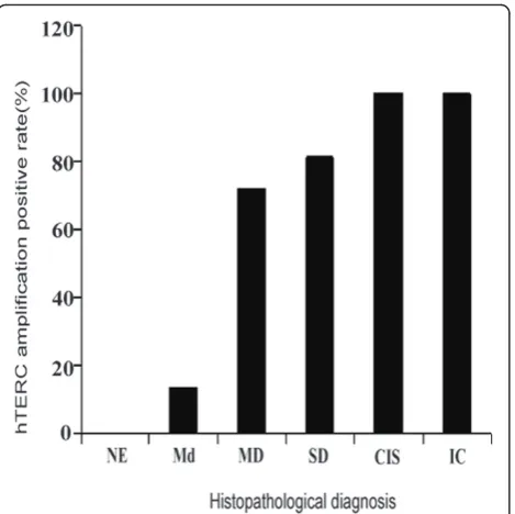
Results:By histopathologic examination, hTERC amplification rates in NE, Md, MD, SD, CIS and IC cases were 0% (0/14), 13.33% (2/15), 72.22% (13/18), 81.25% (13/16), 100% (9/9) and 100% (21/21), respectively. Amplification of hTERC was significantly associated with histopathologic diagnosis (P < 0.0001). The percentage of hTERC
amplification in patients with MD, SD, CIS, and IC was significantly higher than those with NE or Md (P < 0.0001). The number of cells with abnormal signals increased and the abnormal signal patterns were diversified with increasing severity of laryngeal dysplasia (P < 0.0001).
Conclusions:The hTERC amplification is important in the development of laryngeal squamous cell carcinoma (LSCC). FISH detection of hTERC amplification may provide an effective approach in conjunction with
histopathologic evaluation for differential diagnosis of laryngeal lesions.
Virtual Slides:The virtual slide(s) for this article can be found here: http://www.diagnosticpathology.diagnomx.eu/ vs/2226606266791985
Keywords:Laryngeal carcinoma, Human telomerase RNA component gene, Amplification, Fluorescent in situ hybridization
Background
Laryngeal squamous cell carcinoma (LSCC) is the most common type of head and neck squamous cell carci-noma (HNSCC) [1,2]. Despite recent progress in the diagnosis and therapeutic modalities for LSCC, the 5-year survival rate has not improved in more than two decades [3]. Early diagnosis of antecedent lesions in the
risk population may help to validate a prevention regi-men and allow effective intervention that improves sur-vival [1,2]. Therefore, it is necessary to identify target molecules for diagnosis and for effective therapy at the premalignant and/or early stages of LSCC.
It has been generally accepted that carcinogenesis involves the progressive accumulation of genetic abnormalities. Gain at 3q is a common feature of SCC, with an overlapping area of gain at 3q26 having been reported in SCC at different anatomic sites [4], includ-ing lung [5,6], head and neck [7-9], cervix of the uterus
* Correspondence: Liuhg1125@163.com
1
Department of Pathology, Beijing Tongren Hospital, Capital Medical University, Beijing, 100730, China
Full list of author information is available at the end of the article
[10,11], esophagus [12,13] and etc. The human telomer-ase RNA component (hTERC) gene, localized on chro-mosome 3q26, encodes the RNA component of human telomerase, and acts as a template for the addition of the repeat sequence [14]. Amplification of hTERC has been identified in many tumor samples and immorta-lized cell lines using techniques such as fluorescence in situ hybridization (FISH) and Southern blot analysis, suggesting that transcription is upregulated during tumorigenesis [15]. However, the role of the hTERC gene amplification in the carcinogenesis of LSCC has yet to be defined.
In this study, we applied a dual-color fluorescence in situ hybridization (FISH) panel in the detection of hTERC and systematically analyzed FISH results among different groups of laryngeal lesions based on histo-pathologic diagnosis. The aim of the present study was to characterize the amplification of hTERC and evaluate its significance as an adjunctive tool.
Methods Tissue collection
Patients involved in this study included 78 males and 15 females, with ages ranging between 35-78 years. All patients were recruited between October 2007-July 2009, and samples were obtained after patients had pro-vided written, informed consent. 20 formalin-fixed par-affin-embedded blocks of normal laryngeal epithelium were collected to establish the FISH detection threshold of samples. In addition, formalin-fixed paraffin-embedded blocks of 93 laryngeal carcinoma and precur-sor lesions specimens were obtained from the Depart-ment of Pathology of Beijing Tongren Hospital. The cases included 14 normal epithelium (NE), 15 mild plasia (Md), 18 moderate dysplasia (MD), 16 severe dys-plasia (SD), 9 carcinoma in situ (CIS), 21 invasive carcinoma (IC). The patients had received no che-motherapy or radiotherapy prior to surgery. Two pathol-ogists reviewed all hematoxylin and eosin-stained slides to confirm the diagnosis and ensure the presence of representative tissue before FISH was performed. All diagnoses in this research were established using 2005 WHO Classification.
The diagnostic criterias are as follows: Mild dysplasia: the architectural disturbance and cytological atypia is limited to the lower third of the epithelium; Moderate dysplasia:the architectural disturbance and cytological atypia are extended to the middle third of the epithe-lium and the cells show moderately polymorphic, nuclear mitoses are increased, but in the upper third of the epithelium the cells are well differentiated and arrange in lamellar form; Severe dyplasia:the architec-tural disturbance and cytologic atypia extended more than two thirds of the epithelium, there is no mature
morphology, and only in the top layer shows some lamellar forms; Carcinoma in situ (CIS):The dysplasia of the mucous epithelium involves the full thickness but still not break through the basal lamina. There is no characteristic appearance of gross findings, usually pre-senting as thickening and whiteness of local mucosa. Microscopically, the lesions show that the atypical cells with large, hyperchromic nuclei spread to full thickness, and the cell size is different. The pathologically mitotic figures are visible and dyskeratosis, and the cell polarity is disappeared. The lesion can involve the mucosal glands but the basement membrane is intact.
Histological examination and fluorescent in situ hybridization analysis
The tissues were fixed in 4% paraformaldehyde solution, serially sectioned at 5 μm thickness and then stained routinely with hematoxylin and eosin (H&E) following standard procedures. The paraffin-embedded tissue sec-tion was asayed by fluorescent in situ hybridizasec-tion (FISH) with the GP hTERC FISH kit accoding manufa-ture’s instruction (GP Medical Technologies Ltd., Beij-ing, China). Prior to hybridization of the paraffin-embedded tissue sections, slides were treated as follows: slides were rinsed with dimethylbenzene twice for 10 min for deparaffinization, then incubated in 100% etha-nol for 5 min, an ethaetha-nol series (100%, 85% and 70%) in turn for 2 min each for rehydration, deionized water for 3 min, then 90°C deionized water once for 30 min, 2× saline sodium citrate (SSC) twice for 5 min, pepsin digestion K solution (200μg/ml) at 37°C for 8 min, 2 × SSC twice for 5 min, and an ethanol series (70%, 85% and 100%) in turn for 2 min each. Slides were then air-dried at room temperature.
The GP hTERC FISH kit (GP Medical Technologies Ltd., Beijing, China) contains a hTERC DNA probe (Spectrum Red) and a control chromosome 3 centro-mere-specific probe (CSP3; Spectrum Green). The probe mixture (2 μl probe, 7 μl hybridizing buffer and 1 μl deionized water for each slide) was dropped onto the slides, followed by denaturation at 83°C for 5 min. Hybridization was subsequently performed in a humidi-fied box at 42°C overnight. Slides were then washed in 2 × SSC containing 0.1% (v/v) NP-40 for 5 min, followed by 70% (v/v) ethanol for 3 min and the air-dried slides were restained with 40,6-diamidino-2-phenylindole (DAPI) and mounted with cover slips.
evaluated. In a normal cell, the signal ratio of CSP3 to hTERC is 2:2, whereas in abnormal cells the ratio will be 2:3, 2:4, 2:5, 3:3, 4:4, and so on. Therefore, a cell with 3 or 4 hTERC signals, regardless of the signal numbers of CSP3, will be considered as having an abnormal sig-nal pattern. For a positive result with gain of the hTERC gene, the percent of nuclei with a combination of all the abnormal signal patterns should be more than the cutoff value. The cutoff value was defined as the mean plus three standard deviations of the percentage of nuclei with a combination of all possible abnormal signal pat-terns [16-18]. The cutoff value was calculated using 20 normal epithelium, confirmed by histopathology. In this study, the cut-off value was 7.287%.
Statistical analysis
Chi-square test or Fisher’s exact test were used to evalu-ate the results. A p value < 0.05 (two-sided) was consid-ered to indicate statistically significant. All analyses were performed with the use of the Statistical Package for the Social Sciences, version 12.0 (SPSS Inc, Chicago, IL, USA).
Results
hTERC amplification in association with histopathologic evaluations
hTERC gene amplification was not observed in any of the 14 normal epithelium analyzed (Figure 1). In con-trast hTERC gene amplification was observed in 13.3% (2/15) of mild dysplasia (Figure 2), 72.2% (13/18) of moderate dysplasia (Figure 3), 81.3% (13/16) of severe dysplasia (Figure 4), and in all 9 cases of carcinoma in situ (Figure 5) and all 21 cases of invasive carcinoma (Figure 6). The rate of hTERC gene amplification in
moderate dysplasia, severe dysplasia, carcinoma in situ or invasive carcinoma was significantly higher than that in normal mucosa and mild dysplasia (P < 0.001)(Table 1, Figure 7).
hTERC amplification patterns in association with different types of lesions
The fluorescent signal pattern of CSP3 versus hTERC is 2:2 in normal cells. For the 14 cases classified as normal epithelium, all of the 22 abnormal cells exhib-ited the 2:3 signal pattern; for the 15 mild dysplasia and 18 moderate dysplasia cases, the 2:3 and 2:4 signal pattern were the most common; for the 15 sereve dys-plasia and 9 carcinoma in situ cases, the 2:3 and 2:4 signal pattern were the most common, followed by the 3:3 and 4:4 patterns; for the 21 Invasive carcinoma cases, in the 988 abnormal cells observed the 4:4 signal pattern was the most common, followed by the 3:3 and 5:5 patterns (Table 1). The numbers of hTERC ampli-fied cases increased with the severity of laryngeal dis-eases, as well as with the complexity of abnormal signal (P < 0.001).
Discussion
In the present study, we investigated the relation between the hTERC amplification and laryngeal lesions using fluorescent in situ hybridization (FISH) in Chinese Han population. We found that the percentage of hTERC amplification in patients with moderate dyspla-sia (MD), severe dyspladyspla-sia (SD), carcinoma in situ (CIS), and invasive carcinoma (IC) was significantly higher than those with normal epithelium (NE) or mild dyspla-sia (Md). The amplification of hTERC was significantly associated with the histopathologic diagnosis.
Emerging evidence suggest that transition from nor-mal epithelium to dysplasia and squamous cell carci-noma is related to the progressive accumulation of genetic changes leading to a clonal population of trans-formed epithelial cells [19]. Despite extensive research into these genetic changes in laryngeal carcinogenesis, reliable genetic markers with diagnostic and prognostic value are still lacking.
Telomerase is a ribonucleoprotein enzyme that adds the TTAGGG repeats to the chromosomal ends and maintains chromosome integrity during DNA replica-tion. Activation of telomerase can lead to cellular immortalization, prevent from apoptosis and potentially promote tumorigenesis [20]. The human telomerase RNA component (hTERC) gene, localized on chromo-some 3q26, encodes the RNA component of human
telomerase, constituting one of two major subunits, and acts as a template for the addition of the repeat sequence [21]. When hTERC is overexpressed, the cells with critically short telomeres avoid undergoing apopto-sis, potentially leading to tumorigenesis [15].
In our study, we detected the amplification of hTERC by FISH in all cases of invasive carcinoma of larynx. This result indicates demonstrated that amplification of the hTERC gene is one of the most frequent amplifica-tions described in LSCC, and it was increased with increasing stage of dysplasia and had a high incidence in invasive carcinoma. These results support a previous study by Soder et al, which detected more than two copies of hTERC per cell in 97% (29/30) of head and neck squamous cell carcinoma (HNSCC) by FISH [22].
While studies employing comparative genomic
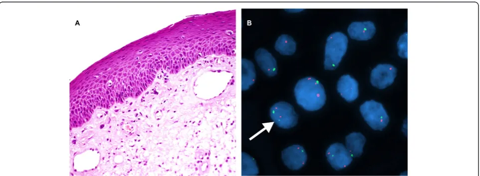
Figure 2A case of mild dysplasia of the larynx. A. H&E stain. (×200). B. Interphase FISH displaying a CSP3: hTERC signal pattern of 2:2 (green: red). (×1000).
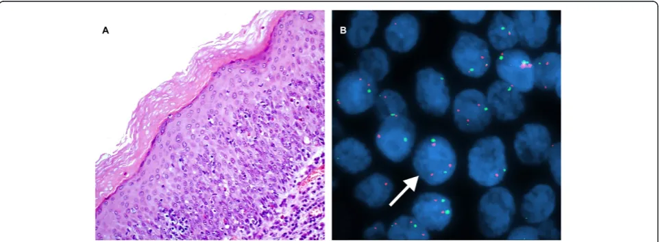
Figure 4A case of sereve dysplasia of the larynx. A. H&E stain. (×200). B. Interphase FISH displaying a CSP3: hTERC signal pattern is 2:3, 2:5, 3:3, and etc. (×1000).
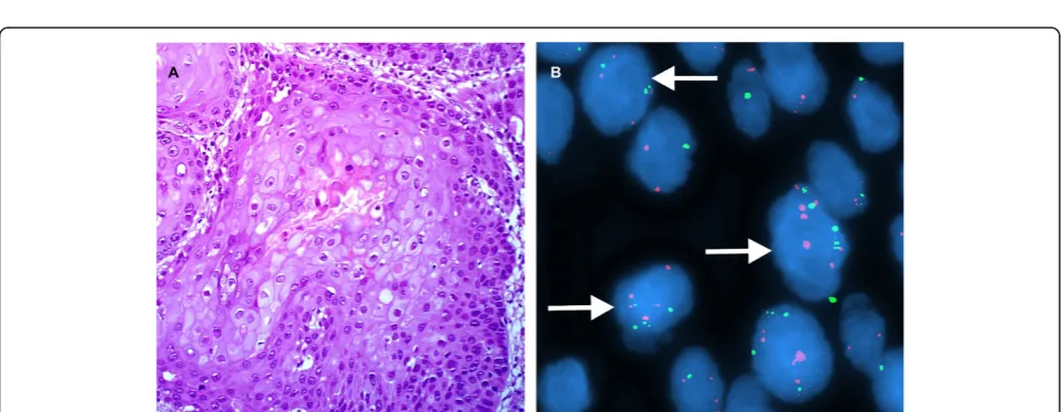
Figure 5A case of carcinoma in situ. A. H&E stain. (×200). B. Interphase FISH displaying a CSP3: hTERC signal pattern is 2:3, 4:4, 6:6, and etc. (×1000).
hybridization (CGH) have reported lower frequency for 3q26 gains (between 50-82%) in HNSCC [23-26], this difference is likely attributable to the different sensitivity of the respective techniques. Detection of trisomy using CGH requires this numerical aberration to be present in at least 40% of the cells. In contrast, FISH detects changes on a single-cell basis and is therefore not sensi-tive to dilution. Furthermore, small, regional, low-copy number amplification may escape detection by CGH [27].
Interestingly, the amplification of hTERC was observed at low frequency in normal epithelium and mild dysplasia, compared with a significantly higher fre-quency in cases of the moderate dysplasia, severe dys-plasia, carcinoma in situ and invasive carcinoma. This finding supports that the amplification of hTERC may be a transition event in the progression to invasive
squamous cell carcinoma of larynx. It is in agreement with previous studies, the gain of chromosome 3q was reported as the most common site of genetic overrepre-sentation in mucosal squamous cell carcinomas and has suggested it is a pivotal transition event in cancer patho-genesis [10,11,28]. The finding also supports the possibi-lity for hTERC amplification as a clinically useful genetic marker assisting histopathologic analysis for the differential diagnosis of low-grade (< mild dysplasia) ver-sus high-grade (> moderate dysplasia) of laryngeal dys-plasia. The finding was consistent with results reported by other investigators. Luzar et al demonstrated that the presence and relative quantity of Human telomerase reverse transcriptase (hTERT) mRNA increases progres-sively with the degree of laryngeal epithelial abnormal-ities. Statistical analysis revealed two groups of laryngeal epithelial changes, with significant differences in the levels of hTERT mRNA expression (P < 0.0033): (i) nor-mal and reactive hyperplastic laryngeal epithelium (squamous and basal-parabasal hyperplasia), and (ii) aty-pical hyperplasia (potentially malignant lesion), CIS and invasive laryngeal SCC [29]. They also found the telo-merase catalytic protein immunohistochemistry parallels well with hTERT mRNA relative quantities in laryngeal carcinogenesis. The results indicate that telomerase reactivation is an early event in laryngeal carcinogenesis, already detectable at the stage of precancerous laryngeal epithelial changes [30].
Gene mutations and chromosomal abnormalities are commonly present in tumor cells. Chromosome aneu-ploidy and structural anomalies lead to genomic instability, resulting in tumorigenesis. In the present study, the percentages of cells with hTERC amplification increased with increasing severity of disease, and the amplification patterns got more diverse and complex as well. Notably, more complicated patterns of hTERC amplification other than 2:3 were found only in the high-grade lesions. The study demonstrated that chro-mosome aneuploidy was a common event in laryngeal lesions. Whether amplification patterns can be used as diagnostic or prognostic markers warrants further investigation.
Table 1 hTERC Amplification According to Histopathologic Evaluation
Laryngeal lesions Cases, no. hTERC+, no. (%) Abnormal cells, no. (%)* Proportion of amplification types, % of cases 2:3 2:4 2:5 3:3 4:4 5:5 6:6 Others 3:4,3:5, 4:5 etc.
Normal epithelium 14 0(0.0) 22(1.6) 100
Mild dysplasia 15 2 (13.3) 68 (4.5) 85.3 14.7
Moderate dysplasia 18 13 (72.2) 409 (22.7) 47.6 39.8 1.5 4.8 3.6 2.7
Sereve dysplasia 16 13 (81.3) 506(31.6) 38.6 34.6 2.7 9.8 7.6 2.8 0.4 3.5
Carcinoma in situ 9 9(100.0) 353(39.2) 28.1 24.6 3.8 17.8 14.5 6.4 1.6 3.2
Invasive carcinoma 21 21(100.0) 988 (47.0) 14.2 12.3 6.2 21.6 24.7 15.4 2.3 3.3
*Number of abnormal cells (% abnormal cells out of total cells examined, and 100 cells were observed in each case)
In conclusion, We have analyzed the amplification of hTERC in different stages of laryngeal carcinogenesis. The present study suggests that FISH analysis can be performed on histological specimens in order to detect the amplification of hTERC for the diagnosis of laryn-geal lesions. The hTERC amplification assay may be an adjunct to histopathologic screening and may be useful in assessing the potential of individual lesions to progress.
Abbreviations
LSCC: Laryngeal squamous cell carcinoma; HNSCC: Head and neck squamous cell carcinoma; hTERC: Human telomerase RNA component (hTERC) gene; CSP3: Chromosome 3 centromere-specific probe; FISH: Fluorescence in situ hybridization; NE: Normal epithelium; Md: Mild dysplasia; MD: Moderate dysplasia; SD: Sereve dysplasia; CIS: Carcinoma in situ; IC, Invasive carcinoma
Acknowledgements
The authors thank Hui-hua Li (Department of Pathology, Capital Medical University, Beijing, China) for his excellent manuscript modification assistance, We also thank Li-ping Gong (Department of Pathology, Capital Medical University, Beijing, China) for her excellent technical assistance. This study was supported by grants from the Health System High Level Health Technical Personnel Training Plan of Beijing city (2009-3-31) and the National Natural Science Foundation of China (No.81070769).
Author details
1
Department of Pathology, Beijing Tongren Hospital, Capital Medical University, Beijing, 100730, China.2Department of Pathology, Capital Medical
University, Beijing, 100069, China.
Authors’contributions
YL conceived, designed and coordinated the study, evaluated FISH, performed the statistical analysis and drafted the manuscript. X-LD and CT performed data collection, reviewed the histological diagnosis, participated in the study design and helped to draft the manuscript. H-GL revised manuscript critically for important intellectual content and gave final approval of the version to be published. All authors read and approved the final manuscript.
Competing interests
The authors declare that they have no competing interests.
Received: 25 January 2012 Accepted: 30 March 2012 Published: 30 March 2012
References
1. Licitra L, Bernier J, Grandi C, Locati L, Merlano M, Gatta G, Lefebvre JL: Cancer of the larynx.Crit Rev Oncol Hematol2003,47:65-80. 2. Mao L, Hong WK, Papadimitrakopoulou VA:Focus on head and neck
cancer.Cancer Cell2004,5:311-316.
3. Jemal A, Murray T, Ward E, Samuels A, Tiwari RC, Ghafoor A, Feuer EJ, Thun MJ:Cancer statistics, 2005.CA Cancer J Clin2005,55:10-30. 4. Sugita M, Tanaka N, Davidson S, Sekiya S, Varella-Garcia M, West J,
Drabkin HA, Gemmill RM:Molecular definition of a small amplification domain within 3q26 in tumors of cervix, ovary, and lung.Cancer Genet Cytogenet2000,117:9-18.
5. Yokoi S, Yasui K, Iizasa T, Imoto I, Fujisawa T, Inazawa J:TERC identified as a probable target within the 3q26 amplicon that is detected frequently in non-small cell lung cancers.Clin Cancer Res2003,9:4705-4713. 6. Fan YB, Ye L, Wang TY, Wu GP:Correlation between morphology and
human telomerase gene amplification in bronchial brushing cells for the diagnosis of lung cancer.Diagn Cytopathol2010,38:402-406.
7. Hermsen M, Guervos MA, Meijer G, Baak J, van Diest P, Marcos CA, Sampedro A:New chromosomal regions with high-level amplifications in
squamous cell carcinomas of the larynx and pharynx, identified by comparative genomic hybridization.J Pathol2001,194:177-182. 8. Singh B, Stoffel A, Gogineni S, Poluri A, Pfister DG, Shaha AR, Pathak A,
Bosl G, Cordon-Cardo C, Shah JP, Rao PH:Amplification of the 3q26.3 locus is associated with progression to invasive cancer and is a negative prognostic factor in head and neck squamous cell carcinomas.Am J Pathol2002,161:365-371.
9. Ashman JN, Patmore HS, Condon LT, Cawkwell L, Stafford ND, Greenman J: Prognostic value of genomic alterations in head and neck squamous cell carcinoma detected by comparative genomic hybridisation.Br J Cancer2003,89:864-869.
10. Hopman AH, Theelen W, Hommelberg PP, Kamps MA, Herrington CS, Morrison LE, Speel EJ, Smedts F, Ramaekers FC:Genomic integration of oncogenic HPV and gain of the human telomerase gene TERC at 3q26 are strongly associated events in the progression of uterine cervical dysplasia to invasive cancer.J Pathol2006,210:412-419.
11. Caraway NP, Khanna A, Dawlett M, Guo M, Guo N, Lin E, Katz RL:Gain of the 3q26 region in cervicovaginal liquid-based pap preparations is associated with squamous intraepithelial lesions and squamous cell carcinoma.Gynecol Oncol2008,110:37-42.
12. Yen CC, Chen YJ, Pan CC, Lu KH, Chen PC, Hsia JY, Chen JT, Wu YC, Hsu WH, Wang LS, Huang MH, Huang BS, Hu CP, Chen PM, Lin CH:Copy number changes of target genes in chromosome 3q25.3-qter of esophageal squamous cell carcinoma: TP63 is amplified in early carcinogenesis but down-regulated as disease progressed.World J Gastroenterol2005,11:1267-1272.
13. Imoto I, Yuki Y, Sonoda I, Ito T, Shimada Y, Imamura M, Inazawa J: Identification of ZASC1 encoding a Kruppel-like zinc finger protein as a novel target for 3q26 amplification in esophageal squamous cell carcinomas.Cancer Res2003,63:5691-5696.
14. Meyerson M:Role of telomerase in normal and cancer cells.J Clin Oncol
2000,18:2626-2634.
15. Guilleret I, Yan P, Guillou L, Braunschweig R, Coindre JM, Benhattar J:The human telomerase RNA gene (hTERC) is regulated during carcinogenesis but is not dependent on DNA methylation.Carcinogenesis2002, 23:2025-2030.
16. Tu Z, Zhang A, Wu R, Jiang J, Li Y, Wulan N, Li J, Zhang Y, Li Y, Chen Z, Wei L:Genomic amplification of the human telomerase RNA gene for differential diagnosis of cervical disorders.Cancer Genet Cytogenet2009, 191:10-16.
17. Jiang J, Wei LH, Li YL, Wu RF, Xie X, Feng YJ, Zhang G, Zhao C, Zhao Y, Chen Z:Detection of TERC amplification in cervical epithelial cells for the diagnosis of high-grade cervical lesions and invasive cancer: a multicenter study in China.J Mol Diagn2010,12:808-817.
18. Xiang L, Yang H, Li J, Wu X, Zhou X:Different amplification patterns of the human telomerase RNA gene in invasive cervical carcinomas and cervical intraepithelial neoplasia grade III.Diagn Cytopathol2011, doi: 10.1002/dc.21635.
19. Ha PK, Califano JA:The molecular biology of laryngeal cancer.Otolaryngol Clin North Am2002,35:993-1012.
20. Cohen SB, Graham ME, Lovrecz GO, Bache N, Robinson PJ, Reddel RR: Protein composition of catalytically active human telomerase from immortal cells.Science2007,315:1850-1853.
21. Cao Y, Bryan TM, Reddel RR:Increased copy number of the TERT and TERC telomerase subunit genes in cancer cells.Cancer Sci2008, 99:1092-1099.
22. Soder AI, Hoare SF, Muir S, Going JJ, Parkinson EK, Keith WN:Amplification, increased dosage and in situ expression of the telomerase RNA gene in human cancer.Oncogene1997,14:1013-1021.
23. Sasiadek M, Stembalska-Kozlowska A, Smigiel R, Krecicki T, Blin N, Mirghomizadeh F:Microsatellite and chromosome instability in squamous cell laryngeal carcinoma.Int J Oncol2001,19:401-405. 24. Huang Q, Yu GP, McCormick SA, Mo J, Datta B, Mahimkar M, Lazarus P,
Schaffer AA, Desper R, Schantz SP:Genetic differences detected by comparative genomic hybridization in head and neck squamous cell carcinomas from different tumor sites: construction of oncogenetic trees for tumor progression.Genes Chromosomes Cancer2002,34:224-233. 25. Hashimoto Y, Oga A, Kawauchi S, Furuya T, Shimizu N, Nakano T, Imate Y,
26. Patmore HS, Ashman JN, Stafford ND, Berrieman HK, MacDonald A, Greenman J, Cawkwell L:Genetic analysis of head and neck squamous cell carcinoma using comparative genomic hybridisation identifies specific aberrations associated with laryngeal origin.Cancer Lett2007, 258:55-62.
27. Taubald A, Liehr T, Ries J, Girod S, Hassfurter E, Gebhart E:CGH-detected DNA sequence copy number amplifications can be confirmed by interphase-FISH: new possiblities for prognostic approaches in oral squamous cell carcinomas.Int J Mol Med1998,2:555-560. 28. Bockmuhl U, Schluns K, Kuchler I, Petersen S, Petersen I:Genetic
imbalances with impact on survival in head and neck cancer patients. Am J Pathol2000,157:369-375.
29. Luzar B, Poljak M, Marin IJ, Gale N:Telomerase reactivation is an early event in laryngeal carcinogenesis.Mod Pathol2003,16:841-848. 30. Luzar B, Poljak M, Gale N:Telomerase catalytic subunit in laryngeal
carcinogenesis-an immunohistochemical study.Mod Pathol2005, 18:406-411.
doi:10.1186/1746-1596-7-34
Cite this article as:Liuet al.:Human telomerase RNA component (hTERC) gene amplification detected by FISH in precancerous lesions and carcinoma of the larynx.Diagnostic Pathology20127:34.
Submit your next manuscript to BioMed Central and take full advantage of:
• Convenient online submission
• Thorough peer review
• No space constraints or color figure charges
• Immediate publication on acceptance
• Inclusion in PubMed, CAS, Scopus and Google Scholar
• Research which is freely available for redistribution