R E V I E W
Open Access
Traumatic brain injury: pathophysiology for
neurocritical care
Kosaku Kinoshita
Abstract
Severe cases of traumatic brain injury (TBI) require neurocritical care, the goal being to stabilize hemodynamics and systemic oxygenation to prevent secondary brain injury. It is reported that approximately 45 % of dysoxygenation episodes during critical care have both extracranial and intracranial causes, such as intracranial hypertension and brain edema. For this reason, neurocritical care is incomplete if it only focuses on prevention of increased intracranial pressure (ICP) or decreased cerebral perfusion pressure (CPP). Arterial hypotension is a major risk factor for secondary brain injury, but hypertension with a loss of autoregulation response or excess hyperventilation to reduce ICP can also result in a critical condition in the brain and is associated with a poor outcome after TBI. Moreover, brain injury itself stimulates systemic inflammation, leading to increased permeability of the blood–brain barrier, exacerbated by secondary brain injury and resulting in increased ICP. Indeed, systemic inflammatory response syndrome after TBI reflects the extent of tissue damage at onset and predicts further tissue disruption, producing a worsening clinical condition and ultimately a poor outcome.
Elevation of blood catecholamine levels after severe brain damage has been reported to contribute to the regulation of the cytokine network, but this phenomenon is a systemic protective response against systemic insults. Catecholamines are directly involved in the regulation of cytokines, and elevated levels appear to influence the immune system during stress. Medical complications are the leading cause of late morbidity and mortality in many types of brain damage. Neurocritical care after severe TBI has therefore been refined to focus not only on secondary brain injury but also on systemic organ damage after excitation of sympathetic nerves following a stress reaction.
Keywords:Traumatic brain injury, Pathophysiology, Neurocritical care, Catecholamine, Hyperglycemia
Introduction
When a patient needs neurocritical care after a trau-matic brain injury (TBI), several factors must be given focus, such as primary and secondary brain injuries. Pri-mary brain injury is defined by the direct mechanical forces which occur at the time of the traumatic impact to the brain tissue. These forces and the injury they cause to the brain tissue trigger secondary brain injury over time. The impact of secondary brain injury caused by dysautoregulation of brain vessels and blood–brain barrier (BBB) disruption may be magnified by these processes, leading to the development of brain edema, increased intracranial pressure (ICP), and finally,
decreased cerebral perfusion pressure (CPP; difference between systemic arterial pressure and ICP; normally ranges approximately between 60 and 70 mmHg). How-ever, these brain injury processes incorporate many clin-ical factors: depolarization and disturbance of ionic homeostasis [1], neurotransmitter release (e.g., glutamate excitotoxicity) [2], mitochondrial dysfunction [3], neur-onal apoptosis [4], lipid degradation [5], and initiation of inflammatory and immune responses [6]. However, the extremely complex nature of these brain injury mecha-nisms makes it difficult to simply and clearly differenti-ate between the factors in patients with TBI [7, 8].
The central mechanisms of dysregulation after brain injury may contribute to the development and progres-sion of extracerebral organ dysfunction by promoting systemic inflammation that have the potential for med-ical complications. Complications such as pneumonia, Correspondence:kinoshita.kosaku@nihon-u.ac.jp
Division of Emergency and Critical Care Medicine, Department of Acute Medicine, Nihon University School of Medicine, 30-1 Oyaguchi Kamimachi, Itabashi-ku, Tokyo 173-8610, Japan
sepsis, or multiple organ dysfunction syndrome are the leading causes of late morbidity and mortality in many types of brain damage [9–13]. Indeed, the catecholamine surge following systemic insult is directly involved in the regulation of cytokine expression in situations of acute stress [11, 12, 14], producing a worsening clinical condi-tion and, ultimately, a poor outcome [11, 15]. The trauma-induced catecholamine surge affects systemic organs and contributes to organ damage [16]. Neurocri-tical care after severe TBI has therefore been refined to focus not only on secondary brain injury but also on sys-temic organ damage after excitation of sympathetic nerves following a stress reaction, including hypergly-cemia [17, 18]. This article reviews the pathophysiology with a focus on neurocritical care linked to systemic responses in patients with severe TBI.
Review
Regulatory systems of the brain
The normal brain has several mechanisms for regulating pressure and volume. The purpose of these mechanisms is to maintain a continuous cerebral blood flow (CBF) and adequate oxygen supply, despite changes in both systemic arterial pressure (SAP) and cerebral metabolic requirements [19]. The key mechanism is the change in cerebrovascular resistance through vasoconstriction and dilatation that are adjusted using many different media-tors [20]. Cerebral pressure reactivity is one of the crit-ical systems in cerebral autoregulation and allows smooth vascular muscle response to changes in SAP. Under physiological conditions, an increase in SAP caused by a compensatory vasoconstriction will lead to increased cerebrovascular resistance, thus keeping the CBF constant [21].
Small vessels in the brain thus react to hydrostatic pressure and regulate the vascular tone to maintain a constant CBF between mean arterial pressures (MAP) of 60 and 160 mmHg. When the autoregulation mechan-ism fails and the BBB is also disrupted, the CBF becomes dependent on SAP, resulting in a critical condition for the injured brain. As can be observed from the pressure regulation curve’s rightward shift in the severely injured brain, accidental changes in SAP can cause severe and linear changes in CBF that lead to harmful and irrevers-ible conditions, such as hypoperfusion (brain ischemia) or hyperperfusion (e.g., hyperemia). These may lead to an irreversible and catastrophic increase in ICP (Fig. 1).
Vasodilation and vasoconstriction cascade in cerebral vasculature
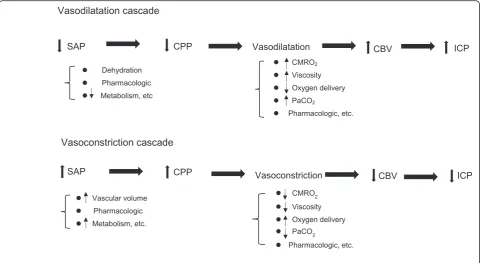
With a normally responding cerebral autoregulatory mechanism, the maximum cerebral vasoconstriction re-sponse would drive the vascular mechanism to minimize the cerebral blood volume (CBV). Changes in CBV or SAP would lead to vasodilation or constriction of brain vessels as a response in line with the previously reported vasodilation and vasoconstriction cascades [22, 23]. Many factors can initiate the vasodilation and vasocon-striction cascades, including SAP, systemic blood vol-ume, blood viscosity, oxygen delivery/metabolism, hypo/ hypercapnia, and pharmacologic agents (Fig. 2).
Cerebral vasodilation could result in decreased SAP, leading to increased CBV and ICP. If the SAP re-mains low, the CPP will drop further, accelerating the vasodilation cascade until the maximum cerebral vasodilation is attained or SAP can be stabilized. The cascade could also be initiated by hypoxemia, dehy-dration, or hypercapnia.
Conversely, stimulating a vasoconstriction cascade can sometimes be strategically useful for severe TBI patients. An increase in SAP could stimulate the cerebral vasoconstriction cascade that potentially drives a drop in CBV with a subsequent drop in ICP. If the volume regulatory response is intact (i.e., brain responds normally), an increase in CBV will also ac-celerate the vasoconstriction cascade, thereby redu-cing ICP. The vasoconstriction cascade will also contribute to fluid loading, red cell transfusion, vis-cosity reduction (this means fluid replacement in a clinical setting), or improved oxygen delivery for sys-temic management in critical care. This cascade could be clinically effective for small volume replace-ment in low-CPP patients who may be potentially dehydrated. These pressure or volume regulatory cas-cades may hint at opportunities for the next step in treatment strategies for TBI patients. However, trau-matized patients will require careful management since SAP might be maintained due to increased sys-temic vascular resistance (neurogenic hypertension) after TBI, a condition that often masks a potentially dehydrated condition.
Hyperemia after TBI
Hyperemia is associated with elevated CBV and a drop in distal cerebrovascular resistance [24] and fre-quently observed as “luxury perfusion” following is-chemia [25, 26] and/or TBI [24]. Many drivers, such as lactic acid, neuropeptides, and adenosine, generated by vasodilatory metabolites, have been considered to be part of the mechanism for causing a drop in distal cerebrovascular resistance. When pressure autoregula-tion is intact, a suitable coupling has been observed between a small rise in CBF and metabolism [27, 28]. Alternatively, dysfunctional pressure or volume auto-regulation may elicit hyperemia that is associated with intracranial hypertension and an unfavorable outcome [29–31]. If hyperemia combines with BBB disruption, capillary leakage in the dilated vascular bed may cause a brain edema to occur [32]. In the latter process, increased CBF and CBV due to vessel dila-tion with BBB disrupdila-tion may lead to aggravated
vas-cular engorgement and brain edema, ultimately
leading to “malignant brain swelling,” the develop-ment of irreversible intracranial hypertension. If the vasoconstriction cascade is intact and responding Fig. 2Vasodilation and vasoconstriction cascade in the cerebral vasculature. This cascade model was first described by Rosner in the 1990s (see references 22, 23). A cascade of this type is often trigged by changes in CPP. Any step in the cascade, however, can be triggered as the starting point. There are many triggering factors such as dehydration, vascular volume, systemic metabolism, CMRO2, blood viscosity, systemic oxygen
delivery, PaCO2, or certain pharmacologic agents.SAPsystemic arterial pressure,CPPcerebral perfusion pressure,ICPintracranial pressure,CBV
normally, hyperventilation therapy has been proposed to reduce PaCO2 levels, which might be effective for
treating brain swelling.
Management of patients with TBI
Respiratory care
The clinically critical aspect to manage patients with TBI is the minimization of secondary cerebral damage. Hyperventilation therapy for acute-phase patients with severe TBI reduces ICP and improves outcome [33, 34]. However, excessive hyperventilation induces vasocon-striction and subsequent CBF decrease that leads to brain ischemia. Unfortunately, this phenomenon is diffi-cult to detect without any neuromonitoring. A report that discusses the disturbance of cerebral oxygen metab-olism balance mentioned the following as causes: (1) hypoxia; (2) hypotension; (3) hypo/hyper PaCO2; and (4)
anemia. These were extracranial causes comprising 45 % of all causes and were equal to the incidence of dysoxy-genation caused by intracranial causes (48 %) that include increased ICP [35]. Therefore, achieving respiratory and hemodynamic stabilization is essential for preventing the progression of secondary brain injury in TBI patients.
ICP is significantly influenced by PaCO2. Based on the
cerebrovascular CO2 reactiveness, a brain blood vessel
dilatation caused by a rise in PaCO2may induce an ICP
increase and contribute to an increase in CBV (brain swelling), likely resulting in a poor outcome for patients with severe TBI. In contrast, when PaCO2 drops, the
brain blood vessel shrinks, leading to a decrease in CBV and ultimately to a drop in ICP. When hypercapnia develops after a TBI, such as an airway obstruction or respiratory insult, hyperventilation therapy may be ef-fective for decreasing the ICP when the patient’s CO2
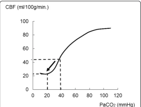
reactivity in the cerebral vasculatures is preserved. As this specific condition often occurs in a pre-hospital set-ting or an emergency room, paramedics or physicians must carefully observe the patients’ respiratory condi-tions. However, if the PaCO2value falls to 20 mmHg or
less from about 40 mmHg, the CBF might fall to half of what it was at 40 mmHg (Fig. 3, arrow), accelerating brain ischemia and causing increased ICP [36–38]. Therefore, excessive hyperventilation therapy should be avoided after TBI, especially within 24 h of the injury [39, 40].
Positive end-expiratory pressure (PEEP) is one key fac-tor for maintaining oxygenation. Application of PEEP may decrease the cerebral venous drainage by raising the intrathoracic pressure and thereby increase the CBV and ICP. PEEP may also increase ICP when the baseline ICP is lower than PEEP, but it has less effect on cerebral per-fusion when ICP is above the highest applied PEEP [41]. Hence, mild to moderate PEEP could be effective in pre-venting ventilator-associated lung injury and increased
ICP [42]. The lowest level of PEEP that maintains ad-equate oxygenation and prevents end-expiratory col-lapse, usually 5 to 8 cm H2O, is recommended. Higher
PEEP, up to 15 cm H2O, may be used in cases of
refrac-tory hypoxemia [43] in spite of its controversial effects on ICP after TBI.
Hemodynamic care
In patients with severe TBI and hypotension, acute brain swelling is often observed after SAP elevation efforts using vasopressors or excessive fluid resuscitation. Elevating SAP with large-volume fluid resuscitation or blood transfusion is one critical approach for patients with severe TBI. Although these approaches aggravate brain swelling and increase ICP, identifying dysautoregu-lation or/and BBB disruption is very difficult. BBB dis-ruption also leads to the formation of brain edema. Brain edema after TBI can be of cytotoxic or vasogenic origin [44, 45] or may be caused by capillary leakage, a risk in TBI that also leads to brain edema. Under these conditions, a high CPP may be harmful even in the case of a relatively intact autoregulation response [45].
Hemodynamic management for patients with TBI has been discussed at length [46, 47]. CPP management is one of the critical strategies that focuses on pressure response [48]. During CPP management with norepin-ephrine for increasing MAP, the risk of hyperemia could be reduced if pressure autoregulation is preserved [49]. While there is no standard regimen for patients in hemorrhagic shock with TBI complications, the goal of fluid resuscitation for these patients is 60 mmHg of CPP or greater, or if CPP of patients with severe TBI is
Fig. 3Changes in CBF related to PaCO2level variation. In the case
of respiratory acidosis, the effect of PaCO2on the cerebral vasculature
can augment cerebral blood flow (CBF). Conversely, CBF would be reduced by vasoconstriction after a drop in PaCO2. When PaCO2values
measurable, the target systolic SAP is 90–100 mmHg in-stead of achieving normal SAP.
Hypotension is frequently observed after TBI [50, 51] and might affect the outcome. An increase in endogen-ous catecholamines (sympathetic-excited catecholamine surge) causes vasoconstriction of peripheral vessels that elevates SAP (neurogenic hypertension) after TBI. As a result, SAP is maintained even if the hypovolemia exists. Mannitol has historically been used for patients with ele-vated ICP as an osmotic diuretic [52, 53]. However, excessive intravascular dehydration by inappropriate man-nitol use leads to dehydration and degrades hemodynamics to an unstable state, whereupon unanticipated hypotension occurs [51]. If intracranial hypertension is also suddenly relieved by surgical decompression craniotomy, the sympa-thetic response is eliminated, which may elicit systemic hypotension caused by reduced vascular resistance (vaso-dilation) [45]. Under conditions where the BBB is disrupted or/and cerebrovascular permeability increases after TBI, brain swelling may occur when massive fluid resuscitation and blood transfusion is administered to treat hypotension [50, 51]. To prevent catastrophic hypotension and brain swelling after TBI during critical care or surgery, the routine use of mannitol administration and intravascular dehydration should be avoided. Normovolemia must be maintained during critical care.
Monitoring CBF and metabolism balance
Jugular bulb oxygen saturation (SjO2) provides
informa-tion on global cerebral oxygen delivery and metabolism, which is used for detecting cerebral hypoperfusion, hyperperfusion, or secondary ischemic brain injury [54–56].
The normal SjO2 level is approximately 60 %. SjO2
values under 50 % is considered to be cerebrally ische-mic when accompanied by low CBF or/and CPP [54]. High SjO2 values may reflect hyperemia (higher CBF
and dilatation of blood vessels; increased CBV) or severe metabolic depression due to severe brain damage. Con-tinuous SjO2 monitoring is effective for detecting
cere-bral ischemia after TBI [57]. SjO2 monitoring is most
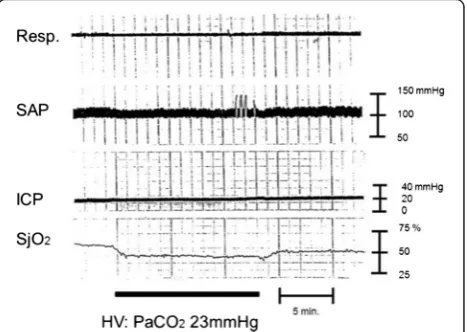
commonly used for severely brain-injured patients to de-tect post-injury brain ischemia and to monitor the effi-cacy of mannitol injection or hyperventilation therapy. If hyperventilation becomes excessive, cerebral vasocon-striction will occur and ultimately lead to further aggra-vation of cerebral perfusion of the already injured brain (reduced CPP that leads to brain ischemia). Figure 4 indicates the relationship between hyperventilation and sequential changes in SjO2. Excessive hyperventilation
can cause a drop in PaCO2, leading to vasoconstriction,
and then result in brain ischemia, based on the SjO2
level (the SjO2 value drops during excess
hyperventila-tion as demonstrated in Fig. 4). Conversely, heightened
PaCO2 values lead to higher SjO2 levels (Fig. 5). This
phenomenon is caused by the effect of greater CBV on vasodilation (vascular bed enhancement).
The vasodilatation of brain vessels is triggered by a drop in CPP with a subsequent CBV increase [22]. The drop in CPP is often associated with a decrease in SAP. CPP can be boosted by infusing fluids or by administer-ing mannitol (as a volume expander) or vasopressors, with a subsequent vasoconstriction of brain blood ves-sels [58] (Fig. 6). Finally, ICP can be lowered as a result of reduced CBV after vasoconstriction [22, 58]. Above the upper autoregulated limit, hyperperfusion may be a risk for hyperemia. Conversely, a drop in SAP at the lower limit for autoregulation response may reduce CPP and cause brain ischemia. Increased ICP levels may lead to further reductions in CPP.
Catecholamine surge after severe brain injury
Catecholamine surge is a well-known phenomenon that is observed after subarachnoid hemorrhage [59], sepsis Fig. 4Brain ischemia after hyperventilation. A female in her 40s with traumatic brain injury was transferred to the hospital by ambulance. Brain CT scan revealed acute subdural hematoma. Surgical interventions were performed, and the patient’s ICP and SjO2were monitored. The
SjO2value drops after hyperventilation. This phenomenon can be
explained by the vasoconstriction effect from reduced PaCO2. Cerebral
perfusion pressure changes might not have any remarkable effect because SAP and ICP values have been constant. Clinically, physicians would not be able to detect brain ischemia only from vital signs in this case without monitoring for brain oxygenation, such as SjO2monitoring. The ICP will stay constant even if there are changes in
the intracranial volume (e.g., the change in the volume of the vascular bed during the space compensatory phase). While the ICP will spread to the CSF space or any similar space until the compensatory effect is lost, no remarkable changes in the ICP are seen during the space compensatory phase. As a consequence, hyperventilation therapy for ICP control will not be effective in this phase. It may even cause harm via the decrease in CBF induced by excess vasoconstriction.Resp. respiration,SAPsystemic arterial pressure,ICPintracranial pressure,SjO2
[10], or TBI [13], where such elevated levels appear to influence the immune system during stress. In particular, the results from stressed subjects have highlighted a close relationship between the cytokine network,
sys-temic inflammatory response syndrome, and the
immune response [60, 61], while pro-inflammatory cyto-kines (e.g., interleukin (IL)-1) can enhance the sympa-thetic nerve activity [62, 63]. Remarkably, in vitro studies have demonstrated that epinephrine or norepin-ephrine upregulated the endotoxin-induced release of anti-inflammatory cytokine IL-10 from human periph-eral blood mononuclear cells (macrophages/monocytes), whereas tumor necrosis factor-alpha production was downregulated [64–66]. Indeed, the catecholamine surge could suppress mononuclear cell functions, which are upregulated by immunostimulatory cytokines. Such functional suppression is also observed in patients with sepsis [67, 68], burns [69], and trauma [12, 70]. This phenomenon may play an important role in early immunosuppression in patients suffering an acute stressful event.
Brain injury and hyperglycemia
Hyperglycemia is also a well-known phenomenon that is observed after stressful events such as severe brain dam-age. The adverse effects of hyperglycemia on ischemic brain injury have been well established in both the clin-ical and experimental settings. While the clinclin-ical evi-dence indicates that high blood glucose levels following TBI are linked to a greater severity of injury and poor neurological outcome [17, 18], the role of blood glucose in the secondary mechanisms of neuronal damage after TBI has not yet been clarified. Data from brain ischemia models suggest that hyperglycemia has a deleterious effect, probably due to enhanced lactic acidosis. Previous studies have demonstrated that hyperglycemia causes a Fig. 5Effect on cerebral blood flow caused by augmentation of PaCO2. A male in his 30s suffered a traffic accident. Initial CT scan demonstrated
acute subdural hematoma. Increased PaCO2could stimulate the vasodilation cascade in the brain. As a result of an increase in PaCO2, the brain
vasculature goes through vasodilation, with a subsequent increase in cerebral blood flow (and cerebral blood volume), leading to increased ICP. Physicians would be able to detect this from increased SjO2in the clinical setting.Resp.respiration,SAPsystemic arterial pressure,ICPintracranial
pressure,SjO2jugular bulb oxygen saturation,CPPcerebral perfusion pressure. Data were obtained from brain injury patient monitored at our
hospital in the 1990s
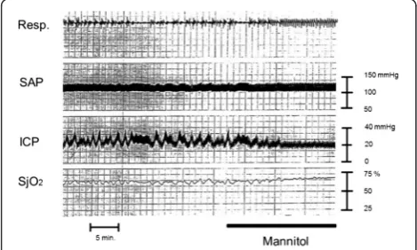
Fig. 6Effect of mannitol administration on patient with intracranial hypertension. A male in his 60s suffered traumatic brain injury. Brain CT scan demonstrated cerebral contusion. Mannitol administration is a potentially effective volume replacement method in the early phase and can stimulate the vasoconstriction cascade. SjO2values gradually
increase after mannitol administration. This phenomenon is likely caused by the volume expansion effect of mannitol, which could stimulate the vasoconstriction cascade leading to decreased CBV. Mannitol will then work as a hyperosmotic diuretic agent at the late phase resulting in decreased ICP and increased CPP.Resp. respiration,SAPsystemic arterial pressure,ICPintracranial pressure,SjO2
variety of pathological changes in the small vessels, ar-teries, and peripheral nerves. Vascular endothelial cells are a significant target of hyperglycemic damage [71], but the mechanisms underlying such damage to the cerebral microvasculature are not fully understood. Several authors have reported that hyperglycemia leads to endothelial dysfunction [72] and cerebrovascular changes both during ischemia and reperfusion [73]. Recently, nuclear factor-kappa B activation has been identified as an early event brought about by elevations in glucose, which may elicit multiple pathways contrib-uting to the initiation of hyperglycemia- or diabetes-induced endothelial cell injury. It also plays a pivotal role in early gene responses following hyperglycemia by pro-moting messenger RNA synthesis for various cell-adhesion molecules, inducible nitric oxide synthase, and cytokines or chemokines [74]. These inflammatory events are believed to contribute to the observed out-comes through secondary injury mechanisms [75, 76]. Additionally, acute inflammatory responses lead to the activation of infiltration and accumulation of poly-morphonuclear leukocytes [77].
It has been proposed that hyperglycemia may contrib-ute to endothelial cell damage in brain ischemia models [78] and TBI [79]. We have yet to gain a clear under-standing, however, of the exact mechanisms by which the neutrophil transmigration across the BBB is en-hanced under the hyperglycemic condition following TBI. Experimental studies have shown that a hypergly-cemic condition activates the intracellular signal trans-duction [80, 81] and protrans-duction of interleukin (IL)-8 [82]. The presence of tumor necrotic factor (TNF) in a high-glucose condition could enhance the production of IL-8 from endothelial cells [82]. We speculate that the hyperglycemic environment and the severe trauma asso-ciated with elevated TNF might work in combination to promote IL-8 production by vascular endothelial cells and foster neutrophil accumulation at the injury site. This, together with the hyperglycemia after TBI, may ag-gravate the endothelial cell damage and enhance the in-flammatory process, leading to neutrophil infiltration into the injured brain.
In the clinical setting, however, a frequent post-hospitalization event in patients with severe brain injury is a rapid and large increase in blood glucose concentra-tion that occurs in various situaconcentra-tions. Several quesconcentra-tions also remain as to when patients with severe brain injury should be started on glucose-containing IV fluids for maintenance alimentation, since acute hyperglycemia may influence the neurological outcome. However, the potential for acute hyperglycemia on its own to cause in-flammation in brain tissue following an acute critical ill-ness, including neutrophil accumulation, has not been investigated much.
Conclusions
Severe brain injury involves impaired autoregulation and responses in the injured brain through many mecha-nisms that lead to secondary brain injuries. Arterial hypotension, hypertension, or excess hyperventilation intended to reduce ICP in patients with damaged auto-regulation response also lead to secondary brain injury and critical brain conditions after TBI that are associated with a poor outcome. The central dysregulation mecha-nisms after brain injury could contribute to the develop-ment and progression of extracerebral organ dysfunction by promoting systemic inflammation that may cause medical complications. Neurocritical care after severe TBI has therefore been refined to focus not only on sec-ondary brain injury but also on systemic organ damage after excitation of sympathetic nerves following stress reactions.
Key points of the“pathophysiology for neurocritical care”in traumatic brain injury
Cerebral autoregulation is one of the important pressure reactivity systems in the brain. The small vessels in the brain react to hydrostatic pressure and regulate the vascular tone to maintain a constant cerebral blood flow between the mean arterial pressures of 60 and 160 mmHg. As the pressure regulation curve shifts rightward in the severely injured brain, accidental changes in systemic arterial pressure can cause severe and linear changes in cerebral blood flow that lead to harmful and irreversible conditions such as hypoperfusion (brain ischemia) or hyperperfusion (e.g., hyperemia).
Changes in cerebral blood volume or systemic arterial pressure lead to vasodilation or constriction of brain vessels. Cerebral vasodilation may result in decreased systemic arterial pressure leading to increased cerebral blood volume and intracranial pressure. The response could also be initiated by hypoxemia, dehydration, or hypocapnia due to hyperventilation therapy.
A drop in cerebral perfusion pressure triggers vasodilation of the cerebral blood vessels and subsequent increase of the cerebral blood volume. The drop in cerebral perfusion pressure is often associated with a decrease in systemic arterial pressure. Above the upper autoregulated limit, hyperperfusion may heighten the risk of hyperemia. Conversely, a drop in systemic arterial pressure at the lower limit for autoregulation response may reduce cerebral perfusion pressure and cause brain ischemia.
that leads to brain ischemia. Based on the cerebrovascular CO2 reactivity, a brain blood
vessel dilatation caused by a rise in PaCO2
may increase the intracranial pressure and contribute to an increase in the cerebral blood volume (brain swelling). The outcome is likely to be poor for patients with severe traumatic brain injury when this occurs. When PaCO2
drops, on the other hand, the brain blood vessel shrinks, leading to a decrease in cerebral blood volume and ultimately a drop in
intracranial pressure.
An increase in endogenous catecholamines (sympathetic-excited catecholamine surge) causes vasoconstriction of peripheral vessels that elevates systemic arterial pressure (neurogenic hypertension) after traumatic brain injury. As a result, systemic arterial pressure is maintained even if the
hypovolemia exists. Mannitol has historically been used for patients with elevated intracranial pressure as an osmotic diuretic. When used inappropriately, however, mannitol induces excessive intravascular dehydration. The resulting dehydration and degraded hemodynamics lead to an unstable state and unanticipated hypotension. To prevent unexpected catastrophic hypotension after TBI, the routine use of mannitol and intravascular dehydration should be avoided.
Hyperglycemia also frequently develops after severe brain damage or similarly stressful events. High blood glucose levels following traumatic brain injury are apparently associated with more severe injuries and poor neurological outcomes. Little is still known, however, about the action of blood glucose in the secondary mechanisms of neuronal damage after traumatic brain injury. The best time to commence glucose-containing IV fluids for maintenance alimentation is also uncertain, since acute hyperglycemia may alter the neurological outcome. It remains to be determined, however, if hyperglycemia alone can readily cause brain tissue inflammation after an acute critical illness involving neutrophil accumulation.
Abbreviations
BBB:blood–brain barrier; CBF: cerebral blood flow; CBV: cerebral blood volume; CPP: cerebral perfusion pressure; ICP: intracranial pressure; MAP: mean arterial pressure; SAP: systemic arterial pressure; SjO2: jugular
bulb oxygen saturation; TBI: traumatic brain injury.
Competing interests
The author declares that he has no competing interests.
Received: 31 August 2015 Accepted: 4 February 2016
References
1. Gentile NT, McIntosh TK. Antagonists of excitatory amino acids and endogenous opioid peptides in the treatment of experimental central nervous system injury. Ann Emerg Med. 1993;22:1028–34.
2. Faden AI, Demediuk P, Panter SS, Vink R. The role of excitatory amino acids and NMDA receptors in traumatic brain injury. Science. 1989;19(244): 798–800.
3. Xiong Y, Gu Q, Peterson PL, Muizelaar JP, Lee CP. Mitochondrial dysfunction and calcium perturbation induced by traumatic brain injury. J Neurotrauma. 1997;14:23–34.
4. Yakovlev AG, Knoblach SM, Fan L, Fox GB, Goodnight R, Faden AI. Activation of CPP32-like caspases contributes to neuronal apoptosis and neurological dysfunction after traumatic brain injury. J Neurosci. 1997;17:7415–24. 5. Hall ED, Detloff MR, Johnson K, Kupina NC. Peroxynitrite-mediated protein
nitration and lipid peroxidation in a mouse model of traumatic brain injury. J Neurotrauma. 2004;21:9–20.
6. Morganti-Kossmann MC, Satgunaseelan L, Bye N, Kossmann T. Modulation of immune response by head injury. Injury. 2007;38:1392–400.
7. Loane DJ, Faden AI. Neuroprotection for traumatic brain injury: translational challenges and emerging therapeutic strategies. Trends Pharmacol Sci. 2010;31:596–604.
8. McIntosh TK, Smith DH, Meaney DF, Kotapka MJ, Gennarelli TA, Graham DI. Neuropathological sequelae of traumatic brain injury: relationship to neurochemical and biomechanical mechanisms. Lab Invest. 1996;74:315–42.
9. Gruber A, Reinprecht A, Illievich UM, Fitzgerald R, Dietrich W, Czech T, et al. Extracerebral organ dysfunction and neurologic outcome after aneurysmal subarachnoid hemorrhage. Crit Care Med. 1999;27:505–14.
10. Johansson PI, Haase N, Perner A, Ostrowski SR. Association between sympathoadrenal activation, fibrinolysis, and endothelial damage in septic patients: a prospective study. J Crit Care. 2014;29:327–33.
11. Hamill RW, Woolf PD, McDonald JV, Lee LA, Kelly M. Catecholamines predict outcome in traumatic brain injury. Ann Neurol. 1987;21:438–43.
12. Woiciechowsky C, Asadullah K, Nestler D, Eberhardt B, Platzer C, Schöning B, et al. Sympathetic activation triggers systemic interleukin-10 release in immunodepression induced by brain injury. Nat Med. 1998;4:808–13.
13. Schroeppel TJ, Fischer PE, Zarzaur BL, Magnotti LJ, Clement LP, Fabian TC, et al. Beta-adrenergic blockade and traumatic brain injury: protective? J Trauma. 2010;69:776–82.
14. Clifton GL, Ziegler MG, Grossman RG. Circulating catecholamines and sympathetic activity after head injury. Neurosurgery. 1981;8:10–4. 15. Yoshimoto Y, Tanaka Y, Hoya K. Acute systemic inflammatory response
syndrome in subarachnoid hemorrhage. Stroke. 2001;32:1989–93. 16. Dunser MW, Hasibeder WR. Sympathetic overstimulation during critical
illness: adverse effects of adrenergic stress. J Intensive Care Med. 2009;24: 293–316.
17. Michaud LJ, Rivara FP, Longstreth Jr WT, Grady MS. Elevated initial blood glucose levels and poor outcome following severe brain injuries in children. J Trauma. 1991;31:1356–62.
18. Rovlias A, Kotsou S. The influence of hyperglycemia on neurological outcome in patients with severe head injury. Neurosurgery. 2000;46:335–43. 19. Aaslid R, Lindegaard KF, Sorteberg W, Nornes H. Cerebral autoregulation
dynamics in humans. Stroke. 1989;20:45–52.
20. Czosnyka M, Brady K, Reinhard M, Smielewski P, Steiner LA. Monitoring of cerebrovascular autoregulation: facts, myths, and missing links. Neurocrit Care. 2009;10:373–86.
21. Lang EW, Chesnut RM. A bedside method for investigating the integrity and critical thresholds of cerebral pressure autoregulation in severe traumatic brain injury patients. Br J Neurosurg. 2000;14:117–26.
22. Rosner MJ, Rosner SD, Johnson AH. Cerebral perfusion pressure: management protocol and clinical results. J Neurosurg. 1995;83:949–62. 23. Rosner MJ, Daughton S. Cerebral perfusion pressure management in head
injury. J Trauma. 1990;30:933–41.
24. Martin NA, Patwardhan RV, Alexander MJ, Africk CZ, Lee JH, Shalmon E, et al. Characterization of cerebral hemodynamic phases following severe head trauma: hypoperfusion, hyperemia, and vasospasm. J Neurosurg. 1997; 87:9–19.
26. De Salles AA, Muizelaar JP, Young HF. Hyperglycemia, cerebrospinal fluid lactic acidosis, and cerebral blood flow in severely head-injured patients. Neurosurgery. 1987;21:45–50.
27. Sakas DE, Bullock MR, Patterson J, Hadley D, Wyper DJ, Teasdale GM. Focal cerebral hyperemia after focal head injury in humans: a benign phenomenon? J Neurosurg. 1995;83:277–84.
28. Dias C, Maia I, Cerejo A, Varsos G, Smielewski P, Paiva JA, et al. Pressures, flow, and brain oxygenation during plateau waves of intracranial pressure. Neurocrit Care. 2014;21:124–32.
29. Kelly DF, Martin NA, Kordestani R, Counelis G, Hovda DA, Bergsneider M, et al. Cerebral blood flow as a predictor of outcome following traumatic brain injury. J Neurosurg. 1997;86:633–41.
30. Kelly DF, Kordestani RK, Martin NA, Nguyen T, Hovda DA, Bergsneider M, et al. Hyperemia following traumatic brain injury: relationship to intracranial hypertension and outcome. J Neurosurg. 1996;85:762–71.
31. Ojha BK, Jha DK, Kale SS, Mehta VS. Trans-cranial Doppler in severe head injury: evaluation of pattern of changes in cerebral blood flow velocity and its impact on outcome. Surg Neurol. 2005;64:174–9.
32. Chieregato A, Noto A, Tanfani A, Bini G, Martino C, Fainardi E. Hyperemia beneath evacuated acute subdural hematoma is frequent and prolonged in patients with an unfavorable outcome: a xe-computed tomographic study. Neurosurgery. 2009;64:705–18.
33. Ghajar JB, Hariri RJ, Patterson RH. Improved outcome from traumatic coma using only ventricular CSF drainage for ICP control. Adv Neurosurg. 1993;21: 173–7.
34. Becker DP, Miller JD, Ward JD, Greenberg RP, Young HF, Sakalas R. The outcome from severe head injury with early diagnosis and intensive management. J Neurosurg. 1977;47:491–502.
35. Woodman T, Robertson CS. Jugular venous oxygen saturation monitoring; In: Narayan RK, Wilberger JE, Povlishock JT, editors. Neurotrauma. New York, NY: McGraw-Hill; 1996. P. 519-537.
36. Sato M, Pawlik G, Heiss WD. Comparative studies of regional CNS blood flow autoregulation and responses to CO2in the cat. Effects of altering
arterial blood pressure and PaCO2 on rCBF of cerebrum, cerebellum, and spinal cord. Stroke. 1984;15:91–7.
37. Smith AL, Wollman H. Cerebral blood flow and metabolism: effects of anesthetic drugs and techniques. Anesthesiology. 1972;36:378–400. 38. Souter MJ, Lam AM. Neurocritical Care. In: Miller RD, Eriksson LI, Fleisher L,
Wiener-Kronish JP, Young WL, editors. Miller’s anesthesia, 7th Edition. Philadelphia, PA: Churchill Livingstone; 2009. p. 2899–2921.
39. Muizelaar JP, Marmarou A, Ward JD, Kontos HA, Choi SC, Becker DP, et al. Adverse effects of prolonged hyperventilation in patients with severe head injury: a randomized clinical trial. J Neurosurg. 1991;75:731–9.
40. Sheinberg M, Kanter MJ, Robertson CS, Contant CF, Narayan RK, Grossman RG. Continuous monitoring of jugular venous oxygen saturation in head-injured patients. J Neurosurg. 1992;76:212–7.
41. McGuire G, Crossley D, Richards J, Wong D. Effects of varying levels of positive end-expiratory pressure on intracranial pressure and cerebral perfusion pressure. Crit Care Med. 1997;25:1059–62.
42. Mascia L, Grasso S, Fiore T, Bruno F, Berardino M, Ducati A. Cerebro-pulmonary interactions during the application of low levels of positive end-expiratory pressure. Intensive Care Med. 2005;31:373–9.
43. Haddad SH, Arabi YM. Critical care management of severe traumatic brain injury in adults. Scand J Trauma Resusc Emerg Med. 2012;20:12. 44. Barzo P, Marmarou A, Fatouros P, Hayasaki K, Corwin F. Contribution of
vasogenic and cellular edema to traumatic brain swelling measured by diffusion-weighted imaging. J Neurosurg. 1997;87:900–7.
45. Asgeirsson B, Grände PO, Nordström CH. A new therapy of post-trauma brain oedema based on haemodynamic principles for brain volume regulation. Intensive Care Med. 1994;20:260–7.
46. Czosnyka M, Smielewski P, Piechnik S, Schmidt EA, Seeley H, Al-Rawi P, et al. Continuous assessment of cerebral autoregulation—clinical verification of the method in head injured patients. Acta Neurochir Suppl. 2000;76:483–4.
47. Steiner LA, Czosnyka M, Piechnik SK, Smielewski P, Chatfield D, Menon DK, et al. Continuous monitoring of cerebrovascular pressure reactivity allows determination of optimal cerebral perfusion pressure in patients with traumatic brain injury. Crit Care Med. 2002;30:733–8.
48. Howells T, Elf K, Jones PA, Ronne-Engström E, Piper I, Nilsson P, et al. Pressure reactivity as a guide in the treatment of cerebral perfusion pressure in patients with brain trauma. J Neurosurg. 2005;102:311–7.
49. Mascia L, Andrews PJ, McKeating EG, Souter MJ, Merrick MV, Piper IR. Cerebral blood flow and metabolism in severe brain injury: the role of pressure autoregulation during cerebral perfusion pressure management. Intensive Care Med. 2000;2:202–5.
50. Chesnut RM, Gautille T, Blunt BA, Klauber MR, Marshall LF. Neurogenic hypotension in patients with severe head injuries. J Trauma. 1998;44:958–64. 51. Kinoshita K, Kushi H, Sakurai A, Utagawa A, Saito T, Moriya T, et al. Risk
factors for intraoperative hypotension in traumatic intracranial hematoma. Resuscitation. 2004;60:151–5.
52. Cruz J, Minoja G, Okuchi K, Facco E. Successful use of the new high-dose mannitol treatment in patients with Glasgow Coma Scale scores of 3 and bilateral abnormal pupillary widening: a randomized trial. J Neurosurg. 2004; 100:376–83.
53. Cruz J, Minoja G, Okuchi K. Improving clinical outcomes from acute subdural hematomas with the emergency preoperative administration of high doses of mannitol: a randomized trial. Neurosurgery. 2001;49:864–71. 54. Dunn IF, Ellegala DB, Kim DH, Litvack ZN. Neuromonitoring in neurological
critical care: neuromonitoring in neurological critical care. Neurocrit Care. 2006;4:83–92.
55. Macmillan CS, Andrews PJ. Cerebrovenous oxygen saturation monitoring: practical considerations and clinical relevance. Intensive Care Med. 2000;26: 1028–36.
56. Cruz J. The first decade of continuous monitoring of jugular bulb oxyhemoglobinsaturation: management strategies and clinical outcome. Crit Care Med. 1998;26:344–51.
57. Latronico N, Beindorf AE, Rasulo FA, Febbrari P, Stefini R, Cornali C, et al. Limits of intermittent jugular bulb oxygen saturation monitoring in the management of severe head trauma patients. Neurosurgery. 2000;46: 1131–8.
58. Rosner MJ, Coley I. Cerebral perfusion pressure: a hemodynamic mechanism of mannitol and the postmannitol hemogram. Neurosurgery. 1987;21: 147–56.
59. Kinoshita K, Yamaguchi J, Sakurai A, Ebihara T, Furukawa M, Tanjoh K. Inhibition of lipopolysaccharide stimulated interleukin-1beta production after subarachnoid hemorrhage. Neurol Res. 2007;29:47–52.
60. Elrifai AM, Bailes JE, Shih SR, Dianzumba S, Brillman J. Characterization of the cardiac effects of acute subarachnoid hemorrhage in dogs. Stroke. 1996;27: 737–42.
61. Severn A, Rapson NT, Hunter CA, Liew FY. Regulation of tumor necrosis factor production by adrenaline and beta-adrenergic agonists. J Immunol. 1992;148:3441–5.
62. Ichijo T, Katafuchi T, Hori T. Central interleukin-1 beta enhances splenic sympathetic nerve activity in rats. Brain Res Bull. 1994;34:547–53. 63. Madden KS, Sanders VM, Felten DL. Catecholamine influences and
sympathetic neural modulation of immune responsiveness. Annu Rev Pharmacol Toxicol. 1995;35:417–48.
64. Chelmicka-Schorr E, Kwasniewski MN, Czlonkowska A. Sympathetic nervous system and macrophage function. Ann N Y Acad Sci. 1992;650:40–5. 65. Meisel C, Vogt K, Platzer C, Randow F, Liebenthal C, Volk HD. Differential
regulation of monocytic tumor necrosis factor-alpha and interleukin-10 expression. Eur J Immunol. 1996;26:1580–6.
66. van der Poll T, Coyle SM, Barbosa K, Braxton CC, Lowry SF. Epinephrine inhibits tumor necrosis factor-alpha and potentiates interleukin 10 production during human endotoxemia. J Clin Invest. 1996;97:713–9. 67. Munoz C, Carlet J, Fitting C, Misset B, Blériot JP, Cavaillon JM. Dysregulation
of in vitro cytokine production by monocytes during sepsis. J Clin Invest. 1991;88:1747–54.
68. Volk HD, Reinke P, Krausch D, Zuckermann H, Asadullah K, Müller JM, et al. Monocyte deactivation—rationale for a new therapeutic strategy in sepsis. Intensive Care Med. 1996;22 Suppl 4:S474–81.
69. Zedler S, Bone RC, Baue AE, von Donnersmarck GH, Faist E. T-cell reactivity and its predictive role in immunosuppression after burns. Crit Care Med. 1999;27:66–72.
70. Flohé S, Lendemans S, Schade FU, Kreuzfelder E, Waydhas C. Influence of surgical intervention in the immune response of severely injured patients. Intensive Care Med. 2004;30:96–102.
71. Nishikawa T, Edelstein D, Du XL, Yamagishi S, Matsumura T, Kaneda Y, et al. Normalizing mitochondrial superoxide production blocks three pathways of hyperglycaemic damage. Nature. 2000;404:787–90.
73. Kawai N, Keep RF, Betz AL. Hyperglycemia and the vascular effects of cerebral ischemia. Stroke. 1997;28:149–54.
74. Pieper GM, Riaz-ul-Haq. Activation of nuclear factor-kappaB in cultured endothelial cells by increased glucose concentration: prevention by calphostin C. J Cardiovasc Pharmacol. 1997;30:528–32.
75. Clark RS, Schiding JK, Kaczorowski SL, Marion DW, Kochanek PM. Neutrophil accumulation after traumatic brain injury in rats: comparison of weight drop and controlled cortical impact models. J Neurotrauma. 1994;11:499–506. 76. Chatzipanteli K, Alonso OF, Kraydieh S, Dietrich WD. Importance of
posttraumatic hypothermia and hyperthermia on the inflammatory response after fluid percussion brain injury: biochemical and
immunocytochemical studies. J Cereb Blood Flow Metab. 2000;20:531–42. 77. Soares HD, Hicks RR, Smith D, McIntosh TK. Inflammatory leukocytic
recruitment and diffuse neuronal degeneration are separate pathological processes resulting from traumatic brain injury. J Neurosci. 1995;15:8223–33. 78. Lin B, Ginsberg MD, Busto R, Li L. Hyperglycemia triggers massive neutrophil
deposition in brain following transient ischemia in rats. Neurosci Lett. 2000; 278:1–4.
79. Kinoshita K, Kraydieh S, Alonso O, Hayashi N, Dietrich WD. Effect of posttraumatic hyperglycemia on contusion volume and neutrophil accumulation after moderate fluid-percussion brain injury in rats. J Neurotrauma. 2002;19:681–92.
80. Wilmer WA, Dixon CL, Hebert C. Chronic exposure of human mesangial cells to high glucose environments activates the p38 MAPK pathway. Kidney Int. 2001;60:858–71.
81. Igarashi M, Wakasaki H, Takahara N, Ishii H, Jiang ZY, Yamauchi T, et al. Glucose or diabetes activates p38 mitogen-activated protein kinase via different pathways. J Clin Invest. 1999;103:185–95.
82. Kinoshita K, Tanjoh K, Noda A, Sakurai A, Yamaguchi J, Azuhata T, et al. Interleukin-8 production from human umbilical vein endothelial cells during brief hyperglycemia: the effect of tumor necrotic factor-alpha. J Surg Res. 2008;144:127–31.
• We accept pre-submission inquiries
• Our selector tool helps you to find the most relevant journal
• We provide round the clock customer support
• Convenient online submission
• Thorough peer review
• Inclusion in PubMed and all major indexing services
• Maximum visibility for your research
Submit your manuscript at www.biomedcentral.com/submit