R E S E A R C H
Open Access
Modification of platinum sensitivity by
KEAP1/NRF2 signals in non-small cell lung
cancer
Yijun Tian
†, Kongming Wu
†, Qian Liu, Na Han, Li Zhang, Qian Chu
*and Yuan Chen
*Abstract
Background:The objective of this study was to evaluate the effect of platinum-based drugs on nuclear-factor erythroid2 like 2 (NRF2) signaling in non-small cell lung cancer cell lines with or without Kelch-like ECH-associated protein 1 (KEAP1) mutations and to determine the role of NRF2 and KEAP1 on platinum-based drug treatment.
Methods:We used real-time PCR to assess relative mRNA expression and used western blotting and
immunofluorescence assays to assess protein expression. Small interfering RNA and shuttle plasmids were used to modulate the expression of NRF2, wild-type KEAP1, and mutant KEAP1. Drug sensitivity to platinum-based drugs was evaluated with Cell Count Kit-8.
Results:We found that platinum-based therapies modified the NRF2 signaling pathway differently in KEAP1-mutated non-small cell lung cancer (NSCLC) cell lines compared with wild-type KEAP1 cell lines. The reactive degree of NRF2 signaling also varies between nedaplatin and cisplatin. The modification of NRF2 or KEAP1 expression in NSCLC cell lines disrupted downstream gene expression and cell sensitivity to platinum-based drugs. Finally, gene expression data retrieved from The Cancer Genome Atlas (TCGA) consortium indicated that KEAP1 mutation significantly affects NRF2 signaling activity in patients with NSCLC.
Conclusions:Our findings suggest that NRF2 signaling plays an indispensable role in NSCLC cell sensitivity to platinum-based treatments and provides a rationale for using NRF2 as a specific biomarker for predicting which patients will be most likely to benefit from platinum-based treatment.
Keyword:NRF2, KEAP1, Non-small cell lung cancer, HO1, NQO1, GCLM, Nedaplatin, Cisplatin
Abbreviations:KEAP1, Kelch-like ECH-associated protein 1; NRF2, Nuclear-factor erythroid2 like 2; NSCLC, Non-small cell lung cancer; BTB, Broad complex, tramtrack, and bric à brac; IVR, Intervention region; wt, Wild type;
mut, Mutation; HO1, Heme oxygenase-1; NQO1, NAD (P) H-quinone oxidoreductase 1; GCLM, Glutamate-cysteine ligase modifier subunit; TCGA, The Cancer Genome Atlas; CDS, Coding sequence; EGFR-TKI, Epidermal growth factor receptor tyrosine kinase inhibitor; EML4-ALK-TKI, Echinoderm microtubule-associated protein-like 4-anaplastic lymphoma kinase fusion tyrosine kinase inhibitor; ARE, Antioxidant responsive element
* Correspondence:[email protected];[email protected]
†Equal contributors
Department of Oncology, Tongji Hospital of Tongji Medical College, Huazhong University of Science and Technology, 1095 Jiefang Avenue, Wuhan 430030, China
Background
Non-small cell lung cancer (NSCLC) is associated with considerably high mortality worldwide [1]. Surgery can ensure local disease control in patients with early stages of disease, and patients unsuitable for surgery at diagnosis, primarily those with advanced stage dis-ease, receive chemotherapy or targeted therapy as a first line of treatment. Epidermal growth factor recep-tor tyrosine kinase inhibirecep-tor (EGFR-TKI) and echino-derm microtubule-associated protein-like 4-anaplastic lymphoma kinase fusion tyrosine kinase inhibitor (EML4-ALK-TKI) exert substantial therapeutic effects in lung adenocarcinomas with specific commonly oc-curring mutations [2–5]. Platinum-based treatment regimens remain the standard of care recommended by the lung cancer guidelines for squamous lung can-cer or adenocarcinoma with undefined mutation status [6]. Recently, studies [7, 8] have revealed that a nedaplatin-based treatment regimen is associated with a favorable response in patients with squamous cell lung cancer. Platinum-based drugs are defined as DNA adduct-forming agents that exert antineoplastic effects by inducing the formation of double-stranded DNA cross-links, thus resulting in irreversible DNA damage [9]. Accumulating DNA damage eventually leads to apoptosis [10, 11].
Nuclear-factor (NF)-E2-related factor 2 (NRF2) is a core transcription factor involved in the cellular re-sponse to oxidative stress [12]. Under basal conditions, NRF2 forms a complex with Kelch-like ECH-associated protein-1 (KEAP1), which is targeted for ubiquitination. Upon exposure to stress or electrophilic regents, NRF2 evades the ubiquitination process and translocates from the cytoplasm to the nucleus [13]. By binding to antioxi-dant responsive element (ARE), NRF2 increases the transcription of multiple cytoprotective enzymes [14] such as heme oxygenase-1(HO1), NAD (P) H dehydro-genase (quinone) 1 (NQO1), and glutamate-cysteine lig-ase modifier (GCLM) [15].
In urethane-induced lung carcinogenesis model, NRF2 activation suppresses tumorigenesis, but somatic activation of NRF2 in cancer cells enhanced growth of tumors [16]. Studies have demonstrated that NRF2 acti-vation contributes to chemo-resistance [17–19] or radio-resistance [20] in lung cancers. Investigators have also focused on the relationship between NRF2 activity and platinum treatment in NSCLC cell line [21, 22]. However, studies designed to observe and compare NRF2 signaling response under different platinum treatment are rarely reported [23]. In this study, we sought to elucidate the role of NRF2 in platinum-based chemotherapy for NSCLC, and we identified KEAP1 mutation as a key factor contributing to platinum sensitivity.
Methods
Chemicals and cell culture
Unless otherwise stated, all chemicals were purchased from Sigma-Aldrich (Shanghai, China). Antibodies against NRF2 (ab62352) were purchased from Abcam; antibodies against HO1 (#5853) and NQO1 (#3187) were purchased from Cell Signaling Technology; and antibodies against KEAP1 (10503-2-AP), GCLM (14241-1-AP), andβ-actin were purchased from Proteintech.
The A549 (human lung adenocarcinoma), H292 (human mucoepidermoid carcinoma), H460 (human large cell lung cancer), and SKMES-1 (human squamous lung cancer) cell lines were obtained from the Cell Resource Center, Peking Union Medical College. The cells were cultured in RPMI-1640, with the exception of SKMES-1 cells, which were cul-tured in MEM. All media were supplemented with 10 % fetal bovine serum (GIBCO, NY, USA). No antibiotics were added to the media.
Plasmid and siRNA design
The wild-type KEAP1 coding sequence (CDS) was cloned into the pEnter shuttle plasmid. The Fast Mutagenesis sys-tem (Transgene, Beijing, China) was used to introduce the G333C and D236H mutations. The primers used were:
G333C-forward, TGATCTACA CCGCGGGCTGCTACTTCCGACAGTC G333C-reverse,
AGCCCGCGGTGTAGATCAGGCGGCCCACC D236H-forward,
AGCCCGCGGTGTAGATCAGGCGGCCCACC D236H-reverse,
GGTCCCGGCTGATGAGGGTCACCAGTTGG
The small interfering RNAs used to silence NRF2 were: Sense, GGUUGAGACUACCAUGGUUTT
Antisense, AACCAUGGUAGUCUCAACCTT
Transfection was conducted with Lipofectamine 2000 (Invitrogen) according to a previously published method [24].
Real-time PCR analysis
expression fold-changes according to the 2−△△CTalgorithm. The primers used for real-time PCR are:
NRF2, forward, TCCAGTCAGAAACCAGTGGAT Reverse, GAATGTCTGCGCCAAAAGCTG
HO1, forward, AAGATTGCCCAGAAAGCCCTGGAC Reverse, AACTGTCGCCACCAGAAAGCTGAG NQO1, forward, GAAGAGCACTGATCGTACTGGC Reverse, GGATACTGAAAGTTCGCAGGG
GCLM, forward, TGTCTTGGAATGCACTGTATCTC Reverse, CCCAGTAAGGCTGTAAATGCTC
KEAP1 mutation sequencing
Total RNA was extracted and used to synthesize the first chain cDNA according to the procedure described in the real-time PCR analysis description. cDNA was used as a template to synthesize six segments of the KEAP1 gene using the following cycling conditions: initial denaturing step at 94 °C for 5 min followed by 30 cycles at 94 °C for 40 s, 60 °C for 40 s, and 72 °C for 55 s. Products were separated on 1 % agarose gels, and bands were visualized with ethidium bromide. The products were sequenced using the ABI3730 XL DNA Analyzer (Applied Biosys-tem Japan, Tokyo, Japan) and analyzed with Chromas 2.4.3 software. Primers used to amplify the KEAP1 gene fragments were:
Fragment 1, forward AGAGGTGGTGGTGTTGCTTAT Reverse TGGAGATGGAGGCCGTGTA
Fragment 2, forward CAGGTCAAGTACCAGGATG Reverse GATGAGGGTCACCAGTTG
Fragment 3, forward ATCGGCATCGCCAACTTC Reverse AGGTAGCTGAGCGACTGT
Fragment 4, forward CAGAAGTGCGAGATCCTG Reverse GCTCTGGCTCATACCTCT
Fragment 5, forward GCCCTGGACTGTTACAAC Reverse GTCTCTGTTTCCACATCGTA
Fragment 6, forward GCTGTCCTCAATCGTCTC Reverse AGTTCTGCTGGTCAATCTG
NRF2 exon2, forward TCGTGATGGACTTGGAGCTG Reverse AGCATCTGATTTGGGAATGTG
Sections were spliced together after manual inspection and compared with the reference sequence with BLAST to identify potential mutations.
Western blot analysis
Cytoplasm and nuclear protein were extracted by using NE-PER™ Nuclear and Cytoplasmic Extraction Reagents (ThermoFisher Scientific). For total protein extraction, we used standard protocols for protein isolation and antibody detection, as previously described [24, 25]. Cells were washed twice with ice-cold PBS and then
lysed with RIPA buffer. Each sample was denatured in 100 °C boiling water in the presence of SDS and DTT. A total of 20 μg of protein from each sample was loaded onto a 10 % SDS-PAGE gel for separation by electro-phoresis. A tank system was used to transfer proteins from gels to PVDF membranes. The membranes were blocked with nonfat milk for 1 h and incubated with pri-mary antibodies overnight. All pripri-mary antibodies were used at a dilution of 1:1000, with the exception of anti-β-actin (1:5000) and anti-NQO1 (1:3000). Blots were sufficiently washed in 0.1 % Tris-buffered saline and Tween20 (TBST) four to six times for at least 5 min per wash. Washed membranes were incubated with second-ary antibodies at a dilution of 1:5000 for 1 h at room temperature. After incubation with secondary antibodies, the membranes were washed three times for at least 5 min per wash. SuperSignal West Pico Chemilumines-cent Substrate (ThermoFisher Scientific) was applied for exposure using Syngene G: BOX F3 Fluorescence Im-aging System. All images of the blots were processed in Photoshop without gray value modification and spliced in Microsoft PowerPoint.
Immunofluorescence staining
Immunofluorescence was performed as previously re-ported with some modifications [26]. Cells were seeded onto a 24-well plate until they reached a confluence of 50~60 % and were treated with the indicated reagents. After treatment, cells were fixed in 4 % paraformalde-hyde at 4 °C for 10 min. The cells were then washed in PBS for 5 min and permeabilized and blocked with 0.1 % Triton X-100 and 1 % bovine serum albumin PBS for 20 min. Cells were incubated with primary antibody against HO1 at a dilution of 1:200 at 4 °C overnight. The cells were then washed in PBS three times for 5 min per wash. Cells were incubated with Alexa Fluor 488® sec-ondary antibody (Invitrogen) at a dilution of 1:1000 for 1 h at room temperature. DAPI solution was added after incubation with secondary antibody. Cells were washed three times for 5 min per wash before imaging. Fluores-cence images were captured using a Leica digital micro-scope (Leica Microsystems, Wetzler, Germany). Images were merged using Adobe Photoshop CS6, and no other modifications were made.
Cytotoxic assay
was added to each well. Absorbance at 490 nm was mea-sured after a 2-h incubation at 37 °C. Cell survival was cal-culated as the ratio between the treated group and control groups. Ratios were graphed in line charts with Graphpad Prism 6 software.
Public database analysis
Expression data from The Cancer Genome Atlas (TCGA) squamous lung cancer patients was retrieved using the package TCGA-Assembler (http://www.compgenome.org/ TCGA-Assembler/) in an R program. KEAP1 mutation status was defined as previously described [28]. Specimens with a confirmed KEAP1 mutation status were included for evaluation of relative gene expression values.
Statistical methods
Parametric tests were used to evaluate data with a nor-mal distribution. Non-parametric tests were used to evaluate data with unknown distribution patterns. mRNA fold-changes were compared using Student’s t tests. Survival comparisons were conducted using paired t tests. Comparisons between the mutant and wild-type KEAP1 groups were conducted using Mann-Whitney tests. Significance was set atP< 0.05.
Results
KEAP1 mutation status influenced the expression of NRF2 downstream target genes in cells exposed to platinum-based treatment
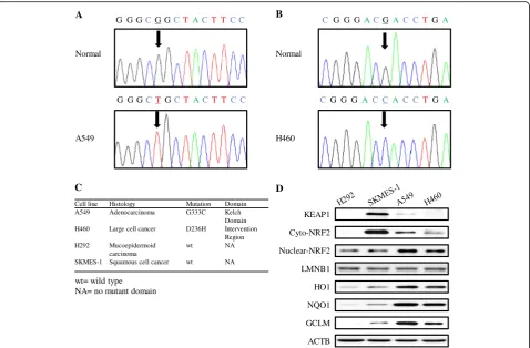
KEAP1 is a key regulator of NRF2 function, and the role of wild-type KEAP1 in negatively regulating NRF2 sig-naling has been well characterized. We first sequenced the KEAP1 gene in four NSCLC cell lines to identify po-tential mutations and found that the A549 cell line har-bors a G333C mutation within the first Kelch domain (KLD) (Fig. 1a) and that the H460 cell line harbors a D236H mutation in the intervention region (IVR) (Fig. 1b). No KEAP1 mutations were identified in the H292 and SKMES-1 cell lines. Sequencing of NRF2 exon 2 in the four NSCLC cell lines demonstrated that no mutations existed in this locus. We then evaluated basal protein levels of KEAP1 and components of the NRF2 signaling pathway in the four NSCLC cell lines. NRF2 protein in nuclear extracts and the proteins encoded by the three genes downstream of NRF2, HO1, NQO1, and GCLM were more highly expressed in A549 and H460 cells than in H292 and SKMES-1 cells (Fig. 1d).
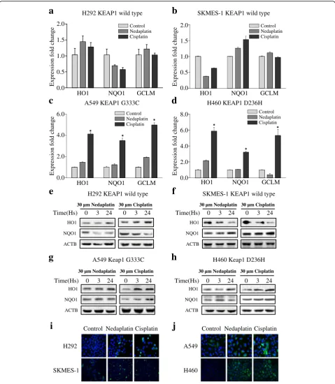
To profile the expression of NRF2 signaling pathway components in response to platinum treatment, the four NSCLC cell lines (H292, SKMES-1, A549, and H460) were treated with 30 μM nedaplatin or 30μM cisplatin for 24 h. Total RNA from each group was extracted, and HO1, NQO1, and GCLM gene expression levels were assessed with real-time PCR. In the wild-type KEAP1
cell lines H292 (Fig. 2a) and SKMES-1 (Fig. 2b), neda-platin and cisneda-platin slightly enhanced the expression of NRF2 downstream target genes after 24 h of treatment. In the mutant KEAP1 cell line A549 (Fig. 2c, *p< 0.01, **p< 0.05) and H460 (Fig. 2d, *p< 0.01, **p< 0.05), neda-platin induced a weak but significant increase in three downstream gene expression. Cisplatin treatment, how-ever, significantly enhanced HO1, NQO1, and GCLM expression in both A549 and H460 cell lines after 24 h of treatment.
To evaluate protein levels, the four cell lines were treated with 30 μM nedaplatin or 30 μM cisplatin for 3 or 24 h. Total proteins were extracted, and HO1, NQO1, GCLM protein expression was evaluated by using western blot analysis. In the wild-type KEAP1 H292 and SKMES-1 cell lines (Fig. 2e, f ), a mild increase or no change of NQO1 and HO1 protein was observed after 3- or 24-h treatment with either platinum-based drug. In the mutant KEAP1 cell lines A549 (Fig. 2g) and H460 (Fig. 2h), a mild increase or no change was ob-served in NQO1 and HO1 protein levels after nedaplatin treatment, whereas the expression of these proteins was substantially enhanced after 24 h of treatment with cisplatin.
The results of immunofluorescence analysis of HO1 protein expression were consistent with the results of the western blot analysis. Over a 24-h treatment period, 30μM nedaplatin or cisplatin induced a mild increase in HO1 protein expression in H292 and SKMES-1 cells (Fig. 2i). In the A549 and H460 cell lines (Fig. 2j), both platinum-based drugs enhanced the expression of HO1 to a greater degree over the same time period.
KEAP1 mutations modified NRF2 signaling and influenced platinum sensitivity
To investigate the impact of KEAP1 mutations status on NRF2 signaling, the KEAP1 CDS was cloned into the pEnter vector and transfected into the NSCLC cell lines. In the mutant KEAP1 cell lines A549 (Fig. 3a, b, *p< 0.001, **p< 0.05) and H460 (Fig. 3e, f, *p< 0.001), consti-tutive expression of wild-type KEAP1 over a 72-h period reduced the expression of the NRF2 downstream target genes HO1, NQO1, and GCLM at the mRNA and pro-tein levels. Overexpression of wild-type KEAP1 sensi-tized A549 cells (Fig. 3c, d, paired t test p= 0.0118 for nedaplatin, paired t test p= 0.0046 for cisplatin) and H460 cells (Fig. 3g, h, paired ttestp= 0.0089 for neda-platin, pairedttestp= 0.0134 for cisplatin) to nedaplatin and cisplatin over a 24-h treatment period.
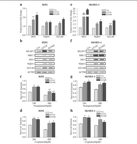
(Fig. 4e, *p< 0.001, **p< 0.05) transfected with the mu-tant KEAP1 construct. An identical trend was observed at the protein level (Fig. 4b, f ). Transfection of the mu-tant KEAP1 plasmid significantly increased survival in H292 cells (Fig. 4c, d, *p< 0.001, **p< 0.05) and SKMES-1 (Fig. 4g, h, *p< 0.001) treated with 60 μM cisplatin or nedaplatin.
NRF2 activity altered downstream gene expression and influenced platinum sensitivity
To assess the effects of NRF2 expression on NSCLC cells exposed to platinum-based drugs, we designed a small interfering RNA (siRNA) construct targeting NRF2 mRNA to inhibit NRF2 activity. In the wild-type KEAP1 cell lines H292 (Fig. 5a, b, *p< 0.001, **p< 0.05) and SKMES-1 (Fig. 5e, f, *p< 0.001, **p< 0.05), siRNA trans-fection over a 72-h period reduced the expression of NRF2 and its downstream targets HO1, NQO1, and GCLM at the mRNA and protein levels. Pretreatment with siRNA targeting NRF2 increased sensitivity to neda-platin and cisneda-platin in H292 cells (Fig. 5c, d, pairedttest p= 0.0042 for nedaplatin, paired t test p= 0.0053 for
cisplatin) and SKMES-1 (Fig. 5g, h, pairedttestp= 0.0116 for nedaplatin, pairedttestp= 0.0057 for cisplatin) over a 24-h treatment period.
In addition, NSCLC cell lines were treated with t-BHQ, a specific activator of NRF2. In the wild-type KEAP1 NSCLC cell lines H292 (Fig. 6a, b, *p< 0.001, **p< 0.05) and SKMES-1 (Fig. 6e, f, *p< 0.001, **p< 0.05), administration of the NRF2 activator over a 24-h period significantly increased the expression of the NRF2 downstream genes HO1, NQO1, and GCLM at the mRNA and protein levels.
Pretreatment with 60 μm t-BHQ conferred
chemo-resistance to nedaplatin and cisplatin in H292 cells (Fig. 6c, d, pairedttestp= 0.0089 for nedaplatin, paired ttestp= 0.0105 for cisplatin) and in SKMES-1 cells (Fig. 6g, h, paired t test p= 0.0218 for nedaplatin, paired t test p= 0.0057 for cisplatin) over a 24-h treatment period.
KEAP1 mutation enhanced NRF2 signals expression in NSCLC patients
A total of 100 individuals diagnosed with NSCLC were se-lected from the TCGA group for analysis. KEAP1 mutation status was defined as previously described [28]. We found
NQO1 KEAP1
Cyto-NRF2
HO1
ACTB D
GCLM Nuclear-NRF2
LMNB1 Normal
G G GCG GCT ACT T C C
G G GCTGCT ACT T C C
G G G
C ACGAC CTGA
G G G
C A C C AC CT GA
A
A549 H460
B
Normal
Cell line Histology Mutation Domain A549 Adenocarcinoma G333C Kelch
Domain H460 Large cell cancer D236H Intervention
Region H292 Mucoepidermoid
carcinoma
wt NA
SKMES-1 Squamous cell cancer wt NA C
wt= wild type NA= no mutant domain
H292 KEAP1 wild type
a
Expression fold change
HO1 NQO1
0.0 0.5 1.0 1.5 2.0
GCLM
SKMES-1 KEAP1 wild type
b
Expression fold change
HO1 NQO1
0.0 0.5 1.0 1.5 2.0
GCLM
A549 KEAP1 G333C
c
Expression fold change
HO1 NQO1
0.0 2.0 4.0 6.0
GCLM
H460 KEAP1 D236H
d
Expression fold change
HO1 NQO1
0.0 2.0 4.0 6.0
GCLM 8.0
NQO1 HO1
ACTB
0 3
30 µm Nedaplatin
24
Time(Hs) 0 3
30 µm Cisplatin
24
e
H292 KEAP1 wild typeNQO1 HO1
ACTB
0 3
30 µm Nedaplatin
24
Time(Hs) 0 3
30 µm Cisplatin
24
f
SKMES-1 KEAP1 wild typeNQO1 HO1
ACTB
0 3
30 µm Nedaplatin
24
Time(Hs) 0 3
30 µm Cisplatin
24
g
A549 Keap1 G333CNQO1 HO1
ACTB
0 3
30 µm Nedaplatin
24
Time(Hs) 0 3
30 µm Cisplatin
24
h
H460 Keap1 D236HH292
Control Nedaplatin Cisplatin
SKMES-1
A549
H460
j
Control Nedaplatin Cisplatini
Control Nedaplatin Cisplatin
Control Nedaplatin Cisplatin
Control Nedaplatin Cisplatin
Control Nedaplatin Cisplatin
that 86 patients harbored the wild-type KEAP1 allele, and 16 patients harbored a KEAP1 mutation. Gene expression data from patients with mutant or wild-type KEAP1 were used to generate a scatter plot diagram. No significant dif-ferences in the mRNA expression levels of NRF2 (Fig. 7a Mann-Whitney testp= 0.193) and KEAP1 (Fig. 7b Mann-Whitney test p= 0.265) were observed between the mu-tated patients with mutant KEAP1 cases and with wild-type KEAP1. However, expression levels of the NRF2 down-stream target genes HO1 (Fig. 7c Mann-Whitney testp= 0.0083), NQO1 (Fig. 7d Mann-Whitney test p< 0.0001), and GCLM (Fig. 7e Mann-Whitney test p< 0.0001) were
significantly enhanced in patients with mutant KEAP1 cases compared with those with wild-type KEAP1.
Discussion
A large genomic study has demonstrated that multiple oncogenic and tumor suppressor pathways are in-volved in the initiation and progression of lung cancer [29]. In addition to the well-known oncogenic path-ways, such as those mediated by retrovirus-associated DNA sequences (RAS), epidermal growth factor re-ceptor (EGFR), and anaplastic lymphoma kinase (ALK), novel signaling pathways continue to be
d
h
f
e
a
b
c
g
HO1 NRF2
ACTB GCLM KEAP1
NQO1
Vec KEAP1
A549
HO1 NRF2
ACTB GCLM KEAP1
NQO1
Vec KEAP1
H460
0.3 0.0
NQO1 HO1
Expression fold change
0.6 0.9
GCLM 1.2
1.5
A549
30 60 90 120 150
0 0.2 0.4 0.6 0.8 1.0
Cisplatin(µM)
H460
0.0
Survival fraction p=0.0134
30 60 90 120 150
0 0.2 0.4 0.6 0.8 1.0
Nedaplatin(µM)
H460
0.0
Survival fraction p=0.0089
Survival fraction
30 60 90 120 150 0
0.2 0.4 0.6 0.8 1.0
Cisplatin(µM)
A549
0.0 p=0.0046
30 60 90 120 150
0
Survival fraction0.2
0.4 0.6 0.8 1.0
Nedaplatin(µM)
A549
0.0
p=0.0118
0.3 0.0
NQO1 HO1
Expression fold change
0.6 0.9
GCLM 1.2
1.5
H460
Vec KEAP1
Vec KEAP1
Vec KEAP1 Vec KEAP1
Vec KEAP1
Vec KEAP1
identified, such as those mediated by NOTCH [30, 31] and DACH1 [26, 32]. Targeted drugs already serve as a first-line therapy for NSCLC patients with specific mutations. However, for the many NSCLC patients with an unknown mutation status or with no gene
mutations, platinum-based regimens remain the
standard of care proposed by NCCN guideline. Previ-ous studies [20, 33] have revealed that NRF2 and its downstream genes play pivotal protective roles in NSCLC chemotherapy. A recent genomic analysis has revealed that disruptions in the KEAP1/NRF2 pathway are observed in nearly 30 % of squamous lung cancer
g
c
H292
ACTB HO1
GCLM KEAP1
NQO1 NRF2 0.0 1.0 2.0 3.0
a
2.5 2.0 1.5 1.0 0.5 0.0 10 15 20
e
ACTB HO1
GCLM KEAP1
NQO1 NRF2
SKMES-1
b
f
d
h
Expression fold change Expression fold change
0.2 0.4 0.6 0.8 1.0
Survival fraction
0.0
Nedaplatin(60µM)
H292
24h 48h
HO1 NQO1 GCLM
H292
HO1 NQO1 GCLM
SKMES-1
0.2 0.4 0.6 0.8 1.0
Survival fraction
0.0
Nedaplatin(60µM)
24h 48h
SKMES-1
0.2 0.4 0.6 0.8 1.0
Survival fraction
0.0
Cisplatin(60µM)
H292
24h 48h
0.2 0.4 0.6 0.8 1.0
Survival fraction
0.0
Cisplatin(60µM)
24h 48h
SKMES-1
Vec G333C D236H
Vec G333C D236H
Vec G333C D236H
Vec G333C D236H
Vec G333C D236H Vec
G333C D236H
patients [28]. KEAP1 mutations were more common in smoked NSCLC. Research by Takahashi et al. [34] demonstrated all four KEAP1 mutated lung cancer pa-tients were heavy smoker with a mean pack year value of 79.0, while no KEAP1 mutation was detected in no-smoked patients. Platinum-induced DNA damage and cellular response to stress might be associated with the efficacy of chemotherapy and might contribute to resistance [10, 12, 35].
Functional KEAP1 represses NRF2 activity by recruit-ing the NRF2 protein to the actin cytoskeleton [36]. Se-quencing experiments revealed that the A549 cell line
harbors a mutation (glycine 333 to cysteine) in the Kelch domain and that the H460 cell line harbors a mutation (aspartic acid 236 to histidine) in the intervention region of KEAP1. No mutations in exon 2 of the NRF2 gene, the site associated with most NRF2 mutations [37], have been identified in sequencing analysis. We observed in-creased basal levels of NRF2 downstream target genes in the NSCLC cell lines harboring KEAP1 mutations com-pared with the wild-type KEAP1 NSCLC cell lines. Inter-estingly, an increased protein level of cytoplasm NRF2 in squamous cell lung cancer cell line SKMES-1 was ob-served. But the high cytoplasm NRF2 protein did not
e
d
h
a
0.5 1.0 1.5
0.0
NRF2 HO1 GCLM
Expression fold change
NQO1
H292
b
0.5 1.0 1.5
0.0
NRF2 HO1 NQO1
Expression fold change
GCLM
SKMES-1
f
NRF2
NQO1 GCLM
NC siNRF2
HO1
ACTB
H292
g
c
0.2 0.4 0.6 0.8 1.0
Nedaplatin(µM)
SKMES-1
Survival fraction 0.0 p=0.0116
30 60 90 120 150 0
0.2 0.4 0.6 0.8 1.0
Cisplatin(µM)
H292
Survival fraction 0.0 p=0.0053
30 60 90 120 150
0
30 60 90 120 150 0
Survival fraction
0.2 0.4 0.6 0.8 1.0
Nedaplatin(µM)
H292
0.0 p=0.0042
GCLM
ACTB HO1
SKMES1
NRF2
NQO1
NC siNRF2
SKMES-1
0.2 0.4 0.6 0.8 1.0
Cisplatin(µM)
Survival fraction 0.0 p=0.0057
30 60 90 120 150
0
NC siNRF2
siNRF2 NC siNRF2 NC
siNRF2 NC
siNRF2 NC NC siNRF2
lead to increased NRF2 nuclear accumulation and ex-pression of NRF2 downstream target genes, suggesting that NRF2 function is tightly regulated by wild-type KEAP1. Our findings provide a visual profile of the
functional and aberrant KEAP1-NRF2 interactions ob-served in NSCLC.
After exposure to cisplatin, the KEAP1 mutant cell lines A549 and H460 exhibited considerably increased
p=0.0089
a
f
g
e
d
h
b
c
1.0 2.0 3.0
0.0
NQO1 HO1
Expression fold change
4.0
GCLM
SKMES-1
H292
NRF2
HO1
NQO1
ACTB GCLM
0 1 3 6 12 24
60 µM t- BHQ
Time(Hs) 2.0
0.0
NQO1 HO1
Expression fold change
4.0 6.0
GCLM
H292
0 1 3 6 12 24
60 µM t- BHQ
NRF2
HO1
NQO1
SKMES-1
ACTB GCLM Time(Hs)
Nedaplatin(µM) 30 60 90 120 150 0
Survival fraction
0.2 0.4 0.6 0.8 1.0
H292
0.0
Cisplatin(µM)
Survival fraction
0.2 0.4 0.6 0.8 1.0
H292
0.0
p=0.0105
30 60 90 120 150 0
p=0.0218
Survival fraction
0.2 0.4 0.6 0.8 1.0
Nedaplatin(µM)
SKMES-1
0.0
30 60 90 120 150 0
Survival fraction
0.2 0.4 0.6 0.8 1.0
Cisplatin(µM)
SKMES-1
0.0
p=0.0057
30 60 90 120 150 0
tBHQ DMSO
tBHQ DMSO
tBHQ DMSO tBHQ DMSO DMSO
tBHQ
DMSO tBHQ
levels of NRF2 signaling, whereas when the KEAP1 wild-type cell lines H292 and SKMES-1 exhibited no changes or only mild changes. Cisplatin is a weak in-ducer of ARE in the MCF-7 human breast cancer cell line (without KEAP1 mutation [38]) and induces a 1.3-fold change in ARE expression levels after 24 h of expos-ure [15], consistently with our observations in wild-type KEAP1 NSCLC cells. Interestingly, in KEAP1 mutant cell lines, two platinum drugs induced different level of increasing extents of NRF2 signaling. That is less than 2.5-fold induction for NRF2 by nedaplatin and more
than 3.5-folds by cisplatin were observed. This
phenomenon was mostly seen on HO1 gene. HO1 blots in Fig. 2g, h demonstrated that nedaplatin induced lower while cisplatin induced higher elevation of HO1 gene in A549 and H460. Reactive oxygen species (ROS) play a role in cisplatin induced and activated by a variety of sig-nals [39, 40]. KEAP1 perceives cellular ROS levels via its multiple amino acid domains [38, 41, 42]. KEAP1 muta-tions disrupt the interaction between KEAP1 and NRF2 [22]; however, the kinetics of this interaction in KEAP1 mutant cells exposed to cisplatin have not previously
been described. Our findings suggest that NRF2 signal-ing is upregulated in NSCLC cells harborsignal-ing KEAP1 mutations.
However, nedaplatin induces only weak activation or no activation of NRF2 signaling. The antineoplastic ac-tivity of nedaplatin, a cisplatin derivate developed in 1983 [43], might be mediated by mechanisms distinct from those of p53-dependent early apoptosis [44]. Sev-eral studies have demonstrated that a nedaplatin-based treatment regimen [45–47] for squamous cell lung can-cer is superior to a cisplatin or carboplatin-based regi-men [48]. Our work provides a potential rationale for nedaplatin as the optimal choice for NSCLC patients with KEAP1 mutations. Platinum-based drugs are cap-able of binding DNA [49], explaining why we observed a decrease in the expression of NRF2 downstream genes during early phases (3 h) of platinum exposure in wild-type KEAP1 H292 cell line.
An early study by Devling et al. [50] has revealed that inhibition of KEAP1 function markedly enhances en-dogenous levels of NRF2. Our results demonstrated that transfection of wild-type KEAP1 potently attenuated
KEAP1-wt KEAP1-mut
Relative expression
GCLM
-5 0 5 10
p<0.0001
e
NRF2
-2 0 2 6 4
Relative expression p=0.194
KEAP1-wt KEAP1-mut
a
f
p=0.0083 HO1
-4 0 4 6
Relative expression
KEAP1-wt KEAP1-mut
c
2
-2
Relative Expression
NQO1
-4 0 4
p<0.0001
KEAP1-wt KEAP1-mut
d
-2 2
KEAP1
5 10
Relative expression p=0.265
KEAP1-wt KEAP1-mut
b
119 8 7 6
Kelch Domain
IVR IVR IVR IVR
HO1 NQO1 GCLM NRF2
wildtype KEAP1 mutated KEAP1
Chemoresistance Cytoplasm
Nuclear
Ubiquitin degradation
BTB BTB
CUL3 CUL3
CUL3
DLG ETGE BTB BTB
CUL3 CUL3
CUL3
DLG ETGE Neh4/5
Neh4/5
Kelch Domain
Kelch Domain
Kelch Domain
NRF2 signaling and sensitized A549 and H460 cells to platinum-based treatment. In addition, expression of G333C and D236H mutant KEAP1 increased the expres-sion of NRF2 downstream genes at the mRNA and pro-tein levels and resulted in increased cell survival after exposure to platinum-based drugs. Most KEAP1 muta-tions enhance the nuclear localization of NRF2, thereby leading to the constitutive activation of downstream gene expression [51]. Although nedaplatin induced lower activation of NRF2 signal in KEAP1 mutant cell line, its sensitivity can be influenced by intervention on NRF2 activity. As NRF2 upregulate a series of detoxification genes and protect cancer cells against insults, our results demonstrated that the sensitivity of nedaplatin may be influenced by other NRF2 downstream genes, including but not limited to AKR1C3, GST, and PSAT1 [52]. In addition, in Fig. 4c, d, Keap1 mutation in H292 cell line conferred indeed less resistance to nedaplatin than cis-platin. But this superiority for nedaplatin was not that significant in SKMES-1 cells (Fig. 4g, h). The underneath mechanism requires validation. In summary, we demon-strated that KEAP1 mutations influence NRF2 signaling and platinum sensitivity in NSCLC cells.
NRF2 activity is involved in chemosensitivity in breast cancer [20] and ovarian cancer [33]. Clinical evidence [53, 54] has also suggested that NRF2 signaling confers chemo-resistance in NSCLC. We found that siRNA knockdown of NRF2 or NRF2 activation significantly dis-rupted NRF2 signaling in vitro and led to sensitization or resistance of NSCLC cells to platinum-based drugs. It demonstrated again that NRF2 signals had a significant impact on platinum sensitivity in H292. We expected to see that NRF2 activity have no impact on nedaplatin sensi-tivity, but repeated experiment demonstrated a minor but significant effect of NRF2 on nedaplatin sensitivity (paired t test p value 0.0116). This effect was also observed in Fig. 6g (paired ttestpvalue 0.0218). We speculated that under siRNA intervention or activator treatment, some other pathways such as NF-kappa B and BACH1 are com-pensatorily involved in the process [55]. Our work pro-vides some new insights into the relationship between NRF2 signaling and platinum-based chemotherapy.
To confirm the significance of KEAP1 mutation in vivo, we evaluated public gene expression data from the publically available TCGA consortium. As ex-pected, KEAP1 mutation did not significantly change alter the mRNA expression of NRF2 and KEAP1. How-ever, mRNA levels of NRF2 downstream target genes were significantly higher in patients with KEAP1 muta-tions compared with patients with wild-type KEAP1, suggesting that KEAP1 and NRF2 interact primarily at the protein level and that KEAP1 mutations strongly affect NRF2 signaling (Fig. 7f ). The association of clinical response with these mutations was not well
defined before. One mentioned study evaluated the im-pact of KEAP1 alteration on NSCLC patients’survival [34]. In this study, KEAP1 mutation predicted a worse overall survival. As to disease-free survival, this study demonstrated a vague trend toward significance.
Conclusions
In summary, the mRNA and protein expression profile of NRF2 and components of the NRF2-associated genes in four NSCLC cell lines were characterized and their responses to platinum-based therapies were analyzed. A causative effect of KEAP1-NRF2 signaling on platinum-based treatment was defined by engineered expressing either wild-type or mutant KEAP1 and knocking-down or activating NRF2. It is reasonable to hypothesize that KEAP1/NRF2 plays a key role in the cellular response to platinum chemotherapy in NSCLC and that KEAP1 could be explored as a specific biomarker for predicting a patient’s response in personalized therapy.
Acknowledgements Not applicable. Funding
This work was supported by National Natural Science Foundation of China (Grant No. 81572608, 81502209, and 81301929). The funders had no role in the study design, data collection and analysis, decision to publish, or preparation of the manuscript.
Availability of data and materials
The dataset supporting the conclusions of this article was retrieved by using the TCGA-Assembler repository (http://www.compgenome.org/TCGA-Assem-bler/). KEAP1 mutation status was defined as previously described (Nature. 2012 Sep 27;489(7417):519–25. doi: 10.1038/nature11404).
Authors’contributions
YT and KW conceived and designed the experiments; YT, LZ, and NH analyzed the data; QL and QC collected the data; YT, KW, and YC discussed and wrote the manuscript. All the authors contributed to the writing of the manuscript. All authors read and approved the final manuscript.
Competing interests
The authors declare that they have no competing interests. Consent for publication
Not applicable.
Ethics approval and consent to participate
The research protocol was approved by the Tongji Hospital Ethics Committee for research in health.
Received: 24 June 2016 Accepted: 25 August 2016
References
1. Siegel RL, Miller KD, Jemal A. Cancer statistics, 2015. CA Cancer J Clin. 2015; 65(1):5–29.
2. Sun W, Yuan X, Tian Y, Wu H, Xu H, Hu G, et al. Non-invasive approaches to monitor EGFR-TKI treatment in non-small-cell lung cancer. J Hematol Oncol. 2015;8:95.
4. Niu F, Wu Y. Novel agents and strategies for overcoming EGFR TKIs resistance. Exp Hematol Oncol. 2014;3(1):2.
5. Iragavarapu C, Mustafa M, Akinleye A, Furqan M, Mittal V, Cang S, et al. Novel ALK inhibitors in clinical use and development. J Hematol Oncol. 2015;8:17. 6. Zornosa C, Vandergrift JL, Kalemkerian GP, Ettinger DS, Rabin MS, Reid M, et
al. First-line systemic therapy practice patterns and concordance with NCCN guidelines for patients diagnosed with metastatic NSCLC treated at NCCN institutions. J Natl Compr Canc Netw. 2012;10(7):847–56.
7. Masago K, Fujita S, Kim YH, Hatachi Y, Fukuhara A, Irisa K, et al. Phase I study of the combination of nedaplatin and gemcitabine in previously untreated advanced squamous cell lung cancer. Cancer Chemother Pharmacol. 2011;67(2):325–30. 8. Shukuya T, Yamanaka T, Seto T, Daga H, Goto K, Saka H, et al. Nedaplatin
plus docetaxel versus cisplatin plus docetaxel for advanced or relapsed squamous cell carcinoma of the lung (WJOG5208L): a randomised, open-label, phase 3 trial. Lancet Oncol. 2015;16(16):1630–8.
9. Siddik ZH. Cisplatin: mode of cytotoxic action and molecular basis of resistance. Oncogene. 2003;22(47):7265–79.
10. Yip HT, Chopra R, Chakrabarti R, Veena MS, Ramamurthy B, Srivatsan ES, et al. Cisplatin-induced growth arrest of head and neck cancer cells correlates with increased expression of p16 and p53. Arch Otolaryngol Head Neck Surg. 2006;132(3):317–26.
11. Mandic A, Hansson J, Linder S, Shoshan MC. Cisplatin induces endoplasmic reticulum stress and nucleus-independent apoptotic signaling. J Biol Chem. 2003;278(11):9100–6.
12. Hayes JD, Dinkova-Kostova AT. The Nrf2 regulatory network provides an interface between redox and intermediary metabolism. Trends Biochem Sci. 2014;39(4):199–218.
13. Niture SK, Khatri R, Jaiswal AK. Regulation of Nrf2—an update. Free Radic Biol Med. 2014;66:36–44.
14. Aleksunes LM, Goedken MJ, Rockwell CE, Thomale J, Manautou JE, Klaassen CD. Transcriptional regulation of renal cytoprotective genes by Nrf2 and its potential use as a therapeutic target to mitigate cisplatin-induced nephrotoxicity. J Pharmacol Exp Ther. 2010;335(1):2–12.
15. Wang XJ, Hayes JD, Wolf CR. Generation of a stable antioxidant response element-driven reporter gene cell line and its use to show redox-dependent activation of nrf2 by cancer chemotherapeutic agents. Cancer Res. 2006;66(22):10983–94.
16. Satoh H, Moriguchi T, Saigusa D, Baird L, Yu L, Rokutan H, et al. NRF2 intensifies host defense systems to prevent lung carcinogenesis, but after tumor initiation accelerates malignant cell growth. Cancer Res. 2016;76(10):3088–96. 17. Jiang T, Chen N, Zhao F, Wang XJ, Kong B, Zheng W, et al. High levels of
Nrf2 determine chemoresistance in type II endometrial cancer. Cancer Res. 2010;70(13):5486–96.
18. Hu L, Miao W, Loignon M, Kandouz M, Batist G. Putative chemopreventive molecules can increase Nrf2-regulated cell defense in some human cancer cell lines, resulting in resistance to common cytotoxic therapies. Cancer Chemother Pharmacol. 2010;66(3):467–74.
19. Kim HR, Kim S, Kim EJ, Park JH, Yang SH, Jeong ET, et al. Suppression of Nrf2-driven heme oxygenase-1 enhances the chemosensitivity of lung cancer A549 cells toward cisplatin. Lung Cancer. 2008;60(1):47–56. 20. Liu X, Sun C, Liu B, Jin X, Li P, Zheng X, et al. Genistein mediates the selective
radiosensitizing effect in NSCLC A549 cells via inhibiting methylation of the keap1 gene promoter region. Oncotarget. 2016;7(19):27267–79. 21. Singh A, Misra V, Thimmulappa RK, Lee H, Ames S, Hoque MO, et al.
Dysfunctional KEAP1-NRF2 interaction in non-small-cell lung cancer. PLoS Med. 2006;3(10):e420.
22. Wang XJ, Sun Z, Villeneuve NF, Zhang S, Zhao F, Li Y, et al. Nrf2 enhances resistance of cancer cells to chemotherapeutic drugs, the dark side of Nrf2. Carcinogenesis. 2008;29(6):1235–43.
23. Wang XJ, Li Y, Luo L, Wang H, Chi Z, Xin A, et al. Oxaliplatin activates the Keap1/Nrf2 antioxidant system conferring protection against the cytotoxicity of anticancer drugs. Free Radic Biol Med. 2014;70:68–77. 24. Chu Q, Han N, Yuan X, Nie X, Wu H, Chen Y, et al. DACH1 inhibits cyclin D1
expression, cellular proliferation and tumor growth of renal cancer cells. J Hematol Oncol. 2014;7:73.
25. Suwei D, Liang Z, Zhimin L, Ruilei L, Yingying Z, Zhen L, et al. NLK functions to maintain proliferation and stemness of NSCLC and is a target of metformin. J Hematol Oncol. 2015;8:120.
26. Han N, Yuan X, Wu H, Xu H, Chu Q, Guo M, et al. DACH1 inhibits lung adenocarcinoma invasion and tumor growth by repressing CXCL5 signaling. Oncotarget. 2015;6(8):5877–88.
27. Liu Y, Zhou R, Yuan X, Han N, Zhou S, Xu H, et al. DACH1 is a novel predictive and prognostic biomarker in hepatocellular carcinoma as a negative regulator of Wnt/beta-catenin signaling. Oncotarget. 2015;6(11):8621–34.
28. Cancer Genome Atlas Research N. Comprehensive genomic characterization of squamous cell lung cancers. Nature.
2012;489(7417):519–25.
29. Govindan R, Ding L, Griffith M, Subramanian J, Dees ND, Kanchi KL, et al. Genomic landscape of non-small cell lung cancer in smokers and never-smokers. Cell. 2012;150(6):1121–34.
30. Yuan X, Wu H, Xu H, Han N, Chu Q, Yu S, et al. Meta-analysis reveals the correlation of Notch signaling with non-small cell lung cancer progression and prognosis. Sci Rep. 2015;5:10338.
31. Yuan X, Wu H, Han N, Xu H, Chu Q, Yu S, et al. Notch signaling and EMT in non-small cell lung cancer: biological significance and therapeutic application. J Hematol Oncol. 2014;7(1):87.
32. Liu Y, Han N, Zhou S, Zhou R, Yuan X, Xu H, et al. The DACH/EYA/SIX gene network and its role in tumor initiation and progression. Int J Cancer. 2016; 138(5):1067–75.
33. Cho JM, Manandhar S, Lee HR, Park HM, Kwak MK. Role of the Nrf2-antioxidant system in cytotoxicity mediated by anticancer cisplatin: implication to cancer cell resistance. Cancer Lett. 2008;260(1–2):96–108. 34. Takahashi T, Sonobe M, Menju T, Nakayama E, Mino N, Iwakiri S, et al.
Mutations in Keap1 are a potential prognostic factor in resected non-small cell lung cancer. J Surg Oncol. 2010;101(6):500–6.
35. Brozovic A, Ambriovic-Ristov A, Osmak M. The relationship between cisplatin-induced reactive oxygen species, glutathione, and BCL-2 and resistance to cisplatin. Crit Rev Toxicol. 2010;40(4):347–59.
36. Kang MI, Kobayashi A, Wakabayashi N, Kim SG, Yamamoto M. Scaffolding of Keap1 to the actin cytoskeleton controls the function of Nrf2 as key regulator of cytoprotective phase 2 genes. Proc Natl Acad Sci U S A. 2004; 101(7):2046–51.
37. Hu Y, Ju Y, Lin D, Wang Z, Huang Y, Zhang S, et al. Mutation of the Nrf2 gene in non-small cell lung cancer. Mol Biol Rep. 2012;39(4):4743–7. 38. Loignon M, Miao W, Hu L, Bier A, Bismar TA, Scrivens PJ, et al. Cul3
overexpression depletes Nrf2 in breast cancer and is associated with sensitivity to carcinogens, to oxidative stress, and to chemotherapy. Mol Cancer Ther. 2009;8(8):2432–40.
39. Casares C, Ramirez-Camacho R, Trinidad A, Roldan A, Jorge E, Garcia-Berrocal JR. Reactive oxygen species in apoptosis induced by cisplatin: review of physiopathological mechanisms in animal models. Eur Arch Otorhinolaryngol. 2012;269(12):2455–9.
40. Yang Y, Karakhanova S, Werner J, Bazhin AV. Reactive oxygen species in cancer biology and anticancer therapy. Curr Med Chem. 2013;20(30):3677–92. 41. Itoh K, Tong KI, Yamamoto M. Molecular mechanism activating Nrf2-Keap1
pathway in regulation of adaptive response to electrophiles. Free Radic Biol Med. 2004;36(10):1208–13.
42. Padmanabhan B, Tong KI, Ohta T, Nakamura Y, Scharlock M, Ohtsuji M, et al. Structural basis for defects of Keap1 activity provoked by its point mutations in lung cancer. Mol Cell. 2006;21(5):689–700.
43. Kanzawa F, Matsushima Y, Nakano H, Nakagawa K, Takahashi H, Sasaki Y, et al. Antitumor activity of a new platinum compound (glycolate-o, o’) diammineplatinum (II) (254-S), against non-small cell lung carcinoma grown in a human tumor clonogenic assay system. Anticancer Res. 1988;8(3):323–7. 44. Nakamura Y, Hasegawa M, Hayakawa K, Matsuura M, Suzuki Y, Nasu S, et al.
Induction of p53-dependent apoptosis in vivo by nedaplatin and ionizing radiation. Oncol Rep. 2000;7(2):261–5.
45. Naito Y, Kubota K, Ohmatsu H, Goto K, Niho S, Yoh K, et al. Phase II study of nedaplatin and docetaxel in patients with advanced squamous cell carcinoma of the lung. Ann Oncol. 2011;22(11):2471–5.
46. Hirose T, Sugiyama T, Kusumoto S, Shirai T, Nakashima M, Yamaoka T, et al. Phase II study of the combination of nedaplatin and weekly paclitaxel in patients with advanced non-small cell lung cancer. Anticancer Res. 2009;29(5):1733–8. 47. Sekine I, Nokihara H, Horiike A, Yamamoto N, Kunitoh H, Ohe Y, et al. Phase
I study of cisplatin analogue nedaplatin (254-S) and paclitaxel in patients with unresectable squamous cell carcinoma. Br J Cancer.
2004;90(6):1125–8.
48. Wakelee HA, Wang W, Schiller JH, Langer CJ, Sandler AB, Belani CP, et al. Survival differences by sex for patients with advanced non-small cell lung cancer on Eastern Cooperative Oncology Group trial 1594. J Thorac Oncol. 2006;1(5):441–6. 49. Ang WH, Myint M, Lippard SJ. Transcription inhibition by platinum-DNA
50. Devling TW, Lindsay CD, McLellan LI, McMahon M, Hayes JD. Utility of siRNA against Keap1 as a strategy to stimulate a cancer chemopreventive phenotype. Proc Natl Acad Sci U S A. 2005;102(20):7280–5A.
51. Hast BE, Cloer EW, Goldfarb D, Li H, Siesser PF, Yan F, et al. Cancer-derived mutations in KEAP1 impair NRF2 degradation but not ubiquitination. Cancer Res. 2014;74(3):808–17.
52. Denicola GM, Chen PH, Mullarky E, Sudderth JA, Hu Z, Wu D, et al. NRF2 regulates serine biosynthesis in non-small cell lung cancer. Nat Genet. 2015;47(12):1475–81.
53. Yang H, Wang W, Zhang Y, Zhao J, Lin E, Gao J, et al. The role of NF-E2-related factor 2 in predicting chemoresistance and prognosis in advanced non-small-cell lung cancer. Clin Lung Cancer. 2011;12(3):166–71.
54. Cescon DW, She D, Sakashita S, Zhu CQ, Pintilie M, Shepherd FA, et al. NRF2 pathway activation and adjuvant chemotherapy benefit in lung squamous cell carcinoma. Clin Cancer Res. 2015;21(11):2499–505.
55. Tian Y, Liu Q, He X, Yuan X, Chen Y, Chu Q, et al. Emerging roles of Nrf2 signal in non-small cell lung cancer. J Hematol Oncol. 2016;9:14.
• We accept pre-submission inquiries
• Our selector tool helps you to find the most relevant journal
• We provide round the clock customer support
• Convenient online submission
• Thorough peer review
• Inclusion in PubMed and all major indexing services
• Maximum visibility for your research
Submit your manuscript at www.biomedcentral.com/submit