INTRODUCTION
Th e expansion of an in vitro model for the early stages of neurodegenerative disease is a current inevitability. Neurodegenerative disease is one of the leading causes of death throughout the world [1]. It has been considered as one of the major problems for our aging society and well-defi ned as a group of illnesses of the nervous system, which com-prises of brain, spinal cord, as well as peripheral nerves [1]. Degenerative nerve diseases result in the deterioration of several human body activities like talking, balancing, moving, breathing, and cardiac function [2]. Oxidative stress and reac-tive oxygen species (ROS) have been implicated in the devel-opment of neurodegenerative diseases [3]. An in vitro model of these processes would improve our understanding of the development of neurodegenerative diseases, and enhance the development of further treatments.
Astrocytes are predominant cell types in the brain [4] and play a critical role in maintaining synaptic transmission, anti-oxidant defense, metabolic and ionic homeostasis, and trophic support, as well as protection of neurons [5]. Glutamate is a principal excitatory amino acid neurotransmitter, which is a messenger molecule that is released when nerve cells pass sig-nals to each other and to their target organ. Like all neurotrans-mitters, glutamate harbor at specifi c recognition molecules on the receiving neuron, and plays an important role in most forms of neurodegenerative diseases, especially when there is an increased concentration of extracellular glutamate [6].
Since the brain consists of easily oxidized lipid and has a large oxygen consumption rate, they are consistently defi cient of antioxidant contents. Brain is susceptible towards oxidative injury, which will further damage the cell lipid, protein, and DNA [7]. Oxidative stress also plays a role in the modulation of critical cellular functions, such as apoptosis program acti-vation, ion transport, and calcium mobilization, which lead to cell death [8,9]. Th us, several studies have been carried out to prevent nerve cell death caused by oxidative stress through the administration of free radical scavenging antioxidant, such as vitamin E. Vitamin E is a well-known chain-breaking
*Corresponding author: Huzwah Khaza’ai, Department of Biomedical Science, Faculty of Medicine and Health Sciences, UPM,
Tel.: +603-8947-2436, Fax.: 03-89472537 E-mail: [email protected]
Submitted: 14 August 2014 / Accepted: 12 September 2014
alpha tocopherol against glutamate injury in astrocytes
Th ilaga Rati Selvaraju1, Huzwah Khaza’ai1*, Sharmili Vidyadaran2, Mohd Sokhini Abd Mutalib3,
Ramachandran Vasudevan4
1Biochemistry Unit, Department of Biomedical Science, Faculty of Medicine and Health Sciences, University Putra Malaysia, Serdang,
Malaysia. 2Department of Immunology, Faculty of Medicine and Health Sciences, University Putra Malaysia, Serdang, Malaysia. 3Department of Nutrition and Dietetic, Faculty of Medicine and Health Sciences, University Putra Malaysia, Serdang, Malaysia. 4Institute of
Gerontology, University Putra Malaysia, Serdang, Malaysia
Abstract
Tocotrienol rich fraction (TRF) is an extract of palm oil, which consists of 25 alpha tocopherol (α-TCP) and 75 tocotrienols. TRF has been shown to possess potent antioxidant, anti-infl ammatory, anticancer, neuroprotection, and cholesterol lowering activities. Glutamate is the main excitatory amino acid neurotransmitter in the central nervous system of mammalian, which can be excitotoxic, and it has been suggested to play a key role in neurodegenerative disorders like Parkinson’s and Alzheimer’s diseases. In this present study, the eff ects of vitamin E (TRF and α-TCP) in protecting astrocytes against glutamate injury were elucidated. Astrocytes induced with 180 mM of glutamate lead to signifi cant cell death. However, glutamate mediated cytotoxicity was diminished via pre and post supplementation of TRF and α-TCP. Hence, vitamin E acted as a potent antioxidant agent in recovering mitochondrial injury due to elevated oxidative stress, and enhanced better survivability upon glutamate toxicity.
KEY WORDS: vitamin E, tocotrienol rich fraction, alpha tocopherol, glutamate; astrocytes
antioxidant, with the ability to increase the viability of neuro-nal cells that had undergone glutamate injury [10].Vitamin E is composed of eight diff erent isoforms, four tocopherols (α-, β-, γ-, δ-), and four tocotrienols (α-, β-, γ-, δ-), which have been identifi ed with neuroprotective properties. In human, the presence of alpha-tocopherol transfer pro-tein (α-TTP) renders the bioavailability of alpha tocopherol (α-TCP) to be higher than α-tocotrienol. Despite its low con-centration, tocotrienol is more eff ective than tocopherol in protecting cells from oxidative stress [11]. It is of a particular interest that the slight structural diff erences between tocoph-erol and tocotrienol can account for the greater physiological activities found in tocotrienol.
Tocotrienol rich fraction (TRF) is an extract of palm oil, and consists of 25 of α-TCP and 75 tocotrienols. TRF has been shown to possess potent antioxidant [12,13], anti-in-fl ammatory [14], anticancer [15-17], neuroprotection [10,18], and cholesterol-lowering [19-21] activities. Crude palm oil extracted from the oil palm fruits (Elaeis guineensis) mostly comprises huge volume of tocotrienols (about 800 mg/kg), which mostly contain γ-tocotrienol, α-tocotrienol, and δ-to-cotrienol. Other sources for tocotrienols are from barley, rice bran, rye, and wheat germ. Refi ned palm oil contains about 350-440 ppm of vitamin E, which consists of tocopherol (30) and tocotrienol (70) [22].
Th e eff ects of tocotrienols and tocopherols against gluta-mate injury in neuronal cells have been extensively studied [22]; however, to our knowledge, there is still lack of information on their eff ects in astrocytes. It is expected that the prophylactic and preventive functions of tocotrienols and tocopherol in neurodegeneration could be achieved and there could be pos-sibilities of eff ective nutrition based therapeutics usages.
In addition, the use of vitamin E for the management of Alzheimer’s disease is progressively becoming a topic of inter-est. Vitamin E treatment has been shown to slow the devel-opment of Alzheimer’s disease [23], and might off er a thera-peutically relevant solution. Vitamin E also prevents oxidative stress related cell death [24]. Although the eff ects of vitamin E on neuronal cells have been well documented, knowledge on astrocytes is still lacking and the concern on astrocytes could promote better protection to neuronal cells. Our brain is made up of billions of neurons, which are loyally supported by glial cells (astrocytes). Th erefore, in order to function well, these neurons need astrocytes, as astrocytes support the function of neuronal in transmitting messages.
MATERIALS AND METHODS
Media, chemicals, and reagents
Human glioblastoma cells (DBTRG-05MG) were obtained from ATCC (Manassas, VA, USA). RPMI 1640 culture media,
trypsin, fetal bovine serum (FBS), penicillin/streptomycin, and phosphate buff ered saline (PBS) were acquired from Invitrogen (Carlsbad, CA, USA). 3-(4,5-dimethylthiazol-2-yl)-5-(carboxy-methoxyphenyl)-2-(4-sulfophenyl)-2H-tetrazolium powder was gained from Phytotechnology Laboratories (Flint St, Overland Park, KS). Dimethyl sulfoxide (DMSO), Rhodamine 123, propidium iodide (PI), and other chemicals were obtained from Sigma (St. Louis, MO, USA).
MTT assay
Once the treated cells were incubated for 24 hours, MTT assay was carried out to determine the percentage of cells viable upon vitamin E supplementation against glutamate insult. Th is assay was carried out in two diff erent conditions: pre- and post-treatments. Pre-treatment is defi ned as astro-cytes were exposed to 100, 200, and 300 ng/mL of TRF and α-TCP before glutamate injury. Meanwhile, in post-treatment, the cells had undergone glutamate challenge before they were treated with various types and concentrations of vitamin E. Th e procedure of MTT assay was carried out by adding 50 μL of MTT into each well and was incubated for 4 hours. After that, all the contents of the well were removed with a syringe before 100 μL of DMSO was added into each well. Th e data related to absorbance, which refl ected the viability of the cells, were taken at 570 nm with a background value at 630 nm by using the microplate reader [11]. Th e graph of the viability of the cells against vitamin E was plotted.
Mitochondria membrane potential assay (MMP
assay)
Th e treated cells were incubated for 24 hours, and MMP assay was conducted as another indicator for the survival of cell upon the supplementation of vitamin E and glutamate challenge. On the third day (after seeding and treatment), the cells were washed with PBS and were stained with 50 μL Rhodamine 123 for 30 minutes. Rhodamine 123 is known as a fl uorescent detection dye, as it will bind to the mitochondria of cells and inhibit electron transport chain (ETC). Th us, in healthy mitochondria, more Rhodamine 123 was needed in order to stop the process of ETC and to give high density of fl uorescent detection. After 30 minutes of incubation with dye, the cells were washed with PBS and were read through a fl uorescent microplate reader at a wavelength of 485 nm and emission at 530 nm [25]. Th e graph of mitochondrial mem-brane potentiality against vitamin E was plotted.
Th
iobarbituric acid-reactive substance (TBARS)
assay
sonicator for a minute. After the addition of TBA and TCA, the cells were vortexed and were heated at 80oC for 40 min-utes. After heating, the mixture was cooled to room tempera-ture, and 50μL of 1-butanol was added. Th en, the supernatant was transferred to respective eppendorf tubes, and was centri-fuged at 6,000 rpm for 5 minutes at 4oC. After that, the pink-ish supernatant was transferred to respective cuvette, and was read by a spectrophotometer at 535 nm to measure the con-centration of malondialdehyde. Th e purpose of TBARS assay was to study the eff ects of TRF and α-TCP in quenching lipid peroxidation in the glutamate injured astrocytes.Annexin V-FITC and PI staining assay
Quantitative morphological analysis was executed via Annexin V FITC apoptosis detection kit according to the pro-tocol provided by the manufacturer. Astrocytes were seeded in 6 well plates at a density of 5 × 10⁵ cells/mL. On the follow-ing day, the astrocytes were pre- and post-treated with various concentrations of vitamin E. Th e harvested cells were resus-pended in 1x binding buff er. Subsequently, 5 μL of Annexin V-FITC, and 5 μL of PI were added into 100 μL of cells suspen-sion, which were then incubated for 15 minutes in the dark. Next, it was followed by an addition of 400 μL ice-cold 1X binding buff er, and the solution was mixed gently. Th e sam-ples were quantitatively analyzed using a fl ow cytometer (LSR Fortessa, USA).
Scanning electron microscopy
Th e astrocytes that were exposed to 200 ng/mL of TRF and α-TCP were subjected to scanning electron microscopy analysis to observe the morphological changes upon gluta-mate challenge. Th e cells that were grown on coverslips in 6 well plates were transferred to petri dishes and were fi xed in 4 glutaraldehyde (Agar scientifi c, UK) for 4 hours at 4°C. Th en, the samples were washed (0.1 M sodium cacodylate buff er), were post-fi xed (1 osmium tetroxide Agar Scientifi c, UK), and were dehydrated (20-90 alcohol, ChemAR® Systerm, Malaysia) before they were placed on a critical point dryer for 30 mins (Bal-tec CPD 030, Germany). After that, mounting was carried out by sticking the samples onto stubs. Finally, the specimens were gold coated in a sputter coater (Bal-tec SCD 005, Germany), prior to view under a variety of pressures via scanning electron microscope (Leo 1455 VPSEM attached with energy dispersive X-ray (EDX).
Cell cycle analysis (RNase/PI assay)
Astrocytes were collected 24 hours after pre- and post-treatment of vitamin E and were washed with PBS. Th e pellets were fi xed in 70 ice cold ethanol and were kept overnight. Ethanol was removed through centrifugation
(1200 rpm, 5 mins) and the pellets were washed thoroughly with PBS twice. Th e astrocytes pellets were resuspended in 425 μL of PBS, 50 μL of 1mg/mL RNase, and 25 μL of 1 mg/mL PI. Th is was followed by incubation of pellet mixture for 30 mins in dark, and the DNA contents of the cells were analyzed using a fl ow cytometer (BD Facs Calibur, USA) with cell quest pro software.
Data analysis
All the data retrieved were reported as the mean ± SEM. As for statistical analysis, one way analysis of variance (ANOVA) was used and Tukey’s test was carried out for comparison in each treatment concentration using SPSS (Version 17.0, SPSS Inc., Chicago, IL, USA). A p-value less than 0.05 was consid-ered as statistically signifi cant.
RESULTS
Cell viability of glutamate-injured astrocytes
against vitamin E treatment
Th e microvolume of tetrazolium test (MTT) assay is a potential indicator of the viability of cells, as it was used to evaluate the activity of enzyme within the mitochon-dria, which can reduce the yellow MTT solution to purple formazan [26]. Th e eff ects of TRF and α-TCP upon gluta-mate induced cytotoxicity were evaluated via MTT cell viability assay. Exposure to 180 mM of glutamate in astro-cytes caused inhibition of cell viability approximately about 60. In this study, TRF and α-TCP were pre-incubated for 5 minutes for pre-treatment purposes. Short pre-incubation time (5 mins) was used to compare the effi ciency between TRF and α-TCP uptake. Pre-treatment with 100, 200, and 300 ng/mL of TRF and α-TCP increased the viability of the cells signifi cantly with an average of 50.72, 52.96, 49.02, and 58.94, 60.24, 59.50 respectively (Figure 1). Th is sug-gests that TRF and α-TCP, at low concentration and short pre-incubation period, exert potential prophylactic eff ect against the toxicity of glutamate in astrocyte. Th e prolifer-ation rate for 100, 200, and 300 ng/mL of TRF treated cells were 53.13, 60.81, and 58.79 respectively, and it had been noticed in post-treatment as the TRF and α-TCP were given after 30 minutes incubation of glutamate. Meanwhile, α-TCP exhibited 59.46, 60.12, and 57.29 of cell survival after glu-tamate challenge (Figure 1).
Eff ects of vitamin E in preserving mitochondrial
membrane potential of astrocytes after glutamate
excitotoxicity
200, and 300 ng/mL in preventing mitochondrial injury from glutamate toxicity. Figure 2 shows the results of pre-treatment of this assay with TRF and α-TCP, which indicated that MMP reached at 75.58, 68.17, and 69.92, and at 54.41, 75.69, and 66.56 respectively. TRF and α-TCP, at low concentration and 24 hours of pre-incubation, exerted better prophylac-tic properties against the toxicity of glutamate in astrocytes. Nevertheless, 100 ng/mL of TRF and 200 ng/mL of α-TCP gave the highest MMP value.Next, a post-treatment of MMP assay was carried out to determine recovery eff ects of vitamin E upon glutamate insult in astrocytes. From the analysis carried out (Figure 2), both TRF and α-TCP showed increased MMP upon glutamate challenge. Th e MMP reached 60.81, 66.28, and 54.88 for TRF with 100, 200, and 300 ng/mL respectively. α-TCP treatment showed MMP values of 53.78, 64.44, and 53.77 at 100, 200, and 300 ng/mL respectively. Both TRF and α-TCP at low concen-trations, which were 100 to 300 ng/mL, were able to prevent the decrease in the level of MMP for glutamate injured astrocytes.
Reduction of lipid peroxidation in glutamate
treated astrocytes upon vitamin E treatment
Th e resultant oxidative stress was evaluated by identifying the level of lipid peroxidation via Th iobarbituric acid reactive substances (TBARS) assay. Measurement of malondialdehyde (MDA) was used as an indicator of lipid peroxidation. From the results depicted in Table 1, the concentration of MDA per protein in pre-treated astrocytes decreased signifi cantly to
0.2031, 0.1947, and 0.1061 of MDA (μM/mg) at 100, 200 and 300 ng/mL TRF treatment respectively when compared to pos-itive control. As for 100-300 ng/mL α-TCP, the MDA concen-tration decreased to 0.3040, 0.1643, and 0.1239 (μM/mg). In the pre-treatment study of TBARS assay, 300 ng/mL of TRF exhib-ited high potential in reducing MDA concentration per protein. Overall, Vitamin E pre-treated astrocytes displayed lower MDA concentration per protein than glutamate injured cells, which proved the potential prophylactic eff ects of TRF and α-TCP [27].
Furthermore, the post-treatment of TRF and α-TCP showed reduction in the concentrations of MDA per pro-tein (μM/mg) against glutamate induced injury in astrocytes. Th e concentrations of MDA reduced to 0.5271, 0.4671, and 0.5247 MDA (μM/mg) at 100, 200, and 300 ng TRF treat-ment respectively, as compared to neurotoxic agent glutamate induced sample. As for α-TCP, the values of MDA decreased to 0.7759, 0.4128, and 0.1938 at 100-300ng/mL α-TCP. In the post-treatment study, 200 ng/mL of TRF and 300 ng/mL of α-TCP demonstrated a signifi cant diff erence with the positive control. Post-treated astrocytes showed prominent reduction in the concentration of MDA per protein in both TRF and α-TCP treated groups.
TRF and α-TCP prevented glutamate induced cell
death in astrocytes
In the pre-treatment study of annexin V-FITC apoptosis detection assay, the control showed 90.87 of viable cells, while glutamate treated group exhibited reduction in cell viability to
39.77. Both TRF and α-TCP treated samples showed incre-ment in cell viability compared to glutamate treated group. Concentration of 200 ng/mL of TRF portrayed the highest viability rate of 61.30 in this pre-treatment study. Th e number of cells that had undergone apoptosis and necrosis were lower in the untreated sample, as compared to glutamate induced model. By pre-treating the astrocytes with various concentra-tions of vitamin E, it can be clearly seen from Figure 3 that apoptotic and necrotic rates in vitamin E supplemented sam-ples were lesser than those in glutamate injured group.A post-treatment of this apoptosis test showed 92.47 of cell viability for untreated cells, whereas the cell viability decreased to 43.83 for glutamate induced astrocytes. Both TRF and α-TCP treated samples increased the percentage of cell viability. As for TRF and α-TCP, concentration of 200 ng/mL exhibited higher percentage of cell viability in post-treatment. On the other hand, the glutamate treated cells had undergone higher apopto-sis and necroapopto-sis rates than the untreated cells. Th e post-treated astrocytes with TRF and α-TCP had improved cell viability and
decreased the number of cells that went through apoptosis and necrosis phases, as shown in Figure 4. Representative fl owcyto-metric quadrants are shown in Figure 5
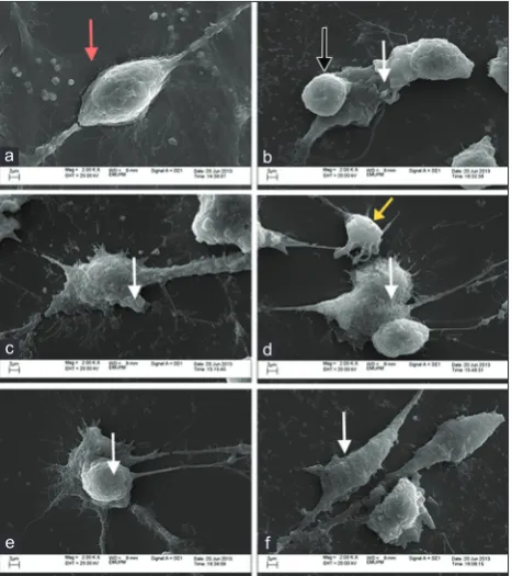
Morphological analysis of astrocytes via scanning
electron microscopy
Apart from fl owcytometric annexin V-FITC apoptosis detection assay analysis, the eff ects of vitamin E were also examined morphologically via scanning electron microscopy. Visible diff erence was noticed in the morphology of untreated sample with glutamate treated and vitamin E supplemented groups. Th e untreated cells possess smooth, fi nite, and rigid surface area with good cell membrane integrity (Figure 6a). Cells treated with 180 mM glutamate alone showed damage evidenced by blebbing, rounded appearance, irregular plas-malemma, and loss of refraction fi bers (Figure 6b). Cells that were pre- and post-treated with 200 ng/mL TRF and α-TCP had similar appearance to control cells (untreated), although
TABLE 1. Eff ect of diff erent doses of vitamin E (ng/mL) on astrocytes injured with 180 mM glutamate
Pre (μM/mg) Post (μM/mg) TRF α-TCP Glutamate
Untreated/negative control 0.1364±0.0212 0.1907±0.0272 - -
-Glutamate/positive control 1.2689±0.0430 1.0594±0.0699 - - +
100 ng/mL TRF 0.2031±0.0276 0.5271±0.0737 + - +
200 ng/mL TRF 0.1947±0.0296 0.4671±0.0238 + - +
300 ng/mL TRF 0.1061±0.0133 0.5247±0.0216 + - +
100 ng/mL TCP 0.3040±0.0126 0.7759±0.0757 - + +
200 ng/mL TCP 0.1643±0.0097 0.4128±0.0743 - + +
300 ng/mL TCP 0.1239±0.0089 0.1938±0.0233 - + +
Data were presented as the MDA concentration±SEM of the samples.
many contained fragmented processes and blebs. Cellular debris was also noted (6c-6f ).Neuroprotective eff ects of vitamin E on cell cycle
phases after exposure to glutamate toxicity
Based on Table 2, the cell cycle is divided into three distinct phases, which are G1, S, and G2 or M phases. In the pre-treat-ment study, the untreated sample illustrated the percentages of cell accumulation for astrocytes with 46.68, 36.63, and 16.69 in G1, S, and G2/M phases respectively. Meanwhile, for glutamate injured cells, the results of cell accumulation were 37.25, 23.11, and 39.64. Vitamin E in pre-treated astrocytes resulted in the increase of cell population in S and G2/M phases when compared to glutamate induced sample.
In the post-treatment study, the control expressed 43.55, 37.39, and 19.74 of cell accumulation, whereas glu-tamate treated sample populated 32.27, 28.59, and 39.14
of astrocytes in G1, S, and G2/M phases accordingly. Cell accumulation with the addition of TRF and α-TCP increased the quantity of cells in G2/M and S phases. Similar pattern of fi ndings were obtained for both pre- and post-treatment studies.
DISCUSSION
Th e MTT fi ndings refl exed that TRF and α-TCP at low concentrations, which were 100 to 300 ng/mL, restored the glutamate-injured astrocytes from injury. Previous stud-ies on neuronal cells reported that 100 nM of α-tocotrienol introduced after 60 minutes of glutamate exposure, but not 90 minutes, showed almost complete protection [10]. In addition, 100 μg/mL and 250 μg/mL of vitamin E pro-tected PC12 neuronal cell from glutamate toxicity in vita-min E co-treatment with increment of more than 20 of cell
FIGURE 3. Pre-treatment of TRF and α-TCP upon glutamate challenge on cell viability, apoptosis and necrosis. Results are the mean ± SEM in triplicates. *p<0.05, vitamin E treated groups compared with glutamate treated group.
viability [11,28]. Furthermore, in both pre- and post-treatment studies, 200 ng/mL of α-TCP showed signifi cant neuroprotec-tive eff ects due to its higher bioavailability and greater uptake via α-TTP. According to a study conducted by Saito et al., 2010 [11], longer incubation time allowed better cytoprotec-tive eff ect of tocopherol than tocotrienol. Injured astrocytes may utilize tocopherol in advance due to higher affi nity to α-TTP. Apart from that, both TRF and α-TCP exerted simi-lar protective eff ects with 20 of increased cell viability. Th is fi nding was consistent with the previous studies that reported tocotrienol and tocopherol showed similar capacities for cyto-protection against glutamate challenge [11].Besides, in both pre- and post-treatment studies of astro-cytes, 100 ng/mL of TRF and 200 ng/mL α-TCP exhibited
signifi cant diff erence in mitochondrial membrane pro-tection compared to glutamate insulted astrocytes. Lipid-soluble antioxidant retained in membrane more eff ectively than hydrophilic antioxidant [11]. Hence, vitamin E can give better neuroprotective eff ects towards damaged mitochon-dria of astrocytes. Moreover, TRF possesses a special confor-mation in the membrane of phospholipid bilayer due to its unsaturated phytyl tail [22]. Other than that, the side chain features also enable more effi cient penetration into tissues with high level of saturated fatty acid, such as the brain. Nanomolar concentrations of α-tocotrienol, in contrast with α-TCP, have the ability to protect against glutamate-induced neuronal death by suppressing inducible pp60 c-Src kinase activation [22].
TABLE 2. Pre and post treatment of TRF and α-TCP upon glutamate toxicity on astrocyte cell population in cell cycle phases. Data is presented as mean±SEM of 3 independent experiments (n=3 in each experiment).
Pre Post
G1 S G2/M G1 S G2/M
Control 46.68±2.13 36.63±2.07 16.69±1.75 43.55±0.88 37.39±0.81 19.74±0.50 Glutamate 37.25±4.48 23.11±1.34 39.64±3.48 32.27±1.43 28.59±0.53 39.14±1.80 100 TRF 32.71±4.20 25.52±0.83 41.79±3.67 33.33±0.65 27.22±0.96 39.45±0.42 200 TRF 32.58±3.78 24.54±0.87 42.87±4.61 35.03±0.45 24.91±0.83 40.05±1.86 300 TRF 30.25±2.52 25.34±1.56 44.41±4.05 30.10±4.72 28.50±1.94 41.41±3.29 100 TCP 32.29±3.37 26.07±1.61 41.64±2.85 28.49±3.83 29.82±2.42 41.69±1.99 200 TCP 31.29±2.59 25.74±1.18 42.96±3.17 34.47±1.24 23.71±0.15 41.82±1.31 300 TCP 32.30±2.89 24.63±1.00 43.06±3.86 29.96±3.77 28.34±3.17 41.69±1.31
Lipid peroxidation assay measured the concentration of MDA as the level of lipid peroxidation. Th e pre- and post-treatments of TRF and α-TCP showed potential protec-tion against glutamate challenge. Both TRF and α-TCP treated cells had low MDA concentration and the diff erence was sig-nifi cant in comparison with glutamate injured astrocytes. Th is study also specifi ed prophylactic eff ect of vitamin E in scav-enging ROS. Concentration of 300 ng/mL of TRF presented high potential in reducing MDA concentration per protein in pre-treatment study of TBARS assay. Th e effi ciency of TRF is highly related to its better distribution in fatty layers of brane. TRF did manage to penetrate through the cell mem-brane effi ciently, hence, could protect the cells from oxidative stress caused by glutamate [29]. A previous study conducted by Long et al., [30] showed that the accumulation of MDA while aging can cause mitochondrial dysfunction by inhibit-ing mitochondrial respiration and enzyme activity. Th us, with supplementation of various doses of TRF and α-TCP, the con-centration of MDA decreases, and subsequently, causes cell viability augmentation.In the study of vitamin E, astrocytes were treated with 180 mM of glutamate that caused approximately 60 of cell death. Flowcytometric annexin V-FITC analysis revealed that
glutamate injured cells showed lower cell viability and higher apoptotic rate, meanwhile the untreated sample exhibited higher cell viability and lower amount of cell death. Th erefore, this results indicated that 180 mM glutamate was toxic to astrocytes. However, the pre- and post-treatments of various concentrations of vitamin E presented better survivability and low cell death rates against glutamate neurotoxicity in astrocytes. α-TCP and TRF protected the cells from rapidly undergoing cell death induced by glutamate by preventing PS translocation, thus, the cell membrane remained intact. Previous research fi ndings showed that at a concentration less than 10 μM, γ-tocotrienol has been reported to improve cell viability signifi cantly against H2O2-induced apoptosis in pri-mary astrocytes [31], pripri-mary cerebellar neurons [32], as well as in primary rat cortical neurons, and human neuroblastoma cell line [33]. Th e morphological fi ndings obtained from scan-ning electron microscopy were well in accordance to the other research studies [34].
In addition, astrocytes treated with 180 mM of glutamate may induce impairment of DNA, protein, and chromatin, and subsequently, result in oxidative stress. Oxidative stress could be one of the mechanisms responsible for cell cycle re-entry [35]. In this study, astrocytes may re-enter the cell cycle to repair the damages occurred due to glutamate insult. Otherwise, badly injured cells might initiate cell death if the damage is too extensive to be repaired [36]. From the results obtained, it showed that vitamin E acts as a potent antioxidant as it can actually enhance the synthesis of DNA (S phase) and the recovery/repair of DNA (G2/M) in glutamate injured cells.
On the other hand, the role of astrocytes in promoting neuronal survival and recovery, following a cerebral insult, is becoming increasingly appreciated. Astrocyte, a subtype of glial cell, is known to protect neuronal cells against oxida-tive stress through transcriptional upregulation of glutathi-one synthesis and removal of extracellular glutamate [37-39]. Besides, studies have shown that the death of astrocytes after ischemia or reperfusion may strongly aff ect neuronal survival due to the absence of trophic and metabolic support to neuro-nal cells and astrocytic glutamate uptake [40]. Th erefore, the cytoprotective eff ect of vitamin E against glutamate induced astrocytes is of our interest in order to maintain homeostasis for the neuronal cells in the brain.
Th ere are several limitations of this study. Mechanisms of action of both TRF and α-TCP in elucidating astrocytes recov-ery upon glutamate insult need to be strongly validated via genomic studies. However, further studies are currently con-ducted by our research group to determine the eff ects of vita-min E in down regulating the expression of traumatic brain injury markers in glutamate induced astrocytes.
In conclusion, revealing a perfect therapy/compound that can protect people who are suff ering from nerve disorders is
FIGURE 6. Scanning electron micrographs. (a) Scanning electron micrograph of control/untreated astrocyte showing retraction fi bers (red arrow). (b) Scanning electron micrograph of 180 mM glutamate treated astrocyte for 24 hours. Cells appear rounded (black arrow) and evident blebbing (white arrow). (c-f ) Scanning electron micrographs for pre and post treatment of 200 ng/mL of TRF and α-TCP upon glutamate challenge. Although cellular debris was noted (yellow arrow), many cells appeared with intact cell membrane but cellular blebbing was noted in some cells (white arrow).
d c
b
f a
the big concern worldwide. Currently, studies have revealed that astrocyte, supportive cell of neuronal, play an important role in the survival of neuronal cells. Earlier, more focus was given to fi nd substance or compound that can provide protec-tion for the neurons against oxidative stress. Only in the last few years, scientists have found out that astrocytes play a more important role rather than just to provide support to the neu-rons. Th is study demonstrated that vitamin E (both TRF and α-TCP) is competent in preventing glutamate induced injury and death in astrocytes. In the nervous system, the astrocytes are in close interaction with the neuronal cells, and therefore, by ensuring the survival of astrocytes from oxidative stress, it is expected that the neurons are also protected. Hence, under-standing the dosages, as well as the prophylactic role of vita-min E, is rather crucial in vita-minimizing neurodegeneration that is off ered by nutrition-based therapy. It is obvious that palm TRF and α-TCP possess the prospective to be developed as a measure for the management of neurodegenerative diseases.DECLARATION OF INTERESTS
Th e authors declare no confl ict of interests.
ACKNOWLEDGMENTS
Th is research was fi nancially supported by Faculty of Medicine and Health Sciences Grant Scheme (vote 9300337), University Putra Malaysia.
REFERENCES
[1] Tabrizi S. Neurodegenerative diseases neurobiology patho-genesis and therapeutics. J Neurol Neurosurg and Psychiatry. 2006;77(2):284. http://dx.doi.org/10.1136/jnnp.2005.072710. [2] Dibble LE, Lange M. Predicting falls in individuals with Parkinson
disease: a reconsideration of clinical balance measures. J Neurol Phys Ther. 2006;30(2):60-7. http://dx.doi.org/10.1097/01. NPT.0000282569.70920.dc.
[3] Shukla V, Mishra SK, Pant HC. Oxidative stress in neurodegenera-tion. Adv Pharmacol Sci. 2011; 2011:572-634.
[4] Rock RB, Gekker G, Hu S, Sheng WS, Cheeran M, Lokensgard JR, et al. Role of microglia in central nervous system infections. Clin Microbiol Rev. 2004;17(4):942-64, table of contents. http://dx.doi. org/10.1128/CMR.17.4.942-964.2004.
[5] Wang Y, Qin ZH. Molecular and cellular mechanisms of excito-toxic neuronal death. Apoptosis. 2010;15(11):1382-402. http://dx.doi. org/10.1007/s10495-010-0481-0
[6] Meldrum BS. Glutamate as a neurotransmitter in the brain: review of physiology and pathology. J Nutr. 2000;130(4S Suppl):1007S-15S. Epub 2000/03/29.
[7] Higuchi Y. Glutathione depletion-induced chromosomal DNA fragmentation associated with apoptosis and necrosis. J Cell Mol Med. 2004;8(4):455-64. http://dx.doi.org/10.1111/j.1582-4934.2004. tb00470.x
[8] Scandalios JG. Oxidative stress responses--what have genome-scale studies taught us? Genome Biol. 2002;3(7):1019. http://dx.doi. org/10.1186/gb-2002-3-7-reviews1019.
[9] Emerit J, Edeas M, Bricaire F. Neurodegenerative diseases and
oxidative stress. Biomed Pharmacother. 2004;58(1):39-46. http:// dx.doi.org/10.1016/j.biopha.2003.11.004.
[10] Sen CK, Khanna S, Roy S, Packer L. Molecular basis of vitamin E action. Tocotrienol potently inhibits glutamate-induced pp60(c-Src) kinase activation and death of HT4 neuronal cells. J Biol Chem. 2000;275(17):13049-55. http://dx.doi.org/10.1074/jbc.275.17.13049. [11] Saito Y, Nishio K, Akazawa YO, Yamanaka K, Miyama A, Yoshida Y,
et al. Cytoprotective eff ects of vitamin E homologues against glu-tamate-induced cell death in immature primary cortical neuron cultures: Tocopherols and tocotrienols exert similar eff ects by anti-oxidant function. Free Radic Biol Med. 2010;49(10):1542-9. http:// dx.doi.org/10.1016/j.freeradbiomed.2010.08.016.
[12] Maniam S, Mohamed N, Shuid AN, Soelaiman IN. Palm tocotrie-nol exerted better antioxidant activities in bone than alpha-tocoph-erol. Basic Clin Pharmacol Toxicol. 2008;103(1):55-60. http://dx.doi. org/10.1111/j.1742-7843.2008.00241.x.
[13] Serbinova E, Kagan V, Han D, Packer L. Free radical recycling and intramembrane mobility in the antioxidant properties of alpha-tocopherol and alpha-tocotrienol. Free Radic Biol Med. 1991;10(5):263-75. http://dx.doi.org/10.1016/0891-5849(91)90033-Y [14] Wu SJ, Liu PL, Ng LT. Tocotrienol-rich fraction of palm oil
exhib-its anti-infl ammatory property by suppressing the expression of infl ammatory mediators in human monocytic cells. Molecular nutrition & food research. 2008;52(8):921-9. http://dx.doi. org/10.1002/mnfr.200700418.
[15] Wu SJ, Ng LT. Antioxidant and antihepatoma activities of palm oil extract. Journal of Food Lipids. 2007;14(2):122-37. http://dx.doi. org/10.1111/j.1745-4522.2007.00075.x.
[16] Takahashi K, Loo G. Disruption of mitochondria during tocotrien-ol-induced apoptosis in MDA-MB-231 human breast cancer cells. Biochem Pharmacol. 2004;67(2):315-24. http://dx.doi.org/10.1016/j. bcp.2003.07.015.
[17] Goh SH, Hew NF, Norhanom AW, Yadav M. Inhibition of tumour promotion by various palm-oil tocotrienols. Int J Cancer. 1994;57(4):529-31. http://dx.doi.org/10.1002/ijc.2910570415 [18] Osakada F, Hashino A, Kume T, Katsuki H, Kaneko S, Akaike
A. Alpha-tocotrienol provides the most potent neuropro-tection among Vitamin E analogs on cultured striatal neu-rons. Neuropharmacology. 2004;47(6):904-15. http://dx.doi. org/10.1016/j.neuropharm.2004.06.029.
[19] Minhajuddin M, Beg ZH, Iqbal J. Hypolipidemic and antioxidant properties of tocotrienol rich fraction isolated from rice bran oil in experimentally induced hyperlipidemic rats. Food Chem Toxicol. 2005;43(5):747-53. http://dx.doi.org/10.1016/j.fct.2005.01.015. [20] Mutalib MSA, Khaza'ai H, Wahle KWJ. Palm-tocotrienol rich
fraction (TRF) is a more eff ective inhibitor of LDL oxidation and endothelial cell lipid peroxidation than α-tocopherol in vitro. Food Research International. 2003;36(5):405-13. http://dx.doi. org/10.1016/S0963-9969(02)00173-4.
[21] Qureshi AA, Bradlow BA, Brace L, Manganello J, Peterson DM, Pearce BC, et al. Response of hypercholesterolemic subjects to administration of tocotrienols. Lipids. 1995;30(12):1171-7. http://dx. doi.org/10.1007/BF02536620.
[22] Sen CK, Rink C, Khanna S. Palm oil-derived natural Vitamin E alpha-tocotrienol in brain health and disease. J Am Coll Nutr. 2010;29(3 Suppl):314S-23S. http://dx.doi.org/10.1080/07315724.20 10.10719846.
[23] Mangialasche F, Kivipelto M, Mecocci P, Rizzuto D, Palmer K, Winblad B, et al. High plasma levels of vitamin E forms and reduced Alzheimer's disease risk in advanced age. J Alzheimers Dis. 2010;20(4):1029-37.
[24] Aggarwal BB, Sundaram C, Prasad S, Kannappan R. Tocotrienols, the vitamin E of the 21st century: its potential against cancer and other chronic diseases. Biochem Pharmacol. 2010;80(11):1613-31. http://dx.doi.org/10.1016/j.bcp.2010.07.043.
[25] Wang J, Rajakulendran N, Amirsadeghi S, Vanlerberghe GC. Impact of mitochondrial alternative oxidase expression on the response of Nicotiana tabacum to cold temperature. Physiol Plant. 2011;142(4):339-51. Epub 2011/03/16.
MTT assay. Methods Mol Biol. 2011;731:237-45. http://dx.doi. org/10.1007/978-1-61779-080-5_20.[27] Nguyen HT, Olson J, DeVries C, Savage W, Holtzman D. Scanning electron microscopic study of lead eff ects on cerebral astrocytes in primary culture. Scan Electron Microsc. 1982(Pt 2):891-6.
[28] Schubert D, Kimura H, Maher P. Growth factors and vitamin E modify neuronal glutamate toxicity. Proc Natl Acad Sci U S A. 1992;89(17):8264-7. http://dx.doi.org/10.1073/pnas.89.17.8264. [29] Khanna S, Roy S, Parinandi NL, Maurer M, Sen CK. Characterization
of the potent neuroprotective properties of the natural vitamin E alpha-tocotrienol. J Neurochem. 2006;98(5):1474-86. http://dx.doi. org/10.1111/j.1471-4159.2006.04000.x.
[30] Long J, Wang X, Gao H, Liu Z, Liu C, Miao M, et al. Malonaldehyde acts as a mitochondrial toxin: Inhibitory eff ects on respiratory func-tion and enzyme activities in isolated rat liver mitochondria. Life Sci. 2006;79(15):1466-72. http://dx.doi.org/10.1016/j.lfs.2006.04.024. [31] Mazlan M, Sue Mian T, Mat Top G, Zurinah Wan Ngah W.
Comparative eff ects of alpha-tocopherol and gamma-tocotrienol against hydrogen peroxide induced apoptosis on primary-cul-tured astrocytes. J Neurol Sci. 2006;243(1-2):5-12. http://dx.doi. org/10.1016/j.jns.2005.10.006.
[32] Th en SM, Mazlan M, Mat Top G, Wan Ngah WZ. Is Vitamin E toxic to neuron cells? Cell Mol Neurobiol. 2009;29(4):485-96. http://dx. doi.org/10.1007/s10571-008-9340-8.
[33] S. M. Th en WZWN, G. M. Top, and M. Mazlan. Comparison of the eff ects of α-tocopherol and γ-tocotrienol against oxidative stress in
two diff erent neuronal cultures. Sains Malaysiana. 2010;39(1):145-56. [34] Hardej D, Trombetta LD. Th e eff ects of ebselen on cisplatin and
diethyldithiocarbamate (DDC) cytotoxicity in rat hippocam-pal astrocytes. Toxicol Lett. 2002;131(3):215-26. http://dx.doi. org/10.1016/S0378-4274(02)00056-5.
[35] Folch J, Junyent F, Verdaguer E, Auladell C, Pizarro JG, Beas-Zarate C, et al. Role of cell cycle re-entry in neurons: a common apoptotic mechanism of neuronal cell death. Neurotox Res. 2012;22(3):195-207. http://dx.doi.org/10.1007/s12640-011-9277-4.
[36] Schwartz EI, Smilenov LB, Price MA, Osredkar T, Baker RA, Ghosh S, et al. Cell cycle activation in postmitotic neurons is essential for DNA repair. Cell Cycle. 2007;6(3):318-29. http://dx.doi.org/10.4161/ cc.6.3.3752.
[37] Dringen R, Hirrlinger J. Glutathione pathways in the brain. Biol Chem. 2003;384(4):505-16. http://dx.doi.org/10.1515/BC.2003.059. [38] Iwata-Ichikawa E, Kondo Y, Miyazaki I, Asanuma M, Ogawa N. Glial
cells protect neurons against oxidative stress via transcriptional up-reg-ulation of the glutathione synthesis. J Neurochem. 1999;72(6):2334-44. http://dx.doi.org/10.1046/j.1471-4159.1999.0722334.x.
[39] Gegelashvili G, Schousboe A. High affi nity glutamate transporters: regulation of expression and activity. Mol Pharmacol. 1997;52(1):6-15. [40] Szydlowska K, Gozdz A, Dabrowski M, Zawadzka M,