R E S E A R C H
Open Access
Human linker histones: interplay between
phosphorylation and O-
b
-GlcNAc to mediate
chromatin structural modifications
Waqar Ahmad
1*, Khadija Shabbiri
2, Noreen Nazar
2, Shazia Nazar
2, Saba Qaiser
2and Mirza Abid Shabbir Mughal
2Abstract
Eukaryotic chromatin is a combination of DNA and histone proteins. It is established fact that epigenetic mechanisms are associated with DNA and histones. Initial studies emphasize on core histones association with DNA, however later studies prove the importance of linker histone H1 epigenetic. There are many types of linker histone H1 found in mammals. These subtypes are cell specific and their amount in different types of cells varies as the cell functions. Many types of post-translational modifications which occur on different residues in each subtype of linker histone H1 induce conformational changes and allow the different subtypes of linker histone H1 to interact with chromatin at different stages during cell cycle which results in the regulation of transcription and gene expression. ProposedO-glycosylation of linker histone H1 promotes condensation of chromatin while phosphorylation of linker histone H1 is known to activate transcription and gene regulation by decondensation of chromatin. Interplay between phosphorylation andO-b-GlcNAc modification on Ser and Thr residues in each
subtype of linker histone H1 inHomo sapiensduring cell cycle may result in diverse functional regulation of proteins. Thisin silicostudy describes the potential phosphorylation, o-glycosylation and their possible interplay sites on conserved Ser/Thr residues in various subtypes of linker histone H1 inHomo sapiens.
Introduction
Eukaryotic genome is packaged into a structure known as chromatin. The basic structural unit of chromatin called as nucleosome is composed of DNA and proteins [1]. The major proteins involved in chromatin structure are histone proteins. Histone proteins are of five types: H1, H2A, H2B, H3 and H4 [2-4]. Histone H1 is known as linker histone while the other four histone proteins are collectively known as core histones. This DNA-pro-tein complex is the tempelate for a number of essential cell processes including transcription recombination, repair and replication. Histone H1 is located on the lin-ker DNA that goes between the nucleosomes in chro-matin structure [5]. Linker DNA which is associated with linker histone H1 interconnects core particles, var-ies in length, depending on specvar-ies and tissue [6]. Orga-nization of DNA into nucleosomes by histone proteins and folding of nucleosomes into higher-order chromatin
structure is generally believed to compact DNA and make it inaccessible to transcription factors [7]. Linker histones H1 are necessary for modulating chromatin structure and function at multiple levels [8].
Organisms contain a variety of subtypes of linker his-tone which exhibit significant sequence divergence and distinct patterns of expression differentiation and devel-opment [9]. The H1 linker histones are the most diver-gent group. Usually nine subtypes of linker histone H1 are present in mammals including H1.1, H1.2, H1.3, H1.4, H1.5, H1o, H1Foo, H1.t [10] and H1.x [11]. Linker histone sub-types are classified according to their tightly regulated expression pattern during embronyal develop-ment and cell differentiation [12]. All known sub-types of linker histone contain a common domain structure. Linker histones consist of a shortN-terminal, a highly conserved central globular domain and a long C -term-inal domain [13]. Somatic cells contain almost all sub-types of linker histone H1 [12]. In vitro, H1-containing chromatin shows strong inhibition of transcription [14] and transcriptionally active chromatin typically depleted in H1 compared with inactive chromatin [15].
* Correspondence: [email protected]
1
Centre of Excellence in Molecular Biology, University of the Punjab, Lahore, Pakistan
Full list of author information is available at the end of the article
Wisniewskiet al. showed that many of the mapped mod-ification sites which are thought to be involved in binding to nucleosomal DNA are located within the globular domain region of the different subtypes of the linker his-tone H1 [16]. H1 depletion results in a dramatic length-ening of chromosomes, which suggests an important role in mitotic chromosome condensation [17]. The presence of these large number of various H1 histone subtypes and their possible post-translational modifications, make it very clear that H1 histones play numerous structural and functional roles in chromatin [18]. Until now, no specific role for the various variants has been established but it is known that the mouse histone H1.2 binds prefer-entially to a regulatory sequence within a mouse H3.2 replication-dependent histone gene [19].
Post-translational modifications (PTMs) of linker his-tone H1 play very important role in regulation of chro-matin structure, transcriptional regulation, gene activity [17] and controlling the accessibility of transcription fac-tors to chromatin structure [20]. A working model of the cell cycle has slowly been constructed from the dis-covery of cyclins 22 years ago. This model is composed of protein phosphorylation, acetylation timed expression of cyclins, and well orchestrated cell division. Neverthe-less, a detailed mechanism of the cell cycle is still incomplete [21-23]. Transcriptional activation of genes starts with the dissociation of linker histone H1 from linker DNA [24]. Phosphorylation of linker histone is required for efficient cell cycle progression by enzyme CDK2 [25]. These kinases requires a consensus
sequence (S/T)PXZ or (S/T)PXK for phosphorylation
(where X is any amino acid and Z is a basic amino acid) and this consensus sequence is found in many linker histone H1 variants which become phosphorylated [26]. It is found that PKC is also involved in phosphorylation of linker histone variants during regulation of gene expression in cell cycle [27]. Phosphorylation of linker histone regulates transcription and gene expression by reducing the electrostatic binding of linker histone to DNA in chromatin [28]. In vivophosphorylation of the linker histone tails influence both the binding to mono-nucleosomes and the aggregation of polymono-nucleosomes [29]. The phosphorylation of linker histones at theirN and C-terminal tails during the cell cycle influence its functions for enhancing decondensation which in turn regulate transcription and gene expression. This phos-phorylation and dephosphos-phorylation is a common regula-tory mechanism for protein functions [30].
O-Glycosylation is also very important PTM of pro-teins. During O-Glycosylation one molecule of N-acetyl-glucosamine (O-b-GlcNAc) is introduced on Ser or Thr residue by enzymeO-GlcNAc transferases (OGT). Addi-tion ofO-b-GlcNAc can inhibit phosphorylation on Ser or Thr residue and is reciprocal with phosphorylation
on some well studied proteins, such as RNA polymerase II, estrogen receptor, and the c-Myc proto-oncogene product [31-34]. These studies suggest that O-GlcNAc may function as a global regulator of cell growth and division. Deletion of OGT in mouse embryonic fibro-blasts is associated with delayed growth, increased expression of the cyclin inhibitor p27, and death. A reduction in O-GlcNAc levels results in cell growth defects, by the lowering UDP-GlcNAc levels to 5% of
normal [35,36]. Studies in Xenopus demonstrated
maturation defects in oocytes when microinjected with galactosyltransferase which prevents O-GlcNAc removal.
Meanwhile incubation of Xenopusoocytes with the
O-GlcNAcase inhibitor PUGNAc altered progression of oocytes through progesterone-mediated maturation [37-40].
In 1994, Kim et al. first time observed the o-GlcNAc modification in mouse linker histone H1. They also observed same PTM on core histones [41]. In 2005, Slawsonet al. showed that increased O-GlcNAc resulted in growth defects linked to delay in G2/M progression, altered mitotic phosphorylation, and cyclin expression. Over expression of O-GlcNAcase, the enzyme that removes O-GlcNAc, induces amitotic exit phenotype accompanied by a delay in mitotic phosphorylation, altered cyclin expression, and pronounced disruption in nuclear organization. Overexpression of the O-GlcNAc transferase, the enzyme that adds O-GlcNAc, results in a polyploid phenotype with faulty cytokinesis. Notably, O-GlcNAc transferase is concentrated at the mitotic spindle and mid body at M phase. These data suggest that dynamic O-GlcNAc processing is a pivotal regula-tory component of the cell cycle, controlling cell cycle progression by regulating mitotic phosphorylation, cyclin expression, and cell division [42]. On the basis of above observations, Kaleem et al (2008) used bioinformatics tools to predict o-glycosylation on human core histone H3, even though there was no experimental proof of that PTM on histone H3 [43].
Interplay between O-b-GlcNAc modification and
phosphorylation on the same amino acid residues has been observed in several nuclear and cytoplasmic pro-teins [44]. These PTMs are dynamic and result in tem-porary conformational changes and regulate many functions of the proteins. The alternation of these two modifications on the same or neighboring residue may modulate the specific function of the proteins either by enhancing or inhibiting the functional capacity. Residues
where O-b-GlcNAc and phosphorylation compete for
Although a yin/yang relationship between phosphoryla-tion andO-GlcNAcylation on histone H3 has been pro-posed; the direct evidence forO-glycosylation of histones was never been described. Recent studies by Sakabeet al (2010-2011) first time proved the O-GlcNAc modifica-tion on histones and also mapped glycosylamodifica-tion sites with specific immunological, enzymatic and mass spectro-metric techniques. They also insist to include O-GlcNAc modification as part of histone code. They showed that histone O-GlcNAcylation increases with heat shock and this increase is concomitant with DNA condensation [46,47]. The present work describe potential phosphory-lation,O-Glycosylation and their possible interplay sites which influence condensation, decondensation and tran-scriptional and gene regulation during cell cycle in var-ious subtypes of linker histone H1.
Materials and Methods
The sequences of different types of linker histone H1 of many species mostly mammals have been described by many workers [10,11,16]. The sequence data used for predicting phosphorylation and glycosylation sites for different subtypes of linker histone H1 of human was retrieved from the SWISS-PROT [48] sequence data-base. The primary accession numbers for each subtype of linker histone in human are Q02539 (H1.1), P16403 (H1.2), P16402 (H1.3), P10412 (H1.4), P16401 (H1.5), Q81ZA3 (H1oo), P22492 (H1.T), P07305 (H1.0) and Q 92522 (H1.X). BLAST search was made using the NCBI database of non-redundant sequences [49]. The search was done for all organisms’sequences with expect value set to 10 using blosum 62 matrix and low complexity filter selecting nr database. Hits with highest bits score and zero expect value were selected. The four to five sequences of each subtype of linker histone H1 from different selected species were selected to find out con-served residues inHomo sapienslinker histone H1. All selected sequences were multiple aligned using CLUS-TALW [50]. All the sequences of subtypes of linker
his-tone H1 present in Homo sapiens were aligned to get
the conservation status of subtypes.
Post-translational modifications prediction methods Phosphorylation sites on Ser, Thr and Tyr residues were predicted by using NetPhos 2.0 (http://cbs.dtu.dk/ser-vices/NetPhos/) server [51]. NetPhos 2.0 is a neural net-work-based method for the prediction of potential phosphorylation sites.
NetPhosK 1.0 server (http://cbs.dtu.dk/services/Net-PhosK) [52] was used to predict kinase specific phos-phorylation sites in human histone H1.
Phospho.ELM database (http://phospho.elm.eu.org/) was used for the determination of the experimentally ver-ified phosphorylation sites [53] present on various linker
histone H1 subtypes in different species. The Phospho. ELM database contains a collection of experimentally verified Ser, Thr and Tyr sites in eukaryotic proteins.
To predict potentialO-b-GlcNAc modification sites,
YinOYang 1.2 (http://www.cbs.dtu.dk/services/
YinOYang/) was used. This method is also capable of predicting the potential phosphorylation sites as well and hence predicting the Yin Yang sites [54-56].
Neural networks-based prediction methods
Artificial neural networks based methods have been exten-sively used in biological sequence analysis and predicting the potentials for modifications [57]. The methods devel-oped using machine learning approach includes memoriz-ing the neural networks with the sequence environment windows of phosphorylated/glycosylated and non-phos-phorylated/non-glycosylated sites. During this learning process the input data of phosphorylated/glycosylated and non-phosphorylated/non-glycosylated sites is presented to neural networks in the form of binary codes of 21 digits. A threshold value in form of bits is set for positive hit and zero for negative hits. The learning process and perfor-mance is checked with the data reserved for cross valida-tion using statistical equavalida-tions. During learning, the error is computed and weights given to each neuron are set to get the maximum correct predictions.
Results
Allignment of sequences for determination of conserved status of Ser/Thr residues within different linker histone subtypes
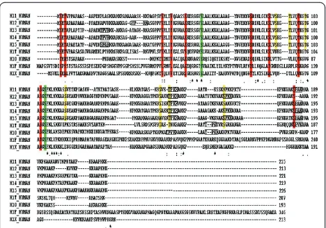
Each human linker histone subtype was aligned with other species. Conserved and conserved substituted Ser and Thr residues within each subtype were determined (Data not shown). These nine subtypes were also aligned with each other to find conserved residues within sub-types (Figure 1).
Prediction of phosphorylated S/T residues with motif (S/T)PXZ and (S/T)PXK motifs were searched for each linker histone H1 subtypes. Sequences within boxes showed the specific motifs (Figure 1). These residues are given in Table 1.
Acquiring of experimentally verified S/T/Y residues Data for experimentally confirmed S/T/Y residues was obtained from Phospho ELM and UniprotKB (http:// www.uniprot.org) is given in Table 1. All histone H1 subtypes phosphorylated during cell cycle except H1oo.
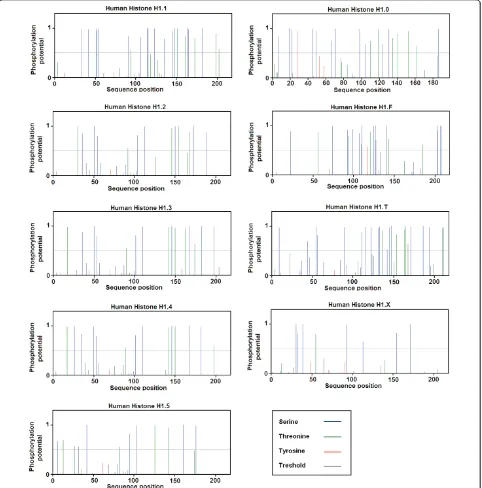
Prediction of Phosphorylation Sites
subtypes of linker histone H1 showed high potential for phosphorylation as shown in Figure 2. The predicted Ser and Thr residues are shown in Table 1.
Prediction of Kinases involved in Phosphorylation
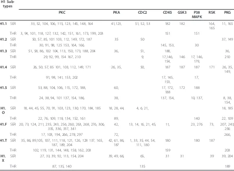
Different kinases are involved in phosphorylation of Ser and Thr residues of linker histone H1 subtypes. Almost each kinase predicted is involved in phosphorylation of two or more residues. The predicted kinases involved in phosphorylation by NetPhos K 1.0 are shown in Table 2.
Prediction of O-Linked Glycosylation Sites
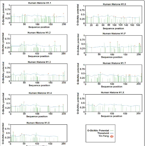
Prediction results forO-linked glycosylation sites showed that all subtypes of linker histone H1 have very high poten-tial forO-b-GlcNAc modification Table 3. There are many predicted Yin Yang sites in each subtype of linker histone which are shown by an asterisk as shown in Figure 3.
Identification of False-Negative Sites
The Ser and Thr residues which were not predicted to be O-b-GlcNAc modified but have very high potential for phosphorylation and very close to threshold value are known as false-negative sites (FN-sites). All the Ser and Thr residues which were predicted false-negatively with high conservation status and phosphorylation potential among different subtypes of linker histone H1 are given in Table 3.
Possible proposed YinYang sites within different subtypes of linker histone H1
The possible proposed Yin Yang sites for the interplay of phosphorylation and O-b-GlcNAc modification are
given in Table 3. These Yin Yang sites are proposed on the basis of conservation status of Ser/Thr residues in each subtype of linker histone H1. The Ser/Thr residues are also proposed for the possible interplay of phosphor-ylation and O-b-GlcNAc modification on the basis of their similarity with other species. These Ser/Thr resi-dues which are predicted “by similarity” are not yet
experimentally known in Homo sapiens but these are
known in other species of vertebrates.
Discussion
Human linker histones have more than eight sub-types, all consisting of a highly conserved globular domain and less conservedN- andC-terminal tails. The sequence of terminal tails of different subtypes of linker histone H1 within a species is much less conserved but the sequence of terminal tails of a specific subtype is well conserved among different species [58]. In addition to heterogeneity of their primary structures, the histone tails are also post-translationally modified under various biological conditions [59]. The proportion of linker his-tone H1 subtypes varies in a tissue- and species-species manner [60], and the expression of each subtype varies throughout development and differentiation [61]. Stu-dies of the structure of different subtypes of linker his-tone H1 and their interaction with the nucleosome and their roles in controlling gene activity indicate that lin-ker histones have both an essential architectural func-tion and an important task in regulating transcripfunc-tion [2]. The precise functions and modifications of linker histones are not yet fully understood, but it is known that different linker histone variants are preferentially localized to particular chromosomal domains. The sequences within the globular domain of linker histone H1 are thought to be responsible for the differential effect of overproduction of different linker histone var-iants on gene expression [62], while the N- andC -term-inal domains of linker histone H1 are responsible for the condensation of chromatin [63]. TheN-terminal of linker histone H1 binds with linker DNA [64] and C-terminal of linker histone H1 has binding affinity with core histones [58]. Different linker histone H1 subtypes have different chromatin condensing abilities [65]. All linker histone H1 subtypes differ not only in primary sequence but also in turnover rate, timing of synthesis during development and extent of phosphorylation and they also have the potential to add a great deal of flex-ibility to chromatin structure and transcriptional activa-tion [66]. Linker histone H1 is required for longitudinal compaction of replicated chromosome. Enrichment of linker histone H1 onto chromatin required passage through interphase, when DNA replication takes place. Thus, linker histone H1 contributes to chromosome condensation in vertebrates [67]. In mouse depletion of Figure 1Sequence alignment of different subtypes of linker
linker histone H1 caused chromatin structure changes which include decreased global nucleosome spacing, reduced chromatin compaction and decreased in certain histone modifications like methylation [68]. In vitro experiments showed that linker histone H1 represses transcriptional promoters and factors by condensing the chromatin material [69] butin vivostudies showed that linker histone H1 does not function as a global tran-scriptional repressor, but instead participates in com-plexes that either activate or repress specific genes [70]. Differences between linker histone H1 subtypes for both binding and the capacity to aggregate polynucleosome into condensed structure implies functional differences between the different linker histone H1 subtypes during cell cycle and development of organism [71].
Sub-fractions of H1 histones differ in their effectiveness in condensing DNA fibers into ordered aggregates. Furthermore, each of linker histone H1 variant has dif-ferences in their binding capacity with DNA [72].
Haleet al. showed that phosphorylation of linker his-tone H1 provides a signal for the disassembly of higher order chromatin structure during cell cycle [73]. Linker histone H1 phosphorylated in a cell-cycle dependent manner, in G1 phase levels of H1 phosphorylation are usually lowest and then rise continuously during S and G2 phase. The M-phase where chromatin is highly con-densed shows the maximum no. of phosphorylated sites [74]. The phosphorylation of linker histone H1 subtypes occurs on specific Ser and Thr residues during cell cycle in the presence of different protein kinases [75].
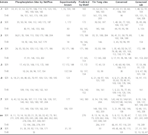
Table 1 Phosphorylation andO-b-GlcNAc site map ofHomo sapiens Substrate Phosphorylation Sites by NetPhos Experimentally
known
Predicted by Motif
Yin Yang sites Conserved Conserved sub
H1.1 SER 33, 41, 51, 52, 53, 91, 106, 114, 115, 123, 135, 145, 148, 164, 165
1, 35, 103, 183 183 33, 52, 53, 114, 164, 165
41, 43, 51, 53, 60, 106, 183
1, 48, 52, 91, 103 THR 94, 151, 161, 173, 199, 203 151 151 161, 173, 199,
203
94 101, 151, 11, 164, 203 H1.2 SER 35, 50, 54, 104, 112, 149, 172, 187 1, 172 172 30, 50, 187 1, 40, 58, 77, 102,
104, 172, 187
35, 85, 88, 112 THR 30, 91, 145, 153, 166 30 30, 145,
153
145, 166 44, 91, 95, 98 3, 153
H1.3 SER 36,51, 55, 104, 113, 150, 173, 188, 204 188 173, 188 35, 51, 188, 204 36, 41, 51, 58, 79, 89, 102, 104, 188
1, 86
THR 18, 92, 146, 154, 167, 179 18 18, 146, 154
146 3, 45, 92, 96, 99 154
H1.4 SER 26, 35, 50, 54, 103, 112, 150, 171, 186 35, 171, 186 171, 186 35, 50, 186 1, 35, 40, 50, 54, 57, 78, 85, 88, 101, 103, 112, 150, 171, 186
172, 188
THR 17, 91, 145, 153, 202 17 17, 145, 153
17, 145, 202 3, 17, 91, 95, 98, 145 141, 153, 202
H1.5 SER 17, 43, 53, 106, 115, 172, 188 17, 172, 188 17, 172 17, 43, 53 1, 43, 60, 80, 104, 106, 115
17, 53, 88, 91, 172 THR 10, 24, 38, 94, 137, 154 137,154 10, 137,
154
10, 38 38 3, 8, 47, 98, 101, 154 H1.0 SER 6, 18, 21, 44, 48, 65, 70, 97, 103, 123, 130, 185 123 6, 21, 44, 97, 103,
123, 130
4, 6, 21, 28, 44, 45, 55, 65, 70, 89, 91, 103, 130, 170, 184,
185
18, 97, 115
THR 109, 118, 134, 140, 152, 161 118, 140, 152
134, 161 1, 5, 22, 76, 77, 83, 109, 118, 123, 134,
140, 152
161
H1.T SER 8, 42, 52, 54, 86, 107, 111, 118, 126, 128, 137, 140, 142, 165, 180, 187, 204
177 142, 180 8, 54, 118, 180, 204
1,42, 44, 52, 54,61,81, 105,107,140, 142,165,
180
8, 35, 126, 128, 137, 187, 189, 204 THR 131, 148, 158, 159, 162, 203 158, 159 148, 158, 159,
162, 203
3, 21, 99, 102, 148, 158
10, 48, 131, 145, 203 H1oo SER 8, 11, 13, 14, 16, 20, 21, 23, 26, 32, 42, 73, 161,
211, 229, 230, 235, 243, 245, 246, 260, 262, 263, 276, 336, 337, 340, 341
276 8, 13, 14, 16, 26, 73, 229, 262, 336,
337, 340, 341
5, 8, 12, 13, 20, 67, 110, 118, 221, 236
7, 122, 219, 231, 241, 249
THR 72, 194, 256, 278, 319 256, 319 66, 81, 97, 116, 231 19, 209 H1.X SER 31, 33, 39, 92, 113, 154, 171 31, 33 33 49, 65, 66, 92, 113, 27, 31, 133
Interphase phosphorylation occurs mainly on Ser resi-dues while during mitosis, Thr phosphorylation takes place [76]. The C-terminal domain of linker histone H1 not only makes up half of the linker histone molecule, but also has the abundant lysine/arginine residues and (S/T)PXK consensus sequences (phosphorylation motifs) [77]. The relative contributions of linker histone H1
binding amino acids and the (S/T)PXZ or (S/T)PXK
protein which demonstrates that the (S/T)PXK motifs are not the sole determinants of the affinity of histone H1 binding [78,79]. It is also very interesting to know that phosphorylation of linker histone also found in N
-terminal regions where no (S/T)PXK consensus
sequence found and so there is no absolute cell cycle specific site for phosphorylation [80]. Linker histone phosphorylation mainly depends upon their specific sub-types which occur during cell cycle at different residues. Linker histone H1.5 phosphorylated in both the C- and N-terminal regions while linker histone H1.2, H1.3 and
H1.4 exclusively phosphorylated in the C-terminal
regions [81].
Linker histones not only regulate gene expression and transcription but also have roles in ageing, DNA repair and apoptosis which suggest their importance in main-taining chromatin and genomic integrity [82]. These regulations are in response to changes in the ionic environment by electrostatic interactions between DNA, histone proteins, and free ions [6]. Decondensation of chromatin mediated through phosphorylation of linker
histone that weakens the electrostatic interactions between the negatively charged DNA and positively charged C-terminal tails of linker histone subtypes and vice versa [83]. During mitosis linker histone H1.1 phos-phorylated on two residues Thr-152 and Ser-182 [79], histone H1.2 phosphorylate on Ser-172, histone H1.3 phosphorylate on Ser-188, histone H1.4 phosphorylate on three residues including two Ser residues 171 and 186, and one Thr residue 145 while linker histone H1.5 phosphorylate on four residues, two Ser 17 and 172, and two Thr 137 and 154 [73]. Linker histone H1.T phos-phorylates on three residues Ser-177, Thr-158 and 159 while H1.X also phosphorylates three residues Ser-2, 31 and 33 [83]. There is no experimental data available about the phosphorylated sites of other two remaining linker histone subtypes H1.F and H1.0 in mammals. It is found that during interphase, phosphorylation of Ser residues occurs while during mitosis Thr residues are phosphorylated. This shows the dual effect of linker his-tones phosphorylation during cell cycle; firstly during interphase the phosphorylation of Ser residues of all
Table 2 Protein kinases invoved in phosphorylation of different subtypes of linker histone H1 inHomo sapiens Histone
H1 Sub-types
Enzymes for Phosphorylation HUMAN
PKC PKA CDC2 CDK5 GSK3 P38
MAPK
RSK PKG
H1.1 SER 33, 52, 104, 106, 115, 123, 145, 148, 164 41,123, 51, 52, 53 182 182 164, 165
11, 165
THR 3, 94, 101, 118, 127, 132, 142, 151, 161, 173, 199, 203 151 151
H1.2 SER 50, 57, 85, 101 103, 112, 149, 172, 187 35 50 37, 149
THR 30, 91, 98, 125 153, 164, 166, 145, 153, 30
H1.3 SER 51, 58, 86, 102 104, 113, 150, 173, 188, 204 36, 51, 188, 36,
THR 29, 92, 99, 154 167, 210 9, 17,146,
154,
146 17, 146, 179,
210
H1.4 SER 26, 50, 57, 85 101, 103, 112, 149, 171 26, 35, 50, 187 187 187 171 26, 35, 149,
THR 91, 98, 141, 153, 202 17, 145,
153,
17,
H1.5 SER 53, 88, 104, 106, 115, 172, 188, 60, 17, 172, 188
172 188
THR 24, 38, 94, 101 137, 154, 186, 38, 137, 154, 10, 137, 8, 38, 154, H1.
O
SER 18, 44, 45, 55, 70, 91, 103, 123, 130, 170, 184, 185 18, 28, 44, 4, 6, 21, 18, 185
THR 22, 76, 109, 118, 134, 152, 161 89, 140 22, 109
H1.F SER 20, 73, 124, 211, 235, 243, 256, 260, 263, 268, 276, 306, 335, 336, 337, 341
42, 13, 14, 16, 21, 45, 11, 23, 276 73, 207, 243, 256
THR 17, 103, 194, 266, 278, 297 72, 266,
H1.T SER 35, 86, 89,105, 107, 111, 118, 121, 126, 128 137, 165, 187, 189, 204
42, 61, 86, 187
1, 33, 35, 44, 54, 111, 180
180 180 187
THR 102, 119, 131, 144, 148, 158, 162, 203 159 203
H1. X
SER 27, 33, 39, 92, 113, 154, 204 39, 49, 66, 65, 31 31 39 39, 204
subtypes of linker histone H1 promotes DNA replica-tion, transcription and gene regulation and then during mitosis phosphorylation of Thr residues of linker his-tone H1.4, H1.5 and H1.T may be required for recruit-ing proteins that are involved in condensation mechanism by unknown mechanism [84].
Our results of NetPhos K 1.0 for the prediction of phosphorylation potential of all Ser and Thr residues (which are experimentally known and described above and also involved in phosphorylation in different sub-types of linker histone H1) showed that these residues are phosphorylated by different kinases during cell cycle as shown in Table 2. These experimentally veri-fied residues are conserved in all subtypes of linker histones in mammals and we can conclude that these phosphorylated sites can be present on linker histones of other mammals “by similarity” where these phos-phorylation sites are not yet experimentally known.
O-b-GlcNAc modification can occur on these Ser and
Thr residues where kinases are involved in phosphory-lation as it is well known that kinases and OGT can compete for same site modification [85]. This shows a possibility for interplay between phosphorylation and OGT on these residues. YinOYang 1.2 prediction results had shown that all subtypes of linker histone H1 of mouse have high potential for O-linked
glycosy-lation (Figure 3). The proteins modified by O-b
-GlcNAc are more concentrated on condensed
chromatin as compared with transcriptionally active regions [86] thus the O-b-GlcNAc modification acts in a reciprocal manner to phosphorylation. Chromatin and several transcription factors are also found to be modified by OGT [87].
The Ser and Thr residues of linker histone H1 which are know to be experimentally phosphorylated and also showed positive potential for O-b-GlcNAc modification are 188 of H1.3, 186 and Thr-145 of H1.4, Ser-17 of H1.5 and Ser-Ser-177 of linker histone H1.T. NetPhos 2.0 prediction results showed that there are many Ser and Thr residues which are not yet experimentally veri-fied but have high potential for phosphorylation, same as; YinOYang 1.2 also predicted such type of residues to have high potential forO-b-GlcNAc modification (Table 1). These predicted sites can also be phosphorylated by different kinases (Table 2) and act as possible Yin Yang
sites for O-b-GlcNAc modification (Table 3). The
remaining Ser and Thr residues of linker histone sub-types which are conserved in different species and either known or predicted to be phosphorylated, showed nega-tive potential forO-b-GlcNAc modification but are very close to threshold value are known as false-negative Yin Yang (FN-Yin Yang) sites (Table 3). These conserved sites can be accessed by different kinases so that these sites have also strong possibility for OGT access and thus can also act as source of interplay for
phosphoryla-tion and O-b-GlcNAc [54-56]. The binding of DNA
with nucleosome can be increased with the mutation of Ser and Thr phosphorylation sites to alanine residues at different subtypes of linker histone H1 [22]. This phe-nomenon has showed that these Ser and Thr residues are involved in transcription and gene regulation during cell cycle through interplay of phosphorylation andO-b -GlcNAc modification.
The above discussion reveals that all the conserved phosphorylated residues which show positive potential for O-b-GlcNAc modification or predicted as FN-Yin Yang sites as shown in Table3 may involved in modulat-ing the functions through interplay between phosphory-lation and O-b-GlcNAc modification among different subtypes of linker histone H1. These linker histone H1 subtypes phosphorylated on specific Ser residues at N -terminal region; enhance the process of DNA replica-tion, transcription and gene regulation by decondensa-tion of chromatin material during interphase. We propose that this decondensation process can be blocked byO-b-GlcNAc modification on these specific Ser residues which may result in chromatin condensa-tion and repress transcripcondensa-tion of DNA. Secondly the
interplay between phosphorylation and O-b-GlcNAc
modification on Thr residues during mitosis may acti-vate proteins which are involved in condensation mechanism. Thus we can conclude that phosphorylation
Table 3 Proposed Ser/Thr residues for interplay of phosphorylation andO-b-GlcNAc modification in different subtypes of linker histone H1 inHomo sapiens SUBSTRATE Proposed Yin Yang
sites
Proposed Fn-Yin Yang sites
H1.1 SER 103, 183 41, 51, 91, 104, 106, 182
THR 203 94, 203
H1.2 SER 187
-THR -
-H1.3 SER 188 104
THR 146 92, 154
H1.4 SER 35, 186 54, 103, 112, 171 THR 17, 45, 202 91, 153 H1.5 SER 17 106, 115, 172
THR -
-H1.0 SER 21, 44, 97, 103, 123, 130
-THR 134, 161
-H1.T SER 54, 180, 204 42, 52, 107, 126, 128, 137, 140, 165, 187
THR 148, 158, 203 31
H1oo SER 8, 13
-THR -
-H1.X SER
-in different subtypes of l-inker histone H1 on proposed Ser/Thr residues is involved in decondensation of chro-matin structure which leads to transcription regulation and gene expression, whereas the O-b-GlcNAc modifi-cation occurring on the same Ser/Thr residues may involved in condensation of chromatin. As histone O-GlcNAcylation is concomitant with DNA condensation,
hyperthermia has been shown to sensitize tumor cells to radiotherapy. Although the mechanism for this sensitiza-tion has not been elucidated, it has been suggested that prior treatment with heat affects the cellular response to DNA damage induced by ionizing radiation and changes in histone O-GlcNAcylation might be another potential mechanism for radio-sensitization [47].
Abbreviations
PTMs: post-translational modifications; Ser: Serine; Thr: Threonine;O-β -GlcNAc: N-acetylglucosamine; OGT: O-GlcNAc transferases; PUGNAc:O -2-acetamide-2-deoxy-D-glucopyranosylideneamino-N-phenylcarbamate.
Acknowledgements
The authors are very grateful to Dr. Nasir-Ud-Din and Ishtiaq Ahmad from Institute of Molecular Sciences and Bioinformatics for their cooperation.
Author details
1Centre of Excellence in Molecular Biology, University of the Punjab, Lahore,
Pakistan.2Department of Chemistry, GC University, Lahore, Pakistan.
Authors’contributions
NN, SN, SQ and MASM collected and analyzed data. WA and KS design the study and wrote the manuscript. All authors read and confirmed the final manuscript.
Authors’information
Ahmad W (M Phil Chemistry) is Research Officer at CEMB, University of the Punjab, Shabbiri K (M Phil Chemistry) and Qaiser S are Lecturers at GC University Lahore. Mughal MAS (MPhil Chemistry) is teaching assistant at GC University Lahore while Nazar N and Nazar S are BSc (Hons) students at GC University, Lahore.
Competing interests
All authors have no any kind of institutional or financial competing interests.
Received: 18 March 2011 Accepted: 12 July 2011 Published: 12 July 2011
References
1. Horn PJ, Peterson CL:Molecular biology. Chromatin higher order folding-wrapping up transcription.Science2002,297:1824-1827.
2. Alami R, Fan Y, Pack S, Sonbuchner TM, Besse A, Lin O, Greally JM, Sokoultchi AI, Bouhassira EE:Mammalian linker-histone subtypes differentially affect gene expressionin vivo..Proc Natl Acad Sci USA2003,
100:5920-5925.
3. Misteli T, Gunjan A, Hock R, Bustin M, Brown DT:Dynamic binding of histone H1 to chromatin in living cells.Nature2000,408:877-881. 4. Ausio J:Histone H1 and evolution of sperm nuclear basic proteins.J Biol
Chem1999,274:31115-31118.
5. Bednar J, Horowitz AR, Grigoryev AS, Carruthers ML, Hansen CJ, Koster JA, Woodcock LC:Nucleosomes, linker DNA, and Linker histone form a unique structural motif that directs the higher-order folding and compaction of chromatin.Cell Biol1998,95:14173-14178.
6. Zhang Y, Reinberg D:Transcription regulation by histone methylation: interplay between different covalent modifications of the core histone tails.Genes Dev2001,15:2343-2360.
7. Dillon N:Heterochromatin structure and function.Biol Cell2004,
96:631-637.
8. Corona DF, Siriaco G, Armstrong JA, Snarskaya N, McClymont SA, Scott MP, Tamkun JW:ISWI Regulates Higher-Order Chromatin Structure and Histone H1 Assembly In Vivo.PLoS Biol2007,5:e232.
9. Saeki H, Ohsumi K, Aihara H, Ito T, Hirose S, Ura K, Kaneda Y:Linker histone variants control chromatin dynamics during early embrogenesis.PNAS
2005,102:5697-5702.
10. Sarg B, Green A, Soderkvist P, Helliger W, Rundquist L, Lindner HH:
Characterization of sequence variations in human histone H1.2 and H1.4 subtypes.FEBS J2005,272:3673-3683.
11. Yamamoto T, Horikoshi M:Cloning of the cDNA encoding a novel subtype of histone H1.Gene1996,173:281-285.
12. Khochbin S:Histone H1 diversity: bridging regulatory signals to linker histone function.Gene2001,271:1-12.
13. Kasinsky HE, Lewis JD, Dacks JB, Ausio J:Origin of H1 linker histones.
FASEB J2001,15:34-42.
14. O’Neill LP, Turner BM:Histone H1.4 acetylation distinguishes coding regions of the human genome from heterochromatin in a
differentiation-dependent but transcription-independent manner.EMBO J14:3946-57, 195.
15. Santisteban MS, Arents G, Moudrianakis EN, Smith MM:Histone octamer function in vivo: mutations in the dimmer-tetramer interfaces disrupt bith gene activation and repression.EMBO J1997,16:2493-2506. 16. Wisniewski JR, Zougman A, Kruger S, Mann M:Mass spectrometric
mapping of linker histone H1 variants reveals multiple acetylations, methylations, and phosphorylation as well as differences between cell culture and tissue.Mol Cell Proteomics2007,6:72-87.
17. Woodcock CL, Skoultchi IA, Fan Y:Role of linker histone in chromatin structure and function: H1 stoichiometry and nucleosome repeat length.
Chromosome Res2006,14:17-25.
18. Talasz H, Helliger W, Puschendorf B, Lindener H:In vivophosphorylation of histone H1 variants during the cell cycle.Biochemistry1996,35:1761-1767. 19. Kaludov NK, Pabon-Pena L, Seavy M, Robinson G, Hurt MM:A mouse
histone H1 variant, H1b, binds preferetionally to a regulatory sequence within a mouse H3.2 replication-dependent histon gene.J Biol Chem
1997,272:15120-15127.
20. Parseghian MH, Luhrs KA:Beyond the walls of the nucleus: the role of histones in cellular signaling and innate immunity.Biochem Cell Biol2006,
84:589-604.
21. Evans T, Rosenthal ET, Youngblom J, Distel D, Hunt T:Cyclin: a protein specified by maternal mRNA in sea urchin eggs that is destroyed at each cleavage division.Cell1983,33:389-396.
22. Murray AM:Recycling the cell cycle: cyclins revisited.Cell2004,
116:221-234.
23. Geng Y, Whoriskey W, Park MY, Bronson RT, Medema RH, Li T, Weinberg RA, Sicinski P:Rescue of cyclin D1 deficiency by knockin cyclin E.Cell1999,
97:767-777.
24. Ashrafi M, Bathaie SZ, Taqhikhani M, Moosavi-Movahedi AA:The effect of carotenoids obtained from saffron on histone H1 structure and H1-DNA interaction.Int J Biol Macromol2005,36:246-252.
25. Contreras A, Hale TK, Stenoien DL, Rosen JM, Mancini MA, Herrera RE:The dynamic mobility of histone H1 is regulated by cyclin/CDK
phosphorylation.Mol Cell Biol2003,23:8626-8636.
26. Paulson JR, Patzlaff JS, Vallis AJ:Evidence that the endogenous histone H1 phosphatase in HeLa mitotic chromosomes is protein phosphatase 1, not protein phosphatase 2A.J Cell Sci1996,109:1437-1447.
27. Zhao M, Sutherland C, Wilson DP, Deng J, Macdonald JA, Walsh MP:
Identification of the linker histone H1 as a protein kinase Cepsilon-binding protein in vascular smooth muscle.Biochem Cell Biol2004,
82:538-546.
28. Dou Y, Gorovsly MA:Phosphorylation of linker histone H1 regulates gene expression in vivo by creating a charge patch.Mol Cell2000,6:225-231. 29. Wolffe AP, Almouzni G, Ura K, Pruss D, Hayes JJ:Transcription factor
access to DNA in the nucleosome.Cold Spring Harb Symp Quant Biol1993,
58:225-235.
30. Chadee DN, Taylor WR, Hurta RA, Allis CD, Wright JA, Davie JR:Increased phosphorylation of histone H1 in mouse fibroblasts transformed with oncogenes or constitutively active mitogen-activated protein kinase kinase.J Biol Chem1995,270:20098-20105.
31. Comtesse N, Maldner E, Meese E:Identification of a nuclear variant of MGEA5, a cytoplasmic hyaluronidase and a
beta-N-acetylglucosaminidase.Biochem Biophysc Res Commun2001,283:634-640. 32. Kelly WG, Dahmus ME, Hart GW:RNA polymerase II is a glycoprotein.
Modification of the COOH-terminal domain by O-GlcNAc.J Biol Chem
1993,268:10416-10424.
33. Cheng X, Cole RN, Zaia J, Hart GW:Alternative O-glycosylation/O-phosphorylation of the murine estrogen receptor beta.Biochemistry2000,
39:11609-11620.
34. Chou TY, Hart GW, Dang CV:c-Myc is glycosylated at threonine 58, a known phosphorylation site and a mutational hot spot in lymphomas.J Biol Chem1995,270:18961-18965.
35. O’Donnell N, Zachara NE, Hart GW, Marth JD:Ogt-dependent X-chromosome-linked protein glycosylation is a requisite modification in somatic cell function and embryo viability.Mol Cell Biol2004,
24:1680-1690.
36. Shafi R, Iyer SPN, Ellies LG, O’Donnell N, Marek KW, Chui D, Hart GW, Marth JD:The O-GlcNAc transferase gene resides on the X chromosome and is essential for embryonic stem cell viability and mouse ontogeny.
Proc Natl Acad Sci USA2000,97:5735-5739.
abrogate proliferation control in EMeg32-deficient cells.EMBO J2000,
19:5092-5104.
38. Fang B, Miller MW:Use of galactosyltansferase to assess the biological function of linked N-acetyl-d-glucosamine: a potential role for O-GlcNAc during cell division.Exp Cell Res2000,263:243-253.
39. Dong DLY, Hart GW:Purification and characterization of an O-GlcNAc selective N-acetyl-beta_D-glucosaminidase from rat spleen cytosol.J Biol Chem1994,269:19321-19330.
40. Haltiwanger RS, Grove K, Philipsberg GA:Modulation of O-linked N-acetylglucosamine levels on nuclear and cytoplasmic proteins in vivo using the peptide O = GlcNAc-beta-N-acetylglucosaminidase inhibitor O-(2-acetamido-2-deoxy-D-glucopyranosylidene) amino-N-phenycarbamate.
J Biol Chem1998,273:3611-3617.
41. Kim TH, Park JY, Lee SH, Chang HI:Possible glycosylation of H1 histone in Murine liver nucleus.Korean Biochem J1994,27:240-244.
42. Salwson C, Zachara NE, Vosseller K, Cheung WD, Lane MD, Hart GE:
Perturbations in O-linkedβ-N-Acetylglucosamine protein modification cause severe defects in mitotic progression and cytokinesis.J Biol Chem
2005,280:32944-32956.
43. Kaleem A, Hoessli DC, Ahmad I, Walker-Nasir E, Nasim A, Shakoori AR, Nasir-ud-Din :Immediate-early gene regulation by interplay between different post-translational modifications on human histone H3.J Cell Biochem
2008,103:835-851.
44. Kamemura K, Hayes BK, Comer FI, Hart GW:Dynamic interplay between O-glycosylation and O-phosphorylation of nucleocytoplasmic proteins: alternative glycosylation/phosphorylation of THR-58, a known
mutational hot spot of c-Myc in lymphomas, is regulated by mitogens.J Biol Chem2002,277:19229-19235.
45. Zachara NE, Hart GW:The emerging significance of O-GlcNAc in cellular regulation.Chem Rev2002,102:431-438.
46. Sakabe K, Hart GW:O-GlcNAc transferase regulates mitotic chromatin dynamics.J Biol Chem2010,285:34460-34468.
47. Sakabe K, Wang Z, Hart GW:β-N-acetylglucosamine (O-GlcNAc) is part of the histone code.Proc Natl Acad Sci USA2010,107:19915-19920. 48. Boeckmann B, Bairoch A, Apweiler R, Blatter MC, Estreicher A, Gasteiger E,
Martin MJ, Michoud K, O’Donovan C, Phan I, Pilbout S, Schneider M:The SWISS-PROT protein knowledgebase and its supplement TrEMBL in 2003.Nucleic Acids Res2003,31:365-370.
49. AltschuL SF, Madden TL, Schaffer AA, Zhang J, Zhang Z, Miller W, Lipman DJ:Gapped BLAST and PSI-BLAST: a new generation of protein database search programs.Nucleic Acids Res1997,25:3389-3402. 50. Thompson JD, Higgins DJ, Gibson TJ:CLUSTAL W: improving the
sensitivity of progressive multiple sequence alignment through sequence weighting, position-specific gap penalties and weight matrix choice.Nucleic Acids Res1994,22:4673-46780.
51. Blom N, Gammeltoft S, Brunk S:Sequence and structure-based prediction of eukaryotic protein phosphorylation sites.J Mol Biol1999,
294:1351-1362.
52. Blom N, Sicheritz-Ponten T, Gupta R, Gammeltoft S, Brunk S:Prediction of post-translational glycosylation and phosphorylation of proteins from the amino acid sequence.Proteomics2004,4:1633-1649.
53. Diella F, Cameron S, Gemund C, Linding R, Via A, Kuster B, Sicheritz-Ponten T, Blom N, Gibson TJ:Phospho.ELM: a database of experimentally verified phosphorylation sites in eukaryotic proteins.BMC Bioinformatics
2004,22:79.
54. Ahmad I, Hoessli DC, Walker-Nasir E, Rafik SM, Shakoori AR, Nasir-ud-Din :
Oct-2 DNA binding transcription factor: functional consequences of phosphorylation and glycosylation.Nucleic Acids Res2006,34:175-184. 55. Kaleem A, Hoessli DC, Haq IU, Walker-Nasir E, Butt A, Iqbal Z, Zamani Z,
Shakoori AR, Nasir-ud-Din :CREB in long-term potentiation in
hippocampus: role of post-translational modifications-studies In silico.J Cell Biochem2011,112:138-146.
56. Ahmad I, Khan TS, Hoessli DC, Walker-Nasir E, Kaleem A, Shakoori AR, Nasir-ud-Din :In silico modulation of HMGN-1 binding to histones and gene expression by interplay of phosphorylation and O-GlcNAc modification.
Protein Pept Lett2008,15:193-199.
57. Baldi P, Brunak S:Bioinformatics: The machine learning Approach.MIT Press.2 edition. Cambridge, MA; 2002.
58. Goytisolo FA, Gerchman SE, Yu X, Rees C, Graziano V, Ramakrishan V, Thomas JO:Identification of two DNA-binding sites on the globular domain of histone H5.EMBO J1996,15:3421-3429.
59. Lennox RW:Differences in evolutionary stability among mammalian H1 subtypes. Implications for the roles of H1 subtypes in chromatin.J Biol Chem1984,259:669-672.
60. Khochbin S, Wolffe AP:Developmentally regulated expression of linker-histone variants in vertebrates.Eur J Biochem1994,225:501-510. 61. Helliger W, Lindner H, Grubl-Knosp O, Puschendorf B:Alteration in
proportions of histone H1 variants during the differentiation of murine erythroleukaemic cells.Biochem J1992,288:747-51.
62. Brown DT, Gunjan A, Alexander BT, Sittman DB:Differential effect of H1 variant overproduction on gene expression is due to differences in the central globular domain.Nucleic Acids Res1997,25:5003-5009. 63. Bharath MM, Ramesh S, Chandra NR, Rao MR:Identification of a 34
amino acid stretch within the C-terminus of histone H1 as the DNA-condensing domain by site-directed mutagenesis.Biochemistry2002,
41:7617-7627.
64. Takeshima H, Suetake I, Tajima S:Mouse Dnmt3a Preferentially Methylates Linker DNA and Is Inhibited by Histone H1.J Mol Biol2008,383:810-821. 65. Lucia De, Faraone-Mennella MR, D’Erme M, Quessada P, Caiafa P, Farina B:
Histone-induced condensation of rat testis chromatin: testis-specific H1 versus somatic H1 variants.Biochem Biophys Res Commun1994,
198:9-32.
66. Khadake JR, Rao MR:Condensation of DNA and chromatin by an SPKK-containing octapeptide repeat motif present in the C-terminus of histone H1.Biochemistry1997,36:1041-1051.
67. Maresca TJ, Heald R:The long and the short of it: linker histone H1 is required for metaphase chromosome compaction.Cell Cycle2006,
5:589-591.
68. Fan Y, Nikitina T, Zhao J, Fleury TJ, Bhattacharyya R, Bouhassira EE, Stein A, Woodcock CL, Skoultchi AI:Histone H1 depletion in mammals alters global chromatin structure but causes specific changes in gene regulation.Cell2005,123:1199-1212.
69. Zlatanova J, van Holde K:Histone H1 and transcription: still an enigma?J Cell Sci1992,103:889-895, 1992.
70. Wolffe AP, Khochbin S, Dimitrov S:What do linker histone do in chromatin?Bioessays1997,19:249-255.
71. Talasz H, Sapojnikova N, Helliger W, Lindner H, Puschendorf B:In vitro binding of H1 histone subtypes to nucleosomal organized mouse mammary tumor virus long terminal repeat promotor.J Biol Chem1998,
273:32236-32243.
72. Hill CS, Rimmer JM, Green BN, Finch JT, Thomas JO:Histone-DNA interactions and their modulation by phosphorylation of -Ser-Pro-X-Lys/ Arg- motifs.EMBO J1991,10:1939-1948.
73. Hale TK, Contreras A, Morrison AJ, Herrera RE:Phosphorylation of the linker histone H1 by CDK regulates its binding to HP1alpha.Mol Cell
2006,22:693-699.
74. Roth SY, Allis CD:Chromatin condensation: does histone H1 dephosphorylation play a role?Trends Biochem Sci1992,17:8-93. 75. Ewen ME:Where the cell cycle and histone meet.Genes Dev2000,
14:2265-2270.
76. Sarg B, Helliger W, Talasz H, Forg B, Lindner HH:Histone H1
phosphorylation occurs site-specifically during interphase and mitosis: identification of a novel phosphorylation site on histone H1.J Biol Chem
2005,281:6573-6580.
77. Ponte I, Vidal-Taboada JM, Suau P:Evolution of the vertebrate H1 histone class: evidence for the functional differentiation of the subtypes.Mol Biol Evol1998,15:702-708.
78. Th’ng JP, Sung R, Ye M, Hendzel MJ:H1 family histones in the nucleus. Control of binding and localization by the C-terminal domain.J Biol Chem2005,280:27809-27814.
79. Hendzel MJ, Lever MA, Crawford E, Th’ng JP:The C-terminal domain of histone H1 binding to chromatinin vivo..J Biol Chem2004,
279:20028-20034.
80. Gurley LR, Valdez JG, Buchanan JS:Characterization of the mitotic specific phosphorylation site of histone H1. Absence of a consensus sequence for the p34cdc2/cyclin B kinase.J Biol Chem1995,270:27653-27660. 81. Hadnagy A, Beaulieu R, Balicki D:Histone tail modifications and
noncanonical functions of histones: perspectives in cancer epigenetics.
Mol Cancer Ther2008,7:740-748.
82. Dou Y, Gorovsky MA:Regulation of transcription by H1 phosphorylation in Tetrahymena is position independent and requires clustered sites.
83. Olsen JV, Blagoev B, Gnad F, Macek B, Kumar C, Mortensen P, Manm M:
Globalin vivo, and site specific phosphorylation dynamics in singling networks.Cell2006,127:635-648.
84. Trollope AF, Sapojnikova N, Thorne AW, Crane-Robinson C, Myers FA:Linker histone subtypes are not generalized gene repressors.Biochim Biophys Acta2010,1799:642-652.
85. Haltiwanger RS, Busby S, Grove K, Li S, Mason D, Medina L, Moloney D, Philipsberg G, Scartozzi R:O-glycosylation of nuclear and cytoplasmic proteins: regulation analogous to phosphorylation.J Biochem Biophys Res Commun1997,231:237-242.
86. Slawson C, Zachara NE, Vosseller K, Cheung WD, Lane MD, Hart GW:
Perturbations in O-linkedβ-N-acetylglucosamine protein modification cause severe defects in mitotic progression and cytokinesis.J Biol Chem
2005,280:32944-32956.
87. Majumdar G, Harmon A, Candelaris R, Marteniz-Hernandez A, Raghow R, Solomon SS:O-glycosylation of Sp1 and transcriptional regulation of the calmodulin gene by insulin and glucagon.Am J Physiol Endocrinol Metab
2003,285:584-591.
doi:10.1186/1747-1028-6-15
Cite this article as:Ahmadet al.:Human linker histones: interplay between phosphorylation and O-b-GlcNAc to mediate chromatin structural modifications.Cell Division20116:15.
Submit your next manuscript to BioMed Central and take full advantage of:
• Convenient online submission
• Thorough peer review
• No space constraints or color figure charges
• Immediate publication on acceptance
• Inclusion in PubMed, CAS, Scopus and Google Scholar
• Research which is freely available for redistribution